https://ebookmass.com/product/diagnostic-ultrasoundmusculoskeletal-2nd-edition-james-f-griffith-md-mrcp-frcrauthor/
Instant digital products (PDF, ePub, MOBI) ready for you
Download now and discover formats that fit your needs...
Diagnostic Ultrasound: Head and Neck, 2nd Edition Ahuja Mbbs (Bom) Md (Bom) Frcr Fhkcr Fhkam (Radiology)
https://ebookmass.com/product/diagnostic-ultrasound-head-and-neck-2ndedition-ahuja-mbbs-bom-md-bom-frcr-fhkcr-fhkam-radiology/
ebookmass.com
Practical Musculoskeletal Ultrasound E Book 2nd Edition, (Ebook PDF)
https://ebookmass.com/product/practical-musculoskeletal-ultrasound-ebook-2nd-edition-ebook-pdf/
ebookmass.com
Diagnostic Ultrasound Abdomen and Pelvis 2nd Edition Aya Kamaya
https://ebookmass.com/product/diagnostic-ultrasound-abdomen-andpelvis-2nd-edition-aya-kamaya/
ebookmass.com
Apprendre la radiologie William Herring
https://ebookmass.com/product/apprendre-la-radiologie-william-herring/
ebookmass.com
Networks of Touch: A Tactile History of Chinese Art, 1790–1840 (Perspectives on Sensory History) 1st Edition Hatch
https://ebookmass.com/product/networks-of-touch-a-tactile-history-ofchinese-art-1790-1840-perspectives-on-sensory-history-1st-editionhatch/
ebookmass.com
eTextbook 978-0134382593 Statics and Mechanics of Materials (5th Edition)
https://ebookmass.com/product/etextbook-978-0134382593-statics-andmechanics-of-materials-5th-edition/
ebookmass.com
Qualitative Research Methods in Human Geography 5th Edition Iain Hay (Editor)
https://ebookmass.com/product/qualitative-research-methods-in-humangeography-5th-edition-iain-hay-editor/
ebookmass.com
A Right Royal Ruse: A Fake Dating Royal Rom-Com Maude Winters
https://ebookmass.com/product/a-right-royal-ruse-a-fake-dating-royalrom-com-maude-winters/
ebookmass.com
Borderland Battles: Violence, Crime, And Governance At The Edges Of Colombia’s War (2019) Annette Idler
https://ebookmass.com/product/borderland-battles-violence-crime-andgovernance-at-the-edges-of-colombias-war-2019-annette-idler/
ebookmass.com
Managerial Accounting: Creating Value in a Dynamic Business Environment 11th Edition, (Ebook PDF)
https://ebookmass.com/product/managerial-accounting-creating-value-ina-dynamic-business-environment-11th-edition-ebook-pdf/
ebookmass.com
SECOND EDITION Griffith LEE | HUNG | NG SECOND EDITION James F. Griffith, MD, MRCP, FRCR Professor
Department of Imaging and Interventional Radiology
The Chinese University of Hong Kong Hong Kong (SAR), China
Ryan K. L. Lee, MBChB, FRCR, FHKCR, FHKAM (Radiology)
Clinical Assistant Professor (Honorary) Department of Imaging and Interventional Radiology
The Chinese University of Hong Kong Hong Kong (SAR), China
Esther H. Y. Hung, MBChB, FRCR, FHKCR, FHKAM (Radiology)
Clinical Associate Professor (Honorary) Department of Imaging and Interventional Radiology
The Chinese University of Hong Kong Hong Kong (SAR), China
Alex W. H. Ng, MBChB, FRCR, FHKCR, FHKAM (Radiology)
Clinical Associate Professor (Honorary) Department of Imaging and Interventional Radiology
The Chinese University of Hong Kong Hong Kong (SAR), China
1600 John F. Kennedy Blvd.
Ste 1800 Philadelphia, PA 19103-2899
DIAGNOSTIC ULTRASOUND: MUSCULOSKELETAL, SECOND EDITION
Copyright © 2019 by Elsevier. All rights reserved.
ISBN: 978-0-323-57013-8
No part of this publication may be reproduced or transmitted in any form or by any means, electronic or mechanical, including photocopying, recording, or any information storage and retrieval system, without permission in writing from the publisher. Details on how to seek permission, further information about the Publisher’s permissions policies and our arrangements with organizations such as the Copyright Clearance Center and the Copyright Licensing Agency, can be found at our website: www. elsevier.com/permissions.
This book and the individual contributions contained in it are protected under copyright by the Publisher (other than as may be noted herein).
Notices Practitioners and researchers must always rely on their own experience and knowledge in evaluating and using any information, methods, compounds or experiments described herein. Because of rapid advances in the medical sciences, in particular, independent verification of diagnoses and drug dosages should be made. To the fullest extent of the law, no responsibility is assumed by Elsevier, authors, editors or contributors for any injury and/or damage to persons or property as a matter of products liability, negligence or otherwise, or from any use or operation of any methods, products, instructions, or ideas contained in the material herein.
Library of Congress Control Number: 2018953139
Cover Designer: Tom M. Olson, BA Printed in Canada by Friesens, Altona, Manitoba, Canada
Dedication To Clara, Isobel and Olivia, Mum, Dad, and all my siblings.
For the very best of times, thank you.
JFG
Additional Contributing Authors Jill M. Abrigo, MD
Gregory E. Antonio, MD, DRANZCR, FHKCR
Stella Sin Yee Ho, RDMS, RVT, PhD
Eric K. H. Liu, PhD, RDMS
Eugene McNally, FRCR, FRCPI
Karen Partington, MRCS, FRCR
Bhawan K. Paunipagar, MBBS, MD, DNB
K. T. Wong, MBChB, FRCR
Jade Wong-You-Cheong, MBChB, MRCP, FRCR
Paula J. Woodward, MD
Philip Yoong, FRCR
Preface I have no doubt whatsoever that musculoskeletal ultrasound will become the most influential imaging modality in the world of musculoskeletal disease diagnosis, treatment, and monitoring. It is already well on its way to becoming that megastar. Those musculoskeletal conditions best imaged by musculoskeletal ultrasound and those best examined by other modalities, such as MR, CT, or radiography, are now quite clearly understood. The vast majority of musculoskeletal conditions outside of deeper joint structures are accessible to adequate evaluation by musculoskeletal ultrasound. Going forward, this range of clinical application is not likely to significantly expand, but the clarity with which these conditions will be seen will no doubt improve even further. Since the emergence of high-resolution transducers in the early 90s, ultrasound image resolution has improved beyond recognition. High musculoskeletal image quality is now achievable on all modern mid- to high-end ultrasound machines.
Ultrasound is different from other modalities in that you alone are the master of your destiny in realizing a high-quality ultrasound examination, acquiring readily understandable images, and arriving at the correct diagnosis. Similar to other imaging modalities, any diagnosis is formulated, not just on a single imaging characteristic, but also on myriad imaging signs evaluated within a particular clinical context. All ultrasound experts will tell you that ultrasound imaging is a lifelong learning skill. Each time you examine a patient with ultrasound, you should finish that examination a teeny weeny bit more skilled than you were before you began it. No other imaging modality seems to afford this capacity for continual improvement quite as much as ultrasound. This requires a pedantic approach armed with a thorough knowledge of ultrasound anatomy, a meticulous ultrasound technique, a clear understanding of the pertinent ultrasound findings, awareness of the most likely diagnosis, the mitigating features, and the potential differential diagnoses. Each of these aspects has been specifically addressed in this book, the 2nd edition of Diagnostic Ultrasound: Musculoskeletal
Anatomy, with a particular emphasis on anatomy relevant to musculoskeletal ultrasound, is comprehensively covered in the 1st section. The next section, Technique, discusses an overall approach to musculoskeletal ultrasound and artifacts, followed by a detailed account of how to undertake a comprehensive examination of the main joints, emphasizing those areas most frequently examined by musculoskeletal ultrasound. Key elements to writing an ultrasound report are also addressed. The 3rd section, Diagnoses, looks at specific musculoskeletal conditions, the range of potential ultrasound appearances encountered, and the key facts required to make a correct diagnosis. The 4th section, Differential Diagnoses, looks at clinical ultrasound from a different perspective, outlining those diagnoses to be considered when faced with a particular ultrasound scenario, such as a hypoechoic muscle mass or a hypervascular subcutaneous mass. The 5th section addresses the fast-growing influence and range of ultrasound-guided interventional techniques in treatment and diagnosis. The overall content of the book, particularly the latter 4 sections, has been updated by at least 20% from the 1st edition, which was published in 2013.
A huge thank you goes out to Rebecca Bluth, Karen E. Concannon, and Rich Coombs at Elsevier for your help in putting this book together. The email equivalent of neural strain was understandably evident on many occasions over the past 18 months. Despite this, and having never met in person, we have nevertheless, I feel, become good friends and I remain in awe of your professional attitude, good nature, and dedication. A big, big thank you for your efforts.
A huge thank you also goes out to my close colleagues, Alex Ng, Ryan Lee, Esther Hung, and Cina Tong, as well as all the staff in the Department of Imaging and Interventional Radiology, Prince of Wales Hospital, The Chinese University of Hong Kong. Your dedication to maintaining a high standard of musculoskeletal ultrasound is admirable.
And to you, the readers, I do hope you enjoy this book and find it helpful in your daily practice. Please do persevere with musculoskeletal ultrasound. 100% guaranteed, you will not be disappointed.
James F. Griffith, MD, MRCP, FRCR Professor
Department of Imaging and Interventional Radiology
The Chinese University of Hong Kong Hong Kong (SAR), China
Acknowledgments Lead Editor Rebecca L. Bluth, BA
Text Editors Arthur G. Gelsinger, MA
Nina I. Bennett, BA
Terry W. Ferrell, MS
Matt W. Hoecherl, BS
Megg Morin, BA
Joshua Reynolds, PhD
Image Editors Jeffrey J. Marmorstone, BS
Lisa A. M. Steadman, BS
Illustrations Richard Coombs, MS
Lane R. Bennion, MS
Laura C. Wissler, MA
Art Direction and Design Tom M. Olson, BA
Production Coordinators Emily C. Fassett, BA
Angela M. G. Terry, BA
Sections SECTION 1: Anatomy
SECTION 2: Technique
SECTION 3: Diagnoses
SECTION 4: Differential Diagnoses
SECTION 5: Interventional Procedures
TABLEOFCONTENTS SECTION1:ANATOMY
UPPERLIMB
4
SternoclavicularandAcromioclavicularJoints
JamesF.Griffith,MD,MRCP,FRCRandBhawanK.
Paunipagar,MBBS,MD,DNB
10 Shoulder
JamesF.Griffith,MD,MRCP,FRCRandBhawanK.
Paunipagar,MBBS,MD,DNB
28 Axilla
JamesF.Griffith,MD,MRCP,FRCRandBhawanK.
Paunipagar,MBBS,MD,DNB
36 Arm
JamesF.Griffith,MD,MRCP,FRCRandBhawanK.
Paunipagar,MBBS,MD,DNB
44 ArmVessels
JamesF.Griffith,MD,MRCP,FRCRandBhawanK.
Paunipagar,MBBS,MD,DNB
52 Elbow
JamesF.Griffith,MD,MRCP,FRCRandBhawanK.
Paunipagar,MBBS,MD,DNB
72 Forearm
JamesF.Griffith,MD,MRCP,FRCRandBhawanK.
Paunipagar,MBBS,MD,DNB
80 ForearmVessels
JamesF.Griffith,MD,MRCP,FRCRandBhawanK.
Paunipagar,MBBS,MD,DNB
88 Wrist
JamesF.Griffith,MD,MRCP,FRCRandBhawanK.
Paunipagar,MBBS,MD,DNB
102 Hand
JamesF.Griffith,MD,MRCP,FRCRandBhawanK.
Paunipagar,MBBS,MD,DNB
114 HandVessels
JamesF.Griffith,MD,MRCP,FRCRandBhawanK.
Paunipagar,MBBS,MD,DNB
120 Thumb
JamesF.Griffith,MD,MRCP,FRCRandBhawanK.
Paunipagar,MBBS,MD,DNB
130 Fingers
JamesF.Griffith,MD,MRCP,FRCRandBhawanK.
Paunipagar,MBBS,MD,DNB
142 RadialNerve
JamesF.Griffith,MD,MRCP,FRCRandBhawanK.
Paunipagar,MBBS,MD,DNB
150 MedianNerve
JamesF.Griffith,MD,MRCP,FRCRandBhawanK.
Paunipagar,MBBS,MD,DNB
160 UlnarNerve
JamesF.Griffith,MD,MRCP,FRCRandBhawanK. Paunipagar,MBBS,MD,DNB LOWERLIMB
170 Hip
GregoryE.Antonio,MD,DRANZCR,FHKCRandEricK.H. Liu,PhD,RDMS
180 ThighMuscles
GregoryE.Antonio,MD,DRANZCR,FHKCRandEricK.H. Liu,PhD,RDMS
192 FemoralVesselsandNerves
GregoryE.Antonio,MD,DRANZCR,FHKCRandEricK.H. Liu,PhD,RDMS
206 Knee
GregoryE.Antonio,MD,DRANZCR,FHKCRandEricK.H. Liu,PhD,RDMS
224 LegMuscles
GregoryE.Antonio,MD,DRANZCR,FHKCRandEricK.H. Liu,PhD,RDMS
236 LegVessels
GregoryE.Antonio,MD,DRANZCR,FHKCR,EricK.H.Liu, PhD,RDMS,andPaulaJ.Woodward,MD
254 LegNerves
JamesF.Griffith,MD,MRCP,FRCR,GregoryE.Antonio, MD,DRANZCR,FHKCR,andEricK.H.Liu,PhD,RDMS
258 Ankle
GregoryE.Antonio,MD,DRANZCR,FHKCRandEricK.H. Liu,PhD,RDMS
276 Tarsus
GregoryE.Antonio,MD,DRANZCR,FHKCRandEricK.H. Liu,PhD,RDMS
290 FootVessels
GregoryE.Antonio,MD,DRANZCR,FHKCRandEricK.H. Liu,PhD,RDMS
296 MetatarsalsandToes
GregoryE.Antonio,MD,DRANZCR,FHKCRandEricK.H. Liu,PhD,RDMS
TRUNK 302 BrachialPlexus
JamesF.Griffith,MD,MRCP,FRCR,K.T.Wong,MBChB, FRCR,andPaulaJ.Woodward,MD
310 RibsandIntercostalSpace
JamesF.Griffith,MD,MRCP,FRCR,GregoryE.Antonio, MD,DRANZCR,FHKCR,andEricK.H.Liu,PhD,RDMS
314 AbdominalWall
JadeWong-You-Cheong,MBChB,MRCP,FRCR
TABLEOFCONTENTS 326 AbdominalWallandParaspinalStructures
EstherH.Y.Hung,MBChB,FRCR,FHKCR,FHKAM (Radiology),StellaSinYeeHo,RDMS,RVT,PhD,andJillM. Abrigo,MD
338 Groin
AlexW.H.Ng,MBChB,FRCR,FHKCR,FHKAM(Radiology), GregoryE.Antonio,MD,DRANZCR,FHKCR,andEricK.H. Liu,PhD,RDMS
348 GlutealMuscles
RyanK.L.Lee,MBChB,FRCR,FHKAM(Radiology), GregoryE.Antonio,MD,DRANZCR,FHKCR,andEricK.H. Liu,PhD,RDMS
SECTION2:TECHNIQUE 360 ApproachtoMusculoskeletalUltrasound
JamesF.Griffith,MD,MRCP,FRCR
364 MusculoskeletalUltrasoundArtifacts
JamesF.Griffith,MD,MRCP,FRCR
372 ShoulderUltrasound
JamesF.Griffith,MD,MRCP,FRCR
380 ElbowUltrasound
JamesF.Griffith,MD,MRCP,FRCR
388 WristUltrasound
JamesF.Griffith,MD,MRCP,FRCR
396 HandUltrasound
JamesF.Griffith,MD,MRCP,FRCR
400 GroinHerniaUltrasound
AlexW.H.Ng,MBChB,FRCR,FHKCR,FHKAM(Radiology)
408 HipUltrasound
JamesF.Griffith,MD,MRCP,FRCR
416 KneeUltrasound
JamesF.Griffith,MD,MRCP,FRCR
424 AnkleUltrasound
JamesF.Griffith,MD,MRCP,FRCR
432 FootUltrasound
JamesF.Griffith,MD,MRCP,FRCR
438 WritinganUltrasoundReport
JamesF.Griffith,MD,MRCP,FRCR
SECTION3:DIAGNOSES
TENDONDISORDERS
442 RotatorCuff/BicepsTendinosis
EstherH.Y.Hung,MBChB,FRCR,FHKCR,FHKAM (Radiology)andJamesF.Griffith,MD,MRCP,FRCR
454 RotatorCuff/BicepsTendonTear
EstherH.Y.Hung,MBChB,FRCR,FHKCR,FHKAM (Radiology)andJamesF.Griffith,MD,MRCP,FRCR
462 NonrotatorCuffTendinosis
AlexW.H.Ng,MBChB,FRCR,FHKCR,FHKAM(Radiology) andJamesF.Griffith,MD,MRCP,FRCR
472 NonrotatorCuffTendonTears
AlexW.H.Ng,MBChB,FRCR,FHKCR,FHKAM(Radiology) andJamesF.Griffith,MD,MRCP,FRCR
484 Tenosynovitis
AlexW.H.Ng,MBChB,FRCR,FHKCR,FHKAM(Radiology) andJamesF.Griffith,MD,MRCP,FRCR
494 ElbowEpicondylitis
RyanK.L.Lee,MBChB,FRCR,FHKAM(Radiology)and JamesF.Griffith,MD,MRCP,FRCR
SOFTTISSUE,BONE,ANDJOINTINJURY
502 FatInjury
EstherH.Y.Hung,MBChB,FRCR,FHKCR,FHKAM (Radiology)andJamesF.Griffith,MD,MRCP,FRCR
510 MuscleInfarction
JamesF.Griffith,MD,MRCP,FRCR 514 MuscleInjury
JamesF.Griffith,MD,MRCP,FRCR
524 Hematoma/Seroma
AlexW.H.Ng,MBChB,FRCR,FHKCR,FHKAM(Radiology) andJamesF.Griffith,MD,MRCP,FRCR 532 LigamentInjury
AlexW.H.Ng,MBChB,FRCR,FHKCR,FHKAM(Radiology) andJamesF.Griffith,MD,MRCP,FRCR 540 BoneFracture
JamesF.Griffith,MD,MRCP,FRCR
ARTHROPATHIES 550 Osteoarthritis
JamesF.Griffith,MD,MRCP,FRCR 558 InflammatoryArthritis
JamesF.Griffith,MD,MRCP,FRCR 568 GoutandPseudogout
RyanK.L.Lee,MBChB,FRCR,FHKAM(Radiology)and JamesF.Griffith,MD,MRCP,FRCR 576 DevelopmentalHipDysplasia
AlexW.H.Ng,MBChB,FRCR,FHKCR,FHKAM(Radiology) andJamesF.Griffith,MD,MRCP,FRCR
NEUROVASCULARABNORMALITIES
582 NerveInjury
JamesF.Griffith,MD,MRCP,FRCR
590 NerveSheathTumors
EstherH.Y.Hung,MBChB,FRCR,FHKCR,FHKAM (Radiology)andJamesF.Griffith,MD,MRCP,FRCR
602 CarpalTunnelSyndrome
AlexW.H.Ng,MBChB,FRCR,FHKCR,FHKAM(Radiology) andJamesF.Griffith,MD,MRCP,FRCR
610 CubitalTunnelSyndrome
RyanK.L.Lee,MBChB,FRCR,FHKAM(Radiology)and JamesF.Griffith,MD,MRCP,FRCR
614 TarsalTunnelSyndrome
JamesF.Griffith,MD,MRCP,FRCR
620 VascularDilatationorInflammation
RyanK.L.Lee,MBChB,FRCR,FHKAM(Radiology)and JamesF.Griffith,MD,MRCP,FRCR
INFECTION
628 SoftTissueInfection
EstherH.Y.Hung,MBChB,FRCR,FHKCR,FHKAM (Radiology)andJamesF.Griffith,MD,MRCP,FRCR
636 BoneInfection
EstherH.Y.Hung,MBChB,FRCR,FHKCR,FHKAM (Radiology)andJamesF.Griffith,MD,MRCP,FRCR
646 JointInfection
TABLEOFCONTENTS EstherH.Y.Hung,MBChB,FRCR,FHKCR,FHKAM (Radiology)andJamesF.Griffith,MD,MRCP,FRCR
652 PostoperativeInfection
EstherH.Y.Hung,MBChB,FRCR,FHKCR,FHKAM (Radiology)andJamesF.Griffith,MD,MRCP,FRCR
ARTICULARANDPARAARTICULARMASSES 658 HemarthrosisandLipohemarthrosis
RyanK.L.Lee,MBChB,FRCR,FHKAM(Radiology)and JamesF.Griffith,MD,MRCP,FRCR
662 BakerCyst
RyanK.L.Lee,MBChB,FRCR,FHKAM(Radiology)and JamesF.Griffith,MD,MRCP,FRCR
668 Bursitis
AlexW.H.Ng,MBChB,FRCR,FHKCR,FHKAM(Radiology) andJamesF.Griffith,MD,MRCP,FRCR
680 GanglionCyst
RyanK.L.Lee,MBChB,FRCR,FHKAM(Radiology)and JamesF.Griffith,MD,MRCP,FRCR
690 ParameniscalCyst
RyanK.L.Lee,MBChB,FRCR,FHKAM(Radiology)and JamesF.Griffith,MD,MRCP,FRCR
694 SynovialTumor
JamesF.Griffith,MD,MRCP,FRCR
SOFTTISSUEANDBONETUMORS
706 PlantarFasciitisandFibromatosis
RyanK.L.Lee,MBChB,FRCR,FHKAM(Radiology)and JamesF.Griffith,MD,MRCP,FRCR 712 Lipoma
EstherH.Y.Hung,MBChB,FRCR,FHKCR,FHKAM (Radiology)andJamesF.Griffith,MD,MRCP,FRCR 720 EpidermoidCyst
EstherH.Y.Hung,MBChB,FRCR,FHKCR,FHKAM (Radiology)andJamesF.Griffith,MD,MRCP,FRCR 726 Pilomatricoma
EstherH.Y.Hung,MBChB,FRCR,FHKCR,FHKAM (Radiology)andJamesF.Griffith,MD,MRCP,FRCR 730 DermatofibrosarcomaProtuberans
RyanK.L.Lee,MBChB,FRCR,FHKAM(Radiology)and JamesF.Griffith,MD,MRCP,FRCR 734 VascularLeiomyoma
RyanK.L.Lee,MBChB,FRCR,FHKAM(Radiology)and JamesF.Griffith,MD,MRCP,FRCR
738 SuperficialMetastases,Lymphoma,andMelanoma
EstherH.Y.Hung,MBChB,FRCR,FHKCR,FHKAM (Radiology)andJamesF.Griffith,MD,MRCP,FRCR
744 VascularAnomaly
EstherH.Y.Hung,MBChB,FRCR,FHKCR,FHKAM (Radiology)andJamesF.Griffith,MD,MRCP,FRCR
752 ForeignBodyandInjectionGranuloma
RyanK.L.Lee,MBChB,FRCR,FHKAM(Radiology)and JamesF.Griffith,MD,MRCP,FRCR
760 LymphNodeAbnormality
EstherH.Y.Hung,MBChB,FRCR,FHKCR,FHKAM (Radiology)andJamesF.Griffith,MD,MRCP,FRCR
766 SoftTissueSarcoma
AlexW.H.Ng,MBChB,FRCR,FHKCR,FHKAM(Radiology) andJamesF.Griffith,MD,MRCP,FRCR
774 BoneTumor
JamesF.Griffith,MD,MRCP,FRCR
786 LocalTumorRecurrence
AlexW.H.Ng,MBChB,FRCR,FHKCR,FHKAM(Radiology) andJamesF.Griffith,MD,MRCP,FRCR
HERNIA
796 AbdominalWallHernia
RyanK.L.Lee,MBChB,FRCR,FHKAM(Radiology)and JamesF.Griffith,MD,MRCP,FRCR
804 GroinHernia
RyanK.L.Lee,MBChB,FRCR,FHKAM(Radiology)and JamesF.Griffith,MD,MRCP,FRCR
SECTION
4:DIFFERENTIALDIAGNOSES GENERALLUMPSANDBUMPS
812
HypoechoicSubcutaneousMass
EstherH.Y.Hung,MBChB,FRCR,FHKCR,FHKAM (Radiology)andJamesF.Griffith,MD,MRCP,FRCR
818 HyperechoicSubcutaneousMass
EstherH.Y.Hung,MBChB,FRCR,FHKCR,FHKAM (Radiology)andJamesF.Griffith,MD,MRCP,FRCR
824 HypoechoicMuscleMass
JamesF.Griffith,MD,MRCP,FRCR
830 HyperechoicMuscleMass
JamesF.Griffith,MD,MRCP,FRCR
836 CysticSoftTissueMass
RyanK.L.Lee,MBChB,FRCR,FHKAM(Radiology)and JamesF.Griffith,MD,MRCP,FRCR
842 CalcifiedSoftTissueMass
AlexW.H.Ng,MBChB,FRCR,FHKCR,FHKAM(Radiology) andJamesF.Griffith,MD,MRCP,FRCR
846 HypervascularSoftTissueMass
EstherH.Y.Hung,MBChB,FRCR,FHKCR,FHKAM (Radiology)andJamesF.Griffith,MD,MRCP,FRCR
TENDONABNORMALITIES
852 PeritendinousMass
AlexW.H.Ng,MBChB,FRCR,FHKCR,FHKAM(Radiology) andJamesF.Griffith,MD,MRCP,FRCR
856 TendonHypoechogenicity
AlexW.H.Ng,MBChB,FRCR,FHKCR,FHKAM(Radiology) andJamesF.Griffith,MD,MRCP,FRCR
860 TendonHyperechogenicity
AlexW.H.Ng,MBChB,FRCR,FHKCR,FHKAM(Radiology) andJamesF.Griffith,MD,MRCP,FRCR
864 TendonSwelling
AlexW.H.Ng,MBChB,FRCR,FHKCR,FHKAM(Radiology) andJamesF.Griffith,MD,MRCP,FRCR
NERVE,FASCIA,ANDBONE 870 SwollenNerve
JamesF.Griffith,MD,MRCP,FRCR
876 FascialLesion
TABLEOFCONTENTS JamesF.Griffith,MD,MRCP,FRCR
882 BoneSurfaceLesion
JamesF.Griffith,MD,MRCP,FRCR
JOINTABNORMALITIES
888 ParaarticularCysticMass
JamesF.Griffith,MD,MRCP,FRCR
894 SynovialSwelling
JamesF.Griffith,MD,MRCP,FRCR
900 JointEffusion
RyanK.L.Lee,MBChB,FRCR,FHKAM(Radiology)and JamesF.Griffith,MD,MRCP,FRCR
CHESTANDABDOMINALWALL
904 ChestWallLesion
RyanK.L.Lee,MBChB,FRCR,FHKAM(Radiology)and JamesF.Griffith,MD,MRCP,FRCR
910 AbdominalWallMass
RyanK.L.Lee,MBChB,FRCR,FHKAM(Radiology)and JamesF.Griffith,MD,MRCP,FRCR
SECTION5:INTERVENTIONAL PROCEDURES
BIOPSY
918 SoftTissueTumorBiopsy
EstherH.Y.Hung,MBChB,FRCR,FHKCR,FHKAM (Radiology)andJamesF.Griffith,MD,MRCP,FRCR
924 BoneTumorBiopsy
JamesF.Griffith,MD,MRCP,FRCR
JOINTPROCEDURES
932 JointInjection:UpperLimb
RyanK.L.Lee,MBChB,FRCR,FHKAM(Radiology)
942 JointInjection:LowerLimb
RyanK.L.Lee,MBChB,FRCR,FHKAM(Radiology)
950 ShoulderProcedures
EstherH.Y.Hung,MBChB,FRCR,FHKCR,FHKAM (Radiology),KarenPartington,MRCS,FRCR,andEugene McNally,FRCR,FRCPI
958 ElbowProcedures
AlexW.H.Ng,MBChB,FRCR,FHKCR,FHKAM(Radiology), PhilipYoong,FRCR,andJamesF.Griffith,MD,MRCP, FRCR
964 HandandWristProcedures
RyanK.L.Lee,MBChB,FRCR,FHKAM(Radiology),Philip Yoong,FRCR,andJamesF.Griffith,MD,MRCP,FRCR
970 HipandPelvisProcedures
JamesF.Griffith,MD,MRCP,FRCRandPhilipYoong, FRCR
978 KneeProcedures
AlexW.H.Ng,MBChB,FRCR,FHKCR,FHKAM(Radiology), KarenPartington,MRCS,FRCR,andEugeneMcNally, FRCR,FRCPI
986 AnkleandFootProcedures
RyanK.L.Lee,MBChB,FRCR,FHKAM(Radiology),Karen Partington,MRCS,FRCR,andEugeneMcNally,FRCR, FRCPI
SECOND EDITION Griffith LEE | HUNG | NG Upper Limb Lower Limb Trunk SternoclavicularandAcromioclavicularJoints TERMINOLOGY Abbreviations
•Sternoclavicular(SC)joint
•Acromioclavicular(AC)joint
GROSSANATOMY SternoclavicularJoint
•Betweenmedialendofclavicle&manubrium
○Synovialsellar-type(saddle)joint
○Medialendofclavicle=large& bulbous
○Muchlargerthanmanubrialconcavity
○<1/2ofmedialclaviclearticulateswithmanubrium
–Stabilitythroughcapsuloligamentousstructures
•Intraarticulardisc
○Attachedtojointcapsuleanteriorly& posteriorly
○Completeorincomplete±perforations
○Thickestposterosuperiorly(3mm)
•LigamentsofSCjoint
○Capsularligaments
–Coveranterosuperior& posterioraspectsofSC joint
–Preventupwarddisplacementofmedialclavicle, whichmaybecausedbydownwardforceonshoulder
–Anteriorstrongerthanposteriorportion
○Interclavicularligament
–Connectssuperomedialaspectofclavicletocapsular ligaments& uppermanubrium
–Coversanterosuperior& posterioraspectsofjoint
–Preventsexcessiveupwardmotionofclavicle
○Costoclavicularligaments
–Uniteinferiorsurfacemedialendclavicletoupper surfaceof1strib
–Anteriorfibersarisefromanteromedialsurfaceof1st rib& resistupwardmotion
–Posteriorfibersariselateraltoanteriorfibers& resist downwardmotion
•Muscleattachmentstomedialclavicle&sternum
○Pectoralismajorfromanterioraspectmedial2/3clavicle (clavicularhead)
○Sternocleidomastoidfromposteriorsurfacemedial1/3 ofclavicle(clavicularhead)
○Sternohyoid&sternothyroidmusclesseparategreat vesselsfromSC joint
AcromioclavicularJoint
•Synovialjointbetweenlateralendofclavicle&medialend ofacromion
○Articularsurfaceofclavicleorientedposterolaterally whereasarticularsurfaceofacromionoriented anteromedially
–Angleofinclinationbetweenopposingarticular surfacesvarieswithclavicleoverridingacromion (50%),verticalorientationbetweenacromion & clavicle(25%),clavicleunderridingacromion(5%), & mixedpattern(20%)
–MaximumwidthofnormaljointonUS=5mmif<35 years&<4.4mmif>35years
–Maximumthicknessofcapsulefrombonysurface= 2.7mmif<35years&<3.6mmif>35years
•Intraarticulardisc
○Undergoesrapiddegenerationbeginningin2nddecade
→markeddegenerationofdiscby4thdecade
•LigamentsofACjoint
○SuperiorACligament
–Stronger& thicker(2.0-5.5mm)thanthinorabsent inferiorACligament
–Insertsalonglateralclavicle(8mm)& medialacromion (10mm)
○Coracoclavicularligaments
–Conoid&trapezoidligaments
–Varysignificantlyinlength&width
–Conoidligamentlocatedposteromedially
–Insertstoconoidtubercle,whichislocatedwhere middle1/3ofclaviclecurvesintolateral1/3ofclavicle
–Mainlypreventsupwardmovementofclavicle
–Trapezoidligamentlocatedanterolaterally
–Insertstotrapezoidridge,whichrunsalonginferior surfaceoflateral1/3ofclavicle
–Mainlypreventslateralcompressionofclavicleagainst acromion
○Muscleattachmentstolateralclavicle
–Deltoidattachedtoanteriorsurfacelateral1/3of clavicle
–Trapeziusattachedtoposteriorsurfacelateral1/3of clavicle
ANATOMYIMAGINGISSUES ImagingRecommendations
•High-resolutionlineartransducer
•AligntransducertransverselyalongSCorACjoints
•ACjointlaxitycanbeassessedbypullingdownonarm whileobservingchangeinjointwidthonUS
○Comparewithcontralateralside
•MainclinicalpresentationofSCjointispainlesslump
○Milddegreesofcapsularthickeningisreadilyapparent clinicallysincejointjustbeneathskinsurface
–Clinicalswellingoftenduetorelativeforward positioningofapparentlyswollenSCjointduetoaxial rotationofuppertrunk
–Occasionallyduetomildcapsularswelling±mild subluxationsecondarytoSCosteoarthritis
○MainclinicalpresentationofACjointispaindueto osteoarthritis,ACjointimpingement,inflammatory arthropathy,&subluxation/dislocation
ImagingPitfalls
•SCorACjoints
○Normallystep-offbetweenmedialclavicle&manubrium &,tolesserdegree,betweenlateralclavicle&acromion
○Shouldnotbeinterpretedassubluxation
○Acromionnormallyelevatesfromrestpositionduring armadduction
○ACjointindex=ACjointwidthofuninjuredside/ACjoint widthofinjuredside=1.0normally
○DeterminewhetherACjointis
–Notsubluxed(similartooppositeside):Grade1
–Partiallysubluxed(claviclesubluxed<50%depthof ACjoint):Grade2
–Severelysubluxedordislocated(claviclesubluxed> 50%depthofACjoint):Grade3
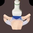
SternoclavicularandAcromioclavicularJoints TRANSVERSEUS,STERNOCLAVICULARJOINT Interclavicularl.
1strib
Anteriorsternoclavicularl.
Costoclavicularl.
Clavicle
Articulardisc
1stcostalcartilage
Manubriumsternum
Medialendofclavicle
Sternoclavicularjoint
Manubrium,sternum
Interclavicularl.
Medialendofclavicle
Jointcapsule
Interclavicularl.
Manubrium,sternum
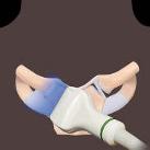
(Top)Graphicshowstheanterioraspectofthesternoclavicularjoint.Notethejointcapsule,articulardisc,andinterclavicularligament. (Middle)TransversegrayscaleUSshowstheanterosuperioraspectofthesternoclavicularjoint.Themedialclavicleismuchlargerthan thearticulatingsurfaceofthemanubrium.Thethininterclavicularligamentiscloselyappliedtothesuperioraspectofmanubrium,and itsconnectionwiththemedialendsofbothclaviclesisdepicted.(Bottom)TransversegrayscaleUSshowsthesuperioraspectofthe sternoclavicularjoint.Thecostoclavicularligamentpreventsupwardmovementofthemedialclaviclewhenthelateralclavicleor shoulderisdepressed.
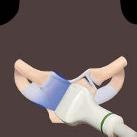
SternoclavicularandAcromioclavicularJoints LONGITUDINALUS,STERNOCLAVICULARJOINT Medialendofclavicle
Pectoralismajorm.
Costoclavicularl.
1strib
Sternohyoid,sternothyroid,t.
Subclaviana.
Sternocleidomastoidm.,sternalend
Sternum
Sternocleidomastoidm.,sternalinsertion
Sternocleidomastoidm.
Subclaviana.
Sternocleidomastoid,clavicularinsertion
Medialendofclavicle
Subclavianv.
(Top)LongitudinalgrayscaleUSshowssternoclavicularjoint.Costoclavicularligamentpreventsupwardmovementofthemedial claviclewhenshoulderisdepressed.Pectoralismajormusclearisesfromthemedial1/2oftheanteriorsurfaceoftheclavicleaswellas fromthesternum,uppercostalcartilages,andupperpartofexternalobliqueaponeurosis.(Middle)LongitudinalgrayscaleUSshows thesternoclavicularjointregion.Thesternocleidomastoidisattachedtotheuppersurfaceofthemedialendoftheclavicleaswellas theupperanteriorsurfaceofthemanubrium.Thesternohyoidandsternothyroidareattachedtotheposterioraspectofthesternumas wellastheclavicleand1stcostalcartilage.(Bottom)LongitudinalgrayscaleUSshowsthesternoclavicularjoint.Greatvesselslie posteriortothesternoclavicularjointandmaygetinjuredinposteriordislocation.Alltendinousattachmentsshouldbeassessedif dislocationispresent,astheymayalsobeinjured.
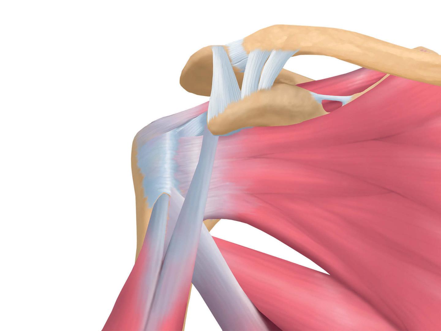
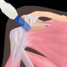
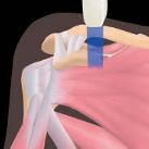
SternoclavicularandAcromioclavicularJoints Superioracromioclavicularl.
Inferioracromioclavicularl.
Coracoacromiall.
Coracoclavicularl.,trapezoidcomponent
Coracohumerall.
Transversehumerall.
Bicepst.,longhead
Bicepst.,shorthead
Latissimusdorsim.
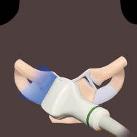
US,ACROMIOCLAVICULARJOINT Clavicle,distal
Coracoclavicularl.,conoidband
Coracoidprocess
Subscapularism.
Lateralendofclavicle
Teresmajorm.
Coracoclavicularl.,trapezoidcomponent
Coracoidprocess
Deltoidm.
Coracoacromiall.
Acromion
Supraspinatusm.
Humeralhead
Coracoidprocess
(Top)Anteriorgraphicshowstheshoulderinsuperficialdissection.(Middle)LongitudinalgrayscaleUSshowstheacromioclavicularjoint region.ThecoracoclavicularligamentisdemonstratedbutisnotasclearlydepictedonUSasitisonMRexam.Theseligamentsprevent upwardandlateralmovementoftheclavicle.(Bottom)TransversegrayscaleUSoftheacromioclavicularjointregionshowsthe coracoacromialligament.Thesupraspinatustendonandinterveningbursacanimpingeagainstthecoracoacromialligamentduringarm abduction.