Diagnostic pathology: bone, 3rd edition gunnlaugur petur nielsen - Download the ebook today to explo

Page 1


https://ebookmass.com/product/diagnostic-pathology-bone-3rd-

Instant digital products (PDF, ePub, MOBI) ready for you

Download now and discover formats that fit your needs...

Bone 2nd Edition G. Petur Nielsen

https://ebookmass.com/product/bone-2nd-edition-g-petur-nielsen/

ebookmass.com

Diagnostic Pathology: Soft Tissue Tumors 3rd Edition

Matthew R. Lindberg

https://ebookmass.com/product/diagnostic-pathology-soft-tissuetumors-3rd-edition-matthew-r-lindberg/

ebookmass.com

Gastrointestinal and Liver Pathology: A Volume in the Series: Foundations in Diagnostic Pathology 3rd Edition Amitabh Srivastava

https://ebookmass.com/product/gastrointestinal-and-liver-pathology-avolume-in-the-series-foundations-in-diagnostic-pathology-3rd-editionamitabh-srivastava/

ebookmass.com

The Room Where It Happened: A White House Memoir John Bolton

https://ebookmass.com/product/the-room-where-it-happened-a-whitehouse-memoir-john-bolton/

ebookmass.com

Preadjusted Edgewise Fixed Orthodontic Appliances: Principles and Practice Farhad B. Naini

https://ebookmass.com/product/preadjusted-edgewise-fixed-orthodonticappliances-principles-and-practice-farhad-b-naini/

ebookmass.com

Laying Bones Reavis Z. Wortham

https://ebookmass.com/product/laying-bones-reavis-z-wortham/

ebookmass.com

The Bodyguard: A Novel Katherine Center

https://ebookmass.com/product/the-bodyguard-a-novel-katherine-center/

ebookmass.com

A Politics of Melancholia George Edmondson

https://ebookmass.com/product/a-politics-of-melancholia-georgeedmondson/

ebookmass.com

Pathophysiology of Disease: An Introduction to Clinical Medicine, 8th Edition Gary D. Hammer

https://ebookmass.com/product/pathophysiology-of-disease-anintroduction-to-clinical-medicine-8th-edition-gary-d-hammer/

ebookmass.com

https://ebookmass.com/product/shes-gone-david-bell/

ebookmass.com

Third Edition

G. Petur Nielsen, MD

Professor of Pathology

Harvard Medical School

Pathologist, Department of Pathology

Director of Bone & Soft Tissue Pathology

Director of Electron Microscopy Unit

Massachusetts General Hospital Boston, Massachusetts

Andrew E. Rosenberg, MD

Professor and Vice Chair

Director, Bone and Soft Tissue Pathology

Department of Pathology and Laboratory Medicine

Miller School of Medicine

University of Miami Miami, Florida

Elsevier

1600 John F. Kennedy Blvd. Ste 1800 Philadelphia, PA 19103-2899

DIAGNOSTIC PATHOLOGY: BONE, THIRD EDITION

Copyright © 2021 by Elsevier. All rights reserved.

ISBN: 978-0-323-76533-6

Inkling:

978-0-323-76535-0

No part of this publication may be reproduced or transmitted in any form or by any means, electronic or mechanical, including photocopying, recording, or any information storage and retrieval system, without permission in writing from the publisher. Details on how to seek permission, further information about the Publisher’s permissions policies and our arrangements with organizations such as the Copyright Clearance Center and the Copyright Licensing Agency, can be found at our website: www.elsevier.com/permissions.

This book and the individual contributions contained in it are protected under copyright by the Publisher (other than as may be noted herein).

Notices

Practitioners and researchers must always rely on their own experience and knowledge in evaluating and using any information, methods, compounds or experiments described herein. Because of rapid advances in the medical sciences, in particular, independent verification of diagnoses and drug dosages should be made. To the fullest extent of the law, no responsibility is assumed by Elsevier, authors, editors or contributors for any injury and/or damage to persons or property as a matter of products liability, negligence or otherwise, or from any use or operation of any methods, products, instructions, or ideas contained in the material herein.

Previous edition copyrighted 2017.

Library of Congress Control Number: 2021932241

Printed in Canada by TC Transcontinental, Beauceville, Quebec, Canada

Last digit is the print number: 9 8 7 6 5 4 3 2 1

Dedications

To my wife and family.

GPN

To my daughters, Olivia and Miranda, who are my lifelong joy; my parents, Philip and Evelyn, who did their best; my siblings, David, Stuart, and Elaine, who have been supportive; my friend and colleague, Al, who always has my back; Corinne, who is always with me; my teachers, who have helped show me the way; my colleagues, with whom I have had the honor to be in the trenches; and the patients who have given me their trust.

AER

Contributing Authors

Ivan Chebib, MD

Director of Immunohistochemistry Laboratory

Massachusetts General Hospital

Assistant Professor of Pathology

Harvard Medical School

Boston, Massachusetts

Yin P. (Rex) Hung, MD, PhD

Assistant Pathologist

Department of Pathology

Massachusetts General Hospital

Assistant Professor of Pathology

Harvard Medical School

Boston, Massachusetts

Michael J. Klein, MD

Pathologist in Chief Emeritus

Hospital for Special Surgery

Professor of Pathology and Laboratory Medicine

Weill Cornell Medicine

Consultant in Pathology

Memorial Sloan Kettering Cancer Center

New York, New York

Daniel I. Rosenthal, MD

Massachusetts General Hospital Professor of Radiology

Harvard Medical School

Boston, Massachusetts

Jaylou M. Velez Torres, MD, FCAP

Assistant Professor of Pathology

Associate Director of Head and Neck Fellowship Program

Head & Neck and Cytopathology

University of Miami Hospital

Miller School of Medicine

Miami, Florida

Preface

The pathology of the skeleton is complex and is the morphologic expression of a broad spectrum of diseases, including those caused by genetic (sporadic and inherited), malformative, inflammatory, metabolic, circulatory, traumatic, iatrogenic, and neoplastic disorders. Bone tumors, including both neoplasms and various conditions that may simulate them, are the focus of our book. This topic is one of the most challenging areas in surgical pathology for several reasons: Bone tumors are uncommon, making it challenging to acquire the necessary experience with their histological variants and mimics. The correct diagnosis usually requires the careful integration of radiological imaging studies and clinical findings; the implications of a diagnosis on a patient can be lifechanging, and medical schools and pathology training programs often have insufficient expertise to provide medical students and young pathologists with the skills needed to diagnose these lesions accurately and precisely.

In this 3rd edition, we updated the text and added many new images or replaced old ones. We also added several new chapters on nonneoplastic orthopedic pathology and a chapter on the radiologic approach to bone tumors.

This book reflects the philosophy and high standards practiced by the truly multidisciplinary team of physicians at the Massachusetts General Hospital, the University of Miami, and the Hospital of Special Surgery, who have diagnosed and surgically treated tens of thousands of patients with bone tumors and nonneoplastic conditions for many decades. Also important to acknowledge are the contributions of the many fellows and residents who participated in the efforts of patient care.

The authors are subspecialized physicians who have dedicated their professional lives to the diagnosis and surgical management of neoplastic and nonneoplastic bone lesions. As a result, the figures include beautiful and classic examples and unusual variants of many of the diseases discussed and are the product of painstaking correlations between the clinical, imaging, macroscopic, histological, immunohistochemical, and molecular characteristics of bone tumors. The text synthesizes the literature and our combined extensive experience, and the images have been selectively culled from the patient files of the Massachusetts General Hospital, the University of Miami Miller School of Medicine, the Hospital of Special Surgery, and the private consultations of the authors. The book is constructed in a thematic format with sections representing groups of related diseases and chapters discussing individual entities and their differential diagnosis.

Accordingly, this textbook serves as an excellent resource for medical students, residents, fellows, and practicing physicians in the disciplines of pathology, radiology, and orthopedics. Medical and radiation oncologists who treat bone tumors will also find it valuable. Our opportunity to participate in the care of patients with bone tumors has been our call and honor, and we hope to do it justice by sharing our experience with the medical community—our goal is to enhance diagnostic accuracy and to provide the biological basis for optimal treatment.

G. Petur Nielsen, MD

Professor of Pathology

Harvard Medical School

Pathologist, Department of Pathology

Director of Bone & Soft Tissue Pathology

Director of Electron Microscopy Unit

Massachusetts General Hospital Boston, Massachusetts

Andrew E. Rosenberg, MD

Professor and Vice Chair

Director, Bone and Soft Tissue Pathology

Department of Pathology and Laboratory Medicine

Miller School of Medicine

University of Miami Miami, Florida

Acknowledgments

LEAD EDITOR

Arthur G. Gelsinger, MA

LEAD ILLUSTRATOR

Laura C. Wissler, MA

TEXT EDITORS

Rebecca L. Bluth, BA

Nina I. Bennett, BA

Terry W. Ferrell, MS

Megg Morin, BA

Kathryn Watkins, BA

IMAGE EDITORS

Jeffrey J. Marmorstone, BS

Lisa A. M. Steadman, BS

ILLUSTRATIONS

Lane R. Bennion, MS

Richard Coombs, MS

ART DIRECTION AND DESIGN

Tom M. Olson, BA

PRODUCTION EDITORS

Emily C. Fassett, BA

John Pecorelli, BS

Sections

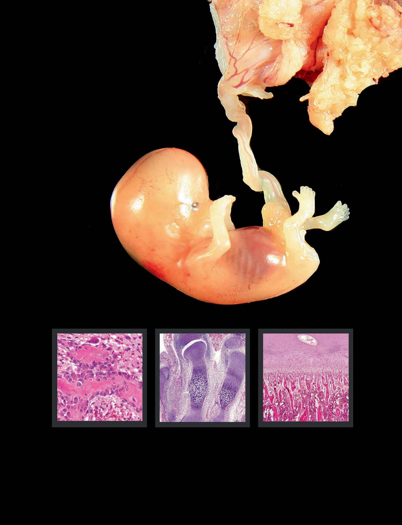
SECTION 1: Growth and Development

SECTION 2: Radiologic Approach to Bone Tumors

SECTION 3: Benign Bone-Forming Tumors

SECTION 4: Malignant Bone-Forming Tumors

SECTION 5: Benign Cartilage Tumors

SECTION 6: Malignant Cartilage Tumors

SECTION 7: Fibrous and Fibrohistiocytic Tumors

SECTION 8: Fibroosseous and Fibroosseous-Epithelial Tumors

SECTION 9: Malignant Small Round Cell Tumors

SECTION 10: Notochordal Tumors

SECTION 11: Giant Cell-Rich Tumors

SECTION 12: Cystic Lesions of Bone

SECTION 13: Vascular Tumors

SECTION 14: Hematopoietic Tumors

SECTION 15: Miscellaneous Mesenchymal Tumors

SECTION 16: Metastatic Tumors

SECTION 17: Bone Tumor Mimics

SECTION 18: Common Nonneoplastic Orthopedic Specimens

SECTION 19: Synovial Tumors

TABLEOFCONTENTS

SECTION

andDevelopment

G.PeturNielsen,MDandAndrewE.Rosenberg,MD

SECTION2:RADIOLOGICAPPROACHTO BONETUMORS

12 RadiologicApproachtoBoneTumors

DanielI.Rosenthal,MD

SECTION3:BENIGNBONE-FORMING TUMORS 22 BoneIsland/Osteopoikilosis

G.PeturNielsen,MDandAndrewE.Rosenberg,MD

G.PeturNielsen,MDandAndrewE.Rosenberg,MD

OsteoidOsteoma

G.PeturNielsen,MDandAndrewE.Rosenberg,MD

Osteoblastoma

G.PeturNielsen,MDandAndrewE.Rosenberg,MD

SECTION4:MALIGNANTBONE-FORMING TUMORS

50 ConventionalOsteosarcoma

G.PeturNielsen,MDandAndrewE.Rosenberg,MD

70 Low-GradeCentralOsteosarcoma

G.PeturNielsen,MDandAndrewE.Rosenberg,MD

76 ParostealOsteosarcoma

G.PeturNielsen,MDandAndrewE.Rosenberg,MD 86 PeriostealOsteosarcoma

G.PeturNielsen,MDandAndrewE.Rosenberg,MD

90 High-GradeSurfaceOsteosarcoma

G.PeturNielsen,MDandAndrewE.Rosenberg,MD

94 SecondarySarcoma

G.PeturNielsen,MD,AndrewE.Rosenberg,MD,andIvan Chebib,MD

SECTION5:BENIGNCARTILAGETUMORS

102 ChondromesenchymalHamartomaofChestWall

G.PeturNielsen,MDandAndrewE.Rosenberg,MD

104 Osteochondroma

G.PeturNielsen,MDandAndrewE.Rosenberg,MD

114 MultipleOsteochondromas

G.PeturNielsen,MD,AndrewE.Rosenberg,MD,andIvan Chebib,MD

118 Enchondroma

G.PeturNielsen,MDandAndrewE.Rosenberg,MD

130 Enchondromatosis

G.PeturNielsen,MDandAndrewE.Rosenberg,MD

138 PeriostealChondroma

G.PeturNielsen,MDandAndrewE.Rosenberg,MD

142 Chondroblastoma

G.PeturNielsen,MDandAndrewE.Rosenberg,MD

150 ChondromyxoidFibroma

G.PeturNielsen,MDandAndrewE.Rosenberg,MD

158 BizarreParostealOsteochondromatous ProliferationandSubungualExostosis

G.PeturNielsen,MDandAndrewE.Rosenberg,MD

SECTION6:MALIGNANTCARTILAGE TUMORS

166 ConventionalChondrosarcoma

G.PeturNielsen,MDandAndrewE.Rosenberg,MD

180 DedifferentiatedChondrosarcoma

G.PeturNielsen,MDandAndrewE.Rosenberg,MD

188 ClearCellChondrosarcoma

G.PeturNielsen,MDandAndrewE.Rosenberg,MD

196 MesenchymalChondrosarcoma

G.PeturNielsen,MDandAndrewE.Rosenberg,MD

SECTION7:FIBROUSAND FIBROHISTIOCYTICTUMORS

206 NonossifyingFibroma

G.PeturNielsen,MDandAndrewE.Rosenberg,MD

212 DesmoplasticFibroma

G.PeturNielsen,MDandAndrewE.Rosenberg,MD

218 MyofibromaandMyofibromatosis

G.PeturNielsen,MDandAndrewE.Rosenberg,MD

222 Fibrosarcoma

G.PeturNielsen,MDandAndrewE.Rosenberg,MD

230 SolitaryFibrousTumor

G.PeturNielsen,MDandAndrewE.Rosenberg,MD

SECTION8:FIBROOSSEOUSAND FIBROOSSEOUS-EPITHELIALTUMORS

234 FibrousDysplasia

G.PeturNielsen,MDandAndrewE.Rosenberg,MD

248 OsteofibrousDysplasia

G.PeturNielsen,MD,AndrewE.Rosenberg,MD,andYin

P.(Rex)Hung,MD,PhD

252 Adamantinoma

G.PeturNielsen,MDandAndrewE.Rosenberg,MD

TABLEOFCONTENTS

SECTION9:MALIGNANTSMALLROUND CELLTUMORS

262 EwingSarcomaandRelatedTumors

G.PeturNielsen,MD,AndrewE.Rosenberg,MD,andYin

P.(Rex)Hung,MD,PhD

280 MelanoticNeuroectodermalTumor

G.PeturNielsen,MDandAndrewE.Rosenberg,MD

SECTION10:NOTOCHORDALTUMORS

284 Ecchordosis

G.PeturNielsen,MD,AndrewE.Rosenberg,MD,andYin

P.(Rex)Hung,MD,PhD

286 BenignNotochordalCellTumor

G.PeturNielsen,MDandAndrewE.Rosenberg,MD

292 Chordoma

G.PeturNielsen,MDandAndrewE.Rosenberg,MD

SECTION11:GIANTCELL-RICHTUMORS

308 GiantCellTumor

G.PeturNielsen,MDandAndrewE.Rosenberg,MD

322 BrownTumor

G.PeturNielsen,MDandAndrewE.Rosenberg,MD

328 GiantCellReparativeGranuloma

G.PeturNielsen,MDandAndrewE.Rosenberg,MD

SECTION12:CYSTICLESIONSOFBONE

336 IntraosseousGanglion

G.PeturNielsen,MDandAndrewE.Rosenberg,MD

340 SimpleBoneCyst

G.PeturNielsen,MDandAndrewE.Rosenberg,MD

350 AneurysmalBoneCyst

G.PeturNielsen,MDandAndrewE.Rosenberg,MD

364 EpidermoidInclusionCyst

G.PeturNielsen,MDandAndrewE.Rosenberg,MD

SECTION13:VASCULARTUMORS

368 ConventionalHemangioma

G.PeturNielsen,MDandAndrewE.Rosenberg,MD

376 Lymphangioma/Lymphangiomatosis

G.PeturNielsen,MDandAndrewE.Rosenberg,MD

378 EpithelioidHemangioma

G.PeturNielsen,MDandAndrewE.Rosenberg,MD

386 PseudomyogenicHemangioendothelioma

G.PeturNielsen,MDandAndrewE.Rosenberg,MD

394 EpithelioidHemangioendothelioma

G.PeturNielsen,MDandAndrewE.Rosenberg,MD

400 Angiosarcoma

G.PeturNielsen,MDandAndrewE.Rosenberg,MD

SECTION14:HEMATOPOIETICTUMORS

408 LangerhansCellHistiocytosis(Eosinophilic Granuloma)

G.PeturNielsen,MDandAndrewE.Rosenberg,MD

416 PrimaryLymphoma

G.PeturNielsen,MDandAndrewE.Rosenberg,MD

426 PlasmaCellMyeloma

G.PeturNielsen,MD,AndrewE.Rosenberg,MD,andIvan Chebib,MD

434 MastCellDisease

G.PeturNielsen,MDandAndrewE.Rosenberg,MD

436 Erdheim-ChesterDisease

G.PeturNielsen,MDandAndrewE.Rosenberg,MD

442 Rosai-DorfmanDisease

G.PeturNielsen,MDandAndrewE.Rosenberg,MD

SECTION15:MISCELLANEOUS MESENCHYMALTUMORS

448 AdipocyticTumors

G.PeturNielsen,MDandAndrewE.Rosenberg,MD

456 Leiomyosarcoma

G.PeturNielsen,MDandAndrewE.Rosenberg,MD

460 Myoepithelioma

G.PeturNielsen,MDandAndrewE.Rosenberg,MD

466 Schwannoma

G.PeturNielsen,MDandAndrewE.Rosenberg,MD

470 MyxopapillaryEpendymoma

G.PeturNielsen,MD,AndrewE.Rosenberg,MD,and JaylouM.VelezTorres,MD,FCAP

472 PhosphaturicMesenchymalTumor

G.PeturNielsen,MDandAndrewE.Rosenberg,MD

SECTION16:METASTATICTUMORS

476 MetastaticTumors

G.PeturNielsen,MD,AndrewE.Rosenberg,MD,and JaylouM.VelezTorres,MD,FCAP

SECTION17:BONETUMORMIMICS

486 Melorheostosis

G.PeturNielsen,MDandAndrewE.Rosenberg,MD

490 Amyloidoma

G.PeturNielsen,MD,AndrewE.Rosenberg,MD,and JaylouM.VelezTorres,MD,FCAP

494 GaucherDisease

G.PeturNielsen,MDandAndrewE.Rosenberg,MD

SECTION18:COMMONNONNEOPLASTIC ORTHOPEDICSPECIMENS

500 Osteoarthrosis

MichaelJ.Klein,MD

512 Osteonecrosis

MichaelJ.Klein,MD

522 PathologyofIntervertebralDiscsandMenisci

G.PeturNielsen,MDandAndrewE.Rosenberg,MD

524 CrystalDepositionDiseases

MichaelJ.Klein,MD

536 CommonMetabolicBoneDisorders

MichaelJ.Klein,MD

SECTION19:SYNOVIALTUMORS

548 TenosynovialGiantCellTumors

G.PeturNielsen,MDandAndrewE.Rosenberg,MD

TABLEOFCONTENTS

556 SynovialChondromatosisandChondrosarcoma

G.PeturNielsen,MDandAndrewE.Rosenberg,MD

564 SynovialHemangioma

G.PeturNielsen,MDandAndrewE.Rosenberg,MD

566 SynovialLipomatosis

G.PeturNielsen,MDandAndrewE.Rosenberg,MD

GrowthandDevelopment

Growth and Development

TERMINOLOGY

Definitions

•Developmentofskeletalsystembeginningin embryogenesis

EPIDEMIOLOGY

NaturalHistory

•Skeletalsystemdevelopsindeliberatesequenceduring specifictimeframethatissameinallhumans

•Majorvariancesofskeletonbetweenindividualslargely determinedbygeneticsandinfluencedbyenvironmental factorssuchasnutrition,exposuretosunlight,andphysical activity

•Generally,skeletondevelopscephaladtocaudad

○Boneformationoccursthroughprocessesofenchondral andintramembranousossification

–Enchondralossification:Cartilagemodelisreplacedby bone

□Occursduringgrowthanddevelopment,repair (callus),andneoplasia

□Cancellousbonedevelopsthroughenchondral ossification

–Intramembranousossification:Fibrousmembrane withprogenitorcellsundergoesossification

□Occursduringgrowthanddevelopment,repair (callus),andneoplasiaossification

□Corticalbonedevelopsfromintramembranous ossification

–Allboneshaveintramembranousossificationvia periosteum

□Mostboneshaveenchondralossificationas componentofdevelopment

□Portionsoffrontal,parietal,occipitalandtemporal bonesofskull,andlateralaspectsofclaviclesform fromonlyintramembranousossification

•Firststepisformationofmesenchymalcondensationsat sitesoffuturebones:Developsduring1stseveralweeksof gestation

DevelopingFetus
Skeletonisdevelopinginthisgrowingfetus.Theskeletonformscephaladtocaudalandgoesthroughthestagesofchondrificationand ossification.Eventually,206bonesarecreated.Theblueprintisimprintedinthegeneticcodeandmarkedlyinfluencedbyenvironmental factors.

GrowthandDevelopment

○Mostbonesinitiallyformashyalinecartilageanlage,and chondrificationoccursduringweeks6-7ofgestation

–Cartilageissurroundedbyperichondrium

○Ossificationofcartilageanlagebeginsweek8of gestation

–Inlongbones,ossificationstartsindiaphysisandis knownasprimarycenterofossification

□Secondarycenterofossificationdevelopsin epiphysis

–Perichondriumbecomesperiosteumasboneis deposited

–Asossificationprogresses,eventuallyhorizontalplate ofcartilageanalogueisproduced,forminggrowthor physealplate

□Zonesofgrowthplateincludereservezone,zone ofproliferation,zoneofhypertrophy,andzoneof mineralization

○Growthplateisresponsibleforincreasinglengthoflong bones

–Mineralizationofcartilageinzoneofmineralizationis associatedwithreleaseofmoleculesbyischemic chondrocytes

□Osteoclastsresorbportionsofcartilageleaving verticallyorientedstrutsthatactastemplatesfor depositionofbonebynewlyformedosteoblasts

□Newlyformedstructurescomposedofcartilage corescoveredbyboneareknownasprimary spongiosa

□Eventually,allcartilageisresorbedandstructures arethendesignatedbonetrabeculae

□Processresultsinformationofmedullarycavity thatbecomespopulatedbyhematopoieticmarrow andfat

CLINICALIMPLICATIONS

NormalSkeleton

•Composedof206bonesthatarelargelybilateraland symmetrical

○Bonesdividedaccordingtosizeandshapeintotubular (longandshort),flatandirregularbones

–Tubularbonesdividedintoepiphysis,metaphysis,and diaphysis

○Bonesneighboringjointshavearticulatingendscovered byhyalineandlessfrequentlyfibrocartilage

○Basedonlocation,bonesareclassifiedintoappendicular (limbs)oraxial(portionsofpelvisandspineandcranium)

BoneComposition

•Biphasicadmixtureorganicandinorganicelements

○Majorstructuralproteinistype1collagen

○Inorganicmineraliscalciumhydroxyapatitethat accountsfor99%ofbody'scalciumandphosphate

○Otherorganicmoleculesincludegrowthfactors, receptors,adhesionmolecules,enzymesandregulators ofmineralization

○Organiccomponentofboneisknownasosteoid

•Cellsofboneincludeosteoblastsandosteoclasts

○Osteoblastsarederivedfromtissuefixedmesenchymal stemcells;mayliveformanyyears

–Bone-formingcellsencasedbymatrixareosteocytes

□Osteocytemostnumerousbone-formingcelland functionstotranslatebiomechanicalforceinto biologicalactivityandproduceshormoneFGF23 thatcontrolsphosphatesecretionbykidney

○Osteoclastsarederivedfromgranulocyte-monocyte stemcellinhematopoieticmarrow

–Functionistoresorbbonebyacidifyingresorptionpit thatdissolvescalciumhydroxyapatiteandreleases enzymesthatbreakdownorganicmolecules

○Osteoblastsandosteoclastsworkincoordinationto make,shape,andremodelbone

–Osteoblastscontrolosteoclasticactivitythrough interactionsofmoleculesRANK,RANKligand,and osteoprotegerin

MICROSCOPIC

BoneMatrix

•Bonetissueisarchitecturallyclassifiedintowovenand lamellarbonebasedonarrangementofcollagenmolecules

○Collageninwovenboneisarrangedrandomlyinall directionsandisdepositedrapidly,resistsforcesequally inalldirectionsandisnotverystrong

○Inlamellarbone,collagenfibersareparalleltoone another;strongesttypeofbone

•4typesoflamellarboneinclude3locatedincortex (circumferential,concentric,andinterstitial)andtrabecular lamellae

•Unitsoflamellarbonedelineatedbycementlines

BoneCells

•Osteoblaststhataremetabolicallyactivearepolyhedral, samesizeashistiocyte,andhaveamphophiliccytoplasm withperinuclearhof,eccentricroundnucleiwithfine chromatin,andsmallnucleoli

○Nucleiorientedawayfromboneformingsurface

•Osteoclastsonlyrecognizedwhenbiologicallyresorbing bone

○Multinucleatedcell(<12nuclei) andcytoplasmhas ruffledborderandisattachedtobonesurface

HyalineArticularCartilage

•Chondrocytessynthesizeandcatabolizematrix

○Evenlydistributedandflatneararticularsurfaceand polyhedralatdeeperlevels

•Baseofarticularcartilagedemarcatedbytidemark,which delineatestruearticularcartilagefromtidemarkcartilage

Synovium

•Connectivetissue1-3cellsthick

•Spindletocuboidalshapedwitheosinophiliccytoplasm

•Subsynovialconnectivetissueveryvascularandcomposed ofvaryingamountsoffibrousandadipocytictissue

SELECTEDREFERENCES

1. RoblingAGetal:Theosteocyte:Newinsights.AnnuRevPhysiol.82:485-506, 2020

2. ChenXetal:Osteoblast-osteoclastinteractions.ConnectTissueRes. 59(2):99-107,2018

3. KatsimbriP:Thebiologyofnormalboneremodelling.EurJCancerCare (Engl).26(6),2017

4. Florencio-SilvaRetal:Biologyofbonetissue:Structure,function,andfactors thatinfluencebonecells.BiomedResInt.2015:421746,2015

GrowthandDevelopment

Growth and Development

(Left)Mesenchymal condensationsarecomposed ofdenselycellularaggregates ofspindlecellsthatdevelopat sitesoffuturebone.At~6 weeksgestation,someofthe cellsdifferentiateanddevelop thecharacteristicsof chondrocytesthatmakethe cartilageanlage.(Right)The developinghandiscomposed ofcartilagemodelsofthe futurebones.Thecentral regionofthemetacarpals containscellsthatare enlargedſt,indicating changeinfunction.The primarycenterofossification isforming.

MesenchymalCondensationsandCartilage CartilageAnlageofHand

(Left)Centralportionof metacarpalshowsalayerof osteoblastsſt thathave producedathinseamof eosinophilicbonecoveringthe surfaceofthecartilage containinglargechondrocytes. Thisregionistheprimary centerofossification.(Right) Inthisphalange,thediaphysis islargelycomposedofbone withadevelopingmedullary cavity.Theperiosteum overlyingtheboneſtisin continuitywiththe perichondrium ﬈.

(Left)Thegrowthplateis locatedbetweentheepiphysis andmetaphysis.Itislinear, hasanundulatingcontour, andisofrelativeeven thickness.Thesecondary centerofossificationiswell formedandhasproduced abundantcancellousbone similartothatinthe metaphysis.(Right)The growthplateiscomposedof reserve,proliferation, hypertrophy,and mineralizationzones.Thebase mergeswiththeprimary spongiosaſtfollowedbythe secondaryspongiosa.

PrimaryCenterofOssification
OssificationInProcess
GrowthPlate
GrowthPlateInAction

GrowthandDevelopment

ZoneofGrowthPlate

LongTubularBone

(Left)Chondrocytesare randomlyarrangedinthe reservezone,orientedin columnsinthezoneof proliferation,enlargeinthe zoneofhypertrophy,andthe matrixmineralizesand chondrocytesundergo apoptosisinthezoneof mineralization.Unresorbed cartilageſthelpsformthe primaryspongiosa.(Right) Eventually,enchondral ossificationextendsalongthe entirediaphysisand metaphyses.Themedullary cavityfilledwithmarrowhas formed,andtheepiphysesare composedofcartilageanlage.

(Left)Proximafemurhas developedfinalshape,is coveredbyarticularcartilage, containsgrowthplates,and themedullarycavityisfilled byredhematopoieticmarrow. thecorticesarethick,and thereisabundantcancellous bone.(Right)Crosssection throughthecortexand medullarycavityshowsthe cortexcomposedofcompact boneandcontaininghaversian systems.Theinterconnecting bonytrabeculaemergewith theendostealsurfaceofthe cortex.Hematopoieticmarrow andfatpopulatethe medullarycavity.

(Left)Numerous interconnectingtrabeculae formthecancellousbone, accountingforthegreatest surfaceareaofbone,andare coveredbyosteoblaststhat arealivingcellularenvelope controllingwhatentersand exitsthebonetissue.(Right)A layerofspindle-shaped quiescentosteoblastscover thesurfaceofthisbone trabeculumſt.Theyare intimatelyattachedtothe bonesurfaceandoverliean osteoidseamthatis delineatedbyacementline ﬈.

Well-FormedBone
CorticalandCancellousBone
CancellousBone
SurfaceLiningQuiescentOsteoblasts

Turn static files into dynamic content formats.

Create a flipbook
Issuu converts static files into: digital portfolios, online yearbooks, online catalogs, digital photo albums and more. Sign up and create your flipbook.
Diagnostic pathology: bone, 3rd edition gunnlaugur petur nielsen - Download the ebook today to explo by Education Libraries - Issuu