https://ebookmass.com/product/diagnostic-imaging-
Instant digital products (PDF, ePub, MOBI) ready for you
Download now and discover formats that fit your needs...
Diagnostic Imaging Oncology 2nd Edition Akram M. Shaaban
https://ebookmass.com/product/diagnostic-imaging-oncology-2nd-editionakram-m-shaaban/
ebookmass.com
Diagnostic Imaging: Pediatric Neuroradiology 3rd Edition
Kevin R. Moore
https://ebookmass.com/product/diagnostic-imaging-pediatricneuroradiology-3rd-edition-kevin-r-moore/
ebookmass.com
Diagnostic Imaging: Interventional Radiology 3rd Edition
Brandt C. Wible
https://ebookmass.com/product/diagnostic-imaging-interventionalradiology-3rd-edition-brandt-c-wible/
ebookmass.com
How to Count Animals, More or Less Shelly Kagan
https://ebookmass.com/product/how-to-count-animals-more-or-lessshelly-kagan/
ebookmass.com
Farm Management 8th Edition https://ebookmass.com/product/farm-management-8th-edition/
ebookmass.com
Baking For Dummies 2nd Edition Wendy Jo Peterson
https://ebookmass.com/product/baking-for-dummies-2nd-edition-wendy-jopeterson/
ebookmass.com
Emergency Medical Responder: Your First Response in Emergency Care 6th Edition, (Ebook PDF)
https://ebookmass.com/product/emergency-medical-responder-your-firstresponse-in-emergency-care-6th-edition-ebook-pdf/
ebookmass.com
Neurologie Collège Des Enseignants De Neurologie France
https://ebookmass.com/product/neurologie-college-des-enseignants-deneurologie-france/
ebookmass.com
Coding Art: A Guide to Unlocking Your Creativity with the Processing Language and p5.js in Four Simple Steps 2 / converted Edition Mathias Funk
https://ebookmass.com/product/coding-art-a-guide-to-unlocking-yourcreativity-with-the-processing-language-and-p5-js-in-four-simplesteps-2-converted-edition-mathias-funk/
ebookmass.com
https://ebookmass.com/product/social-studies-for-the-preschoolprimary-child-9th-edition-ebook-pdf-version/
ebookmass.com
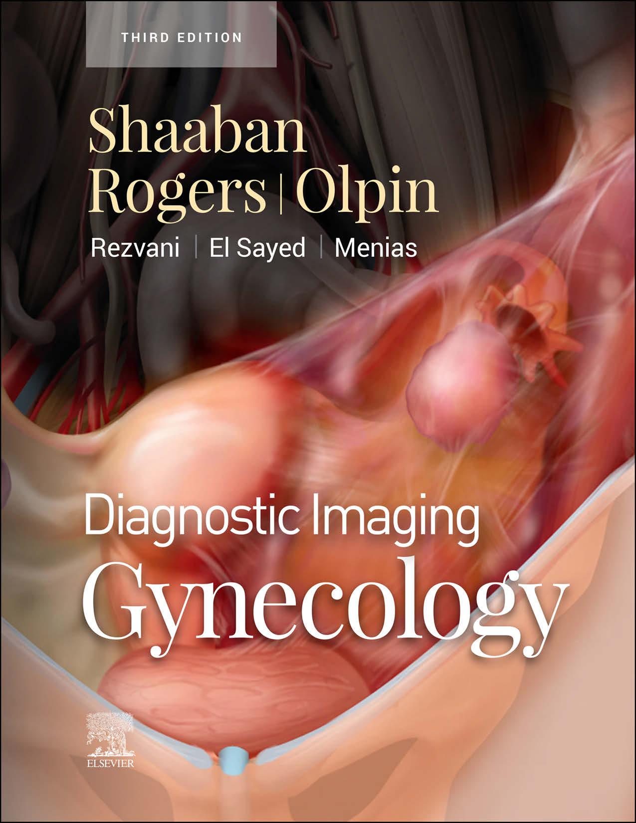
Shaaban Rogers | Olpin | Menias Rezvani | El Sayed
THIRD EDITION Akram M. Shaaban, MBBCh
Professor
Department of Radiology and Imaging Sciences
University of Utah
Salt Lake City, Utah
Douglas Rogers, MD
Assistant Professor
Department of Radiology and Imaging Sciences
University of Utah
Salt Lake City, Utah
Jeffrey Dee Olpin, MD
Professor of Radiology, Abdominal Imaging Division
Department of Radiology and Imaging Sciences
University of Utah
Salt Lake City, Utah
Maryam Rezvani, MD
Associate Professor of Radiology
Department of Radiology
University of Utah School of Medicine
Salt Lake City, Utah
Rania Farouk El Sayed, MD, PhD
Assistant Professor of Radiology
Head of Cairo University MRI Pelvic Floor Center of Excellency and Research
Lab Unit
Department of Radiology
Cairo University Hospitals
Cairo, Egypt
Christine O. Menias, MD
Professor of Radiology
Mayo Clinic School of Medicine
Scottsdale, Arizona
Adjunct Professor of Radiology
Washington University School of Medicine
St. Louis, Missouri
Elsevier
1600 John F. Kennedy Blvd. Ste 1800 Philadelphia, PA 19103-2899
DIAGNOSTIC IMAGING: GYNECOLOGY, THIRD EDITION
Copyright © 2022 by Elsevier. All rights reserved.
ISBN: 978-0-323-79692-7
Inkling: 978-0-323-79693-4
No part of this publication may be reproduced or transmitted in any form or by any means, electronic or mechanical, including photocopying, recording, or any information storage and retrieval system, without permission in writing from the publisher. Details on how to seek permission, further information about the Publisher’s permissions policies and our arrangements with organizations such as the Copyright Clearance Center and the Copyright Licensing Agency, can be found at our website: www.elsevier.com/permissions.
This book and the individual contributions contained in it are protected under copyright by the Publisher (other than as may be noted herein).
Notices Practitioners and researchers must always rely on their own experience and knowledge in evaluating and using any information, methods, compounds or experiments described herein. Because of rapid advances in the medical sciences, in particular, independent verification of diagnoses and drug dosages should be made. To the fullest extent of the law, no responsibility is assumed by Elsevier, authors, editors or contributors for any injury and/or damage to persons or property as a matter of products liability, negligence or otherwise, or from any use or operation of any methods, products, instructions, or ideas contained in the material herein.
Previous edition copyrighted 2015.
Library of Congress Control Number: 2021943237
Printed in Canada by Friesens, Altona, Manitoba, Canada
Dedications To my parents, who taught me the value of perseverance and hard work.
To my wife, Inji, my son, Karim, and my daughters, May and Jena, the jewels of my life, thanks for your understanding and tremendous support.
To all my residents and fellows, whose challenging questions made me a better radiologist.
AMS
To the people who make academic radiology worthwhile, including my mentors, who believe in the merit of my work and continue to teach me, and the residents with passion, who make teaching fulfilling.
DR
Contributing Author Refky Nicola, MS, DO
Associate Professor of Radiology
Additional Contributing Authors Oguz Akin, MD
Nyree Griffin, MD, FRCR
Winnie Hahn, MD
Olga Hatsiopoulou, MD, FRCR
Marcia C. Javitt, MD, FACR
Shephard S. Kosut, MD
Deborah Levine, MD, FACR
Patricia Noël, MD, FRCPC
Caroline Reinhold, MD, MSc
Evis Sala, MD, PhD
Marc S. Tubay, MD
Paula J. Woodward, MD
Preface We are delighted to present Diagnostic Imaging: Gynecology, third edition, the most comprehensive point-of-care imaging resource for gynecologic disorders. The goal of this book is to take the wide range of wonderfully complex topics related to gynecologic imaging and simplify them into a useful and easy-to-understand reference for caretakers at any level of experience, including trainees, general radiologists, gynecology imaging specialists, and gynecologists. This has been achieved using concise, bulleted text and thoughtful grouping of pertinent disease entities by organ, including uterus, cervix, vagina/vulva, ovary, fallopian tubes, multiorgan disorders, and pelvic floor.
Our passionate team of radiologists has thoroughly updated the text and references from the successful second edition, reflecting recent advances in technology and understanding of pathologic conditions as well as changes to TNM/WHO classifications, FIGO staging, and AJCC prognostic groups. Extensive efforts have been made to revamp the already fabulous image galleries with new, high-quality, instructive cases for every entity. More than 2,300 annotated images (and an additional 840 supplemental digital images) exhibit multimodality correlation between ultrasound, sonohysterography, hysterosalpingography, MR, PET/CT, and gross pathology.
The superb radiologic images we present were only possible because of the fine work of our remarkable sonographers and CT/MR technologists. We are also fortunate to collaborate with Laura Wissler, Lane Bennion, and Richard Coombs, who are the most talented and experienced medical illustrators. They possess a rare combination of profound anatomic knowledge and an ability to generate elegant representations of complex structures. Their contributions allow those who contemplate their illustrations to quickly attain a deeper level of comprehension.
This production was especially efficient because of the cohesive efforts of our team, including the image editors (Lisa Steadman and Jeffrey Marmorstone), text editors (Arthur Gelsinger, Rebecca Bluth, Nina Themann, Terry Ferrell, and Megg Morin), graphic designer (Tom Olson), production editors (Emily Fassett and John Pecorelli), lead editor (Kathryn Watkins), and senior manager (Karen Concannon).
Our team is very proud of this work, and we are sure that this new volume will be a rich and oftenused addition to your practice’s collection of resources.
Akram M. Shaaban, MBBCh
Professor
Department of Radiology and Imaging Sciences
University of Utah
Salt Lake City, Utah
Douglas Rogers, MD
Assistant Professor
Department of Radiology and Imaging Sciences
University of Utah
Salt Lake City, Utah
Acknowledgments LEAD EDITOR
Kathryn Watkins, BA
LEAD ILLUSTRATOR
Laura C. Wissler, MA
TEXT EDITORS
Arthur G. Gelsinger, MA
Rebecca L. Bluth, BA
Nina Themann, BA
Terry W. Ferrell, MS
Megg Morin, BA
IMAGE EDITORS
Jeffrey J. Marmorstone, BS
Lisa A. M. Steadman, BS
ILLUSTRATIONS
Richard Coombs, MS
Lane R. Bennion, MS
ART DIRECTION AND DESIGN
Tom M. Olson, BA
PRODUCTION EDITORS
Emily C. Fassett, BA
John Pecorelli, BS
SECTION 1: Techniques
SECTION 2: Uterus
SECTION 3: Cervix
SECTION 4: Vagina and Vulva
SECTION 5: Ovary
SECTION 6: Fallopian Tubes
SECTION 7: Multiorgan Disorders
SECTION 8: Pelvic Floor
TABLEOFCONTENTS SECTION1:
UltrasoundTechniqueandAnatomy
DouglasRogers,MDandMarcS.Tubay,MD 10
14
Sonohysterography
AkramM.Shaaban,MBBChandDouglasRogers,MD
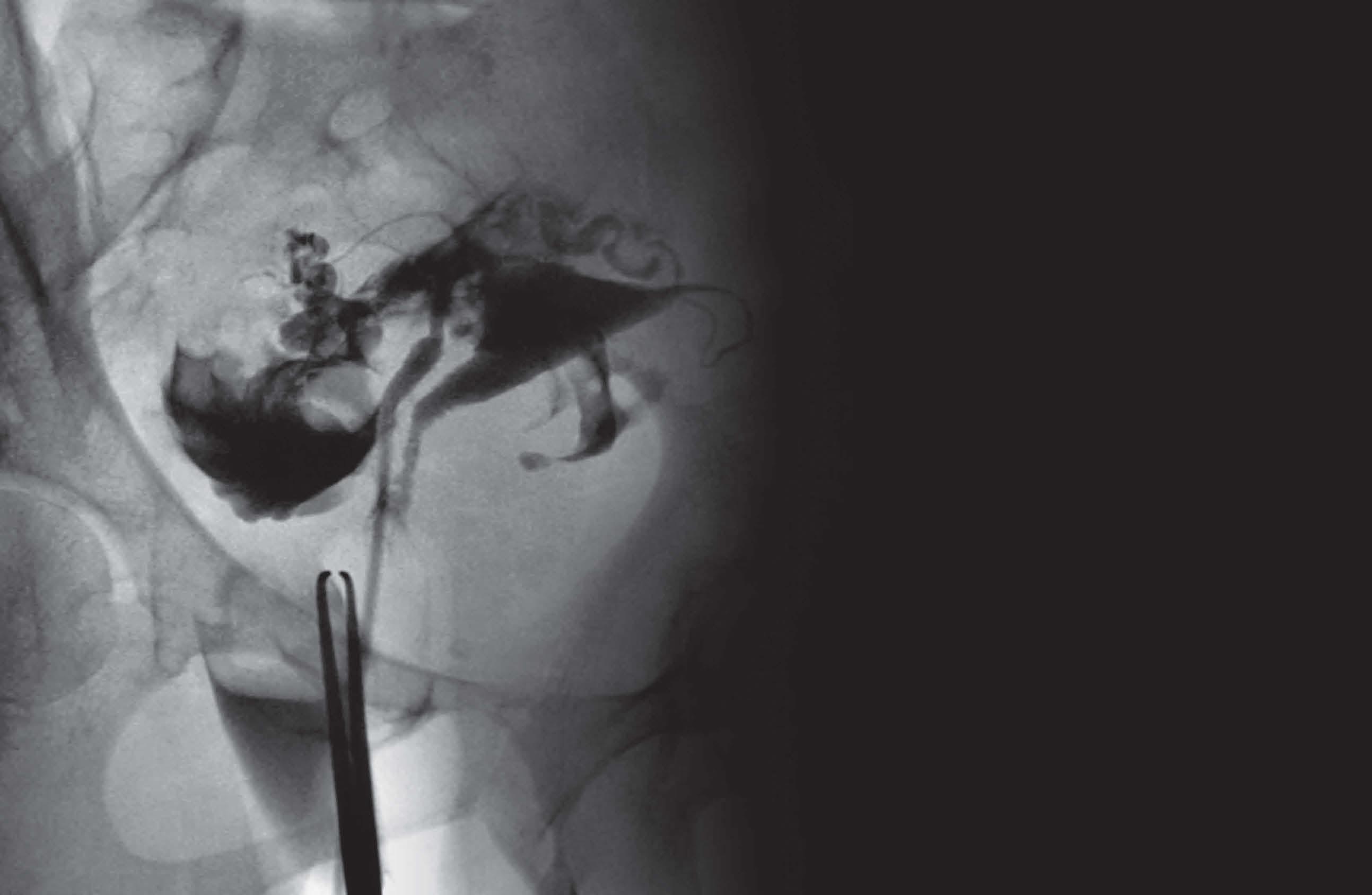
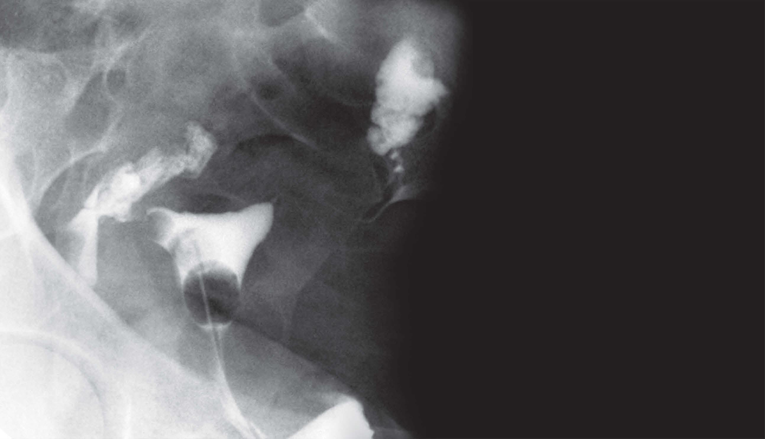
Hysterosalpingography
DouglasRogers,MDandMarcS.Tubay,MD
20 CTTechniqueandAnatomy
24
28
MarcS.Tubay,MDandRefkyNicola,MS,DO
MRTechniqueandAnatomy
MarcS.Tubay,MDandRefkyNicola,MS,DO
PET/CTTechniqueandImagingIssues
MarcS.Tubay,MDandRefkyNicola,MS,DO
SECTION2:UTERUS
INTRODUCTIONANDOVERVIEW
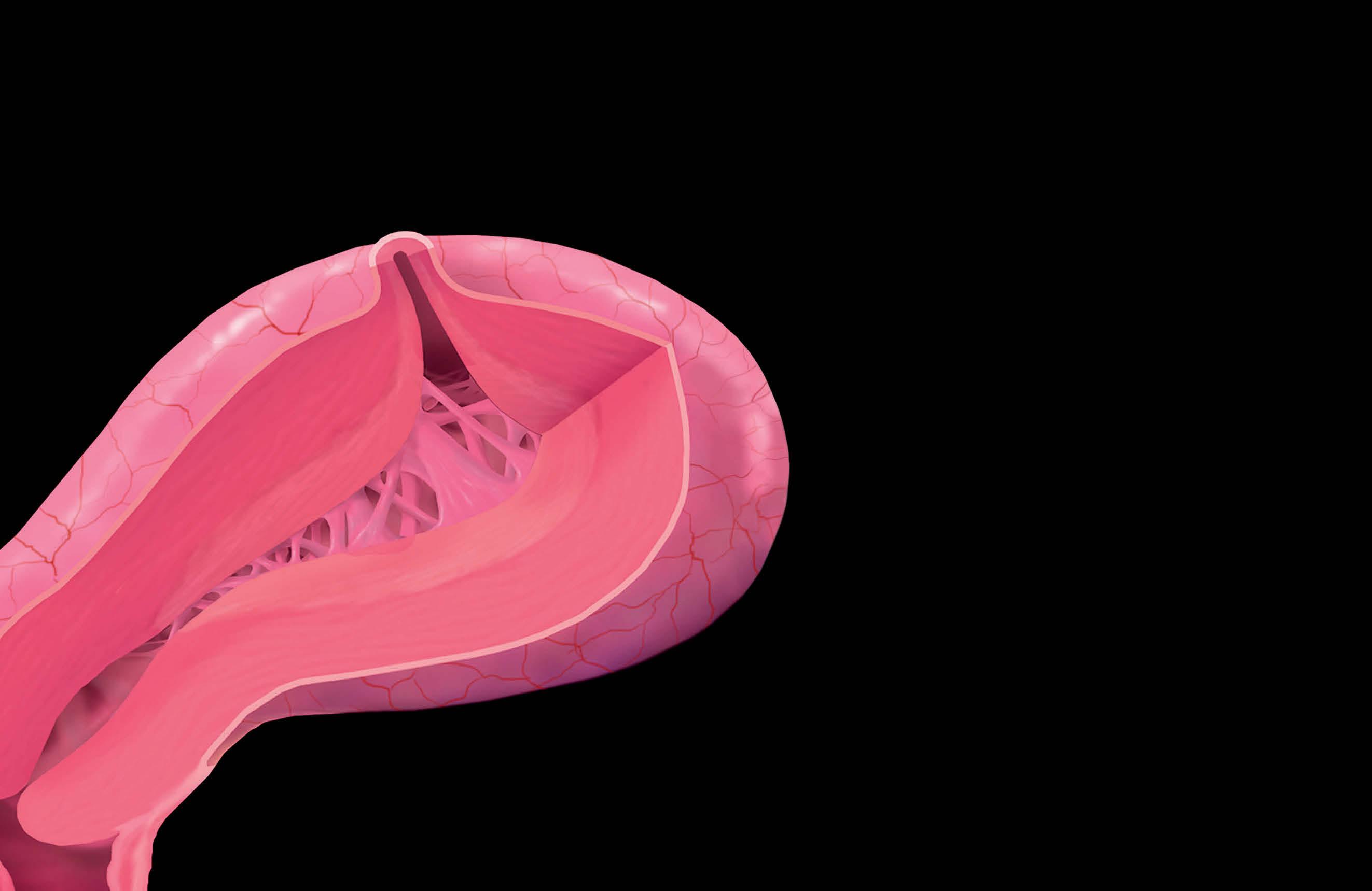
36 AnatomyoftheUterus
PaulaJ.Woodward,MDandAkramM.Shaaban,MBBCh
AGE-RELATEDCHANGES
56 EndometrialAtrophy
JeffreyDeeOlpin,MDandMaryamRezvani,MD
CONGENITAL
58 IntroductiontoMüllerianDuctAnomalies
AkramM.Shaaban,MBBCh
62 MüllerianAgenesis
AkramM.Shaaban,MBBCh
68 UnicornuateUterus
AkramM.Shaaban,MBBCh
74 UterusDidelphys
AkramM.Shaaban,MBBCh,NyreeGriffin,MD,FRCR,and CarolineReinhold,MD,MSc
80 BicornuateUterus
AkramM.Shaaban,MBBCh
84 SeptateUterus
AkramM.Shaaban,MBBCh
90 ArcuateUterus
AkramM.Shaaban,MBBCh
92 DESExposure
AkramM.Shaaban,MBBCh
94 AshermanSyndrome,EndometrialSynechiae
DouglasRogers,MDandChristineO.Menias,MD
98 Endometritis
DouglasRogers,MDandChristineO.Menias,MD
102 Pyomyoma
DouglasRogers,MD
BENIGNNEOPLASMS
MYOMETRIUM
106 UterineLeiomyoma
JeffreyDeeOlpin,MDandMaryamRezvani,MD
112 Leiomyomas:Degeneration,Variants,and Complications
JeffreyDeeOlpin,MDandMarcS.Tubay,MD
120 BenignMetastasizingLeiomyoma
AkramM.Shaaban,MBBChandWinnieHahn,MD
122 DiffuseLeiomyomatosis
DouglasRogers,MDandChristineO.Menias,MD
124 IntravenousLeiomyomatosis
DouglasRogers,MD
128 DisseminatedPeritonealLeiomyomatosis
DouglasRogers,MDandChristineO.Menias,MD
132 LipomatousUterineTumors
DouglasRogers,MDandChristineO.Menias,MD
ENDOMETRIUM
136
EndometrialPolyps
MaryamRezvani,MDandJeffreyDeeOlpin,MD
142 EndometrialHyperplasia
MaryamRezvani,MDandJeffreyDeeOlpin,MD
MALIGNANTNEOPLASMS
ENDOMETRIUM
146 EndometrialCarcinoma
MaryamRezvani,MD
162 UterineAdenosarcoma
DouglasRogers,MD
166 EndometrialStromalSarcoma
DouglasRogers,MD
170 UterineCarcinosarcoma
174
DouglasRogers,MD
GestationalTrophoblasticNeoplasms
AkramM.Shaaban,MBBCh
MYOMETRIUM
184 UterineLeiomyosarcoma
DouglasRogers,MD
VASCULAR
TABLEOFCONTENTS 188 UterineArteriovenousMalformation
MaryamRezvani,MDandJeffreyDeeOlpin,MD
194 UterineArteryEmbolizationImaging
JeffreyDeeOlpin,MDandMaryamRezvani,MD
TREATMENT-RELATEDCONDITIONS
200 Tamoxifen-InducedChanges
JeffreyDeeOlpin,MDandMaryamRezvani,MD
206 ContraceptiveDeviceEvaluation
MaryamRezvani,MDandJeffreyDeeOlpin,MD
214 PostCesareanSectionAppearance
MaryamRezvani,MDandJeffreyDeeOlpin,MD
ADENOMYOSIS
218 Adenomyosis
JeffreyDeeOlpin,MDandMaryamRezvani,MD
224 Adenomyoma
MaryamRezvani,MDandJeffreyDeeOlpin,MD
228 CysticAdenomyosis
MaryamRezvani,MDandJeffreyDeeOlpin,MD
SECTION3:CERVIX INTRODUCTIONANDOVERVIEW
234 AnatomyoftheCervix
MarcS.Tubay,MD
BENIGNNEOPLASMS
240 EndocervicalPolyp
DouglasRogers,MD
244 CervicalLeiomyoma
DouglasRogers,MD
MALIGNANTNEOPLASMS
248 CorpusUteriSarcoma
MaryamRezvani,MD
260 AdenomaMalignum
DouglasRogers,MD
264 CervicalSarcoma
DouglasRogers,MD
268 CervicalMelanoma
AkramM.Shaaban,MBBCh
TREATMENT-RELATEDCONDITIONS
272 PosttrachelectomyAppearances
JeffreyDeeOlpin,MDandMaryamRezvani,MD
MISCELLANEOUS
274 CervicalGlandularHyperplasia
MaryamRezvani,MDandJeffreyDeeOlpin,MD
278 NabothianCysts
MaryamRezvani,MDandJeffreyDeeOlpin,MD
282 CervicalStenosis
DouglasRogers,MD
SECTION4:VAGINAANDVULVA
INTRODUCTIONANDOVERVIEW
288 VaginalandVulvarAnatomy
MarcS.Tubay,MD
CONGENITAL
296 LowerVaginalAtresia
DouglasRogers,MD
298 ImperforateHymen
DouglasRogers,MD
300 VaginalSepta
DouglasRogers,MD
BENIGNNEOPLASMS
302 VaginalLeiomyoma
AkramM.Shaaban,MBBCh,OlgaHatsiopoulou,MD, FRCR,andEvisSala,MD,PhD
308 VulvarSlow-FlowVascularMalformation
DouglasRogers,MD
312 VaginalParaganglioma
DouglasRogers,MD
MALIGNANTNEOPLASMS
316 VaginalCarcinoma
AkramM.Shaaban,MBBCh
328 VaginalLeiomyosarcoma
AkramM.Shaaban,MBBCh,OlgaHatsiopoulou,MD, FRCR,andEvisSala,MD,PhD
330 EmbryonalRhabdomyosarcoma
DouglasRogers,MD
334 VaginalYolkSacTumor
AkramM.Shaaban,MBBCh,OlgaHatsiopoulou,MD, FRCR,andEvisSala,MD,PhD
338 BartholinGlandCarcinoma
DouglasRogers,MD
342 VulvarCarcinoma
MaryamRezvani,MD
354 VulvarLeiomyosarcoma
DouglasRogers,MD
356 VulvarandVaginalMelanoma
AkramM.Shaaban,MBBCh
362 AggressiveAngiomyxoma
DouglasRogers,MD
366 MerkelCellTumor
DouglasRogers,MD
LOWERGENITALCYSTS
368 GartnerDuctCysts
MarcS.Tubay,MDandAkramM.Shaaban,MBBCh
372 BartholinCysts
MarcS.Tubay,MDandAkramM.Shaaban,MBBCh
378 UrethralDiverticulum
MarcS.Tubay,MDandAkramM.Shaaban,MBBCh
382 SkeneGlandCyst
MarcS.Tubay,MDandAkramM.Shaaban,MBBCh
MISCELLANEOUS
386 VaginalForeignBodies
DouglasRogers,MD
394 VaginalFistula
TABLEOFCONTENTS MarcS.Tubay,MDandAkramM.Shaaban,MBBCh
SECTION5:OVARY
INTRODUCTIONANDOVERVIEW
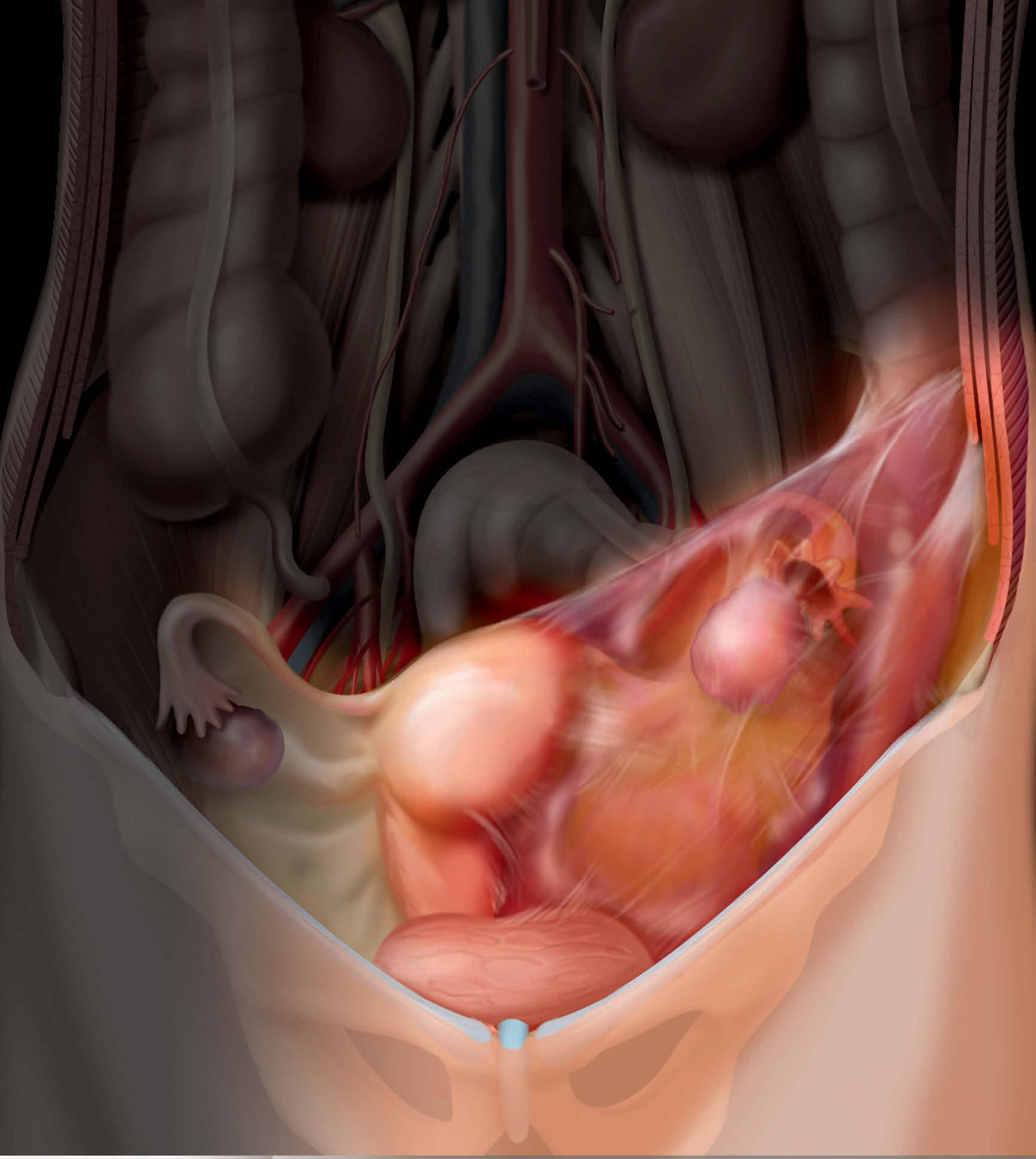
402 AnatomyoftheOvaries
PaulaJ.Woodward,MDandAkramM.Shaaban,MBBCh
PHYSIOLOGICANDAGE-RELATEDCHANGES
410 FollicularCyst
AkramM.Shaaban,MBBCh
414 CorpusLuteum
MarcS.Tubay,MDandAkramM.Shaaban,MBBCh
420 HemorrhagicOvarianCyst
PaulaJ.Woodward,MD
426 OvarianInclusionCyst
MarcS.Tubay,MD
NEOPLASMS
432 OverviewofOvary,FallopianTube,andPrimary PeritonealCarcinoma
AkramM.Shaaban,MBBCh
EPITHELIAL
452 SerousCystadenoma
AkramM.Shaaban,MBBCh,MarciaC.Javitt,MD,FACR, andShephardS.Kosut,MD
458 MucinousCystadenoma
AkramM.Shaaban,MBBCh,WinnieHahn,MD,and DeborahLevine,MD,FACR
464 AdenofibromaandCystadenofibroma
AkramM.Shaaban,MBBCh
470 SerousCarcinoma
AkramM.Shaaban,MBBChandOguzAkin,MD 476 MucinousCarcinoma
AkramM.Shaaban,MBBCh
482 SeromucinousTumors
AkramM.Shaaban,MBBChandChristineO.Menias,MD 488 EndometrioidCarcinoma
AkramM.Shaaban,MBBCh
494 ClearCellCarcinoma
AkramM.Shaaban,MBBChandOguzAkin,MD
500 Carcinosarcoma(MixedMüllerianTumor)
AkramM.Shaaban,MBBCh
504 BrennerTumors
AkramM.Shaaban,MBBCh
GERMCELL
510 MatureCysticTeratoma(DermoidCyst)
AkramM.Shaaban,MBBCh
520 ImmatureTeratoma
AkramM.Shaaban,MBBCh
526 Dysgerminoma
AkramM.Shaaban,MBBChandOguzAkin,MD
532 YolkSacTumor
AkramM.Shaaban,MBBCh,EvisSala,MD,PhD,and ChristineO.Menias,MD
536 Choriocarcinoma
AkramM.Shaaban,MBBChandEvisSala,MD,PhD
540 Carcinoid
AkramM.Shaaban,MBBCh,EvisSala,MD,PhD,and ChristineO.Menias,MD
546 OvarianMixedGermCellTumorandEmbryonal Carcinoma
AkramM.Shaaban,MBBCh
550 StrumaOvarii
AkramM.Shaaban,MBBCh SEXCORD-STROMAL
556 GranulosaCellTumor
AkramM.Shaaban,MBBCh
562 Fibroma,Thecoma,andFibrothecoma
AkramM.Shaaban,MBBCh
568 SertoliandSertoli-LeydigCellTumors
AkramM.Shaaban,MBBChandChristineO.Menias,MD
574 SclerosingStromalTumor
AkramM.Shaaban,MBBChandEvisSala,MD,PhD
METASTASESANDHEMATOLOGIC
578 OvarianMetastases
AkramM.Shaaban,MBBCh
584 OvarianLymphoma
AkramM.Shaaban,MBBCh
NONNEOPLASTICOVARIANLESIONS
590 Endometrioma
MaryamRezvani,MDandJeffreyDeeOlpin,MD
600 Endometriosis
MaryamRezvani,MDandJeffreyDeeOlpin,MD
610 OvarianHyperstimulationSyndrome
MarcS.Tubay,MDandRefkyNicola,MS,DO
614 ThecaLuteinCysts
AkramM.Shaaban,MBBCh,PatriciaNoël,MD,FRCPC, andCarolineReinhold,MD,MSc
618 PolycysticOvarySyndrome
MaryamRezvani,MDandRefkyNicola,MS,DO
624 PeritonealInclusionCysts
MarcS.Tubay,MDandRefkyNicola,MS,DO
VASCULAR 632 OvarianVeinThrombosis
MarcS.Tubay,MDandAkramM.Shaaban,MBBCh
638 PelvicCongestionSyndrome
642
DouglasRogers,MD
AcuteAdnexalTorsion
AkramM.Shaaban,MBBCh
648 MassiveOvarianEdemaandFibromatosis
AkramM.Shaaban,MBBCh
TABLEOFCONTENTS SECTION6:FALLOPIANTUBES
CONGENITAL
656 ParatubalCyst
MaryamRezvani,MDandJeffreyDeeOlpin,MD
INFLAMMATION/INFECTION
660 Hydrosalpinx
MaryamRezvani,MD
664 SalpingitisIsthmicaNodosa
PaulaJ.Woodward,MD
BENIGNNEOPLASMS
668 TubalLeiomyoma
MaryamRezvani,MDandJeffreyDeeOlpin,MD
MISCELLANEOUS
672 Hematosalpinx
MaryamRezvani,MDandJeffreyDeeOlpin,MD
SECTION7:MULTIORGANDISORDERS
PELVICINFLAMMATION
676 PelvicInflammatoryDisease
AkramM.Shaaban,MBBChandMaryamRezvani,MD
686 GenitalTuberculosis
MaryamRezvani,MD
690 Actinomycosis
MaryamRezvani,MD
MALIGNANTNEOPLASMS
694 GenitalLymphoma
DouglasRogers,MDandChristineO.Menias,MD
700 GenitalMetastases
DouglasRogers,MD
ABNORMALSEXUALDEVELOPMENT
704 CompleteAndrogenInsensitivitySyndrome
DouglasRogers,MDandChristineO.Menias,MD
706 DisordersofSexualDevelopment
DouglasRogers,MD
710 GonadalDysgenesis
DouglasRogers,MDandChristineO.Menias,MD
SECTION8:PELVICFLOOR
OVERVIEW
716 AnatomyofthePelvicFloor
RaniaFaroukElSayed,MD,PhD
734 MRofthePelvicFloor
RaniaFaroukElSayed,MD,PhD
PELVICFLOORDYSFUNCTION
ANTERIORCOMPARTMENT
738 AnatomyofBladderandUrethralSupport
RaniaFaroukElSayed,MD,PhD
756 MRofStressUrinaryIncontinence
RaniaFaroukElSayed,MD,PhD
MIDDLECOMPARTMENT
762 AnatomyofUterocervicalandVaginalSupport
RaniaFaroukElSayed,MD,PhD
774 MRofPelvicOrganProlapse
RaniaFaroukElSayed,MD,PhD
POSTERIORCOMPARTMENT
782 AnatomyofAnalCanalandAnalSphincterComplex
RaniaFaroukElSayed,MD,PhD
796 MRofFecalIncontinence
RaniaFaroukElSayed,MD,PhD
804 MRofObstructedDefecation
RaniaFaroukElSayed,MD,PhD
MULTICOMPARTMENTAL
816 MulticompartmentalImaging
RaniaFaroukElSayed,MD,PhD
Shaaban Rogers | Olpin | Menias Rezvani | El Sayed
TERMINOLOGY
UltrasoundTechniqueandAnatomy KEYFACTS •Ultrasoundisimagingmodalitythattransmitshighfrequencysoundwavesintotissuesandgeneratesimages fromreflectedwaves
•Pelvicsonographycanbeperformedusingnumberof techniques(B-mode,M-mode,Doppler,3D,4D)
PREPROCEDURE
•Transabdominalultrasound(TAUS)isusuallyperformed withfullbladder
•Transvaginalpelvicultrasound(TVUS)isperformedwith emptybladder
PROCEDURE
•Mostpelvicsonographicexaminationsutilizeboth transabdominalandtransvaginaltechniques
○TAUSallowsforlargerfieldofviewwithlowerresolution comparedtoTVUS
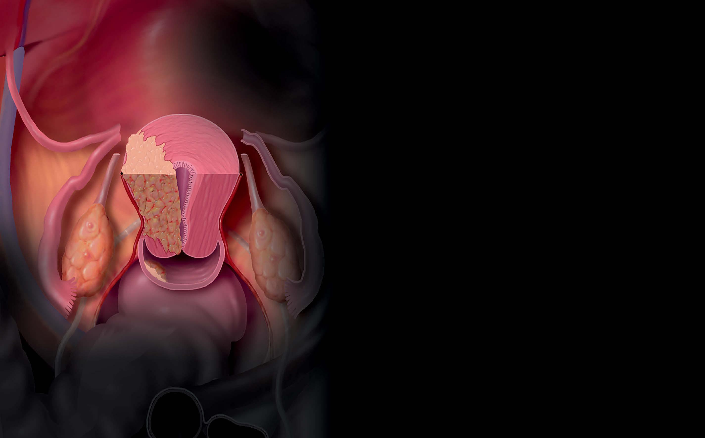
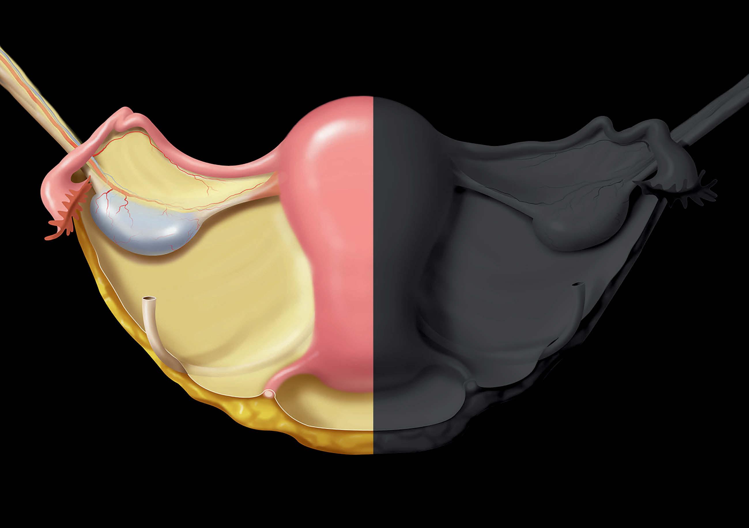
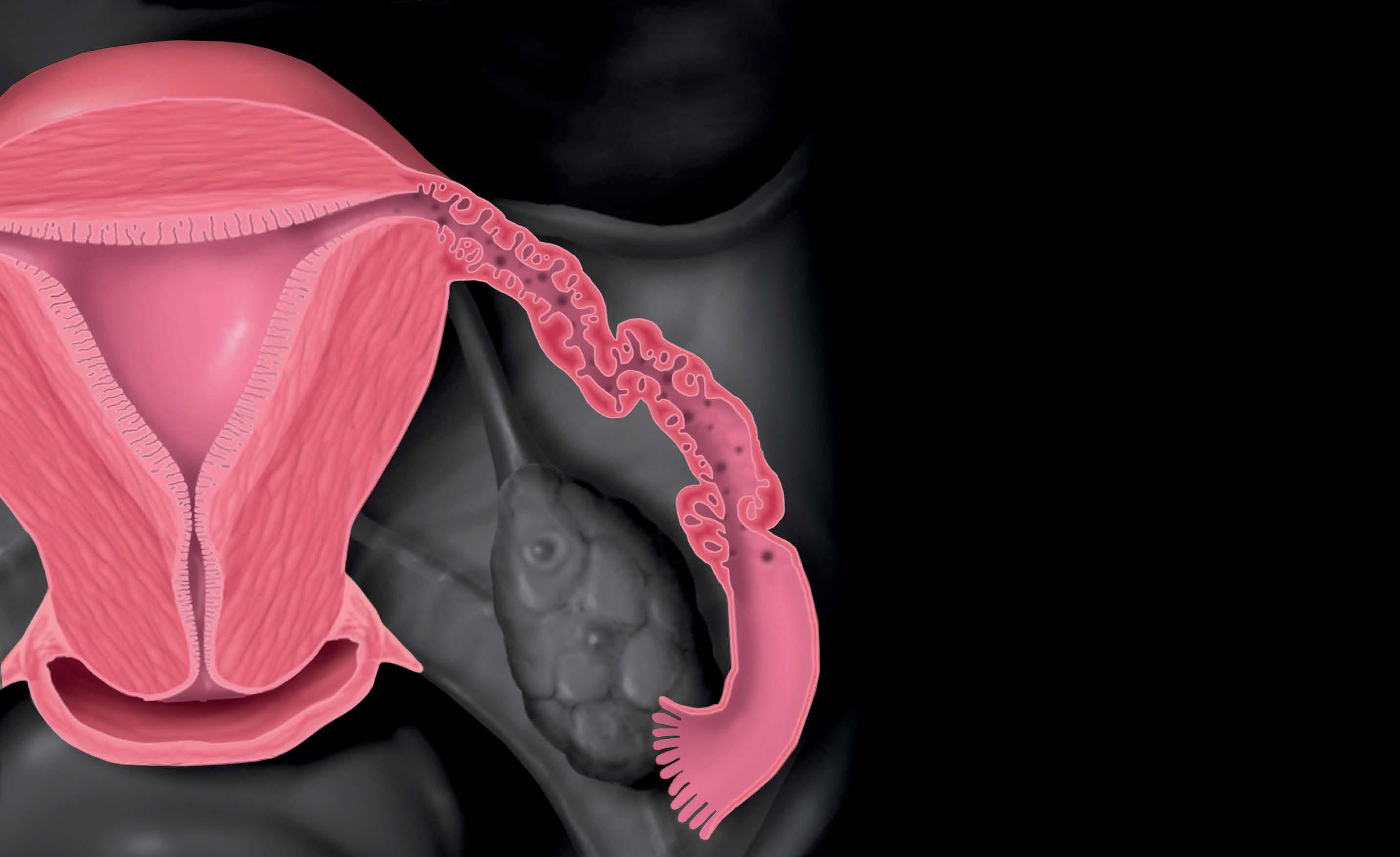
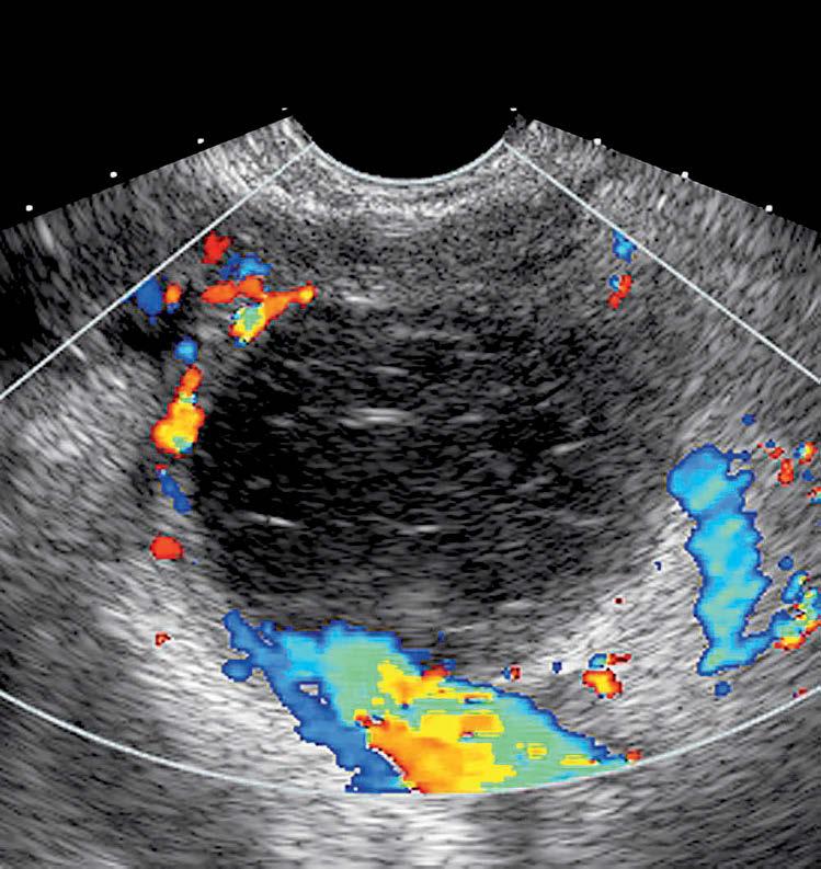
(Left)Longitudinal transvaginalultrasound demonstratesnormal appearanceoftheuterus, includingendometriumſt, myometriumst,cervix, andcul-de-sac.The endovaginalprobeis positionedwithintheanterior vaginalfornix.By convention,theleftsideofthe imageisanterior,andthe rightisposterior.(Right) Transversetransvaginal ultrasounddemonstrates normalappearanceofthe uterus,includingendometrium ſtandmyometriumst.
(Left)Longitudinal transabdominalultrasound showstheuterusſt,vagina st,andurinarybladder. Transabdominalultrasound offersalargerfieldofview butlessresolutioncompared totransvaginalultrasound.
(Right)Transvaginal ultrasoundshowsanormal ovaryſtwithfewsmall folliclesstandadjacent bowel.
○TVUSprovideshigh-resolutionimagesofuterus,cervix, andadnexawithconstrainedfieldofviewcomparedto TAUS
•Pelvicultrasoundrequiresdedicatedevaluationand reportingof
○Uterus:Size,contour,positioning,myometrial echotexture/masses
○Endometrium:Thickness,appearance, presence/positioningofIUD
○Adnexa:Ovariansize,presenceofcystic/solidmass, ovarianvascularflow,tubalabnormalities
○Cul-de-sac:Presenceoffluidormass,uterinesliding
•Probesmustbethoroughlycleansedaccordingto manufacturerandlocalinstitutionguidelines
LongitudinalTransvaginalUltrasound TransverseTransvaginalUltrasound
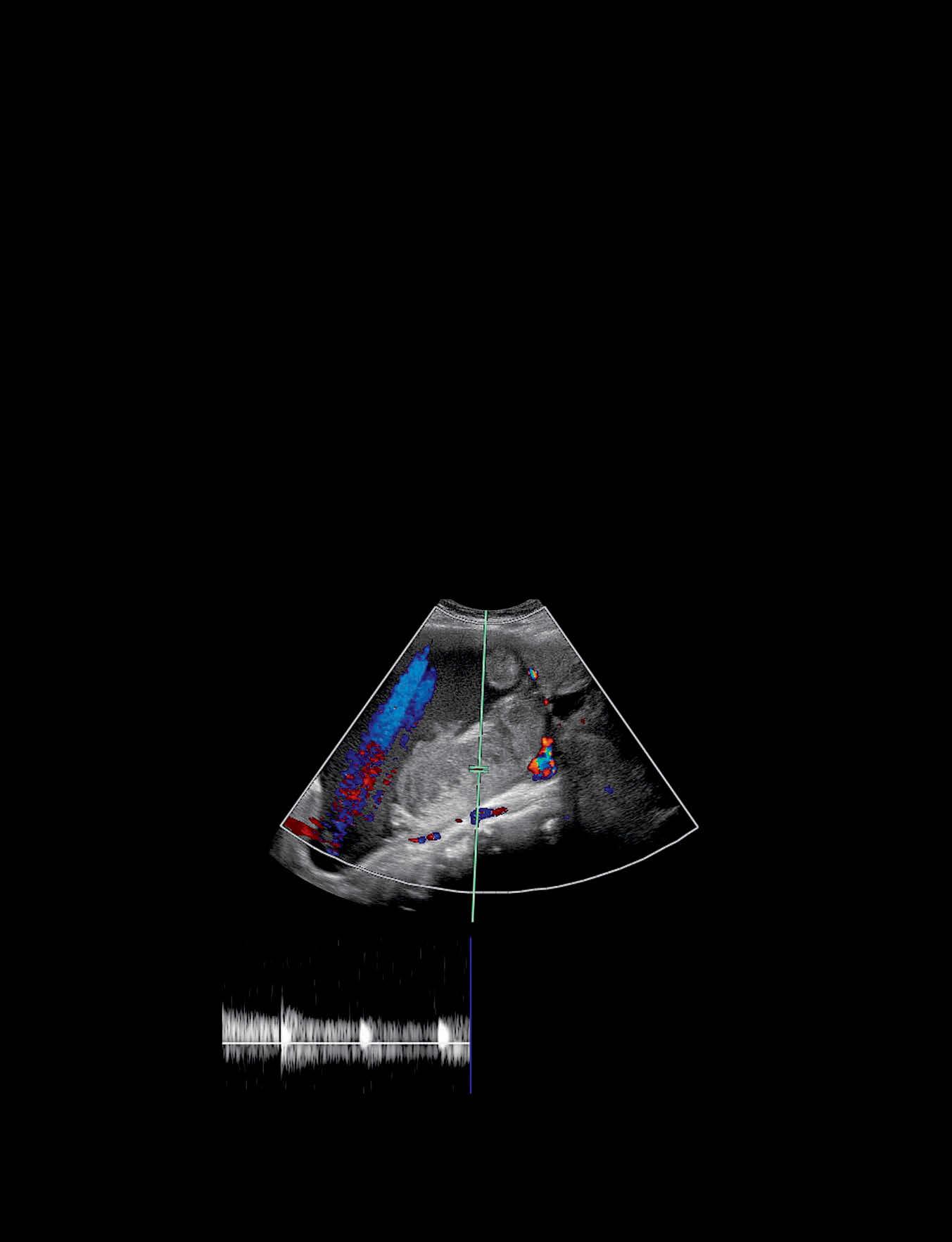
LongitudinalTransabdominalUltrasound OvaryTransvaginalUltrasound
UltrasoundTechniqueandAnatomy TERMINOLOGY Abbreviations
•Transabdominalultrasound(TAUS)
•Transvaginalultrasound(TVUS)
•Saline-infusedsonohysterogram(SIS)
Definitions
•Ultrasoundisimagingmodalitythattransmitshighfrequencysoundwavesintotissuesandgeneratesimages fromreflectedwaves
○TAUSprovideslargefieldofview
–Lowerfrequenciesareusedtoallowforgreaterdepth ofview
□Resultsinlowerresolutionimages
–Usefulforlargemasses
–Characterizeslesionsthatareoutofrangeofvaginal probe
–Mid-tolategestationsaregenerallybetterevaluated withTAUS
○TVUSprovideshigherresolutionimagesofuterus,cervix, andadnexa
–Higherfrequenciesallowforhigherresolutionimages butwithconstrainedfieldofview
–Keymodalityforuterine,cervical,andadnexal pathology
–Usefultoevaluateearlypregnancy
•B-mode(grayscale,2Dmode)ultrasound
○Reflectedsoundwavedataisreconstructedtoproduce 2Dgrayscaleimageofplaneoftissue
○Mostcommonlyusedmode
•M-modeultrasound
○Columnoftissueperpendiculartoprobeisinterrogated toevaluateformotion/velocity
○Demonstratesembryonic/fetalcardiacactivityandheart rate
•Dopplerultrasoundusesfrequencyshiftsofreflected soundwavestodetectflowingblood
○ColorDoppler:Flowisassignedcolorbasedondirection offlowandoverlaidonB-modeimages
○PowerDoppler:MeasuresintensityofDopplershift overlaidongrayscaleimage;moresensitivethancolor Dopplerfordetectionofslowflow
○Pulsed-wave(spectral)Doppler:Velocitytracingis generated,allowingforwaveformanalysis
–DuplexDoppler:Pulsed-waveDopplerdisplayedwith grayscaleanatomicimages
–TriplexDoppler:Pulsed-waveDopplerdisplayedwith grayscaleimagesoverlaidwithcolorDoppler
○Superbmicrovascularimaging:Newtechniquewithhigh sensitivityforbloodflowwithinsmalldiameterandslowflowvessels
•3Dultrasound
○Acquiresvolumeofultrasounddatathatcanbe manipulatedatultrasoundmachineoratdedicated workstationtoproducemultiplanarimagesor3D reconstructions
○Canproduceimagesofsimilarorientationandqualityto MR
•4Dultrasound:3Dultrasounddataisacquiredcontinuously overtime
○Allowsgenerationof3Dsonographicmovies
PREPROCEDURE Indications
•Commonindicationsforpelvicsonographyinclude abnormaluterinebleeding,pelvicpain,contraception
evaluation,pelvicmass,andpregnancy
Contraindications
•TVUSshouldbeavoidedinpatientswithintacthymenor priortohavinghadintercourse
○Transperineal/translabialsonographycanbeperformed whenneeded
○Patientsmaydeclinestudyduetobeinguncomfortable withprocedure
GettingStarted
•Thingstocheck
○FullbladderforTAUS
–Fullbladderactsasacousticwindowfor uterus/adnexa
–Displacessmallbowelfromfieldofview
○EmptybladderforTVUS
–Describeuseoftransvaginalprobetopatient
–Somesonographersprefertohavepatientinsert endovaginal(EV)probe
–Examshouldberelativelypainless
–Ifbladderistoodistended,itmaypushuterusand ovariesoutoffieldofview
○Inwomenofchildbearingage,serumβ-hCGlevelsmay benecessary
○Havechaperone
•Equipmentlist
○Ultrasoundmachine
○Appropriatetransducers
–3.5-7MHzfortransabdominalscans(curvedorsector)
–5-12MHzforEVscans(dedicatedEVprobe)
–7-15MHzforsuperficialtranslabial/transperineal scans(linearprobe)
○Safetyissues
–Thermalandmechanicalindicesareusedasproxies forbioeffectsofultrasound
□Theseshouldbeminimized,particularlywhen imagingembryos
○CommercialprobecoverorcondomtocoverEVprobe forTVUS
–Iflatexallergy,donotuselatexprobecovers
○DedicatedEVprobecleaningsystemandsolution
PROCEDURE PatientPosition/Location
•Bestprocedureapproach
○TAUS:Supineposition
○TVUS:Lithotomyposition
–Feetinstirrupsifbedisequipped
–Pillowunderbuttockscanbeutilizedifneeded, especiallyifbeddoesnothavestirrups
–Similarpositioningfortranslabialortransperineal examinations
Techniques UltrasoundTechniqueandAnatomy •Inmanycenters,routinepelvicultrasoundexaminations includebothTAUSandTVUS
○PatientundergoesTAUSwithfullbladder
○Aftervoiding,patientundergoesTVUS
•Transperineal/translabialevaluations
○Usesectororlineartransducercoveredwithcondomor commerciallyavailableprobecover
○Usefulforvisualizationoflabial/vulvar,distalurethral, andvaginalabnormalities
○Evaluationofprimaryamenorrheainpatientswithintact hymen
○Evaluationofcervixandloweruterusinlate-term pregnantpatientswhenTVUSiscontraindicated
•Transrectalultrasoundisrarelyusedtoevaluateanal sphincterinsettingofpelvicfloordysfunction
EquipmentPreparation
•Probesmustbemeticulouslycleansedaccordingto manufacturer'sandlocalinstitutionalguidelines
•MusthavegelbothinsideandoutsideofEVprobecoverto preventartifactfrominterposedair
•Postmenopausalwomenwithatrophicvaginitismaynot tolerateTVUS
○Usesmallprobeandextralubricatinggel
○AllowpatienttoinsertEVprobe
•Warmedultrasoundgelisbettertoleratedbypatients
ProcedureSteps
•TAUSandTVUSexaminationsshouldinclude
○Uterineimaging
–Uterineflexion/version
–Uterinemeasurements
□Measureuteruslengthonlongitudinal/sagittal midlineimagefromfundustoexternalcervicalos
□APmeasurementisperpendiculartolength measurement
□Uterinewidthismeasuredon transverse/orthogonalimageofuterus
–Myometriumevaluation
□Longitudinalandtransverseimages/cinesthrough entireuterus
□Myometrialmassesshouldbe documented/measured
□Evaluateforadenomyosis
□Incasesofsuspectedmüllerianductanomalies,3D ultrasoundcandepictexternaluterinecontourto helpcharacterizeanomaly
–Endometriumevaluation
□Measureendometrialthicknessperpendicularto longaxisofuterusonmidlinesagittalimage
□Ifthereisfluidwithinendometrialcavity,itshould beexcludedbymeasuringeachendometriallayer separately
□Evaluatefocalendometrialthickeningormasses (colorDopplermaybehelpfultoevaluatefor vascularstalk)
□IfIUDispresent,dedicatedimaginginlongitudinal andtransverseplanesshouldbeobtained
□Acquisitionof3Dvolumewithcoronalreformatted imageisusefultoevaluateIUDposition
–Cervicalimages
□Transverseandlongitudinalimagesthroughcervix
–Insettingofpriorhysterectomy,vaginalcuffshould beevaluated
○Adnexalimaging
–Ovariesshouldbemeasuredin3orthogonalplanes
–ObtaincolorandpulsedwaveDopplerimagesof ovaries,documentingarterialandvenouswaveforms
–Measureanyabnormaladnexallesionin3planesand evaluateforDopplerflowwithinlesion
–Determineiflesionarisesfromovaryorisseparate fromovary
□GentlypresswithEVprobe;adnexallesionarising fromovarywillmovewithovary,whereas paraovarianlesionwillmoveindependentfrom ovarywithpressure
–Bladderfilling&/oremptyingcanhelpdetermine etiologyandlocationofpelviccystincaseswhere largecystismistakenforurinarybladder
–Ifovariesaredifficulttofind,obtaincoronalviewof uterinefundusandanglelaterallytoregionofbroad ligament
□Alternatively,locateiliacvasculatureinlongitudinal planeandslowlyimagetowardmidline
–Scanbetweenuterusandovariestoassessforother adnexalmasses
□Mayidentifyparaovariancysts/masses,ectopic pregnancy,ordilatedfallopiantube
□3Dultrasoundcanhelpconfirmtubularnatureof suspectedhydrosalpinx
○Posteriorcompartment/cul-de-sacimaging
–Evaluateforfreefluid
–Torusuterinusiscommonlocationforadhesionsfrom deeppelvicendometriosis;mayperform"slidingsign" betweenposterioruterusandanteriorrectum
○Inpatientswithfocaltenderness/pain,thisregionshould bethoroughlyevaluated
•Incasesofpelvicmasses,TAUSmayalsoincludeevaluation ofkidneysforhydronephrosis/hydroureter
•ForTVUSevaluation,EVprobeshouldbeslowlyandgently inserted
○Asprobeisbeinginserted,assessforvaginalwallmasses
○Scangenerallyperformedthroughanteriorvaginalwall withprobepositionedinanteriorfornix
○Ifuterusisretrovertedorretroflexed,scanmaybe performedthroughposteriorvaginalwall
○Somepatientshavepainwhencervixismanipulated,so avoidexcessprobepressure
○Inpatientswithbowelgasobscuringvisualizationof ovary,gentleabdominalpressurecandisplacebowel loopsandallowforbettervisualization
•Transperinealevaluation
○Sagittalmidlineviewsofvagina,cervix,andloweruterus areobtained
○Parasagittalviewsasindicated
○Ifperformedduringpregnancy
–Relationshipbetweeninternalcervicalosand placentalmarginshouldbeevaluated
–Measurecervixandassessforfunneling
FindingsandReporting
•Uterinesize
•Uterinecontour
•Uterinepositioning
UltrasoundTechniqueandAnatomy ○Version:Positioningofuteruswithrelationtovagina
○Flexion:Positioningofuterinefundusinrelationtocervix
•Descriptionofmyometrialechotexture
•Descriptionofmyometrialmasses,includinglocation,size, andpositionwithinuterinewall
•Appearanceofcervix
•Descriptionofendometrium
○Endometrialthickness
○Presenceofendometrialmasses,fluid,cysticchange, IUD,focalthickening,orareasthatareilldefinedornot wellimaged
•Ovariansize
•Ovarianarterialandvenouswaveformsdetectedonduplex
Dopplerevaluation
•Descriptionofadnexalmasses
○Ovariancysts
○Complexorsolidadnexalmasses
○Tubalabnormalities
•Freefluid
•Evaluationfordeeppelvicendometriosis
POSTPROCEDURE
ExpectedOutcome
•Noharmfuleffectsfrompelvicsonography
•TAUSandTVUSaregenerallywelltolerated
ThingstoDo
•Cleanseprobesaccordingtomanufacturerandinstitution guidelines
SELECTEDREFERENCES 1. ShwayderJM:Normalpelvicanatomy.ObstetGynecolClinNorthAm. 46(4):563-80,2019
2. CunninghamRKetal:Adenomyosis:asonographicdiagnosis.Radiographics. 38(5):1576-89,2018
3. VandenBoschTetal:Ultrasounddiagnosisofendometriosisand adenomyosis:stateoftheart.BestPractResClinObstetGynaecol.51:16-24, 2018
4. ArmstrongLetal:Three-dimensionalvolumetricsonographyingynecology: anoverviewofclinicalapplications.RadiolClinNorthAm.51(6):1035-47, 2013
5. SakhelKetal:Beginwiththebasics:roleof3-dimensionalsonographyasa first-lineimagingtechniqueinthecost-effectiveevaluationofgynecologic pelvicdisease.JUltrasoundMed.32(3):381-8,2013
6. AndreottiRFetal:Sonographicevaluationofacutepelvicpain.JUltrasound Med.31(11):1713-8,2012
7. LangerJEetal:Imagingofthefemalepelvisthroughthelifecycle. Radiographics.32(6):1575-97,2012
8. AmericanInstituteofUltrasoundinMedicine:AIUMpracticeguidelinefor theperformanceofpelvicultrasoundexaminations.JUltrasoundMed. 29(1):166-72,2010
9. DietzHP:Pelvicfloorultrasound:areview.AmJObstetGynecol.202(4):32134,2010
10.ForsbergFetal:Comparingimageprocessingtechniquesforimproved3dimensionalultrasoundimaging.JUltrasoundMed.29(4):615-9,2010
11.ValskyDVetal:Three-dimensionaltransperinealultrasonographyofthe pelvicfloor:improvingvisualizationfornewclinicalapplicationsandbetter functionalassessment.JUltrasoundMed.26(10):1373-87,2007
12.Timor-TritschIEetal:Three-dimensionalinversionrendering:anew sonographictechniqueanditsuseingynecology.JUltrasoundMed. 24(5):681-8,2005
13.BegaGetal:Three-dimensionalultrasonographyingynecology:technical aspectsandclinicalapplications.JUltrasoundMed.22(11):1249-69,2003
14.LangerRDetal:Transvaginalultrasonographycomparedwithendometrial biopsyforthedetectionofendometrialdisease.Postmenopausal Estrogen/ProgestinInterventionsTrial.NEnglJMed.337(25):1792-8,1997
15.Lev-ToaffAS:Sonohysterography:evaluationofendometrialand myometrialabnormalities.SeminRoentgenol.31(4):288-98,1996
16.FreimanisMGetal:Transvaginalultrasonography.RadiolClinNorthAm. 30(5):955-76,1992
17.LyonsEAetal:Transvaginalsonographyofnormalpelvicanatomy.Radiol ClinNorthAm.30(4):663-75,1992
18.ForrestTSetal:Cyclicendometrialchanges:USassessmentwithhistologic correlation.Radiology.167(1):233-7,1988
19.FleischerACetal:Sonographicdepictionofnormalandabnormal endometriumwithhistopathologiccorrelation.JUltrasoundMed.5(8):44552,1986
Techniques