Dermatopathology
THIRD EDITION
Edited by
Dirk M. Elston, MD
Professor and Chairman
Department of Dermatology and Dermatologic Surgery
Medical University of South Carolina Charleston, SC, USA
Tammie Ferringer, MD
Section Head and Fellowship Director of Dermatopathology
Departments of Dermatology and Laboratory Medicine
Geisinger Medical Center Danville, PA, USA with
Christine J. Ko, MD
Steven Peckham, MD
Whitney A. High, MD, JD, MEng
David J. DiCaudo, MD
Sunita Bhuta, MD
Chapter 7 Interface dermatitis
Lichenoidinterfacedermatitis
Vacuolarinterfacedermatitis
Still'sdisease
Furtherreading
Chapter 8 Psoriasiform and spongiotic dermatitis
Psoriasis
Inflammatorylinearverrucousepidermalnevus(ILVEN)
Mycosisfungoides
Syphilis
Necrolyticerythemas/nutritionaldeficiencydermatitis
Granularparakeratosis
Porokeratosis
Acutespongioticdermatitis
Seborrheicdermatitis
Subacutespongioticdermatitis
Chronicdermatitis(lichensimplexchronicus)
Pityriasisrosea
Spongioticpigmentedpurpuriceruption(PPE)
Stasisdermatitis
Spongioticdermatitiswithintraepidermaleosinophils
Zoonbalanitis
Pityriasisrubrapilaris
Toxicshocksyndrome
Furtherreading
Chapter 9 Blistering diseases
Subcornealvesiculobullousdisorders
Intraepidermalvesiculobullousdisorders
Subepidermalvesiculobullousdisorders:pauciinflammatorysubepidermalconditions
Inflammatorysubepidermalconditions
Frictionblisters
Furtherreading
Chapter 10 Granulomatous and histiocytic diseases
Granulomaannulare
Actinicgranuloma
Necrobiosislipoidica
Rheumatoidnodule
Lupusmiliarisdisseminatusfaciei(LMDF:acneagminata)
Sarcoidosis
Necrobioticxanthogranuloma(NXG)
Xanthogranuloma
Reticulohistiocyticgranuloma(solitaryreticulohistiocytoma)
Rosai–Dorfmandisease(sinushistiocytosiswithmassivelymphadenopathy)
Langerhanscellhistiocytosis(histiocytosisX)
Xanthomas
Gout
Foreign-bodygranuloma
Furtherreading
Chapter 11 Inflammatory vascular diseases
Leukocytoclasticvasculitis(LCV)
Neutrophilicdermatoses
Perivascularlymphoidinfiltrates
Lymphoidvasculitis
Insectbite
Perniosis
Occlusivevasculardiseases
Noninflammatorypurpura
Furtherreading
Chapter 12 Genodermatoses
Pseudoxanthomaelasticum
Ichthyosisvulgaris
Incontinentiapigmenti(Bloch–Sulzbergersyndrome)
Mastocytosis
Epidermolyticichthyosis(bullouscongenitalichthyosiformerythroderma)
Lipoidproteinosis(hyalinosiscutisetmucosae,Urbach–Wiethedisease)
Goltzsyndrome(focaldermalhypoplasia,Goltz–Gorlinsyndrome)
Dowling–Degosdisease(reticulatedpigmentedanomalyoftheflexures)
Galli–Gallidisease
Furtherreading
Chapter 13 Alterations in collagen and elastin
Lichensclerosus(etatrophicus)
Chronicradiationdermatitis
Morphea/scleroderma
Sclerodermoidgraft-versus-hostdisease
Eosinophilicfasciitis(Shulmansyndrome)
Elastosisperforansserpiginosa
Reactiveperforatingcollagenosis
Scarandkeloid
Chondrodermatitisnodularishelicis
Acrodermatitischronicaatrophicans
Ochronosis
Colloidmilium
Anetoderma
Atrophoderma
Connectivetissuenevus
Aplasiacutiscongenita
Furtherreading
Chapter 14 Metabolic disorders
Mucinoses
Amyloidosis
Cutaneouscalcification
Gout
Erythropoieticprotoporphyria(EPP)
Colloidmilium
Mucocele
Oxalosis
Furtherreading
Chapter 15 Disorders of skin appendages
Noninflammatoryalopecia
Inflammatorynonscarringalopecia
Cicatricialalopecia
AcuteLangerhanscellhistiocytosis(histiocytosisX)
Miliaria
Neutrophiliceccrinehidradenitis
Hidradenitissuppurativa
Pseudocystoftheauricle
Relapsingpolychondritis
Furtherreading
Chapter 16 Panniculitis
Septalpanniculitis
Lobularpanniculitis
Subcutaneousfatnecrosisofthenewborn
Traumaticfatnecrosis(“mobileencapsulatedlipoma”)
Subcutaneouspanniculitis-likelymphoma
Furtherreading
Chapter 17 Bacterial, spirochete, and protozoan infections
Furtherreading
Chapter 23 Vascular tumors
Angiokeratoma
Lymphangioma
Nevusflammeus
Angiomaserpiginosum
Venouslake
Glomustumor
Pyogenicgranuloma
Bacillaryangiomatosis
Cherryangioma
Infantilehemangioma
Angiolymphoidhyperplasiawitheosinophilia
Kimuradisease
IntravascularpapillaryendothelialhyperplasiaofMasson(IPEH)
Arteriovenousmalformation(arteriovenoushemangioma)
Targetoidhemosiderotichemangioma(hobnailhemangioma)
Eccrineangiomatoushamartoma
Glomeruloidhemangioma
Microvenularhemangioma
Tuftedangioma(angioblastoma)
Myopericytoma(perivascularmyoidtumor)
PEComa(perivascularepithelioidcelltumor)
Kaposiformhemangioendothelioma
Pleomorphichyalinizingangiectatictumor(PHAT)
Hemangiopericytoma
Spindlecellhemangioma(spindlecellhemangioendothelioma)
Epithelioidhemangioendothelioma
Retiformhemangioendothelioma
Angiosarcoma
Atypicalvascularlesion(AVL)
Kaposisarcoma
Furtherreading
Chapter 24 Cutaneous T-cell lymphoma, NK-cell lymphoma, and myeloid leukemia
CutaneousT-celllymphomaandNK-celllymphoma
Furtherreading
Chapter 25 B-cell lymphoma and lymphocytic leukemia
CutaneousB-celllymphoproliferativedisorders
Lymphomatoidgranulomatosis
Copyright
© 2019, Elsevier Limited. All rights reserved.
First edition 2009
Second edition 2014
The right of Dirk M. Elston, Tammie Ferringer, Christine J. Ko, Steven Peckham, Whitney A. High, David J. DiCaudo to be identified as authors of this work has been asserted by them in accordance with the Copyright, Designs and Patents Act 1988
No part of this publication may be reproduced or transmitted in any form or by any means, electronic or mechanical, including photocopying, recording, or any information storage and retrieval system, without permission in writing from the publisher Details on how to seek permission, further information about the Publisher's permissions policies and our arrangements with organizations such as the Copyright Clearance Center and the Copyright Licensing Agency, can be found at our website: www.elsevier.com/permissions.
This book and the individual contributions contained in it are protected under copyright by the Publisher (other than as may be noted herein)
Copyright for all original illustrations and online material is retained by the authors.
Portions of the text and images were produced while the authors were government employees are in the public domain
Notices
Practitioners and researchers must always rely on their own experience and knowledge in evaluating and using any information, methods, compounds or experiments described herein Because of rapid advances in the medical sciences, in particular, independent verification of diagnoses and drug dosages should be made. To the fullest extent of the law, no responsibility is assumed by Elsevier, authors, editors or contributors for any injury and/or damage to persons or property as a matter of products liability, negligence or otherwise, or from any use or operation of any methods, products, instructions, or ideas contained in the material herein
ISBN: 978-0-7020-7280-2
E-ISBN: 978-0-7020-7281-9
Content Strategist: Charlotta Kryhl
Content Development Specialists: Joanne Scott, Kim Benson
Project Manager: Joanna Souch
Design: Ashley Miner
Illustration Manager: Muthukumaran Thangaraj
Marketing Manager: Michele Milano
Online Lectures and Atlas Materials
1 Author-narrated lectures – 27 presentations with approximately 2000 slides and over 8 hours running time
2. Clinical image atlas – with approximately 600 images
3 Histopathology atlas – with approximately 400 images
4 Infectious disease atlas – with approximately 2500 histopathologic images
5. Soft tissue tumor atlas – with approximately 300 histopathologic images
6. Lymphoma atlas – with 111 images
Preface
This text is designed to cover the essentials of dermatopathology in a style that is enjoyable and easily understood. Please note that you are holding only a portion of the book in your hands! Much of it is online in the form of online lectures and extensive digital image atlases. Online material also includes a high-quality clinical image atlas, an extensive infectious disease atlas, a soft tissue tumor atlas, a lymphoma atlas, and more Be sure to check out all the online features at http://www.expertconsult.com. For students of dermatopathology, we hope the book and lectures make your way a little easier. For those in practice, we hope the book becomes one of your favorite references and one that you reach for often
Dirk M. Elston
New Haven, CT, USA
Steven Peckham MD Pathologist/Dermatopathologist
Precision Pathology Services
San Antonio, TX, USA
Glossaryofterms
Acantholysis
• Loss of cell–cell adhesion
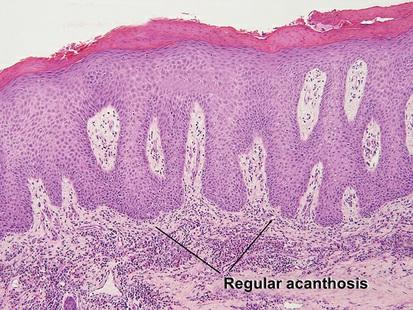
Acanthosis
• Increase in thickness of the epidermis
• Regular (all rete pegs descend to the same level) or irregular (rete pegs descend to different levels in the papillary dermis)
FIG.1.1 Acantholysis,pemphigusvulgaris
FIG 12 Acanthosis,psoriasis
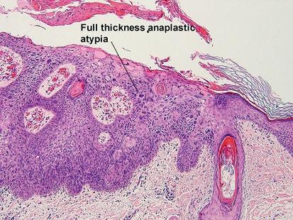
Anaplasia
• Atypical nuclei (abnormal size, shape, staining) and pleomorphism (variation in nuclear characteristics)
Apoptosis(pronouncedapohtosis)
• “Programmed cell death”
• “Dead red” keratinocytes with pyknotic nuclei
• Although the term is often applied to any necrotic or dyskeratotic keratinocyte, it is best reserved for physiologic programmed cell death or pathologic processes that produce death through a similar pathway
FIG.1.3 Anaplasia,Bowendisease