No part of this publication may be reproduced or transmitted in any form or by any means, electronic or mechanical, including photocopying, recording, or any information storage and retrieval system, without permission in writing from the publisher. Details on how to seek permission, further information about the Publisher’s permissions policies, and our arrangements with organizations such as the Copyright Clearance Center and the Copyright Licensing Agency can be found at our website: www.elsevier.com/permissions.
This book and the individual contributions contained in it are protected under copyright by the Publisher (other than as may be noted herein).
ISBN 978-0-7020-6849-2
[eBook 978-0-7020-6854-6]
[Inkling 978-0-7020-6848-5]
Notices
Knowledge and best practice in this field are constantly changing. As new research and experience broaden our understanding, changes in research methods, professional practices, or medical treatment may become necessary.
Practitioners and researchers must always rely on their own experience and knowledge in evaluating and using any information, methods, compounds, or experiments described herein. In using such information or methods they should be mindful of their own safety and the safety of others, including parties for whom they have a professional responsibility. With respect to any drug or pharmaceutical products identified, readers are advised to check the most current information provided (i) on procedures featured or (ii) by the manufacturer of each product to be administered, to verify the recommended dose or formula, the method and duration of administration, and contraindications. It is the responsibility of practitioners, relying on their own experience and knowledge of their patients, to make diagnoses, to determine dosages and the best treatment for each individual patient, and to take all appropriate safety precautions.
To the fullest extent of the law, neither the Publisher nor the authors, contributors, or editors, assume any liability for any injury and/or damage to persons or property as a matter of products liability, negligence or otherwise, or from any use or operation of any methods, products, instructions, or ideas contained in the material herein.
Senior Commissioning Editor: Jeremy Bowes
Senior Content Development Specialist: Ailsa Laing
Project Manager: Joanna Souch
Designer/Design Direction: Miles Hitchen
Illustration Manager: Amy Faith Heyden
Preface to the sixth edition
There has been a near revolution in how a published medical text is handled by its users since we wrote the fifth edition of this work in 2011. For this type of book, there will be considerable usage online. The content and presentation of the sixth edition of this book has been developed to accommodate readers’ likely use in the smartphone age. Our publishers, Elsevier, have been of considerable assistance in this task, providing the platform for the delivery of an interactive, searchable and well illustrated text.
In order to make use of the opportunities offered by online publication, we have increased the flexibility of the educational level of the text. Previously we aimed to write and illustrate a book that was suitable for medical students and general practitioners but which would also appeal to the early
trainee in dermatology. This is still the case, and the printed version has been fully updated, but online publication has given us the chance to broaden the scope of the book. We have added some material at a higher level that can be accessed by those that opt to use it, whilst bolstering the basic strengths of the text through the use of additional illustrations.
Online publishing has allowed us, for each topic covered, to introduce innovations. These include selfassessment questions, flashcards, a picture gallery of dermatological diseases and direct Internet links to other sources of information and reference. It has also been possible to highlight throughout the text, new treatments and dermatological emergencies.
We are aware that the specialty of dermatology is constantly changing.
Preface to the first edition
Recent advances in publishing technology and book presentation demand that a modern text be attractively and concisely presented, in colour and at an affordable price. This is essential for success in a very competitive market. In writing this book, I have attempted to present an introductory dermatology text for the 1990s, using a format of individually designed double-page spreads, generously illustrated with colour photographs, line drawings, tables, bulleted items and ‘key
point’ summaries. This unique approach, which deals with each topic as an educational unit, allows the reader better accessibility to the facts and greater ease in revision than is possible with a conventional textbook.
The book is aimed at medical students but contains sufficient detail to be of use to family practitioners, physicians in internal medicine, registrars or residents in dermatology and dermatological nurses. The contents are divided into
There is presently a greater emphasis than ever before on skin cancer, its recognition and management, and we have reflected this in our content. Other areas that have developed into subspecialisms, which have required additional text, include genital dermatoses, psychodermatology, cosmetic procedures, and advances in dermatologic surgery.
We trust that our target audience of medical students, family doctors, hospital residents, specialty registrars in dermatology or internal medicine, and specialist nurses will continue to find our book helpful in the diagnosis and management of patients with skin diseases.
David J. Gawkrodger and Michael R. Ardern-Jones Sheffield and Southampton, 2015
three sections. The first presents a scientific basis for the understanding of and clinical approach to skin disease. The second details the major dermatological conditions, and the third outlines special topics, such as photoageing and dermatological surgery, that are of current importance or that are poorly dealt with in other textbooks.
David J. Gawkrodger Sheffield 1992
Table e1.1 Diseases associated with genetic deficiency or antibody targeting of ultrastructural components
Target protein Disease associated with gene mutation
Bullous pemphigoid, mucous membrane pemphigoid, pemphigoid gestationis, linear IgA disease
BP200 Bullous pemphigoid
BP230
α6β4 integrin Epidermolysis bullosa simplex with pyloric atresia Junctional epidermolysis bullosa with pyloric atresia
Laminin 332 Junctional epidermolysis bullosa
Collagen VII Dominant dystrophic epidermolysis bullosa
Bullous pemphigoid, mucous membrane pemphigoid, pemphigoid gestationis, linear IgA disease
Mucous membrane pemphigoid
Mucous membrane pemphigoid
Mucous membrane pemphigoid, linear IgA disease, epidermolysis bullosa acquisita, bullous SLE
LAD285 Linear IgA disease
Further reading – online sources
Further information on the skin anatomy, histology and ultrastructural architecture can be accessed via: http:// www.msdmanuals.com/en-gb/professional
formed from epidermis-derived cells and produce an oily sebum, the function of which is uncertain. The glands are small in the child, but become large and active at puberty, being sensitive to androgens. Sebum is produced by holocrine secretion, in which the cells disintegrate to release their lipid cytoplasm
sweat glands
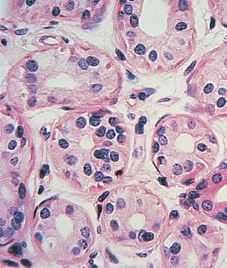
Sweat glands (Fig. 2.4) are tube-like and coiled glands, located within the dermis, which produce a watery secretion. There are two separate types: eccrine and apocrine.
eccrine
Eccrine sweat glands develop from downbudding of the epidermis. The secretory portion is a coiled structure in the deep reticular dermis; the excretory duct spirals upwards to open onto the skin surface. An estimated 2.5 million sweat ducts are present on the skin surface. They are universally distributed, but are most profuse on the palms, soles, axillae and forehead where the glands are under both psychological and thermal control (those elsewhere being under thermal control only). Eccrine sweat glands are innervated by sympathetic (cholinergic) nerve fibres.
apocrine
Also derived from the epidermis, apocrine sweat glands open into hair follicles and are larger than eccrine glands. They are most numerous around the axillae, perineum and areolae. Their sweat is generated by ‘decapitation’ secretion of
the gland’s cells and is odourless when produced; an odour develops after skin bacteria have acted upon it. Sweating is controlled by sympathetic (adrenergic) innervation. The apocrine glands represent a phylogenetic remnant of the mammalian sexual scent gland
Other structures in skin
nerve supply
The skin is richly innervated (Fig. 2.5), with the highest density of nerves being found in areas such as the hands, face and genitalia. All nerves supplying the skin have their cell bodies in the dorsal root ganglia. Both myelinated and non-myelinated fibres are found. The nerves contain neuropeptides, e.g. substance P.
Free sensory nerve endings are seen in the dermis and also encroaching into the epidermis where they may abut onto Merkel cells. These nerve endings detect pain, itch and temperature. Specialized corpuscular receptors are distributed in the dermis, such as the Pacinian corpuscle (detecting pressure and vibration) and touch-sensitive Meissner’s corpuscles, which are mainly seen in the dermal papillae of the feet and hands.
Non-hair-bearing skin (e.g. fingertip)
Autonomic nerves supply the blood vessels, sweat glands and arrector pili muscles. The nerve supply is dermatomal with some overlap.
Blood and lymphatic vessels
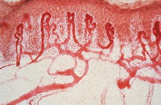
The skin also has a rich and adaptive blood supply. Arteries in the subcutis branch upwards, forming a superficial plexus at the papillary/reticular dermal boundary. Branches extend to the dermal papillae (Fig. 2.6), each of which has a single loop of capillary vessels, one arterial and one venous. Veins drain from
the venous side of this loop to form the mid-dermal and subcutaneous venous networks. In the reticular and papillary dermis, there are arteriovenous anastomoses that are well innervated and concerned with thermoregulation (p. 7). The lymphatic drainage of the skin is important, and abundant meshes of lymphatics originate in the papillae and assemble into larger vessels that ultimately drain into the regional lymph nodes.
Derivatives
• Sebaceous glands, associated with hair follicles, are androgen sensitive.
• Vellus hairs cover most body surfaces; terminal hairs occur on the scalp, beard, axillary and pubic areas.
• Skin has extensive nerve networks with specialized nerve endings.
• Skin has a rich and adaptive blood supply; lymphatics drain to regional lymph nodes.
• Eccrine sweat glands, with sympathetic innervation, are under thermal/ psychological control; apocrine glands are largely vestigial in humans.
Fig. 2.4 sweat gland. A cross-section through the coiled secretory portion of an eccrine sweat gland, situated deep in the dermis.
Fig. 2.5 nerve supply to the skin.
Hair follicle
Free nerve endings
Cutaneous nerve
Hair-bearing skin
Meissner's corpuscle (touch)
Free nerve endings
Pacinian corpuscle (pressure)
Fig. 2.6 superficial dermal blood vessels. Capillary loops branch off the superficial vascular plexus and extend into each dermal papilla.
(see Hyperhidrosis in Ch. 34).
Further reading
Baran, R., Dawber, R.P.R., de Berker, D.A.R., et al., 2001. Baran and Dawber’s Diseases of the Nails and Their Management, 3rd ed. Blackwell Science, Oxford.
Benson, R.A., Palin, R., Holt, P.J., Loftus, I.M., 2013. Diagnosis and management of hyperhidrosis. Br. Med. J. 347, f6800.
Mortimer, P.S., Rockson, S.G., 2014. New developments in clinical aspects of lymphatic disease. J. Clin. Invest. 124 (3), 915–921.
Olszewski, W.L., 2003. The lymphatic system in body homeostasis: physiological conditions. Lymphat. Res. Biol. 1 (1), 11–21.
Schmelz, M., 2011. Neuronal sensitivity of the skin. Eur. J. Dermatol. 21 (Suppl. 2), 43–47.
Shi, V.Y., Leo, M., Hassoun, L., et al., 2015. Role of sebaceous glands in inflammatory dermatoses. J. Am. Acad. Dermatol. 73 (5), 856–863.
Wolfram, L.J., 2003. Human hair: a unique physicochemical composite. J. Am. Acad. Dermatol. 48 (6 Suppl.), S106–S114.
3 Physiology of the skin
The skin is a metabolically active organ with vital functions (Box 3.1), including the protection and homeostasis of the body.
• Presents barrier to physical agents
• Protects against mechanical injury
• Antimicrobial peptides have a bactericidal effect
• Prevents loss of body fluids
• Reduces penetration of UV radiation
• Helps to regulate body temperature
• Acts as a sensory organ
• Affords a surface for grip
• Plays a role in vitamin D production
• Acts as an outpost for immune surveillance
• Communication – cosmetic appearance
Keratinocyte maturation
The differentiation of basal cells into dead, but functionally important, corneocytes is a unique feature of the skin. The horny layer is important in preventing all manner of agents from entering the skin, including micro-organisms, water and particulate matter. Antimicrobial peptides of the defensin and cathelicidin classes, present on the epidermal surface, have anti-bacterial and anti-viral activity. The epidermis also prevents the body’s fluids from getting out.
Epidermal cells undergo the following sequence during keratinocyte maturation (Fig. 3.1):
1. Stem cells in the basal layer divide (continuously) into one new stem cell
and a transit amplifying cell. Transit amplifying cells proliferate briefly and then progress upwards and undergo terminal differentiation.
2. In the prickle cell layer, cells change from being columnar to polygonal. Differentiating keratinocytes synthesize keratins, which aggregate to form tonofilaments. The desmosomes connecting keratinocytes are composed of the structural molecules cadherins, desmogleins and desmocollins. Desmosomes distribute structural stresses throughout the epidermis and maintain a distance of 20 nm between adjacent cells.
3. In the granular layer, enzymes induce degradation of nuclei and organelles. Keratohyalin granules containing filaggrin provide an amorphous protein matrix for the tonofilaments. Membrane-coating granules attach to the cell membrane and release an impervious lipid-containing cement, which contributes to cell adhesion and to the horny layer barrier.
4. In the horny layer, the dead, flattened corneocytes have developed thickened cornified envelopes containing involucrin that encase a matrix of keratin macrofibres aligned by filaggrin. The strong disulphide bonds of the keratin provide strength to the stratum corneum, but the layer is also flexible and can absorb up to three times its own weight in water. However, if it dries out (i.e. water content falls below 10%), pliability fails.
5. The corneocytes are eventually shed from the skin surface after degradation of the lamellated lipid and loss of desmosomal intercellular connections.
rate of maturation
Kinetic studies show that, on average, the dividing basal cells replicate every 200–400 h. The resultant differentiating cells in normal skin take 52–75 days to be shed from the stratum corneum. The epidermal transit time is considerably reduced in keratinization disorders such as psoriasis
Hair growth
In most mammals, hair or fur plays an essential role in survival, especially in the conservation of heat; this is not the case in ‘nude’ humans. Scalp hair in humans does function as a protection against the cancer-inducing effects of ultraviolet (UV) radiation; it also protects against minor injury. However, the main role of hair in human society is as an organ of sexual attraction, and therein lies its importance to the cosmetics industry.
The rate of hair growth differs depending on the site. For example, eyebrow hair grows faster and has a shorter anagen (see later) than scalp hair. On average, there are about 100 000 hairs on the scalp, and the normal rate of growth is 0.4 mm/24 h. Hair growth is cyclical, with three phases, and is randomized for individual hairs, although synchronization does occur during pregnancy. The three phases of hair development (Fig. 3.2) are anagen, catagen and telogen
1. Anagen is the growing phase. For scalp hair, this lasts from 3 to 7 years but, for eyebrow hair, it lasts only 4 months. At any one time, 80–90% of scalp hairs are in anagen, and about 50–100 scalp follicles switch to catagen per day.
Fig. 3.1 Keratinocyte maturation.
Fig. 3.2 The three phases of hair development.
Anagen
Box 3.1 Functions of skin
Melanocytes derive from the neural crest and populate the epidermis in parallel with cutaneous nerves. Abnormal clones of melanocytes therefore often follow patterns consistent with dermatomal innervation (see Fig. 6.4; and Ch. 39 – Pigmentation).
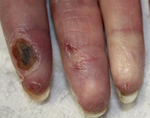
Skin temperature is highly responsive to skin blood flow. Dilatation or contraction of the dermal blood vessels results in vast changes in blood flow, which can vary from 1 to 100 mL/min per 100 g of skin for the fingers and forearms. Arteriovenous anastomoses under the control of the sympathetic nervous system shunt blood to the superficial venous plexuses (Fig. 3.4 and Fig. e3.2), affecting skin temperature. Local factors, both chemical and physical, can also have an effect.
Fig. e3.2 raynaud’s syndrome reflects vascular spasm due to autoimmune disease, and demonstrates the change in skin appearance as a result of altered blood flow. a digital ulcer and tapering of the finger tips can be seen as a result of chronic arterial insufficiency in this case. (From Bolognia JL, Jorizzo JL, Schaffer JV 2012 Dermatology, 3rd Edn. Saunders, with permission.)
Further reading – online sources
Hill MA. 2016. Embryology Neural Crest – Melanocyte Development. Access via https://embryology.med.unsw.edu.au/ embryology/index.php/Neural_Crest_-_Melanocyte _Development
Further reading
Huggenberger, R., Detmar, M., 2011. The cutaneous vascular system in chronic skin inflammation. J. Investig. Dermatol. Symp. Proc. 15 (1), 24–32.
Milstone, L.M., 2004. Epidermal desquamation. J. Dermatol. Sci. 36, 131–140.
4 Biochemistry of the skin
Keratins
The important molecules synthesized by the skin include keratin, melanin, collagen and glycosaminoglycans.
Keratins are high-molecular-weight polypeptide chains produced by keratinocytes (Fig. 4.1). They are the major constituent of the stratum corneum, hair and nails. The stratum corneum comprises 65% keratin (along with 10% soluble protein, 10% amino acid, 10% lipid and 5% cell membrane).
Keratin proteins are of varying molecular weight (between 40 and 67 kDa). Different keratins are found at each level of the epidermis, depending on the stage of differentiation. Epidermal keratin contains less cystine and more glycine than the harder hair keratin
Melanins
Melanin is produced from tyrosine (Fig. 4.2) in melanocytes and takes two forms:
• eumelanin, which is more common and gives a brown–black colour
• phaeomelanin, which is less common and produces a yellow or red colour.
Most natural melanins are mixtures of eumelanin and phaeomelanin. Melanins act as an energy sink and as free radical scavengers, and absorb the energy of ultraviolet (UV) radiation
collagens
Collagens are synthesized by fibroblasts (Fig. 4.3) and are the major structural proteins of the dermis, forming 70–80% of its dry weight. The main amino acids in collagens are glycine, proline and hydroxyproline. Collagens are broken down, e.g. in wound healing, by collagenases, of which the matrix metalloproteinases are important. There are over 22 types of collagen; at least five are found in skin:
• type I – found in the reticular dermis
• type III – found in the papillary dermis
• types IV and VII – found in the basement membrane structures
• type VIII – found in endothelial cells.
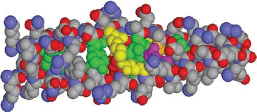
Fig. 4.1 Molecular structure of alpha-keratin. The molecule forms a helical coil which, if stretched, unwinds irreversibly to produce the beta form. The covalent bonds linking the cystine molecules provide extra strength. (From J Invest Dermatol 2001; 116:964–969, with permission of Wiley Blackwell.)
CH2-CH-COO-
Tyrosine
Tyrosinase Cu2+
Polymeric red phaeomelanins
3,4-Dehydroxyphenylalanine (Dopa)
Tyrosinase
Dopaquinone
Leucodopachrome
Non-enzymatic
Cysteine
5,6-Dihydroxyindole
Tyrosinase
Polymeric black eumelanins
Indole-5,6-quinone
Melanochrome
Fig. 4.2 Biosynthesis of melanin. Eumelanin is a high-molecular-weight polymer of complex structure formed by oxidative polymerization. The phaeomelanin polymer is synthesized from dopaquinone and cysteine (via cysteinyl DOPA).
Assembly of polypeptide (procollagen) in ribosomes
Synthesis of glycosaminoglycans in the Golgi complex
Assembly of 3 procollagen chains into 1 procollagen molecule
Tropocollagen molecules aggregate extracellularly to form collagen fibrils
Fibroblast
Fig. 4.3 collagen production. Tropocollagen is formed from three polypeptide chains that are coiled around each other in a triple helix. Assembled collagen fibrils are 100 nm wide, with cross-striations visible with electron microscopy every 64 nm.
(see Table e1.1) (see Melanocyte function in Ch. 3) (see Table e1.1)