No part of this publication may be reproduced or transmitted in any form or by any means, electronic or mechanical, including photocopying, recording, or any information storage and retrieval system, without permission in writing from the Publisher. Details on how to seek permission, further information about the Publisher’s permissions policies and our arrangements with organizations such as the Copyright Clearance Center and the Copyright Licensing Agency, can be found at our website: www.elsevier.com/permissions.
This book and the individual contributions contained in it are protected under copyright by the Publisher (other than as may be noted herein).
Notices
Knowledge and best practice in this field are constantly changing. As new research and experience broaden our understanding, changes in research methods, professional practices, or medical treatment may become necessary.
Practitioners and researchers must always rely on their own experience and knowledge in evaluating and using any information, methods, compounds, or experiments described herein. In using such information or methods they should be mindful of their own safety and the safety of others, including parties for whom they have a professional responsibility.
With respect to any drug or pharmaceutical products identified, readers are advised to check the most current information provided (i) on procedures featured or (ii) by the manufacturer of each product to be administered, to verify the recommended dose or formula, the method and duration of administration, and contraindications. It is the responsibility of practitioners, relying on their own experience and knowledge of their patients, to make diagnoses, to determine dosages and the best treatment for each individual patient, and to take all appropriate safety precautions.
To the fullest extent of the law, neither the Publisher nor the authors, contributors, or editors, assume any liability for any injury and/or damage to persons or property as a matter of products liability, negligence or otherwise, or from any use or operation of any methods, products, instructions, or ideas contained in the material herein.
Library of Congress Cataloging-in-Publication Data
Names: Lin, Harrison W., author. | Roberts, Daniel S. (Daniel Stewart), 1974author. | Harris, Jeffrey P. (Jeffrey Paul), 1949- author.
Title: Cummings review of otolaryngology / Harrison W. Lin, Daniel S. Roberts, Jeffrey P. Harris ; foreword by Charles W. Cummings.
Other titles: Review of otolaryngology
Description: Philadelphia, PA : Elsevier, [2017] | Includes index.
Identifiers: LCCN 2016027590 (print) | LCCN 2016028336 (ebook) | ISBN 9780323401944 (pbk. : alk. paper) | ISBN 9780323427999 ()
University of the Western Cape Cape Town, South Africa
Chapter 11 Head and Neck Pathology
Brian S. Chen, MD
Staff Surgeon, Otology and Neurotology
Department of Otolaryngology—Head and Neck Surgery
William Beaumont Army Medical Center
El Paso, Texas
Chapter 2 Otology and Neurotology
Jennifer Derebery, MD
Associate, House Ear Clinic
Clinical Professor of Otolaryngology
University of Southern California
Los Angeles, California
Chapter 8 Otolaryngic Allergy
Jayme Rose Dowdall, MD
Instructor
Department of Otology and Laryngology
Harvard Medical School
Boston, Massachusetts
Chapter 7 Laryngology
William C. Faquin, MD, PhD
Director, Head and Neck Pathology
Massachusetts General Hospital
Massachusetts Eye and Ear Infirmary
Associate Professor of Pathology
Harvard Medical School
Boston, Massachusetts
Chapter 11 Head and Neck Pathology
Daniel Fink, MD
Assistant Professor, Laryngology and Voice Disorders
Department of Otolaryngology—Head and Neck Surgery
Louisiana State University Health Sciences Center
Baton Rouge, Louisiana
Chapter 7 Laryngology
M. Boyd Gillespie, MD, MSc
Professor
Department of Otolaryngology—Head and Neck Surgery
Medical University of South Carolina Charleston, South Carolina
Chapter 9 Sleep Medicine
Contributors
John L. Go, MD
Associate Professor
Director of Head and Neck Imaging
Department of Radiology
Division of Neuroradiology
University of Southern California
Los Angeles, California
Chapter 12 Head and Neck Radiology
Sachin Gupta, MD
Attending, Otology and Neurotology
Department of Otolaryngology—Head and Neck Surgery
Walter Reed National Military Medical Center
Washington, DC
Chapter 2 Otology and Neurotology
Allen S. Ho, MD
Assistant Professor of Surgery
Department of Surgery
Cedars-Sinai Medical Center
Los Angeles, California
Chapter 5 Head and Neck Surgery
Marc H. Hohman, MD
Facial Plastic and Reconstructive Surgeon
Associate Director, Otolaryngology Residency Program
Madigan Army Medical Center
Tacoma, Washington
Assistant Professor
Department of Surgery
Uniformed Services University of the Health Sciences
Bethesda, Maryland
Chapter 3 Facial Plastic and Reconstructive Surgery
Kiran Kakarala, MD
Assistant Professor
Department of Otolaryngology—Head and Neck Surgery
University of Kansas School of Medicine
Kansas City, Kansas
Chapter 5 Head and Neck Surgery
Elliot Kozin, MD
Department of Otolaryngology
Harvard Medical School
Boston, Massachusetts
Chapter 8 Otolaryngic Allergy
Chapter 9 Sleep Medicine
Shelby Leuin, MD
Assistant Clinical Professor
Pediatric Otolaryngology
Rady Children’s Hospital
University of California, San Diego
San Diego, California
Chapter 6 Pediatric Otolaryngology
Aaron Lin, MD, MA
Pediatric Otolaryngologist
Department of Head and Neck Surgery
Southern California Permanente Medical Group
Downey, California
Chapter 6 Pediatric Otolaryngology
James Lin, MD
Associate Professor
Department of Otolaryngology
Kansas University Medical Center
Kansas City, Kansas
Chapter 2 Otology and Neurotology
Theodore McRackan, MD
Assistant Professor
Director, Lateral Skull Base Surgery
Department of Otolaryngology
Medical University of South Carolina Charleston, South Carolina
Chapter 2 Otology and Neurotology
Anandh G. Rajamohan, MD
Assistant Professor Department of Radiology
Division of Neuroradiology
University of Southern California
Los Angeles, California
Chapter 12 Head and Neck Radiology
Douglas D. Reh, MD
Associate Professor
Otolaryngology—Head and Neck Surgery
Johns Hopkins Medicine
Baltimore, Maryland
Chapter 4 Rhinology and Endoscopic Sinus Surgery
Brian Kip Reilly, MD
Assistant Professor of Otolaryngology
Children’s National Medical Center
Washington, DC
Chapter 6 Pediatric Otolaryngology
Peter M. Sadow, MD, PhD
Associate Director, Head and Neck Pathology
Pathology Service
Massachusetts General Hospital
Associate Professor
Department of Pathology
Harvard Medical School
Associate Director, Head and Neck Pathology
Department of Otolaryngology
Massachusetts Eye and Ear Infirmary
Boston, Massachusetts
Chapter 11 Head and Neck Pathology
Ryan J. Smart, MD, DMD
Oral and Maxillofacial Surgeon
Clinical Instructor of Surgery-University of North Dakota School of Medicine, Staff Surgeon
Essentia Health
West Fargo, North Dakota
Chapter 10 Oral Surgery
Srinivas M. Susarla, MD, DMD
Department of Plastic Surgery
Johns Hopkins Hospital
Baltimore, Maryland
Chapter 3 Facial Plastic and Reconstructive Surgery
Chapter 10 Oral Surgery
Jonathan Ting, MD, MS
Department of Otolaryngology—Head and Neck Surgery
Indiana University School of Medicine
Zionsville, Indiana
Chapter 4 Rhinology and Endoscopic Sinus Surgery
Aaron Wieland, MD
Assistant Professor of Surgery Department of Surgery
Division of Otolaryngology—Head and Neck Surgery
University of Wisconsin
Madison, Wisconsin
Chapter 5 Head and Neck Surgery
The mission of the comprehensive text Cummings Otolaryngology/Head and Neck Surgery is to present, in accessible fashion, information that allows the readership exposure to the most current core content of the specialty. This review mirrors that mission. Congratulations to Drs. Lin, Roberts, and Harris for their excellent work. Medical contributions are expanding at a rate that makes currency of knowledge almost unattainable. Clinical- and compliance-related demands leach away discretionary time previously used to “keep up.” This review book consolidates and highlights the content of its parent to allow rapid review and acquisition of new knowledge. It also presents a vehicle for specialty board examination preparation. Kudos to the editors for this most helpful initiative.
Charles W. Cummings 2016
Preface
Cummings Review of Otolaryngology is a review book designed for physicians taking the otolaryngology written and oral board, recertification, and in-service examinations; the otolaryngology intern, resident, and fellow trainees looking to augment their knowledge base; and medical students preparing for subinternships and residency training. If you are reading this book, you likely have performed at a high level on written examinations for most of your life. Although Cummings Review of Otolaryngology and other texts will prepare you for written examinations in the field of otolaryngology in a systematic and logical way, excelling on clinical rounds and on oral board examinations is a skill that improves with familiarity of oral testing formats and compartmentalizing your fund of knowledge in an organized, easily accessible manner.
We believe that this book is the primary and go-to resource that a medical student or resident will read before clinical rounds with the attending surgeon, a complex surgical case, a mock oral board examination, or the American Board of Otolaryngology examinations. The learning and information conveyed through the book will allow readers to have the most important clinical information—such as a differential diagnosis, clinical algorithm, treatment options, or a list of how-to’s—instantly accessible in their memory to respond quickly to questions in a clinical or testing situation, to facilitate teaching of other residents and medical students, and to assist in patient management. This organized and structured way of thinking is central to success in the oral board format, as well as in clinical rotations and patient care.
Harrison W. Lin
Daniel S. Roberts
Jeffrey P. Harris
We would like to acknowledge all of our teachers and mentors, who have dedicated their lives to both the highest level of patient care and passing on their craft to subsequent generations of surgeons. They serve as continued inspiration toward our academic pursuits. In addition, we wish to thank the students and trainees whose probing questions constantly push us to stay current and to serve as a stimulus for our own creativity.
Harrison W. Lin
Daniel S. Roberts
Jeffrey P. Harris
1 Preparing for Clinical Rounds and Board Examinations, 1
2 Otology and Neurotology, 4
3 Facial Plastic and Reconstructive Surgery, 27
4 Rhinology and Endoscopic Sinus Surgery, 54
5 Head and Neck Surgery, 75
6 Pediatric Otolaryngology, 114
7 Laryngology, 147
8 Otolaryngic Allergy, 176
9 Sleep Medicine, 190
10 Oral Surgery, 200
11 Head and Neck Pathology, 213
12 Head and Neck Radiology, 247 Index, 285
1 Preparing for Clinical Rounds and Board Examinations
INTRODUCTION
Most residents who completed a general surgical internship will remember the “five W’s” that can cause post-operative fever, including wind (pneumonia or atelectasis), water (urinary tract infection), walking (deep venous thrombosis), wound (wound infection), and wonder drug (drug reaction). Those that are undergoing or completed otolaryngology training will furthermore recall being asked to recount the auditory pathway and peaks of the auditory brainstem response, to describe the innervation and actions of the laryngeal muscles, or to discuss the reconstructive ladder, among many other questions in the clinic, on rounds, or in the operating room. Undoubtedly, these well-described and time-tested clinical pearls are critically important in the context of both examinations and everyday patient care, particularly to the resident trainee rotating through busy clinical services while trying to simultaneously read, learn how to operate, and provide the highest quality clinical care. Time is a luxury students and residents unfortunately do not have, and consequently they are best served by learning quickly, efficiently, and effectively.
We wrote the Cummings Review of Otolaryngology in an effort to provide a highly-efficient learning instrument to its readers. Clinical pearls such as the “five W’s”, “E.C.O.L.I.”, and the reconstructive ladder organize and compartmentalize information into packets that most anyone at this level of education and training can swiftly consume, digest and incorporate into their long-term memory. This book provides this compartmentalization of otolaryngology and all of its depth, breadth, and complexity, and offers readers the foundation upon which they can build and expand their fund of knowledge with articles, textbooks, and discussions with senior residents and attendings. Although this text will importantly augment the knowledge base of the reader in a systematic and organized manner, success on clinical rounds and on in-service and board examinations will be further optimized with better understanding of how to prepare for attending rounds and improved familiarity of the testing formats. This chapter provides a systematic approach to these tasks.
ORAL EXAMINATIONS
Familiarity with the format of oral exams will provide the infrastructure upon which the examinee can prepare for success. For most residents and medical students, written exams have been the mainstay of testing, with far less training in the oral exam format. To begin, the American Board of Otolaryngology oral examination takes place over a weekend in a hotel in the city of Chicago, Illinois, near O’Hare International Airport. You have the option of staying in the hotel, in a neighboring hotel walking or shuttle distance away, or in your own accommodations. Based on the first letter of your last name, you will have your exam on Saturday morning, Saturday afternoon, Sunday morning, or Sunday afternoon. At the exam, you receive a
number and a list of five examiners who will likely be among the most prominent and published names in our field. Over 40 minutes, you will be tested on three cases in each of five rooms with different sub-specialty topics, which include head and neck surgical oncology, otology and neurotology, facial plastic and reconstructive surgery, and two general otolaryngology rooms. General cases encompass rhinology, sleep, allergy, pediatric otolaryngology, laryngology, and other general otolaryngology topics. One room will focus on allergy, rhinology, and sleep, and the other will focus on laryngology and pediatric otolaryngology. The examiners will be accessing your ability to discuss eloquently the diagnostic workups, treatment options, complications, and, at times, management of emergencies for each of the presented cases.
Success on the oral board exam depends on an ability to communicate an organized and structured thought process. Oral board formats are similar to the workup of a patient in a clinic. Points are attained by starting with the chief complaint and obtaining a thorough history of the present illness, past medical history, past surgical history, family history, social history, list of medications and allergies, and review of systems. After completing the full history, the examinee should ask to perform a physical exam, first asking for the patient’s vital signs. If there is an emergency, you will be expected to discuss basic life support and assess the patient’s airway, breathing, and circulation. A brief summary statement with pertinent information from the history and physical, along with a comprehensive differential diagnosis, should be provided at some point after the initial assessment is completed. Asking for additional information such as laboratory tests, imaging, and audiograms will also be necessary in many cases. Pathology is typically incorporated into several cases during the testing day. Imaging, pathologic slides, and test results will give you an opportunity to refine your differential diagnosis as needed.
As you discuss the case with your examiner, he or she may focus, narrow, expedite, or redirect your questioning if necessary. When medical treatment or surgery is indicated, you should be able to recite the various options in management, the risks and benefits of each, and the postoperative care protocols. You may need to recognize and manage complications in the postoperative setting.
PEARLS FOR SUCCESS ON ORAL EXAMS
• Stay organized. You are provided a pencil and paper and are welcome to take notes as needed.
• Do not go directly to the diagnosis, even if it may seem obvious to you. Rather, the goal during a case is to obtain points (check marks) by logically moving through the workup of a patient and verbally stating all history questions you should ask, studies you should obtain, diagnoses you should consider, and treatment options you may offer. Demonstrate an organized and linear thought process.
• This review book provides you with the lists to be memorized so that you have easy access to what you want and need to say for many questions that may arise in your oral exam. If you are presented with a patient with hoarseness, simply recite the list of key history questions for a patient with hoarseness. If you are presented with a patient with ear drainage, ask the key questions to ask of all patients presenting to an otology clinic. If your patient has pediatric hearing loss, describe the list of tests you could offer and recommend to the parents. If you are performing parotid surgery and are asked how you would go about finding the facial nerve, simply recount the list of five ways to find the trunk of the facial nerve. If you believe that a rhinoplasty patient could benefit from an increase in tip projection, mention the three ways you could accomplish that. Memorizing the lists in this book will arm you with an organized and comprehensive depth and breadth of knowledge that will help you to remain cool, calm, and collected and succeed in your exam.
• When providing a differential diagnosis use a mnemonic to stay organized. Although this may seem to you artificial, it is an effective way to recite calmly and easily a thorough list of possible diagnoses. Pick one of the examples and stick with it, write it down on your provided piece of paper, and refer to it when needed to guide you
• For pathology questions, describe the finding you see on the image, even if you are unable to provide the correct diagnosis. After all, you are not a pathologist and neither is your examiner. The correct diagnosis is obviously preferred, but some credit may possibly be obtained for an accurate description of the pathological specimen.
• Ask for one test or imaging study at a time. If you are correct in asking for the study, you will be presented the results and will need to interpret them accurately in an organized fashion that demonstrates to your examiner your knowledge and abilities.
• Review computed tomography (CT) and magnetic resonance (MR) images by stating (1) the type of imaging, (2) the view, (3) the type of sequences if applicable, and (4) roughly where the slice is within the series. For example, you could state, “this is an axial CT of the neck, soft-tissue windows, with contrast, at the level of the larynx,” or “this is an MR of the temporal bone, coronal cuts, T1 imaging with contrast, at the level of the internal auditory canal.” You may be provided with and asked to interpret a stack of images from the same scan, and not just one isolated image. Be systematic and help yourself by stating the normal anatomy, which both demonstrates to your examiner your familiarity with the anatomy and helps build a mental image in your brain of where you are. As you should have learned by now, recognition and familiarity with normal help you identify abnormal. Partial credit can be given for an accurate radiologic description of the abnormal findings whether or not the correct diagnosis is obtained.
• Take advantage of any opportunity to take mock examinations. Do not miss formal mock exams that may be provided by your residency programs during your chief year, and if doing a fellowship, consider getting together with your co-fellows in other subspecialties to practice together. If you are not in a fellowship, consider going on Google Talk or
Skype to practice with your co-residents or other residents preparing for the exam. If you take the time to prepare mock exam case files (on Microsoft PowerPoint, for instance) with radiology and pathology images from your own files, publications, or Google searches, and practice going through the formal routine of taking an oral board exam, the actual exam will seem much more familiar and easier.
• Bringing other viewpoints, standards of care, and opinions is another benefit of talking to and studying with co-fellows or residents of other programs. This is especially true for graduates of smaller residency programs who may have only been exposed to a limited number of subspecialty-trained attending physicians. Discussing the “board answers,” that is, what you should say in the board exam, rather than your local surgeon’s independent viewpoint, would serve you well. For instance, your otology-attending physician may have gone forward with a stapedectomy in the setting of a persistent stapedial artery or overhanging facial nerve, but the “board answer” may be to abort. It would be appropriate to bring up the fact that you may have witnessed a successful completion of this case by a more experienced surgeon, but you would abort the case and refer it to a more seasoned otologist.
• Examiners are on your side. If you feel yourself starting to panic or tighten up, ask for a second and relax. You are likely doing better than you think.
• You need to pass the written exam to take the oral exam. If you passed the written exam, chances are heavily in your favor that you will pass the oral exam.
DIFFERENTIAL DIAGNOSIS AND CLINICAL ROUNDS
Cummings Review of Otolaryngology is a powerful tool for rapid learning and review for medical students, interns, and junior residents rounding in the hospital and preparing for “pimp” questions, as well as for residents studying for oral and written in-service and board exams. Even though other review books provide summaries of information of what could be said during an oral examination or in response to a “pimp” question, our book provides a logical, systematic approach that can be applied to any oral exam format; to frequently asked questions by chief residents, fellows, and attending physicians; and to address any clinical situation: what questions should you ask in the history, what findings are you looking for on physical exam, what is the differential diagnosis, what are the critical findings on radiology and pathology studies, what are the treatment options, what is your best option, what are ways to perform this, and what is your postoperative management? Once these lists are reviewed and memorized, the reader will have an armamentarium of knowledge that can be instinctively accessed and effectively used in any clinical or examination scenario. For further detail, you will often need to go to the reference books and other texts to explain in more detail many of the key “buzzwords” that are in this review book. Obviously, the more senior in training you are, the better these terms will make sense to you and stay with you. However, these lists, if memorized early in training, can serve the trainee exceedingly well on rounds, in the clinic, in the operating room, and on exams. For instance, a medical student or intern may have difficulty explaining exactly where the soft-tissue triangles are located, but a sub-intern will routinely be asked to recite the subunits of the nose by facial-plastics attending physicians. Similarly, a student may not know exactly where the greater superficial petrosal nerve travels, but may be asked to name all branches of the facial nerve or surgical landmarks of the middle cranial fossa. Being able to learn such packets of information from this review book and accurately recite them when put on the spot could easily and favorably affect the enthusiasm of the attending and resident physicians for the sub-intern and dramatically elevate the student’s position on the program’s rank list.
PEARLS FOR SUCCESS ON WRITTEN EXAMS AND CLINICAL ROUNDS
• Preparation and repetition are the keys to success. Use the list format to “pimp” question your fellow residents and medical students early and often. Cross off lists that are already memorized, and work to memorize as much of the book as possible. In doing so, the number of humiliatingly awkward silences that follow being asked a “pimp” question will be minimized.
• The American Board of Otolaryngology written examination is a long test, comprising eight 50-minute sections, and is completed on a computer at a local testing center. After signing in, providing all required forms of identification, and putting all of your belongings in a locker, you will be taken to the testing room, where you are provided a whiteboard, marker, and eraser for any notes you may want to take. The clock starts once you log in and click to start a section, and the “break clock” starts once you complete each section. You are given a total of 60 minutes of break time to spend as you wish, and you will need to allot sufficient time to eat lunch, which you will need to bring and store in the locker.
• Undoubtedly, to become a medical student and otolaryngology resident, you will have had to perform at the highest levels on written exams at nearly all points of your education. Most of you will unlikely need to follow the test-taking suggestions in this book, or already have your own effective test-preparation routines. That said, however, it is anecdotally reported that 10-15% of examinees fail the otolaryngology written board exam every year. Accordingly, do not underestimate the difficulty of and level of performance needed to pass the exam. It has been shown that performance on in-service examinations correlates closely with performance on the board examination, and, consequently, you and your program director should be able to determine the intensity at which you should prepare for the written exam.
• Given the length of the exam, prepare your mental endurance as the test date approaches by taking full weekend days during which you study for 3 to 4 consecutive hours without breaks in the morning and again in the afternoon after a brief lunch break. Determine how much caffeine you will need to optimize your attentiveness and minimize mental fatigue as you enter the seventh and eighth hours of study and test taking. Remember that those final questions are worth just as much as the first set of questions.
• Do not perseverate on one or a handful of questions and jeopardize your ability to read and intelligently answer all questions fully. As with most computer tests you have previously taken, you can always go back within a section to questions about which you are unsure. If you have absolutely no idea what the answer may be, just guess, and minimize time wasted. If you have narrowed it down to two or three choices, take a short amount of time to decide what the best guess would be.
• Oftentimes the question will ask for the “best” answer or “next” course of action. Try to avoid overthinking the question; for example, you may be tempted to establish the diagnosis of an unvaccinated child acutely presenting with drooling and stridor, and want to visualize the supraglottis, but the “next” and “best” course of action may be to establish the airway and intubate the child.
• “Pimping” on clinical rounds is often formulated toward one answer, or a short list (eg, what would you need to see on pathology for a follicular thyroid carcinoma diagnosis, or what are the subsites of the hypopharynx?), and because of the limited time of early morning rounds, you will be expected to answer quickly. Unfortunately, the questions can often be in a frustrating “what am I thinking?” format (eg, what exam finding in particular would you expect in a patient with necrotizing otitis externa, or what is special about adenoid cystic carcinoma, or what else could it be?); being able to list off a differential diagnosis or key findings for a particular pathology quickly would maximize your chance of naming the desired answer.
• Remember to include a differential diagnosis in your presentations because this will help you in your learning.
• Remember your patients. Success on written exams and with frequently encountered “pimp” questioning is facilitated by correlating your clinical experiences with your medical knowledge. Use a key figure or a list of pathologic features presented in this book to associate with a clinical experience. You will never forget facts that are associated with patient care.
• The radiology and pathology images in Cummings Review of Otolaryngology are arranged in a high-yield format for exams in otolaryngology. Review these images before your in-service, written, and oral board exams. These images represent some of the most frequently encountered pathologies in our field, and you may find many on your exams.
We wish you success on your written and oral examinations and congratulate you in your choice of a career in otolaryngology, head and neck surgery.
2 Otology and Neurotology
TEMPORAL BONE ANATOMY
Temporal bone portions
a. Squamous
b. Petrous
c. Tympanic
d. Mastoid
LATERAL
VIEW OF THE LEFT TEMPORAL BONE (Fig. 2.1)
Key Points
• The temporal line approximates the floor level of the middle fossa ±5 mm.
• The sigmoid and lateral sinuses are typically anterior and superior to the emissary vein, respectively.
• The Macewen triangle, which is located posterior and superior to the spine of Henle, approximates the lateral landmark of the mastoid antrum.
SUPERIOR VIEW OF THE LEFT TEMPORAL BONE (Fig.
2.2)
Key Points
• The arcuate eminence typically corresponds to the superior semicircular canal, but not always.
• The greater superficial petrosal nerve (GSPN) exits from the geniculate ganglion of the facial nerve; the geniculate ganglion is dehiscent without bony covering in about 10-20% of temporal bones.
MEDIAL VIEW OF LEFT TEMPORAL BONE (Fig. 2.3)
Key Points
• The lateral portion of the internal auditory canal (IAC) is called the fundus, and the medial portion is called the porus.
• Bill’s bar is named after William House. It separates the facial nerve from the superior vestibular nerve at the fundus of
Spine of Henle
Tympanosquamous suture line
Temporal squama
Temporal line
Zygomatic process
Tympanic bone
Mastoid portion
Macewen triangle
Cribriform area
Tympanomastoid suture line
EAC
Zygomatic root
Articular tubercle of zygomatic process
Glenoid fossa
Foramen for mastoid emissary vein
Digastric groove
Mastoid process Styloid process
2.1 Lateral view of left temporal bone surface shows squamous, tympanic, and mastoid portions. (From Francis HW, Niparko JK. Temporal Bone Dissection Guide. New York, NY: Thieme; 2011 and Flint PW, Haughey BH, Lund VJ, et al. Cummings Otolaryngology—Head and Neck Surgery. 6th ed. Philadelphia, PA: Saunders; 2015, fig. 127-1.)
FIGURE
the IAC. Identification of Bill’s bar allows early identification of the facial nerve, likely uninvolved with pathology (ie, a vestibular schwannoma arising from a vestibular nerve).
INFERIOR VIEW OF THE LEFT TEMPORAL BONE (Fig. 2.4)
Key Points
• The styloid process is anteromedial to the stylomastoid foramen.
• Medial and superior to the mastoid tip is the digastric groove, which is the origin of the posterior belly of the
Depression of trigeminal ganglion
Groove for greater superficial petrosal nerve
Groove for middle meningeal artery
Petrous apex Ridge for superior petrosal sinus
Arcuate eminence
FIGURE 2.2 Superior view of left temporal bone shows petrous and squamous portions forming the floor of the middle fossa and anterior limit of the posterior fossa. (From Francis HW, Niparko JK. Temporal Bone Dissection Guide. New York, NY: Thieme; 2011 and Flint PW, Haughey BH, Lund VJ, et al. Cummings Otolaryngology— Head and Neck Surgery. 6th ed. Philadelphia, PA: Saunders; 2015, fig. 127-2.)
Superior petrosal sulcus
Sigmoid sulcus
Petrous portion
Operculum of endolymphatic duct Cochlear aqueduct
digastric muscle. The anterior border of the digastric groove is the stylomastoid foramen.
• The cochlear aqueduct is inferior and parallel to the IAC.
THE AURICLE
BLOOD SUPPLY
• External carotid artery
• Superficial temporal artery (anterior)
• Posterior auricular (posterior)
EAR CANAL
• Lateral one-third is cartilaginous (has cerumen glands and hair follicles)
• Medial two-thirds are bony (no cerumen glands or hair follicles)
• Bony-cartilaginous junction is a route of disease spread
• Fissures of Santorini: natural fissures in anterior cartilaginous ear canal that allows the spread of disease to the superficial parotid
• Foramen of Huschke: anteroinferior bony defect that typically obliterates during development; patency allows the spread of disease to the deep parotid lobe/ temporomandibular joint (TMJ)
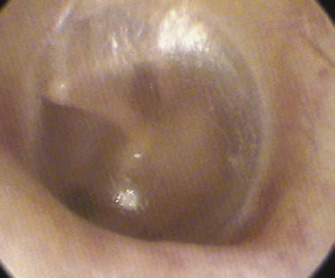
EARDRUM (Fig. 2.5)
1. Manubrium (handle of malleus)
2. Anterior malleolar fold
3. Posterior malleolar fold
4. Pars flaccida (has no fibrous layer between the keratinizing squamous epithelium and middle ear mucosa); space medial to pars flaccida and lateral to malleus neck: Prussack’s space
Temporal squama
Zygomatic process
Subarcuate fossa
Bill bar
Falciform crest IAC c d b a
FIGURE 2.3 The posterior surface features of the left temporal bone include the fundus of the IAC. Foramina for cranial nerve VIII: cochlear (a), inferior vestibular (b), and superior vestibular (c) divisions and cranial nerve VII (d) are shown.
(From Francis HW, Niparko JK. Temporal Bone Dissection Guide. New York, NY: Thieme; 2011 and Flint PW, Haughey BH, Lund VJ, et al. Cummings Otolaryngology—Head and Neck Surgery. 6th ed. Philadelphia, PA: Saunders; 2015, fig. 127-3.)
Keel between carotid and jugular bulb
Jugular spine
Jugular fossa
Stylomastoid foramen
Sulcus for occipital artery
Digastric groove
Mastoid process
Tympanosquamous suture line
Foramen of cochlear aqueduct Carotid canal
fossa
Styloid process
Caroticotympanic canaliculus
Zygomatic root
FIGURE 2.4 Inferior View of the Left Temporal Bone. Note the linear relationship between the stylomastoid foramen, digastric groove, and styloid process. (From Francis HW, Niparko JK. Temporal Bone Dissection Guide. New York, NY: Thieme; 2011 and Flint PW, Haughey BH, Lund VJ, et al. Cummings Otolaryngology—Head and Neck Surgery. 6th ed. Philadelphia, PA: Saunders; 2015, fig. 127-5.)
5. Long process of the incus
6. Promontory behind the eardrum
7. Fibrous tympanic annulus (not present along the pars flaccida)
8. Umbo
9. Lateral or short process of the malleus
10. Opening of the Eustachian tube
MIDDLE EAR
1. Epitympanum (superior to the annulus)
a. Prussack’s space
b. Anterior epitympanum (Supratubal recess) - compartment anterior to the malleus head
c. Posterior epitympanum compartment posterior to the cog (communicates with the mastoid via the aditus ad antrum into the antrum)
2. Mesotympanum (at the level of the annulus, superiorly/ inferiorly)
3. Hypotympanum (below the level of the annulus)
4. Eustachian tube (protympanum): connects and ventilates the anterior mesotympanic space to the nasopharynx
5. Above the Eustachian tube is the supratubal recess (STR)
a. The posterior boundary cochleariform process (inferiorly) and the cog (superiorly); the former is where the tensor tympani tendon takes a 90-degree turn from the medial wall of the middle ear and inserts onto the malleus
b. The medial wall will house the geniculate ganglion; it may be dehiscent by the cholesteatoma
6. Sinus tympani in the posterior mesotympanic space, medial to the descending facial nerve, posterior to the oval window (separated by the ponticulus ) and round window niche (separated by the subiculum ) and of variable posterior extension; cholesteatoma may hide here
7. Facial recess is lateral to the vertical facial nerve but medial to the chorda tympani nerve, which, in turn, is medial to the annulus
FIGURE 2.5 Surface features of the left TM include the manubrium of the malleus (mallear stria, 1); anterior mallear fold (2); posterior mallear fold (3); pars flaccida, or Shrapnell membrane (4); long process of incus (5); pars tensa, through which the promontory and round window are visible (6); tympanic annulus (7); umbo (8); lateral process (9); Eustachian tube opening (10); and anterior and posterior tympanic spines (asterisks). The anterior and posterior tympanic spines are the borders of the bony gateway to the epitympanum (notch of Rivinus). (From Flint PW, Haughey BH, Lund VJ, et al. Cummings Otolaryngology—Head and Neck Surgery. 6th ed. Philadelphia, PA: Saunders; 2015, fig. 127-8.)
8. The suspensory ligaments and mesenteries of the ossicles separate the aeration of the epitympanum and mesotympanum, and connect
a. Anterior to the stapes (isthmus tympani anticus)
b. Posterior to the stapes (isthmus tympani oticus)
9. The long process of the incus has a single blood supply without collaterals predisposing it to erosion
10. The stapedius tendon is innervated by the facial nerve, and it comes out of the pyramidal eminence and inserts onto the neck of the stapes.
Glenoid
EAC
Meatal foramen
Internal auditory artery
Labyrinthine segment
Tympanic segment
Lateral semicircular canal
Second genu
Mastoid segment
Stylomastoid artery
Annular ligament of stylomastoid foramen
Digastric ridge
Posterior belly, digastric muscle
Cochleariform process
Round window
Geniculate ganglion and fossa
First genu
Supratubal recess
Superficial petrosal nerve and artery
Facial nerve
Styloid process
FIGURE 2.6 Anatomy of the Infratemporal Portion of the Facial Nerve and Associated Middle Ear Structures. Shown are sites of vulnerability to injury (arrowheads). Perigeniculate region: Susceptibility of the genicular fossa to fracture also increases the risk of nerve injury via nerve compression and ischemia in the narrow meatal foramen and labyrinthine segment. The first genu of the facial nerve is tethered by the GSPN, which increases susceptibility to shearing injuries; vascular watershed area between branches of the external carotid artery and posterior circulation, the geniculate ganglion is susceptible to injury during surgical dissection in the STR of the anterior epitympanum. Tympanic segment: The nerve is most frequently dehiscent above the oval window and distal tympanic segment; the second genu is susceptible to injury in cholesteatoma surgery because of pathologic dehiscence or distorted anatomy and failure to identify important surgical landmarks. Mastoid segment: In the lower portion of its vertical course and just distal to the stylomastoid foramen, the nerve is positioned lateral to the tympanic annulus and is therefore susceptible to injury during surgery of the EAC. EP, Eminence pyramidale; I, incus; M, malleus; S, stapes. (From Francis HW. Facial nerve emergencies. In Eisele D, McQuone S, eds. Emergencies of the Head and Neck. St. Louis, MO: Mosby; 2000 and Flint PW, Haughey BH, Lund VJ, et al. Cummings Otolaryngology—Head and Neck Surgery. 6th ed. Philadelphia, PA: Saunders; 2015, fig. 127-9.)
11. Tensor tympani innervation by V3 and inserts into the neck of the malleus
COURSE OF THE FACIAL NERVE, RIGHT-EAR PARASAGITTAL VIEW (Fig. 2.6)
KEY POINTS
1. Exits the brainstem (cisternal or the cerebellopontine angle [CPA] portion) 14-17 mm
2. Enters the porus of the IAC and courses to the fundus (meatal portion, 8-10 mm)
4. Labyrinthine segment, 3-5 mm, the narrowest portion, and completely enclosed in bone, leaving it most susceptible to compression from edema (Bell’s) and trauma (temporal bone fracture)
5. Geniculate ganglion at the first genu of the facial nerve
6. Facial nerve in the inner ear enters just posterior and superior to the cochleariform process
7. Courses over the superior border of the oval window (tympanic segment, 10-12 mm)
a. Dehiscent here up to 25-55% of the temporal bones, leaving it susceptible to injury/inflammatory mediators
8. Second or mastoid genu
9. Mastoid or descending facial nerve, 12-15 mm
10. Exits at the stylomastoid foramen
a. Exit is surrounded by the posterior belly of the digastric and skull-base periosteum
11. Facial nerve functions
a. Special visceral efferent (facial nucleus to the stapedius, posterior belly digastric, stylohyoid muscle, and muscles of facial expression)
b. General visceral efferents (superior salivatory nucleus in the nervus intermedius to the geniculate ganglion to the lacrimal gland, nasal mucosa, and submandibular and sublingual glands)
Cochlear vestibule
Stapes
Stapes “piston”
FIGURE 2.7 Schematic of the Middle Ear System. (A) Motion of the ossicular chain along its axis of rotation is illustrated. (B) Area of the TM (ATM) divided by area of the footplate (AFP) represents the area ratio (ATM/AFP). The length of the manubrium (lm) divided by the length of the incus long process (li) is the lever ratio (lm/li). PEC, External canal sound pressure; PV, sound pressure of the vestibule. (From Merchant SN, Rosowski JJ. Auditory physiology. In Glasscock ME, Gulya AJ, eds. GlasscockShambaugh Surgery of the Ear. 5th ed. Hamilton, ON: Decker; 2003:64, fig. 129-2.)
c. Special sensory fibers (anterior two-thirds tongue taste to the solitary nucleus)
d. Somatic sensory fibers (posterior external auditory canal [EAC] and conchal skin of the auricle to the spinal trigeminal nucleus)
e. Visceral afferent fibers (nasal mucosa to solitary nucleus)
MASTOID AND PETROUS APEX
REGIONS OF TEMPORAL BONE PNEUMATIZATION
1. Mastoid
2. Petrous apex
3. Perilabyrinthine
4. Accessory (zygomatic, squamous, occipital, and styloid)
AIR CELL TRACTS
1. Posterosuperior (sinodural)
2. Posteromedial (retrofacial and retrolabyrinthine)
3. Subarcuate
4. Perilabyrinthine
5. Peritubal
AUDITORY ANATOMY AND PHYSIOLOGY
EXTERNAL EAR
1. Auricle resonance frequency 5300 Hz
2. Ear canal resonance frequency 3000 Hz
3. Allows localization via a. Interaural time difference
b. Interaural intensity difference
EARDRUM/OSSICULAR CHAIN (Fig. 2.7)
1. Eardrum: footplate ratio 20:1 (26 dB advantage)
2. Ossicular chain lever ratio 1.3:1 (2.3 dB advantage)
3. Theoretical gain of eardrum/ossicular chain: 28 dB
FIGURE 2.8 Schematic Showing Sound Propagation in the Cochlea. As sound energy travels through the external and middle ears, it causes the stapes footplate to vibrate. The vibration of this footplate results in a compressional wave on the inner ear fluid. Because the pressure in the scala vestibuli is higher than that in the scala tympani, this sets up a pressure gradient that causes the cochlear partition to vibrate as a traveling wave. Because the basilar membrane varies in its stiffness and mass along its length, it is able to act as a series of filters that respond to specific sound frequencies at specific locations along its length. (From Geisler CD. From Sound to Synapse: Physiology of the Mammalian Ear. New York, NY: Oxford University Press; 1998:51 and Flint PW, Haughey BH, Lund VJ, et al. Cummings Otolaryngology—Head and Neck Surgery. 6th ed. Philadelphia, PA: Saunders; 2015, fig. 129-6.)
vestibuli
Phalangeal process
Reticular lamina OHCs
Neul space
Reissner membrane
Stria vascularis
Spiral ligament Scala media
Reticular lamina
Phalangeal process
Nuel space
Claudius cells
Hensen cells
Basilar membrane
Deiters cells
Deiters cells
OHCs
Tunnel of Corti Inner hair cell
Tectorial membrane
Inner spiral sulcus
Border cells of Held
Pillars of Corti Habenula perforata Cochlear nerve Spiral lamina Limbus
FIGURE 2.9 Cross section of the organ of Corti showing the major cellular structures. (From Flint PW, Haughey BH, Lund VJ, et al. Cummings Otolaryngology—Head and Neck Surgery. 6th ed. Philadelphia, PA: Saunders; 2015, fig. 128-2.)
Scala vestibuli with perilymph
Reissner membrane
Inner hair cell
Spiral limbus
Cochlear nerve
Tectorial membrane
OHCs
Scala media with endolymph
Potassium ions
Supporting cells
Basilar membrane
2. Basilar membrane (Fig. 2.9) is
a. Tonotopic: high frequencies at basal region (stiffer)
b. Lower frequencies at apical region (more flexible)
c. Stiffness difference allows it to be a frequency filter
3. Vibratory wave → deflection of hair cell stereocilia → potassium influx → depolarization (resting potential in endolymph +60-100 mV relative to perilymph) → action potential at first level neurons of spiral ganglion ( Fig. 2.10 )
4. Potassium recirculated back through supporting cells and back into the perilymph via the stria vascularis
CENTRAL AUDITORY PATHWAYS (Fig.
2.11)
1. Auditory nerve
2. Cochlear nuclei
a. Dorsal cochlear nucleus
b. Anterior ventral cochlear nucleus
c. Posterior ventral cochlear nucleus (the majority of auditory fibers cross the midline)
FIGURE 2.10 Mechanoelectrical transduction of the auditory signal depends on the recycling of potassium ions in the organ of Corti. (A) Schematic cross-sectional view of the human cochlea. The scala media (cochlear duct) is filled with endolymph, and the scala vestibuli and tympani are filled with perilymph. The endolymph of the scala media bathes the organ of Corti, located between the basilar and tectorial membranes and containing the inner and OHCs. A relatively high concentration of potassium in the endolymph of the scala media relative to the hair cell creates a cation gradient maintained by the activity of the epithelial supporting cells, spiral ligament, and stria vascularis. (B) Cells contain stereocilia along the apical surface and are connected by tip links. The potassium gradient is essential to enable depolarization of the hair cell following influx of potassium ions in response to mechanical vibration of the basilar membrane, deflection of stereocilia, displacement of tip links, and opening of gated potassium channels. Depolarization results in calcium influx through channels along the basolateral membrane of the hair cell, which causes degranulation of neurotransmitter vesicles into the synaptic terminal and propagates an action potential along the auditory nerve. Gap junction proteins between the hair cells (potassium channel, yellow) and epithelial supporting cells (connexin channels, red) allow for the flow of potassium ions back to the stria vascularis, where they are pumped back into the endolymph. (From Willems PJ. Genetic causes of hearing loss. N Engl J Med. 2000;342(15):1101-1109 and Flint PW, Haughey BH, Lund VJ, et al. Cummings Otolaryngology—Head and Neck Surgery. 6th ed. Philadelphia, PA: Saunders; 2015, fig. 129-4.)
Medial geniculate body
Lateral lemniscus
olivary complex
Auditory cortex
Inferior colliculus
Lateral lemniscal nuclei
Auditory nerve
FIGURE 2.11 Illustration of the major central ascending auditory pathways for sound entering via the right cochlea. Commissural pathways and descending feedback projections from higher centers are not depicted. DAS, Dorsal acoustic stria; IAS, intermediate acoustic stria; VAS, ventral acoustic stria. (From Flint PW, Haughey BH, Lund VJ, et al. Cummings Otolaryngology—Head and Neck Surgery 6th ed. Philadelphia, PA: Saunders; 2015, fig. 128-6.)
VESTIBULAR ANATOMY AND PHYSIOLOGY
COPLANAR SEMICIRCULAR CANALS
(ANGULAR ACCELEROMETERS) (Fig. 2.12)
1. Horizontal canals
2. Left anterior (superior), right posterior
3. Right anterior (superior), left posterior
a. Basal firing rate from each ampulla
b. Excitatory with angular acceleration in the direction of the leading canal, and inhibitory in the coplanar, lagging canal; therefore,
i. Ampullopetal flow of the perilymph in the lateral canals is excitatory
ii. Ampullofugal flow of the perilymph in the superior and posterior canals is excitatory
OTOLITHIC ORGANS (LINEAR ACCELEROMETERS)
1. Saccule: vertical acceleration
2. Utricle: horizontal acceleration, head tilt
INNERVATION
1. Superior vestibular nerve
a. Utricle
b. Superior semicircular canal
c. Lateral semicircular canal
2. Inferior vestibular nerve
a. Saccule
b. Posterior semicircular canal
EUSTACHIAN TUBE
1. Two-thirds cartilaginous; one-third bony
2. Tensor veli palatini is the primary dilator
3. Aging leads to increased slope of the tube, as well as increased length, diameter, and efficiency of the opening
4. Ostmann fat pad: metabolically sensitive adipose in the lateral wall of the Eustachian tube distally (rapid weight loss can cause atrophy of the fat pad and results in patulous Eustachian tube syndrome)
AUDIOLOGIC TESTING
AUDIOGRAM (Fig. 2.13)
• O = right-ear air conduction
• X = left-ear air conduction
• Δ = right-ear air masked
• ■ = left-ear air masked
• < = right unmasked bone
• > = left unmasked bone
FIGURE 2.12 Orientation of Semicircular Canals. (A) The horizontal canal is tilted 30 degrees upward from a horizontal plane at its anterior end. (B) Vertical canals are oriented at roughly 45 degrees from the midsagittal plane. AC, Anterior canal; LC, lateral canal; PC, posterior canal. (Modified from Barber HO, Stockwell CW. Manual of Electronystagmography. St. Louis, MO: Mosby-Year Book; 1976 and Flint PW, Haughey BH, Lund VJ, et al. Cummings Otolaryngology—Head and Neck Surgery. 6th ed. Philadelphia, PA: Saunders; 2015, fig. 130-2.)
IMAGING OF THE SKULL BASE AND TEMPORAL BONE COMPUTED
TOMOGRAPHY: THE STUDY OF CHOICE FOR BONY ANATOMY
1. Atresia
2. Canal cholesteatoma
FIGURE 2.13 Audiogram Showing a Range of Hearing Loss. ANSI, American National Standards Institute; dB HL, decibel hearing level. (From Flint PW, Haughey BH, Lund VJ, et al. Cummings Otolaryngology—Head and Neck Surgery 6th ed. Philadelphia, PA: Saunders; 2015, fig. 133-1.)
3. Exostoses
4. Osteoma
5. Glomus tympanicum
6. Aberrant carotid artery
7. Cholesteatoma, uncomplicated
8. Uncomplicated chronic otitis media (OM)
9. Coalescent mastoiditis
10. Glomus tympanicum versus jugulare if visible lesion is below the annulus (examine the jugular plate)
11. Inner ear malformations
a. Lateral canal dysplasia
b. Enlarged vestibular aqueduct
c. Cochlear dysplasia
12. Labyrinthitis ossificans
13. Superior canal dehiscence/inner ear third windows
MAGNETIC RESONANCE IMAGING IS BETTER FOR SOFT-TISSUE ANATOMY: THE STUDY OF CHOICE FOR INTRACRANIAL PATHOLOGY
1. CPA lesions
a. Vestibular schwannoma
b. Meningioma
c. Epidermoid
d. Eighth nerve deficiency (highly weighted T2 images: FIESTA or CISS sequences)
e. Early cochlear fibrosis (ie, early labyrinthitis ossificans before calcification)
WHEN MAGNETIC RESONANCE IMAGING AND COMPUTED TOMOGRAPHY ARE COMPLEMENTARY
1. Complicated OM/cholesteatoma
2. Glomus jugulare tumors
3. Endolymphatic sac tumors
4. Cholesterol granuloma
5. Facial nerve lesions (schwannoma/hemangioma)
IMAGING CHARACTERISTICS OF PETROUS APEX LESIONS (Table 2.1)
The most common petrous apex lesions encountered are: asymmetric pneumatization, retained secretions, cholesterol granuloma, mucoceles and cholesteatomas. Please refer to Table 2.1 for the CT and MRI characteristics of ear entity.
Imaging Characteristics of Cerebellopontine Angle Lesions (Table 2.2)
The most common cerebellopontine angle lesions are acoustic neuromas, meningiomas, arachnoid cysts, epidermoids and lipomas. Please refer to Table 2.2 for the CT and MRI characteristics of each entity.
OTITIS EXTERNA
EXTERNAL AUDITORY CANAL DEFENSE
1. Acidic pH 6.0-6.5
2. Migratory nature of keratin debris (drum is centrifugally out)
a. Mild otitis externa (OE): acidify ½ white vinegar/distilled water ± rubbing alcohol
b. Moderate OE (with more edema and purulence): add antibiotic topical drop ± steroid; wick may be necessary if drum is not visible
c. Severe OE (extension to periauricular and auricular tissues): antibiotic topical drop ± steroid; wick may be necessary; add systemic antibiotic with pseudomonal coverage (quinolone likely)
d. Fungal OE: antibiotics may precipitate and steroids worsen; debride and antifungal (nystatin cream, ampho B powder/cream, and clotrimazole powder/cream)
4. Complications
a. Chondritis (Pseudomonas)
b. Perichondritis
c. Cellulitis
d. Malignant OE: Pseudomonas is the most common at 90%
5. Other acute otitis externa causes
a. Herpes zoster virus (HZV) (Ramsay Hunt)
b. Erysipelas (beta hemolytic strep)
c. Furuncle (S. aureus)
Table 2.1 Imaging Characteristics of Petrous Apex Lesions
Lesion
Asymmetric pneumatization
Bone marrow filled, no air cells
Retained secretions Air cell trabecular preservation, nonexpansile
Cholesterol granuloma Air cell trabecular breakdown, expansile
Mucocele Air cell trabecular breakdown, expansile
Cholesteatoma Air cell trabecular breakdown, expansile
Hyperintense to intermediate intensity
d. Bullous myringitis (viral vs. mycoplasma vs. Streptococcus)
i. Can have sensorineural hearing loss (SNHL) component 65%, resolution with infection resolution 60%
6. Malignant OE buzzwords
a. Diabetic, immunosuppressed
b. Pseudomonas #1; fungal less likely
c. Granulation at the bony-cartilaginous junction or tympanomastoid suture line
d. Biopsy needed: r/o malignancy
e. Imaging
i. Computed tomography (CT) scan: bony erosion
ii. Magnetic resonance imaging (MRI): soft-tissue involvement (likely study of choice)
iii. Technetium scan: can diagnose bony activity and will show long-term change, even after resolution
iv. Follow with gallium scan or indium scan with tagged white blood cells
f. Labs
i. Erythrocyte sedimentation rate: follow disease
ii. C-reactive protein
g. Treatment: intravenous antibiotics, surgery only for definitive biopsy; let infectious disease follow
CHRONIC OTITIS MEDIA, MASTOIDITIS, APICITIS, AND COMPLICATIONS OF THE OTITIS MEDIA
ACQUIRED CHOLESTEATOMA DEVELOPMENT THEORIES
1. Invagination (retraction pocket)
2. Basal cell hyperplasia (a defect in the basal membrane allows ingrowth of epithelial cells)
Hyperintense No enhancement T1 signal disappears with fat suppression
Hypointense Hyperintense No enhancement
Hyperintense Hyperintense No enhancement T2 hyperintensity unchanged with fat saturation
Hypointense Hyperintense Rim enhancement
Hypointense Hyperintense May have rim enhancement if granulation tissue is present