Respiratory Failure
Alexander Goldfarb-Rumyantzev
Pulmonary
The chapter addresses two large areas of critical care medicine, speci¿cally, acute respiratory failure and means of arti¿cial gas exchange, such as mechanical ventilation and extracorporeal membrane gas exchange.
Diagnostic Tests
Chest X-Ray Assessment Algorithm
• Technical issues:
○ view (anterior-posterior/lateral), position/rotation
○ quality and penetration
○ inspiratory eort (number of ribs)
• Evaluate soft tissue
• Evaluate bones: ribs, vertebrae
• Heart, mediastinum, trachea
• Lungs contour: costo-diaphragmal angles, diaphragm, presence of pleural eusion/pneumothorax
• Lungs parenchyma:
○ dilated hila (dilated veins in congestive heart failure [CHF], dilated arteries in congenital defects, lymph nodes, tumor masses)
○ changes in lung parenchyma (e.g., in¿ltrate, pulmonary edema)
ChangesinlungparenchymaonCXR
Airspacedisease/ alveolarpatterns
• Solitaryvs.disseminated changes
• Consolidationofthe lung/lobe/segment
• Infiltrates/patchyinfiltrates
• Cavities/nodules
Pulmonary Function Test Interpretation
Interstitialdisease (interstitial/vascular markings)
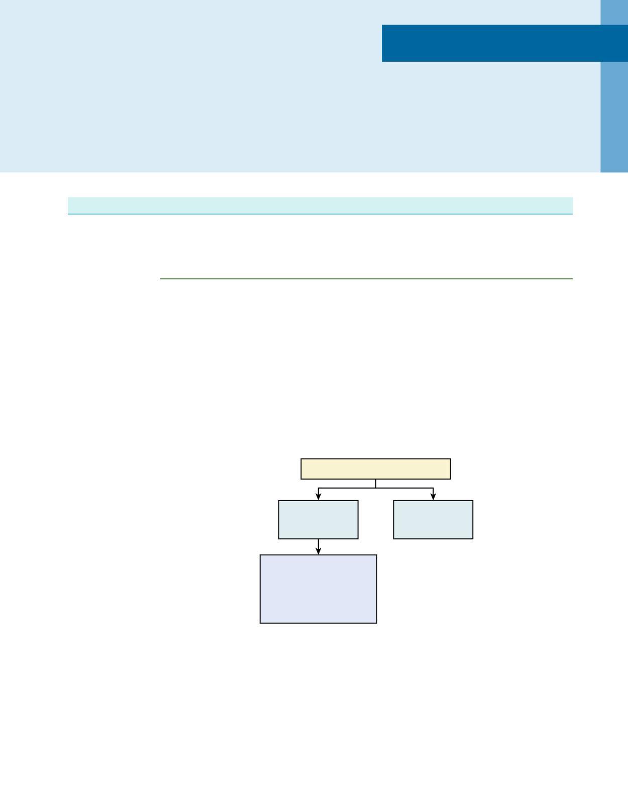
The pulmonary function test is used to diagnose and stage restrictive (caused by extrathoracicor intrathoracic problem) or obstructive lung disease. Restrictive lung diseases cause problems that impair lung expansion, which lead to decreased lung volume (e.g., obesity, interstitial lung disease). On the other hand, in obstructive lung disease, lung volume is usually preserved, but there is an impairment to air Àow, potentially caused by bronchospasm or other airway obstruction.
Interpretationoftheresultsofpulmonaryfunctiontest
Restrictivepattern: ↓FEV1, ↓FVC
Extrathoracic restrictive (obesity,kyphosis): ↓TLC, ↓DLCO
Arterial Blood Gas Analysis
Intrathoracic restrictive (interstitiallung disease): ↓TLC, ↓↓DLCO
Obstructivepattern: ↓↓FEV1
Emphysema: ↓DLCO, ↑orNTLC
Asthma: ↓DLCO
Acid-base disorder diagnostic algorithm
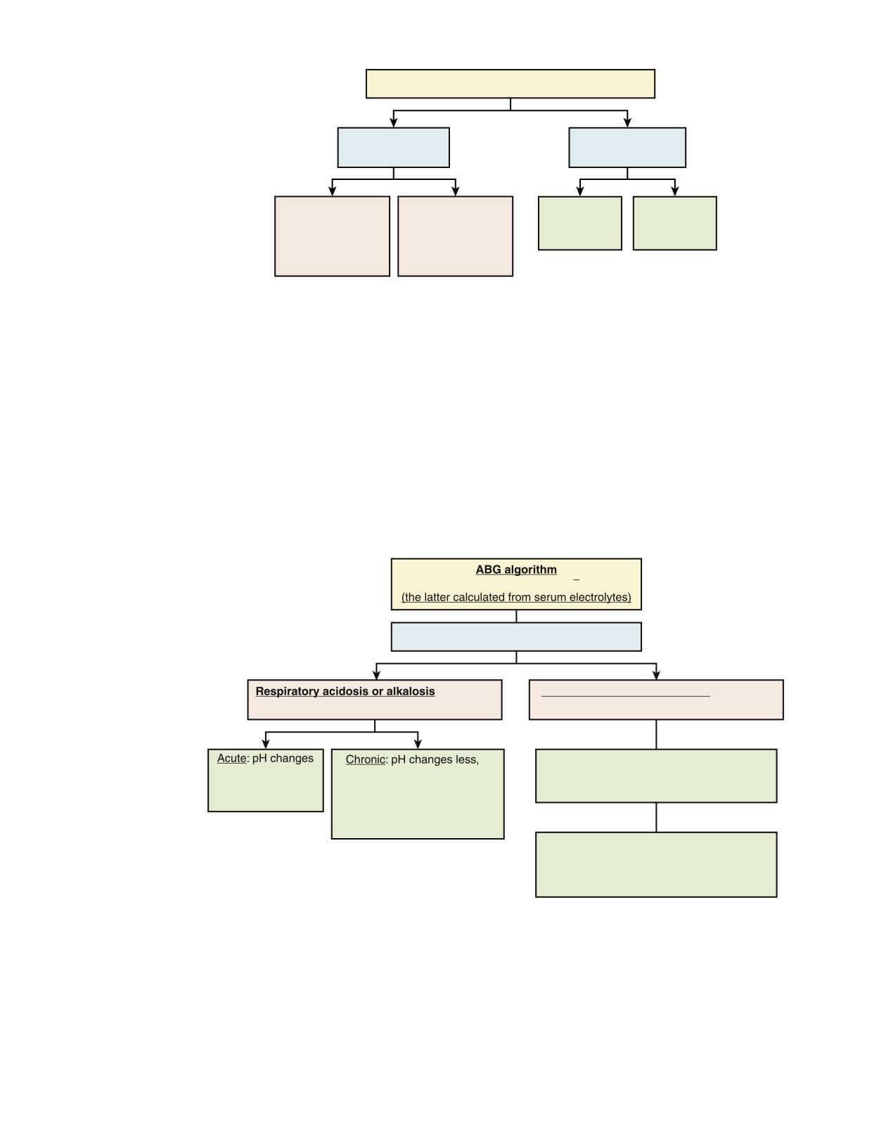
The following diagram provides the algorithm of interpretation of arterial blood gases (ABGs) used in conjunction with plasma chemistry. To use this algorithm, ¿rst examine the pH and identify acidemia or alkalemia, then using the bicarbonate concentration from the serum electrolytes and pCO2, identify whether the primary cause of the disorder is metabolic or respiratory. Finally, perform a calculation to examine if secondary metabolic compensation for a primary respiratory disorder or respiratory compensation for a primary metabolic disorder is appropriate If not, there is a second primary disorder, considered to be a “complex” (meaning more than one) acid-base disorder, rather than a “simple” (meaning single) acid-base disorder underlying the observed changes
Usesfourvalues:pH,pCO2,HCO3 ,aniongap
pH—acidemiaoralkalemia
(pHandpCO2changeintheoppositedirection)
0.08foreach10 mmHgchangein pCO2initially
onlyabout0.04foreach10 mmHgchangeinpCO2due tosecondarymetabolic compensationtakingplace overabout24hours
Metabolicacidosisoralkalosis (pHandpCO2changeinthesamedirection)
Nodifferencebetweenacuteandchronic disorderssincesecondaryrespiratory compensationoccursimmediately
However,metabolicacidosismayoccur withanormalaniongaporwithanincreased aniongap,animportantdistinctionpointing todifferentetiologies
Please note that more extensive discussion on acid-base disorders is available in the Chapter 5 of this book.
Pleural effusion
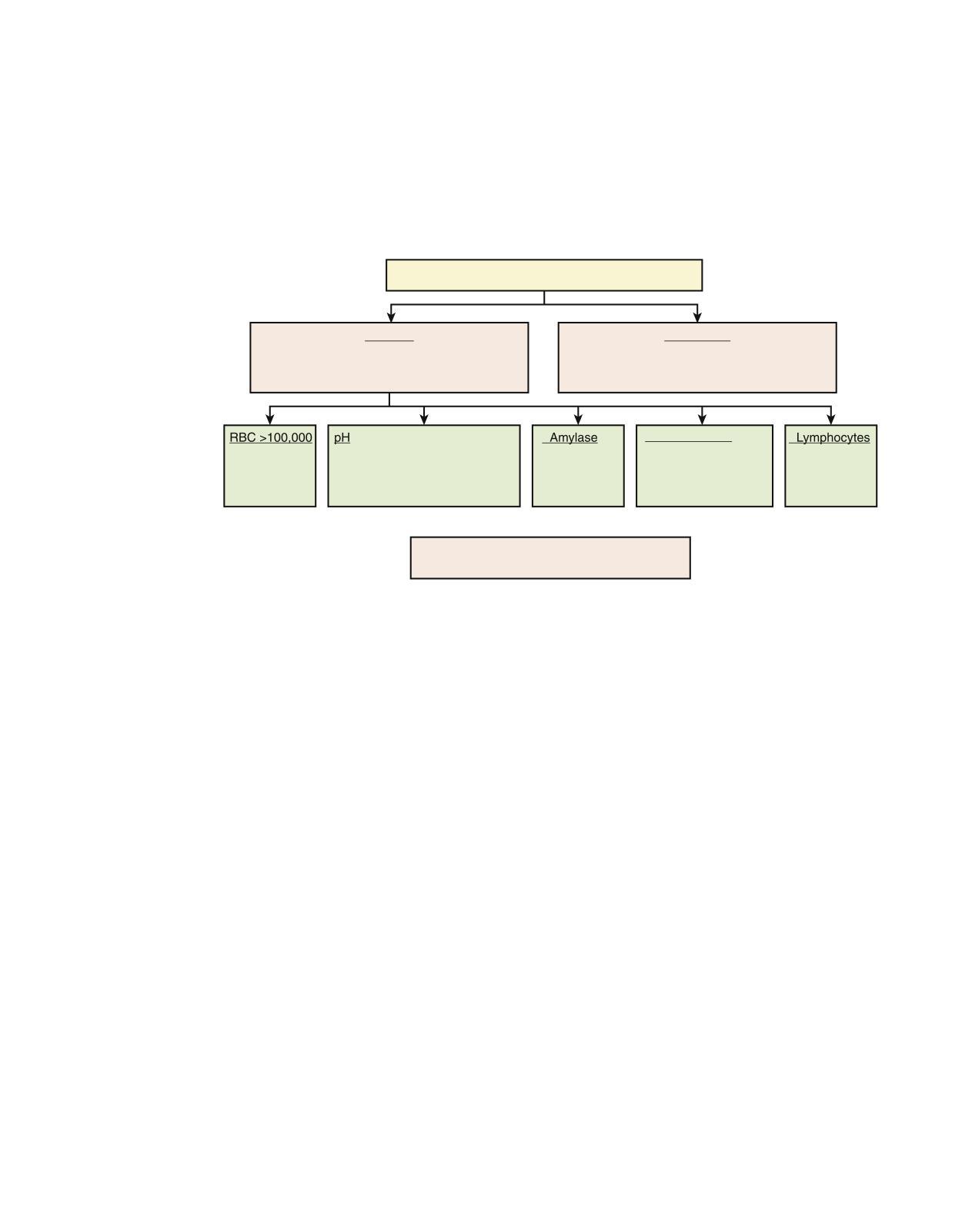
The normal amount of pleural Àuid is about 10 mL. Pleural eusion might be formed due to several potential causes: increased Àuid formation (increased amount of interstitial Àuid in the lungs, increased intravascular pressure in the pleura, decreased pleural pressure, increased permeability of the pleura, increased pleural protein level, increased amount of peritoneal Àuid disruption of blood vessels or lymphatics in the thorax) or decreased Àuid absorption (obstruction of the lymphatics draining pleural Àuid, disruption of the aquaporin system in the pleura, elevated systemic vascular pressure). The ¿rst diagnostic question of pleural Àuid analysis is if it represents a transudate or an exudate.2
Pleuraleffusion
Exudate
(WBC>1000,LDHfluid-to-plasmaratio>0.6, proteinfluid-to-plasmaratio>0.5)
yema
ancreatitis
Transudate
• Nephroticsyndrome
•CHF
•Cirrhosis
Glucose(<60) •Empyema •Malignant • TB •Rheumatoidarthritis
Diagnosticthoracentesisisindicatedifthickness ofpleuralfluidondecubitusx-ray>10mm
Acute Respiratory Failure
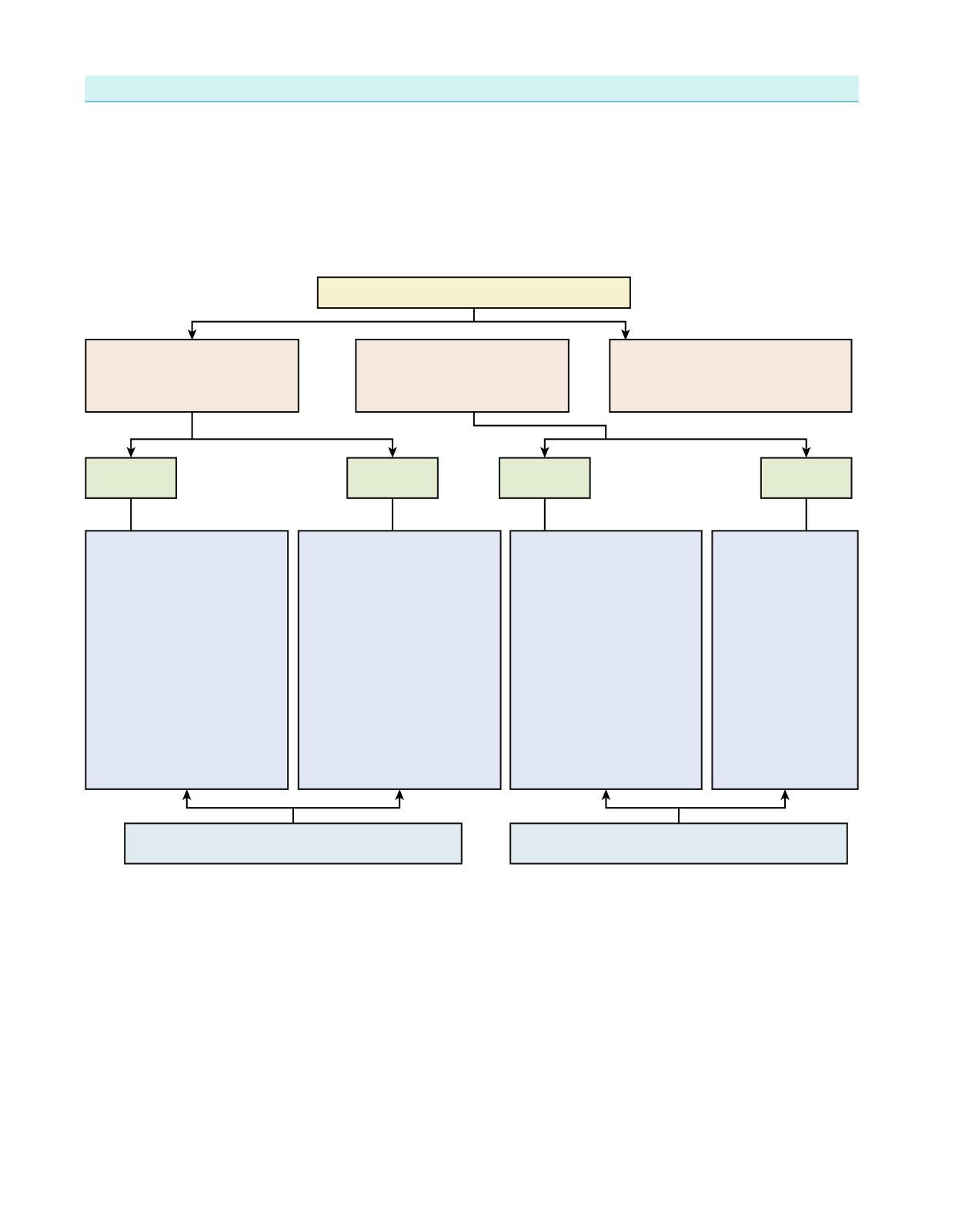
Acute respiratory failure is one of the most common conditions that requires patient to be treated in Intensive Care Unit (ICU). Unlike many other life-threatening conditions requiring ICU admission, respiratory failure presents immediate risk and needs to be addressed promptly In a simpli¿ed format, respiration entails gas exchange with O2 being absorbed and CO2 excreted by the lungs. As a result, respiratory failure could be viewed either as a de¿ciency in oxygenation or as a failure to excrete CO2 Some look at respiratory failure in sepsis as a separate entity, whereas others classify it within either hypoxemic or ventilatory failure. The next chart is a general algorithm describing types of respiratory failure and their mechanisms.3 We provide more details about speci¿c conditions below
Quickoverviewoftypesofrespiratoryfailure
Type1. Oxygenationfailure: hypoxicpattern
↓pO2(<60), ↓SaO2(<90%)
Type2. Ventilatoryfailure: hypercapnicpattern(CO2retention)
↑pCO2(>45-55), ↓pH(<7.35)
Shock:highventilatorydemand, increasedworkofbreathing,hyperpnea, tachypnea,decreasedrespiratorymuscle perfusion,diaphragmfatigue
• Hypoventilation(opiate overdose,COPD,neuromuscular disease,chestwallrigidity,upper airwayobstruction)
• Alveolarhypoxia(lowinspired O2 highaltitude,inhalationof toxicgases,scubadivingmishap, combustionwithinaclosespace)
• Increasedextraction(e.g.,inlow cardiacoutputstate,anemia, whichproduceslowmixed venousoxygenation)
• R-to-Lshunt(anatomic:AV malformation,intracardiac right-to-leftshunt;physiologic shuntthathastodowithalveolar filling:pulmonaryedema,severe ARDS;increasedflowinthe alveolarcapillaries)
• Ventilation-perfusionmismatch (flowobstruction-COPD, asthma;vascularobstruction [pulmonaryembolism]), pneumonia,ARDS,cardiogenic pulmonaryedema, hepatopulmonarysyndrome
• Interstitialinflammationwith ↓diffusion(pneumonia,ARDS, sarcoidosis)
• ↑CO2production(fever,sepsis, seizures,increasedcarbohydrateload,malignant hyperthermia)
• ↓Minuteventilation
• Decreasedrespiratorydrive (drugoverdoses)
• Neuromuscularweakness (CNSdisorder,peripheral nervedisease,metabolic/ electrolyteabnormalities, severefatigue)
• Upperairwayobstruction, dynamicairwayobstruction4
• ↑Deadspace(intrinsic lungdisease-COPD, asthma,cysticfibrosis, pulmonaryfibrosis, emphysema,ARDS, pulmonaryembolism
• Chestwalldisorders (scoliosis,trauma, massiveascites,or pleuraleffusion)
Ventilatory Failure
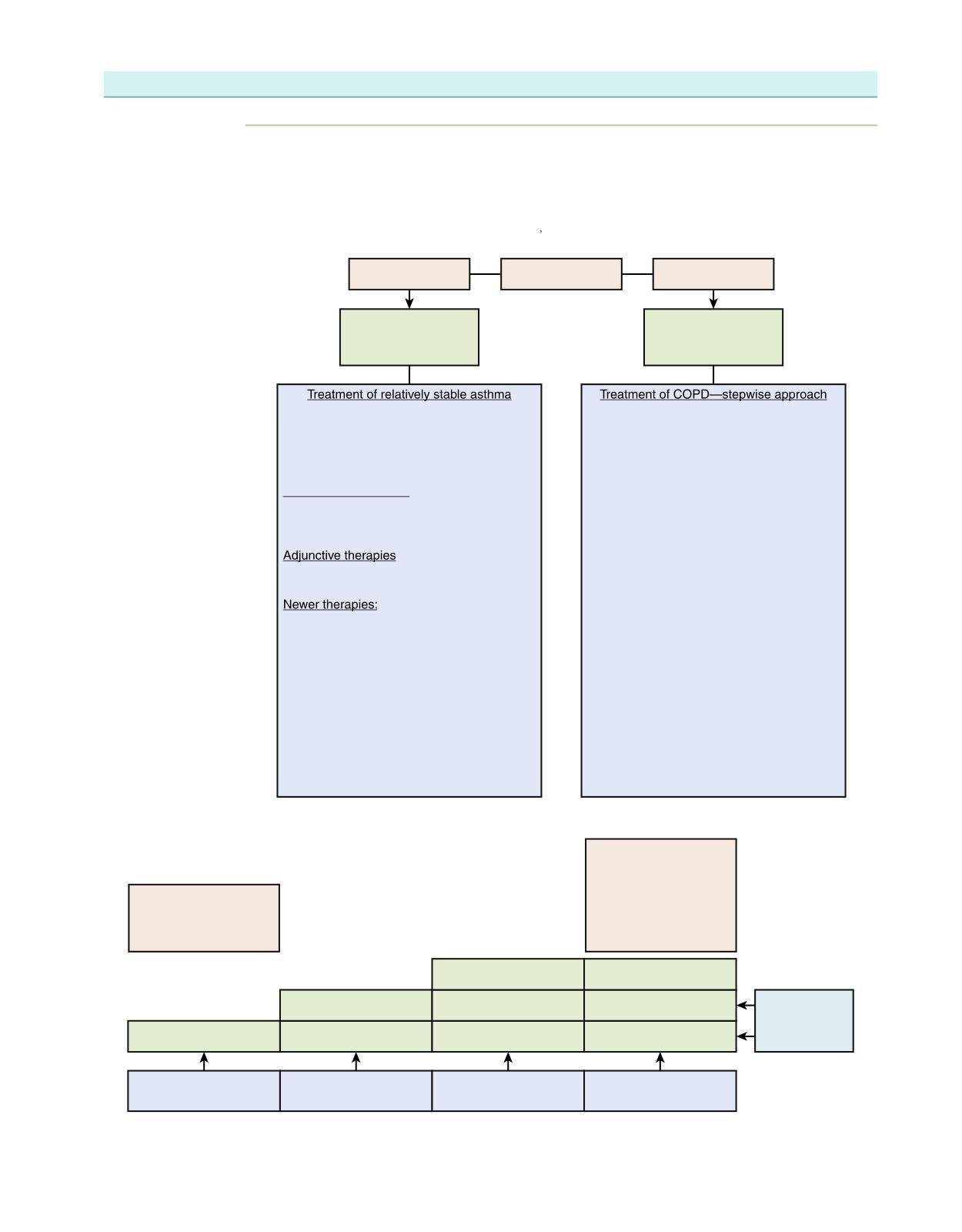
Asthma and Chronic Obstructive Pulmonary Disease
Although pathophysiologies of asthma and chronic obstructive pulmonary disease (COPD) are dierent, the end result leading to ventilator failure is similar and is based on hypoventilation. Therefore whereas approaches to treatment of noncritical stable asthma and COPD might be dierent, once it reaches the stage of respiratory failure, the focus in both conditions is to relieve bronchospasm and provide adequate ventilation. However, one has to be cautious about gas trapping which can precipitate hemodynamic instability and barotrauma.5 6
Asthma Vs COPD
Nolungdestruction, idiopathicattacks, airwayhypersensitivity
IndicationsforhomeO2
• Ht ≥55
• pO2 ≤55
• O2sat ≤85
Dyspneaepisodes: <1h duration,
Long-termsuppressionofairwayinflammation andreliefofsymptomswithquick-acting bronchodilators(primarilyaerosolized beta-agonists)
Inhaledcorticosteroids
Themosteffectiveagentsavailableforthe symptomaticcontrolofasthmaand improvementinpulmonaryfunction
Long-actingbeta-agonists,theophylline,and leukotrieneantagonists
• DiminishingtheproductionofIgEthrough effectsoninterleukin-4oronIgEitself: solublerecombinantIL-4receptor, recombinanthumanizedmonoclonal antibodythatformscomplexeswithfree IgE(rhuMAB,-E25'oromalizumab),blocks theinteractionofIgEwithmastcellsand basophils
• Conventionalallergenimmunotherapy
• DNAvaccinesandothermolecularmethods ofdown-regulatingantigen-specific Th2-mediatedresponses
Underlyinglung disease
Seefigurebelow
POprednisonefor2 weeks,then:
• inhaledsteroids
• alternativedays prednisone
• minimaleffectivedaily doseofprednisone
Antocholinergics Antocholinergics Metered dose inhalers Theophylline Theophylline
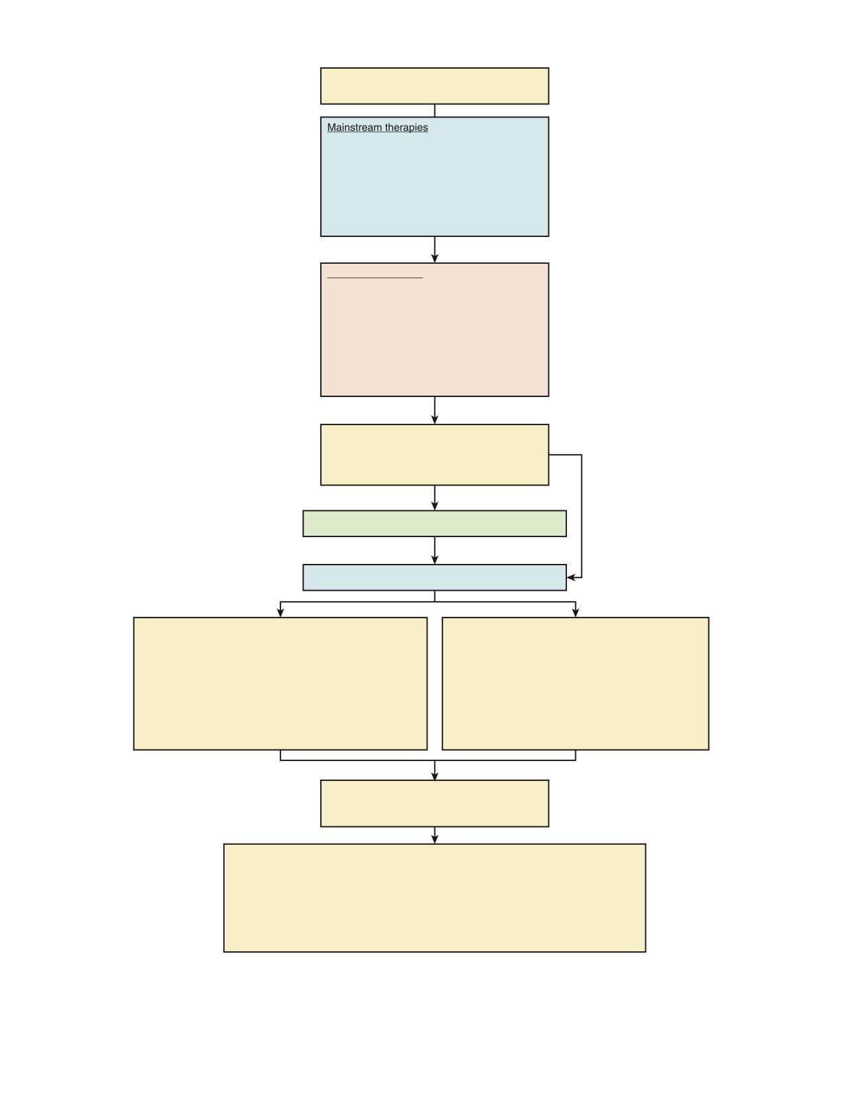
Treatmentofexacerbationsand ventilatoryfailureinasthma
• Beta-2-adrenergicagonists
• Corticosteroids(systemicorinhaled)
• Anticholinergicagents
• Magnesium
• Aminophylline
• Systemiccatecholamines
• Theophylline
• Leukotrieneantagonists
Ifnotbetter
Alternativetherapies
• Heliox
• Ketamine
• Glucagon
• Leukotrieneinhibitors
• Nebulizedclonidine
• Nitroglycerin
• Nebulizedcalciumchannelblockers
• Nebulizedlidocaine
• Externalchestcompression
ContraindicationstoNIPPV? alteredconsciousness,hemodynamicinstability, excessivesecretions,patientis uncooperative,highriskofaspiration
Ifnotbetter–considerNIPPV No
NIPPVstartingat10/5andtitrateupasneeded
Noimprovement Yes
Indicationsforintubation
Clinical
• Cardiacarrest
• Respiratoryarrest/impendingarrestorprofoundbradypnea
• Tachypneaof >40/min
• Alteredsensorium(lethargyoragitation)interfering withO2delivery
• Progressiveexhaustion,fatigue
• Silentchest
• Complicatedbarotrauma
• Unresolvingacidosis
Laboratory
• Severehypoxemiadespitemaximaloxygendelivery (pO2 <60mmHgon100%O2mask)
• Worseningrespiratoryacidosisorfailuretoreverse severerespiratoryacidosisdespiteintensivetherapy Ofnote:itisnothypercapniabutrespiratoryacidosis thattriggerstheintubation.
• ABGcriteria:pH <7.2,pCO2increasing (or>65mmHg) withabnormalpH
Yes
Intubate,mightstartwithvolumecontrol ventilation,setlowrespiratoryrate,paralyze forairtrappingandincreasedpressures
Initialventilatorsettingforasthmaticpatient
• Controlledmechanicalventilationat10breaths/min
• Tidalvolumeat7–8mL/kgofidealbodyweight
• Peakinspiratoryflowat60L/min(constantflow)or80–90L/min(deceleratingflow)
• Fractionofinspiredoxygenat1.0
• Auto-PEEPandPplatshouldbefollowedduringmechanicalventilation
• Hypercapniaispreferabletohyperinflation(notinthecontextofincreasedintracranial pressure).Acceptablehypercapnia:pHaslowas7.15andaPaCO2ofupto80mmHg
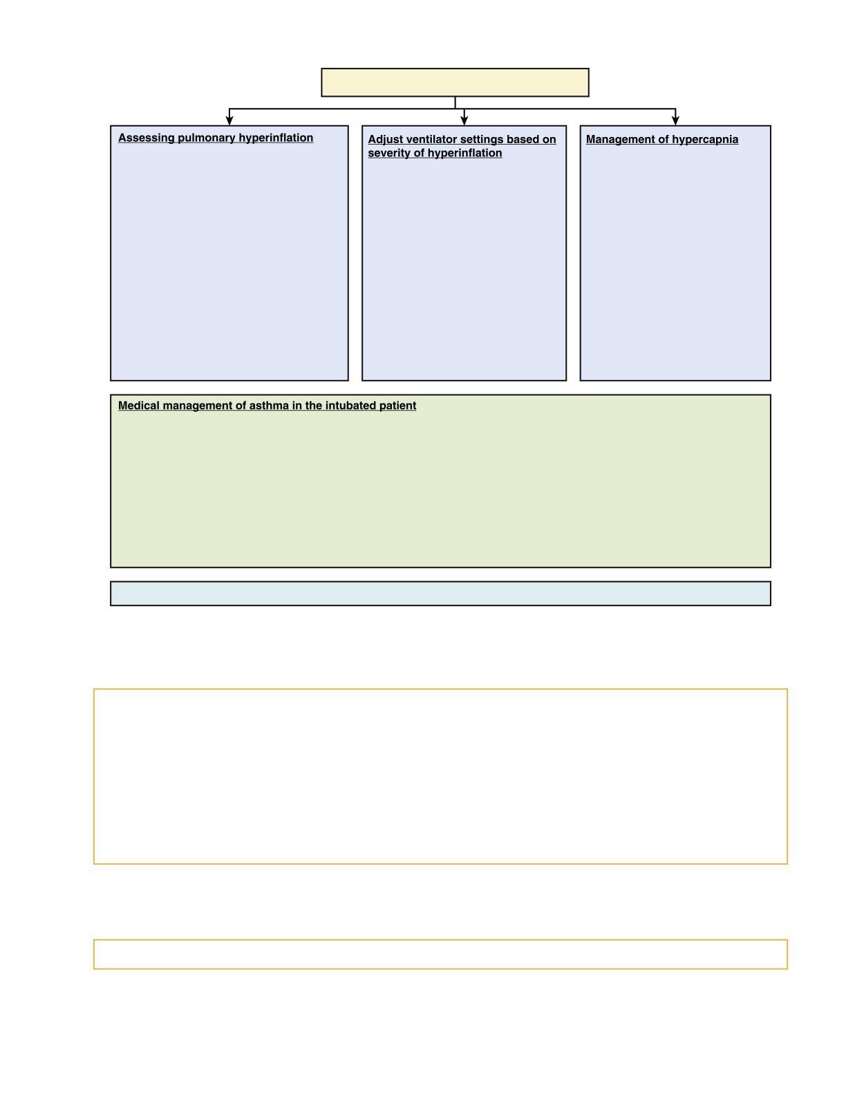
Issueswithmanagingintubatedpatientwithasthma
• Volumeofgasexhaledduring prolongedapnea(lungvolumeat inspirationVEI).VEIisaffectedby severityofairflowobstructionand ventilatorsettings.VEIisthemost reliablepredictorofventilator-related complications.
• Plateauairwaypressure(Pplat;in acutesevereasthmaaverage24–26cm H2O,acceptableupto30)
• Auto-PEEP(10–15cmH2Oinsevere asthma)duringvolume-cycledventilation.
• Peakairwaypressure(Ppk),target <50cm H2O. Ppkdependsoninspiratory flow-resistivepropertiesinadditionto hyperinflation. Ppk >50cmH2Odoes notpredictincreasedriskofbarotrauma.
• Minuteventilation.Increasedminute ventilationincreasestheriskof hypotensionandbarotrauma(when increasedfrom10to16to26L/min).
• MinimalPEEP ≤5cmH2)is recommended
• Consequenceofdeadspace ventilation(causedbyalveolar overdistension)
• Seriousconsequencesof hypercapniaareuncommon
• Neuro:increasedcerebralblood flow,intracranialpressure → cerebraledemaandsubarachnoid hemorrhage.
• Cardiac:decreasedintracellular pH → reducedcontractility
• Consideralkalinizingagentwhen pHispersistentlybelow7.15−7.2 (Nabicarbonateortromethamine).
• Systemiccorticosteroids:anti-inflammatoryeffect(2.5mg/kg/dayofmethylprednisolone)
• Inhaledbeta-agonists(MDIornebulizer):albuterol2.5mgQ4orQ6,ipratropium
• Otherbronchodilators(IVtheophylline)
• Deepsedation:combinationofpropofol(orbenzodiazepine)andfentanyl
• Neuromuscularblockingagent issometimesnecessary(intermittentbolusesratherthancontinuousinfusion) Additionalmeasures(notsupportedbystrongevidence):
• Heliox(amixtureofheliumandoxygen)
• Inhalationalanesthetics(isoflurane)–shouldtheeffectrightaway,andifnotthendiscontinue
• KetamineIV
• Bronchoscopicremovalofimpactedmucus
• Extracorporeallifesupport(membraneoxygenationandCO2removal)
Seemechanicalventilationsectionfordetailsonmanagingintubatedandventilatedpatient
Medical Management of Asthma in the Intubated Patient
• Systemic corticosteroids: antiinammatory effect (2.5 mg/kg per day of methylprednisolone)
• Inhaled beta-agonists (MDI or nebulizer): albuterol 2.5 mg Q4 or Q6, ipratropium
• Other bronchodilators (IV theophylline)
• Deep sedation: combination of propofol (or benzodiazepine) and fentanyl
• Neuromuscular blocking agent is sometimes necessary (intermittent boluses rather than continuous infusion)
MDI, Metered dose inhaler
Additional Measures (not Supported by Strong Evidence)
• Heliox (a mixture of helium and oxygen)
• Inhalational anesthetics (isourane)—the effect should be right away; if not, then discontinue
• Ketamine IV
• Bronchoscopic removal of impacted mucus
• Extracorporeal life support (membrane oxygenation and CO2 removal)
See Mechanical Ventilation section for details on managing intubated and ventilated patient
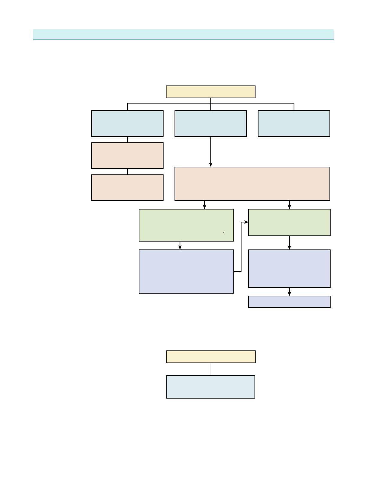
Hypoxemic Respiratory Failure
A number of mechanisms can lead to hypoxemic respiratory failure, resulting either from oxygen delivery problems (acute respiratory distress syndrome [ARDS], pneumonia, pulmonary edema, high altitude) or lung perfusion problems (pulmonary embolism, shunting).
Below is the general approach to treatment of hypoxemic respiratory failure; we also discuss special cases (ARDS, pulmonary embolism) in more detail.
Oxygenationfailure(hypoxemia)
Cardiogenicpulmonaryedema
NIPPV,startwith12/8−10, titrateupPEEP
Ifpersistenthypoxemia, worseningfatigue—intubate andventilate
PneumoniaorARDS Impairedgasexchangeand V-Qmismatch,(decreased ventilation)
V-Qmismatch, e.g.,pulmonaryembolism (decreasedperfusion)
Decisionpoint:intubation/IPPVvs.NIPPVbasedonclinical impression,ifpatientisquicklydeteriorating,lethargic,clearly infected,produceslotsofsecretionandotherwisehaveunfavorable clinicalindicators—donotdelayintubationandIPPV. Thereisno provenevidenceforclinicalbenefitofNIPPVinARDSorpneumonia.
NIPPV,titrateupPEEP
Althoughthereisnoevidenceofbenefit ofNIPPVinARDSandpneumonia, andsuggestionsofworseoutcomes7 somestilladvocateit.
Noimprovementorclinical deterioration;specificallyafter 2hoursofNIPPV: PEEP>10
FiO2>0.6
PaO2<100
PaO2/FiO2(P/Fratio)<200
Intubate,lungprotectivestrategies, titratePEEP,FiO2 PEEPismorephysiologicthan FiO2asit“recruits”lung
IfARDS–increasePEEP Ifrefratoryhypoxemia–tryother stategies:intrapulmonary percussiveventilation,airway pressurereleaseventilation, paralysis,proning,NO
Ifnoimprovement—ECMO
Other than ventilator failure and hypoxemic respiratory failure, some separate respiratory failure in sepsis into a separate entity, whereas in fact it is for the most part a multifactorial combination. Intubation and invasive positive pressure ventilation (IPPV) is the treatment of choice for the respiratory failure in sepsis.
Respiratoryfailureinsepsis
Intubate,IPPV,volume-control ventilation,RR15−18/min
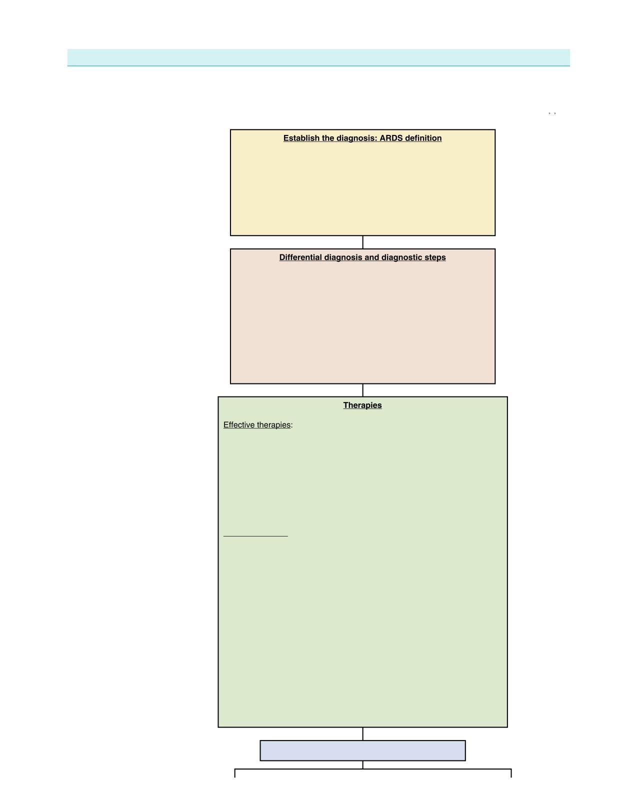
ARDS
ARDS is characterized by increased permeability of the alveolar capillary membrane, diuse alveolar damage, and accumulation of proteinaceous alveolar edema. Mortality remains very high (>40%) and does not seem to decrease between 1994 and 2006.8 That, in addition to high incidence and relatively limited therapeutic options, makes ARDS a serious and mostly unresolved issue in critical care.1 3 9–12
1.Acuteonset,presenceofincitingevent
2.PaO2/FiO2 ≤200(regardlessofPEEPlevel)
3.Bilateralinfiltratesseenonfrontalchestradiograph
4.PCWP ≤18mmHgornoclinicalevidenceofleftatrialhypertension
ClassificationbyPaO2/FiO2ratio
• Acutelunginjury(ALI)ormildARDS:<300
• Moderate >100−200
• Severe ≤100
• Ruleoutothersimilarpresentations:interstitiallungdisease, malignancypresentingsimilartoARDS,acuteeosinophilic pneumonia,diffusealveolarhemorrhage,hypersensitivity pneumonitis,andpulmonaryalveolarproteinosis
• ConsiderBAL:identifyinfectiouscauses(e.g.,bacterialorviral)
• Considerlungbiopsyif
o highclinicalsuspicionfora“contributiveresult”(resultsleadingto additionaltherapy)13
o theriskofempiricaltherapyistoohigh
o whenempiricaltherapyhasbeenunsuccessful14
o notethatlungbiopsypresentssubstantialriskinventilatedpatient onhighPEEPandbenefitsmustclearlyoutweighrisks
• Lungprotectiveventilation(lowertidalvolumeandairwaypressures) (seemechanicalventilationboxbelow)
• Neuromuscularblockade15,16
cisatracuriumshouldbeconsideredforshort-termuse(<48h)in patientswithsevereARDS(definedasPaO2/FiO2<120mmHg) untilfurtherstudiesareavailable17
• EsophagealpressuretoadjustPEEP(improvesoxygenation)
• Fluidconservativevsfluid-liberaltherapy(seefluidmanagementboxbelow)
• Extracorporealmembraneoxygenaton(seeECMOsectionfordetails)
Noprovenbenefit:
• HighPEEP
• Highfrequencyventilation
• Earlypronepositioning
• Continuousadministrationofsurfactanthasnoeffecton30-day survival,durationofmechanicalventilation,orphysiologicfunction
• ActivatedproteinC(APC)
• GM-CSF
• Pulmonaryarterycatheter
• Methylprednisolone/steroids(questionablebenefit). Somerecommend 7−14-daytrialof2−4mg/kgprednisoneinpatientswithsevereARDSwho shownoclinicalsignsofimprovement. Ruleoutortreatsystemicinfections.
• Omega-3fattyacid(maybeharmful)
• Beta-2agonists
• Antioxidants
• Vasodilatortherapy(liposomalprostoglandinE1,nitricoxide). Liposomal PGE1blocksplateletaggregation,downregulatesneutrophil-mediated inflammation,producevasodilatation.
• Ketoconazoleinhibitstromboxanesynthesisandbiosynthesisofleukotriens
• N-acetylcysteine