Table of Contents
Cover
Title page
Copyright
Dedication
Contributors
Preface
Chapter 1: Sensitive methods for detection of SARS-CoV-2 RNA
Abstract
1: Introduction
2: General approaches for the detection of SARS-CoV-2
3: Isothermal amplification methods for sensitive detection of SARS-CoV-2
4: Sensitive detection of SARS-CoV-2 via RT-RPA
5: General considerations for designing an ultrasensitive RTRPA assay
6: Comparative reviews of recently published RT-RPA assays for SARS-CoV-2 detection
7: Methods section
8: Before you begin
9: Key resources table
10: Materials and equipment
11: Step-by-step method details
12: Summary Acknowledgement
References
Chapter 2: The seasonal behaviour of COVID-19 and its galectin-like culprit of the viral spike
Abstract
1: Introduction
2: The seasonal behaviour of viruses
3: The seasonal behaviour of COVID-19
4: Viral genomic make up and seasonality
5: Conclusions and prospects
References
Further reading
Chapter 3: Current molecular diagnostics assays for SARS-CoV-2 and emerging variants
Abstract
1: Introduction
2: SARS-CoV-2 variants of concern
3: COVID-19 diagnostics
4: Diagnostics in the era of COVID-19 vaccination
5: Conclusion References
Further reading
Chapter 4: CRISPR use in diagnosis and therapy for COVID-19
Abstract
1: Introduction
2: Diagnostics and therapeutics for SARS-CoV-2
3: CRISPR/Cas systems
4: CRISPR-based therapeutics for SARS-Cov-2
5: Delivery of CRISPR/Cas components
6: Limitations of CRISPR/Cas system
7: Summary
References
Chapter 5: Recent and advanced nano-technological strategies for COVID-19 vaccine development
Abstract
1: Introduction
2: The structure and infection mechanism of SARS-COV-2
3: Pathogenesis and clinical presentation of COVID-19
4: Vaccine development strategies and platforms
5: Relevant SARS-CoV-2 antigen explored in the design of vaccines
6: Nano-based strategies for COVID-19 vaccine development
7: Benefits and challenges of nanotechnology in COVID-19 vaccine development
8: Conclusion and future perspectives
References
Chapter 6: A review of hypersensitivity methods to detect immune responses to SARS-CoV-2
Abstract
1: Historical perspective
2: General overview, classification and description of hypersensitivity reactions
3: Skin test application of hypersensitivity reactions: In vivo measurements of immune responses
4: In vitro methods to measure immune responses after SARSCov-2 infection
5: A novel application of a DTH method to measure immune responses after SARS-CoV-2 infection
6: DTH to measure immunogenicity elicited by covid vaccines
7: Future prospects of DTH to study SARS-CoV-2 immunogenicity
Acknowledgements
References
Chapter 7: Hesitancy to get vaccinated against COVID-19 and how it might be overcome
Abstract
References
Chapter 8: The emergence of SARS-CoV-2 variants of concern in Australia by haplotype coalescence reveals a continental link to COVID-19 seasonality
Abstract
1: Introduction
2: Methods
3: VOCs in Australia
4: Prevalence of amino acid variants in Australia
5: A network view of haplotype diversity and VOC emergence
6: Continental links to seasonality
7: Discussion
References
Chapter 9: COVID-19 vaccines for high risk and immunocompromised patients
Abstract
1: Introduction
2: Development of COVID-19 vaccines
3: The use of COVID-19 vaccines for the high-risk patient population with the emphasis on HIV-infected patients
References
Chapter 1: Sensitive methods for detection of SARS-CoV-2 RNA
Xi Chen*; Simin Xia The HIT Center for Life Sciences (HCLS), Harbin Institute of Technology, Harbin, Heilongjiang Province, People's Republic of China
* Corresponding author: email address: chenxihit@hit
edu cn
Abstract
The occurrence of the COVID-19 pandemic caused by the SARS-CoV-2 virus since the end of 2019 has significantly affected the entire world. Now SARS-CoV-2 diagnostic tests are not only required for screening of suspected infected people for their medical treatment, but have also become a routine diagnosis for all people at a place where new cases have emerged in order to control spread of the disease from that region For these reasons, sensitive methods for detection of SARS-CoV-2 are highly needed in order to avoid undetected infections In addition, sample pooling that uses pooled specimens has been routinely employed as a time- and cost-effective strategy for community monitoring of SARS-CoV-2. In this regard, the content of each viral RNA sample of an individual will be further diluted in detection; therefore, higher detection sensitivity would be rather preferred. Among nucleic acid-based detection methods, isothermal nucleic acid amplifications are considered quite promising because they typically take less time to complete the test (even less than 20 min) without the need of thermal cycles Hence, it does not necessitate the use of highly costly real-time PCR machines According to recently published isothermal nucleic acid amplification methods, the reverse transcription recombinase polymerase amplification (RTRPA) approach shows outstanding sensitivity with up to single-copy sensitivity in a test reaction. This chapter will mainly focus on how to employ RT-RPA technology to sensitively detect SARSCoV-2 RNA Besides, recently published RT-RPA based detection methods will be summarized and compared regarding their detection parameters and the primers and probes being used In addition, we will also highlight the key considerations on how to design an ultrasensitive RT-RPA assay and the precautions needed to conduct the assay Moreover, based on our recent report, we will also detail the methods we developed to detect SARS-CoV-2 RNA using modified RT-RPA, or RT-ERA, with single-copy sensitivity and the possible extensions beyond this method.
Keywords
RT-RPA; SARS-CoV-2 RNA; Exo probe; Nfo probe; Lateral flow; Single-copy sensitivity
1: Introduction
Since December 2019, an outbreak of COVID-19 caused by severe acute respiratory syndrome coronavirus 2 (SARS-CoV-2) virus occurred and soon spread to the entire world (Wang, Horby, Hayden, & Gao, 2020) The COVID-19 pandemic has led to a dramatic loss of human life worldwide and presents an unprecedented challenge to publish health and food systems; the global economic growth was largely slowed down and social activities were greatly disrupted As of July 1, 2021, COVID-19 has infected over 182 million people worldwide with over 3.9 million reported casualties. COVID-19 is caused by an RNA virus named severe acute respiratory syndrome coronavirus 2 (Chen et al., 2020).
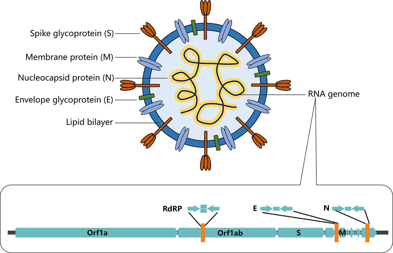
SARS-CoV-2 virus is a coronavirus belonging to the beta family of coronaviruses that also includes SARS-CoV, Middle East respiratory syndrome coronavirus (MERS-CoV), human coronavirus OC43 (HCoV-OC3), and human coronavirus HKU1 (HCoV-HKU1). These beta-viruses are enveloped, positive-sense, single-stranded RNA viruses of zoonotic origin. As for SARS-CoV-2, this virus contains four structural proteins, namely the envelope protein (E), spike protein (S), membrane protein (M), and the nucleocapsid (N) protein (Fig 1) The S, M, and E proteins form the envelope of the virus while the N protein is associated with the single-stranded RNA genome forming nucleocapsid inside the envelope.
FIG 1 Schematic illustration of the structure of coronavirus SARS-CoV-2 and its single-stranded RNA genome. The sequence information of the single-stranded RNA genome of SARS-CoV-2 serves as the basis for the development of nucleic acidbased diagnosis
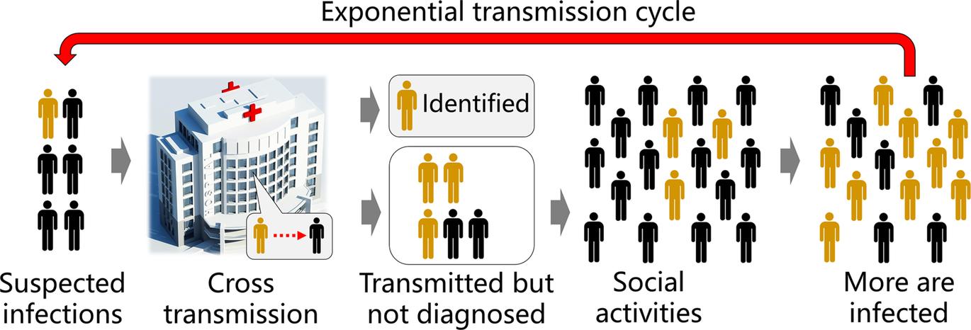
According to initial studies, this disease caused by SARS-CoV-2 showed a high transmissibility with a basic reproduction number R0 = 1.4–5.5 (WHO, 2020; Zhao et al., 2020), likely to be below 5 and above 3 (Chen, 2020) and therefore it is highly desired to perform early testing of SARS-CoV-2 for timely screening of infected individuals and to stop the spread of this disease from a location to its surroundings Because detection sensitivity is the key to reduce false negative results, a detection method with a high level of sensitivity can minimize non-diagnosed infected individuals and reduce the chance of further cross transmission (Fig 2)
FIG 2 Sensitive detection methods will be highly beneficial to prevent the spread of COVID-19 High sensitivity of diagnostic methods ensures low false negative results and therefore reduces the latent cross transmission of non-diagnosed but actually infected individuals to their closely contacted people
2: General approaches for the detection of SARS-CoV-2
There are several different types of diagnostic approaches used in the current pandemic, which can be mainly classified into the following three categories: (i) computed tomography (CT) chest scan, (ii) antigen-antibody interaction-based serological tests, and (iii) nucleic acid (NA)-based tests (Kilic, Weissleder, & Lee, 2020; Qin, Peng, Baravik, & Liu, 2020) The CT chest scan (Ai et al , 2020) detects the pathological change of the respiratory system including the lung by visualization of the transmission of the X-ray through the chest. Typically, a brighter image that reveals a low transmission of the X-ray through the chest could potentially indicate the infection by SARS-CoV-2. However, the specificity of CT chest scan is low because the symptomatic features of COVID-19 CT scan are similar to those of other types of viral pneumonia; and hence this method is generally considered as an auxiliary method for SARS-CoV-2 diagnosis Serological testing is based on the detection of antibodies generated by the immune system which can be used to confirm the infection of SARS-CoV-2 (Amanat et al., 2020; Udugama et al., 2020). However, it typically takes 1–2 weeks before the body can generate detectable antibodies for serological testing; as a result, this approach is not well suited for early-stage diagnosis. Other issues associated with serological tests are the high variability and sometimes low sensitivity and specificity (Tang et al , 2020) Nucleic acid-based detection is the detection of the RNA genomic materials of the SARS-CoV-2, usually using nucleic acid amplification approaches, which has been considered as the gold standard for SARS-CoV-2 viral detection
Reverse transcription-polymerase chain amplification (RT-PCR) has been widely used for the detection of viral RNA for many years. RT-PCR requires a first reverse transcription to produce a cDNA from the viral RNA, and then amplification of cDNA via polymerase chain reaction. During amplification, a fluorescent probe, either a fluorogenic dye (e g SYBR safe) or a rationally designed probe (e g TagMan probe) is included in the PCR reaction which will respond to the amplified DNA to produce fluorescent signals As a result, the amplification could be detected in real-time via fluorescence using a real-time quantitative PCR machine A drawback of RT-PCR diagnosis is the high cost of the PCR machine and the expertise required to design the program and to conduct the analysis. In fact, RTPCR is among the first reported and approved SARS-CoV-2 detection methods (Chu et al., 2020; Corman et al., 2020). At the earliest time after the COVID-19 outbreak, Drosten et al. reported the detection of SARS-CoV-2 by real-time RT-PCR method The PCR primers and probes were designed for the detection of RdRp gene, E-gene and N-gene Although single-copy sensitivity was not achieved, this RT-PCR assay was found to be still quite sensitive with 5 2 copies per reaction at 95% detection probability for E-gene and 3.8 copies per reaction at 95% detection probability for RdRp gene using nonclinical samples; the sensitivity for the N-gene is less and hence was not subjected to intensive evaluation (Corman et al., 2020). Another RT-PCR based approach for the molecular diagnosis of SARS-
CoV-2 introduced in the very beginning of the pandemic detects the ORF1b and N-gene of SARS-CoV-2 with a sensitivity up to ≤ 10 copies per reaction using none-RNA testing sample (Chu et al., 2020).
3: Isothermal amplification methods for sensitive detection of SARS-CoV-2
Isothermal nucleic acid amplification approaches are emerging as new promising methods for detection of viral RNA (James & Alawneh, 2020; Zhao, Chen, Li, Wang, & Fan, 2015). In isothermal nucleic acid amplification, no thermal cycles are needed and therefore, exempted from using sophisticated thermocyclers The respective detection device is hence simpler, more portable and meet the requirement of so-called point of care (POC) testing (Dinnes et al , 2020; Dohla et al , 2020) Sometimes, lateral flow strips can also be used to facilitate the diagnosis so that the testing can be applied in a POC, or even home-based fashion Further, isothermal amplification approaches can achieve a very high sensitivity, with up to single-copy sensitivity per reaction. The ultrahigh sensitivity makes these approaches suitable for early-stage diagnosis of virus infections. Therefore, isothermal nucleic acid amplification has been considered as a promising method for the detection of virus infection and could compensate with the currently widely used RT-PCR methods
Indeed, as for SARS-CoV-2, isothermal amplification detection methods have already been considered as highly promising detection tools (Shen et al , 2020) Early development of isothermal amplification techniques include LAMP (loop-mediated isothermal amplification) (Notomi et al., 2000; Wong, Othman, Lau, Radu, & Chee, 2018), NASBA (nucleic acid sequence-based amplification) (Compton, 1991), HDA (helicase-dependent amplification) (Vincent, Xu, & Kong, 2004), EXPAR (exponential amplification reaction of nucleic acids), and SDA (strand-displacement amplification) (Walker et al , 1992) Among these isothermal detection methods, LAMP coupled with reverse transcription, i e RTLAMP, seems to be the most popular in SARS-CoV-2 analysis (Kashir & Yaqinuddin, 2020) In RTLAMP, four or six primers that target 6–8 regions in the genome are designed in combination with the use of Bsm DNA polymerase. Along with the proceeding LAMP reaction, pairs of primers generate a dumbbell-shaped DNA structure, which functions as the LAMP initiator In this method, around 109 DNA copies can be generated within an hour and the reaction takes place at constant temperature in the range of 60–65 °C. Since magnesium pyrophosphate is generated as a byproduct during LAMP, metalsensitive indicators or pH-sensitive dyes can be employed for visual detection. An advantage of the RTLAMP approach is that regular primers are used which do not necessitate the design of specially functionalized primers or probes; hence this feature is helpful to reduce the cost of a RT-LAMP reaction
On the other hand, LAMP, NASBA and HDA methods do not require thermocycle machines for amplification; however, specifically designed heating devices are still needed In addition, LAMP requires the use of 4–6 primers, which makes primer design more complicated than other methods Furthermore, LAMP, NASBA or HDA typically needs at least 60 min to complete the amplification reaction, which is longer than RT-RPA.
Recently some more contemporary isothermal amplification methods have been introduced, for example, NEAR, DETECR, STOP and so on NEAR (nicking enzyme amplification reaction) or nicking enzyme-assisted amplification (NEAA) uses not only strand-displacement DNA polymerase (e g Bst polymerase), but also nicking endonuclease enzymes to exponentially amplify short oligonucleotides (Wang et al., 2018). Thousands of copies of DNA fragment can be produced from only one restriction site making this approach a unique technique with rather high amplification efficiency. On the other hand, a drawback of NEAR is the formation of non-specific products which limits detection sensitivity. DETECTR (DNA endonuclease-targeted CRISPR trans reporter) method (Chen et al , 2018) is associated with CRISPR-based detection approaches that involves the use of the genome editing Cas12a enzymes DETECTR uses a crRNA-Cas12a complex to recognize amplified DNA targets and binding of the crRNA-Cas12a complex to target DNA induced discrimination cleavage of non-target FQ-DNA reporters. Similar CRISPR-based approaches include SHERLOCK detection (Gootenberg et al., 2017) in one-pot (STOP) that uses Cas13a enzyme.
4: Sensitive detection of SARS-CoV-2 via RT-RPA
Reverse transcription recombinase polymerase amplification (RT-RPA) is a widely recognized isothermal amplification assay for the amplification of RNA, which combines reverse transcription that converts RNA to cDNA, and recombinase polymerase amplification (RPA) that amplifies cDNA under isothermal conditions. RPA sometimes is also called ERA (enzymatic recombinase amplification) as a modified/improved version of RPA according to the commercially supplied kit from the company GenDx (h�p://gendx cn), or RAA (recombinase aided amplification) (Wu et al , 2020; Xue et al , 2020) RPA is a molecular technology introduced by Piepenburg, Williams, Stemple, and Armes (2006) using proteins involved in cellular DNA synthesis, recombination and repair (Piepenburg et al., 2006). The RPA process starts when a recombinase protein binds to primers in the presence of ATP and a crowding agent (e.g. a high molecular weight polyethylene glycol), forming a recombinase-primer complex (step I) The complex then interrogates double-stranded DNA seeking a homologous sequence and facilitates strand hybridization with the primer at the cognate site (step II & III) In the meanwhile, single-stranded binding protein (SSB) is added in the reaction mixture to stabilize the dissociated single-stranded DNA and prevent the ejection of the inserted primer by branch migration, and facilitate the amplification to proceed at room temperature. The DNA polymerase (e.g. Bsu) will bind to the 3′ end of the primer to elongate the primer in the presence of dNTPs (step IV & V). Cyclic repetition of this process results in the exponential amplification of a DNA (Fig. 3) (Piepenburg et al., 2006).
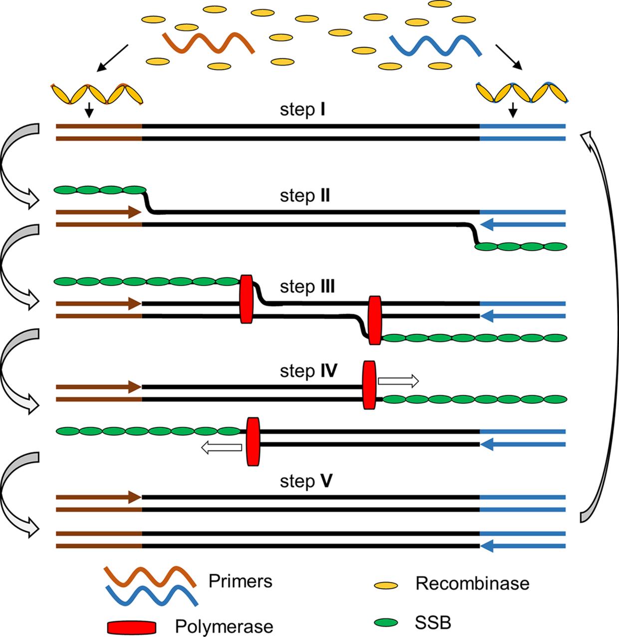
FIG. 3 The general principle of recombinase polymerase amplification. Step I: recombinase and primer form complexes and target homologous DNA; step II: strand exchange forms a D-loop; herein, the brownish and blue-coloured arrows refer to the forward and reverse primers, respectively, upon annealing with their templates; step III: polymerase initiates synthesis; step IV: parental strands separate & synthesis continues; herein, the hollow boxed arrows refer to the directions of the polymerization reaction; step V: two duplexes form. Abbreviation(s): SSB, singlestranded DNA binding protein
In order to employ RPA for the amplification of RNA, additional reverse transcriptase was added in order to convert RNA to cDNA for subsequent RPA amplification. In addition, since RNA is a much less stable species compared to DNA and is highly prone to degradation by the ubiquitously existing RNase, RNase inhibitor protein was added into the RT-RPA reaction In order to facilely detect the amplification product, a probe and a nuclease was used Moreover, creatine kinase, phosphocreatine and ATP were needed to generate energy for the reaction Therefore, a practical RT-RPA based detection reaction requires at least seven enzymes/proteins including: (i) strand-displacing DNA polymerase, (ii) recombinase, (iii) recombination-mediator protein (RMP), (iv) single-strand DNA binding protein, (v)
RNase inhibitor, (vi) creatine kinase, and (vii) nuclease; in addition, multiple necessary reagents are also required like dNTPs, creatine, the crowding agent polyethylene glycol, the activator Mg(OAc)2, the forward and reverse primers, and a probe. For the most reliable test, viral RNA samples need to be purified via a standard RNA purification process rather than using a non-purified viral sample. The list of proteins, enzymes, reagents necessary for conducting an RT-RPA reaction with their recommended concentrations are summarized in Table 1 (Chen et al , 2020) The concentrations of primers, probes, RNase inhibitor and nucleases used in our hand for exo-probe, nfo-probe and multiplexing RT-RPA reaction have also been given (Table 1)
Table 1
List of concentrations of primers, probes, nucleases, inhibitors and Mg(OAc)2 that need to be additionally supplied in a RT-RPA or RT-ERA reaction kit for viral RNA detection.
Components
Exo-probe detection Fw & Rw primers
Concentrations used in our study
500 nM
Exo probe 150 nM
Exonuclease III
Nfo-probe LFD Fw & Rw primers
Recommended ranges by other studies
420 nM, can be varied in the range of 150–600 nM
In the range of 50–150 nM
100 U in 50 µL –
400 nM
Nfo probe 120 nM
420 nM, can be varied in 150–600 nM range
In the range of 50–150 nM
Endonuclease IV 10 U in 50 µL –
Duplexing Rw & Fw Primers
100 nM
Exo probes 30 nM
Exonuclease III 100 U in 50 µL
Subjected to testing and optimization
Other necessary reagents RNase A inhibitor 5 U in 50 µL –
Mg(OAc)2 2 µL
Standard 14 mM, can be varied in the range of 12–30 mM
Note: Other components include (i) RPA enzymes (120 ng μL 1 T4 UvsX recombinase, 60 ng μL 1 T4 UvsY recombinationmediator protein, 600 ng μL 1 T4 gp32 single-stranded binding protein, 30 ng μL 1 Bsu or 8 6–12 8 μg Sau DNA stranddisplacing polymerase), (ii) energy-supply system constituents (50 mM phosphocreatine, 100 ng μL 1 creatine kinase, and 3 mM ATP), (iii) 200 μM each dNTPs, (iv) buffering constituents (typically pH 7 9 50 mM Tris/100 mM KOAc), (v) reducing agent 2 mM DTT, (vi) crowding reagent (5% carbowax 20M), and (vii) reverse transcriptase are generally included in the commercially available RT-RPA or RT-ERA kit with fixed concentration without the need for further adjustment (Li, Macdonald, & von Stetten, 2020)
For the detection of viral RNA using RT-RPA, multiple detecting formats are available. These formats include exo probe, nfo probe, fpg probe, digital (Shen et al., 2011), nesting, microfluidics (Lu� et al., 2010), solid phase, template generation, electrochemistry, colorimetric or SNP detection, and so on (Daher, Stewart, Boissinot, & Bergeron, 2016; Li et al , 2020) Among all these, it seems that using exo probe for fluorescence detection (Behrmann et al , 2020; Tu et al , 2020) and using nfo probe coupled with lateral flow strip detection (Zheng et al , 2021) are the most popular and hence we will mainly focus on these two formats for the detection of SARS-CoV-2 viral RNA by RT-RPA. Herein, either
approach has its own advantages. For the detection of RNA using exo probe, first forward and reverse primers that are around 30–35 nucleotides long are designed; the melting temperature of an oligonucleotide is normally not a critical factor for the performance as a primer. The primer pairs allow the amplification of a relatively short length DNA amplicon ideally around 100–200 bp long that is most suited for RPA amplification In addition to the primer pair, an exo probe was designed for the fluorogenic detection of the amplicon in the presence of exonuclease III (exo enzyme) The exo probe is a modified oligonucleotide featuring an abasic nucleotide analogue tetrahydrofuran residue (THF, sometimes referred to as “dSpacer”) that is prone to cleavage by exo enzyme when the nucleotide is in a double-stranded state. In addition, the exo probe is usually around 46–52 nucleotides long, with at least 30 nucleotides located 5′- to the THF site, and a further at least 15-bp long nucleotide located 3′- to the THF site A blocking group, such as a phosphate, a C3-spacer, a biotin-TEG or an amine, is situated at its 3′ side so that the exo probe cannot act as a primer and block the probe from polymerase extension The entire exo probe is able to specifically hybridize with one chain of the amplicon (THF needs to be counted as one nucleotide residue) In order to allow this probe to detect the amplicon in a fluorogenic way, exo probe features a flanking dT-fluorophore (e.g. fluorescein) on one side of the THF residue and a flanking corresponding dT-quencher group (typically a suitable Black Hole Quencher (BHQ)) on the other side of the THF site. In such a way, the dT-fluorophore is temporally quenched by its FRET quencher (e g BHQ1) and the probe will be in a none or weakly fluorescent state Once RPA reaction produces an amplicon that can hybridize with the exo probe, the exo probe is converted from singlestranded state to double-stranded state that allows the THF residue to be readily cleaved by the exo III enzyme. Hence the quencher moiety is separated from the probe and the fluorescence of the dTfluorophore is restored (Fig. 4). Exo probe allows sensitive detection in RT-RPA based on the enhancement of fluorescence.
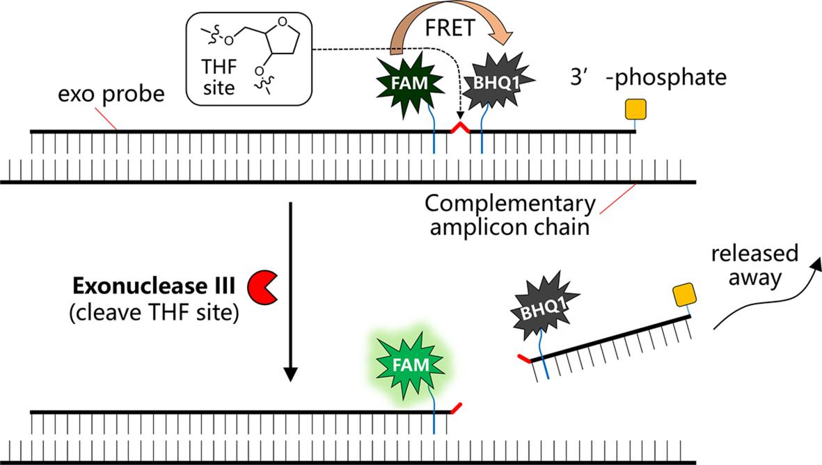
FIG 4 Schematic view of the working principle of a typical exo probe for RT-RPA detection of viral RNA
In order to make the sensitive detection approach more field-deployable with minimal instrumentation, nfo probe could be designed in combination with the use of lateral flow strips for direct visual detection In this regard, nfo probe can hybridize with the amplicon to give a bifunctional amplicon, typically with a biotin antigenic tag at one end and a FAM antigenic tag at the other end Similar to the exo probe, a nfo probe is around 46–52 nucleotides long, features an abasic THF residue in