Associate director of Breast Imaging, Brigham and Women’s Hospital
Associate Professor of Radiology, Harvard Medical School
Julie A. ritner, MD
Staff Radiologist, divisions of Ultrasound and Breast Imaging, Brigham and Women’s Hospital
Instructor in Radiology, Harvard Medical School
Justin r. routhier, MD
Resident in Radiology, Brigham and Women’s Hospital
Ari sacks, MD
Resident in Radiology, Brigham and Women’s Hospital
cheryl A. sadow, MD
Staff Radiologist, division of Abdominal Imaging and Intervention, Brigham and Women’s Hospital
Assistant Professor of Radiology, Harvard Medical School
Asha sarma, MD
Resident in Radiology, Brigham and Women’s Hospital
nehal shah, MD
Staff Radiologist, division of musculoskeletal
Imaging and Intervention, Brigham and Women’s Hospital
Instructor in Radiology, Harvard Medical School
Jeffrey Y. shyu, MD
Resident in Radiology, Brigham and Women’s Hospital
Kirstin M. small, MD
Staff Radiologist, division of musculoskeletal Imaging and Intervention, Brigham and Women’s Hospital
Instructor in Radiology, Harvard Medical School
stacy E. smith, MD
Section Head and Barbara N. Weissman
distinguished Chair of musculoskeletal Imaging and Intervention
Associate director, Radiology Residency Program, Brigham and Women’s Hospital
Assistant Professor of Radiology, Harvard Medical School
Darryl b. sneag, MD
Resident in Radiology, Brigham and Women’s Hospital
shreya sood, MD
Resident in Radiology, Brigham and Women’s Hospital
Michael steigner, MD
Staff Radiologist, division of Non-invasive Cardiovascular Imaging, Brigham and Women’s Hospital Instructor in Radiology, Harvard Medical School
barbara n. Weissman, MD
vice Chair, department of Radiology, Section Head Emeritus, musculoskeletal Imaging director, Radiology Residency Program
Brigham and Women’s Hospital Professor of Radiology, Harvard Medical School
Ged Wieschhoff, MD
Resident in Radiology, Brigham and Women’s Hospital
Jeremy r. Wortman, MD
Resident in Radiology, Brigham and Women’s Hospital
Gregory L. Wrubel, MD
Clinical Fellow in diagnostic Neuroradiology, Brigham and Women’s Hospital
1 Thoracic Imaging
Contents
Introductory concepts 2
Patterns of lung disease 8
Pulmonary infection 20
Infections in the immunocompromised 27
Pulmonary edema and ICU imaging 31
Lung cancer 34
Pulmonary vascular disease 43
Diffuse lung disease 48
Mediastinum 65
Airways 75
Pleura 83
Introductory concepts
Anatomy
Lobar and segmental anatomy
Interlobar fissures
Some references fuse the medial basal and anterior basal segments of the left lower lobe
• The minor fissure separates the right upper lobe (RUL) from the right middle lobe (RML) and is seen on both the frontal and lateral views as a fine horizontal line.
• The major (oblique) fissures are seen only on the lateral radiograph as oblique lines.
On the right, the major fissure separates the RUL and RML from the right lower lobe.
On the left, the major fissure separates the left upper lobe from the left lower lobe.
• The azygos fissure is an accessory fissure present in less than 1% of patients, seen in the presence of an azygos lobe. An azygos lobe is an anatomic variant where the right upper lobe apical or posterior segments are encased in their own parietal and visceral pleura.
Overview of atelectasis
• Atelectasis is loss of lung volume due to decreased aeration. Atelectasis is synonymous with collapse.
• Direct signs of atelectasis are from lobar volume loss and include:
Displacement of the fissures. Vascular crowding.
• Indirect signs of atelectasis are due to the effect of volume loss on adjacent structures and include:
Elevation of the diaphragm.
Rib crowding on the side with volume loss.
Mediastinal shift to the side with volume loss.
Overinflation of adjacent or contralateral lobes. Hilar displacement.
• Air bronchograms are not seen in atelectasis when the cause of the atelectasis is central bronchial obstruction, but air bronchograms can be seen in subsegmental atelectasis. Subsegmental atelectasis is caused by obstruction of small peripheral bronchi, usually by secretions.
• Subsegmental atelectasis and mild fever are both commonly encountered in postsurgical patients, although it has been proposed that there is no causative relationship between atelectasis and postoperative fever.
Mechanisms of atelectasis
• Obstructive atelectasis occurs when alveolar gas is absorbed by blood circulating through alveolar capillaries but is not replaced by inspired air due to bronchial obstruction.
Obstructive atelectasis can cause lobar atelectasis, which is complete collapse of a lobe, discussed on the following page.
Obstructive atelectasis occurs more quickly when the patient is breathing supplemental oxygen since oxygen is absorbed from the alveoli more rapidly than nitrogen.
In general, obstructive atelectasis is associated with volume loss. In critically ill ICU patients, however, there may be rapid transudation of fluid into the obstructed alveoli, causing superimposed consolidation.
In children, airway obstruction is most often due to an aspirated foreign object. In contrast to adults, the affected side becomes hyperexpanded in children due to a ball-valve effect.
Subsegmental atelectasis is a subtype of obstructive atelectasis commonly seen after surgery or general illness, due to mucus obstruction of the small airways.
• Relaxation (passive) atelectasis is caused by relaxation of lung adjacent to an intrathoracic lesion causing mass effect, such as a pleural effusion, pneumothorax, or pulmonary mass.
• Adhesive atelectasis is due to surfactant deficiency.
Adhesive atelectasis is seen most commonly in neonatal respiratory distress syndrome, but can also be seen in acute respiratory distress syndrome (ARDS).
• Cicatricial atelectasis is volume loss from architectural distortion of lung parenchyma by fibrosis.
Lobar atelectasis
• Lobar atelectasis is usually caused by central bronchial obstruction (obstructive atelectasis), which may be secondary to mucus plugging or an obstructing neoplasm.
If the lobar atelectasis occurs acutely, mucus plugging is the most likely cause.
If lobar atelectasis is seen in an outpatient, an obstructing central tumor must be ruled out.
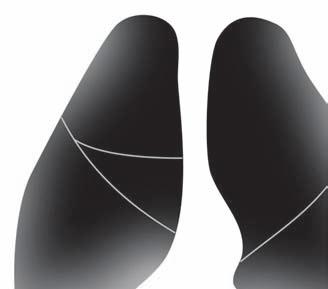
• Lobar atelectasis, or collapse of an entire lobe, has characteristic appearances depending on which of the five lobes is collapsed.
frontal schematic
lateral schematic
• Each of the five lobes tends to collapse in a predictable direction, as shown above.
right lung
right lung left lung left lung
Left
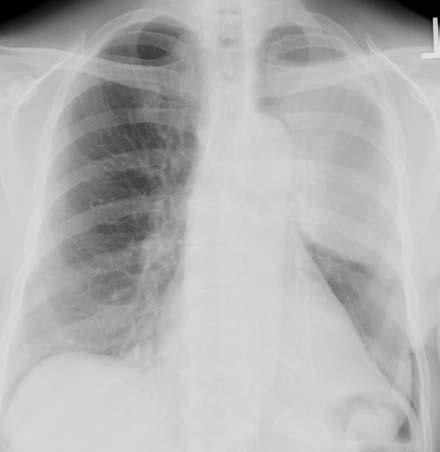
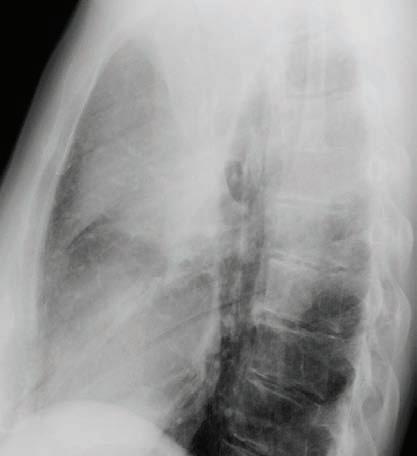
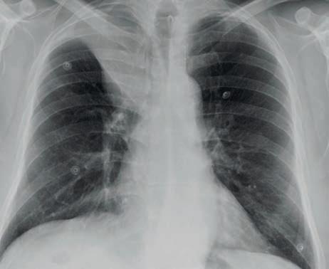
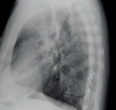
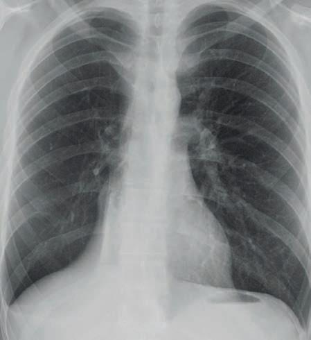
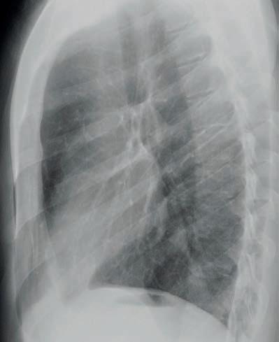
Left upper lobe collapse and luftsichel sign: Frontal radiograph (left image) shows a veil-like left upper lung opacity representing the collapsed left upper lobe (red arrow). A crescent of air lateral to the aortic arch is the luftsichel (yellow arrows). The lateral view (right image) shows the anterior wedge-shaped collapsed left upper lobe (red arrows). Case courtesy Ritu R. Gill, MD, MPH, Brigham and Women’s Hospital.
• The luftsichel (air-sickle in German) sign of left upper lobe collapse is a crescent of air seen on the frontal radiograph, which represents the interface between the aorta and the hyperexpanded superior segment of the left lower lobe.
• It is important to recognize left upper lobe collapse and not mistake the left lung opacity for pneumonia, since a mass obstructing the airway may be the cause of the lobar atelectasis.
Right upper lobe atelectasis
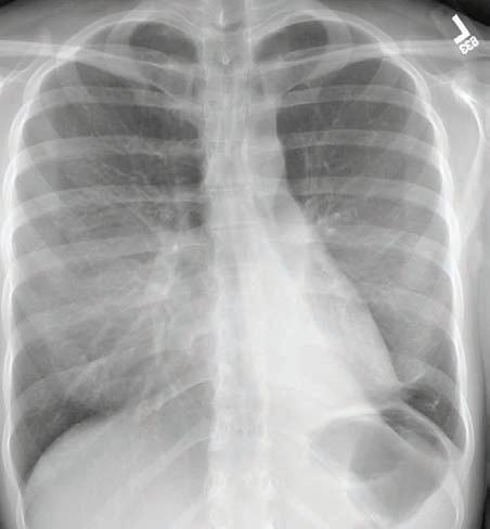
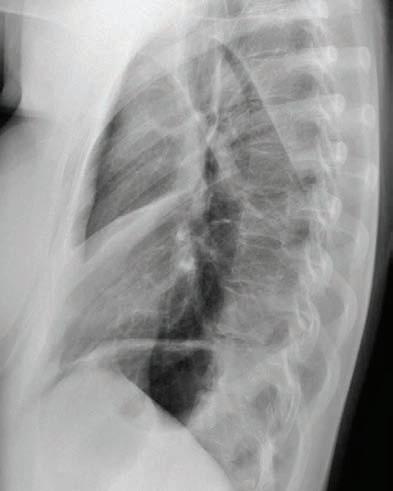
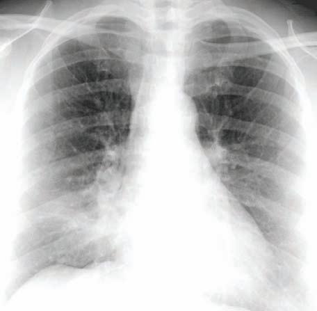
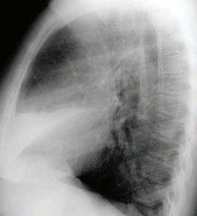
Right upper lobe collapse and Golden’s S sign: Frontal radiograph (left image) shows a right upper lobe opacity with superior displacement of the minor fissure (red arrow) and a convex mass (yellow arrow). Lateral radiograph (right image) shows the wedge-shaped collapsed RUL projecting superiorly (red arrows).
• The reverse S sign of Golden is seen in right upper lobe collapse caused by an obstructing mass. The central convex margins of the mass form a reverse S. Although the sign describes a reverse S, it is also commonly known as Golden’s S sign. Similar to left upper lobe collapse, a right upper lobe collapse should raise concern for an underlying malignancy, especially with a Golden’s S sign present.
• The juxtaphrenic peak sign is a peridiaphragmatic triangular opacity caused by diaphragmatic traction from an inferior accessory fissure or an inferior pulmonary ligament.
Left lower lobe collapse: Frontal and lateral radiographs demonstrate a triangular retrocardiac opacity representing the collapsed left lower lobe (red arrows). There is loss of concavity of the left heart border (the flat waist sign; yellow arrow).
Case courtesy Ritu R. Gill, MD, MPH, Brigham and Women’s Hospital.
• In left lower lobe collapse, the heart slightly rotates and the left hilum is pulled down.
• The flat waist sign describes the flattening of the left heart border as a result of downward shift of hilar structures and resultant cardiac rotation.
Right lower lobe atelectasis
Right lower lobe collapse: Frontal radiograph shows an abnormal vertically oriented interface medial to the right heart border (red arrow), which corresponds to a wedge-shaped opacity projecting over the heart on the lateral view (red arrow) and represents the collapsed right lower lobe. On the frontal radiograph, there is subtle crowding of the ribs (yellow arrows) in the right hemithorax due to volume loss.
• Right lower lobe atelectasis is the mirror-image of left lower lobe atelectasis.
• The collapsed lower lobe appears as a wedge-shaped retrocardiac opacity.
Right middle lobe atelectasis: Frontal chest radiograph shows an indistinct opacity in the right lung with focal silhouetting of the right heart border (arrow). There is elevation of the right hemidiaphragm due to volume loss. The lateral radiograph shows a wedge-shaped opacity (arrow) projecting over the midheart representing the collapsed right middle lobe.
Case courtesy Ritu R. Gill, MD, MPH, Brigham and Women’s Hospital.
• The findings of right middle lobe atelectasis can be subtle on the frontal radiograph. Silhouetting of the right heart border by the collapsed medial segment of the middle lobe may be the only clue. The lateral radiograph shows a wedge-shaped opacity anteriorly.
Round atelectasis
• Round atelectasis is focal atelectasis with a round morphology that is always associated with an adjacent pleural abnormality (e.g., pleural effusion, pleural thickening or plaque, pleural neoplasm, etc.).
• Round atelectasis is most common in the posterior lower lobes.
• All five of the following findings must be present to diagnose round atelectasis:
1) Adjacent pleura must be abnormal.
2) Opacity must be peripheral and in contact with the pleura.
3) Opacity must be round or elliptical.
4) Volume loss must be present in the affected lobe.
5) Pulmonary vessels and bronchi leading into the opacity must be curved — this is the comet tail sign.
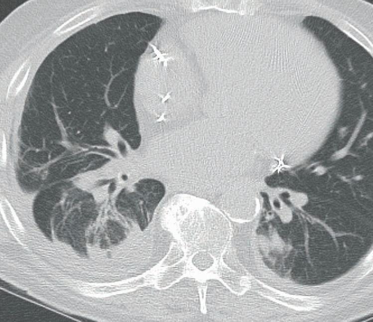
Round atelectasis: Noncontrast CT shows a rounded opacity in the medial right lower lobe (red arrows). This example meets all five criteria for round atelectasis including adjacent pleural abnormality (effusion), opacity in contact with the pleura, round shape, volume loss in the affected lobe, and the comet tail sign (yellow arrows) representing curved vessels and bronchi leading to the focus of round atelectasis.
Patterns of lung disease
Essential anatomy
Secondary pulmonary lobule (SPL)
acinus, not visible on CT (approximately 12 per secondary lobule)
acinar artery and respiratory bronchiole
centrilobular bronchus and artery
pulmonary veins (and lymphatics, not pictured) run in the interlobular septa 1
• The secondary pulmonary lobule (SPL) is the elemental unit of lung function.
• Each SPL contains a central artery (the aptly named centrilobular artery) and a central bronchus, each branching many times to ultimately produce acinar arteries and respiratory bronchioles.
On CT, the centrilobular artery is often visible as a faint dot. The centrilobular bronchus is not normally visible.
The acinus is the basic unit of gas exchange, containing several generations of branching respiratory bronchioles, alveolar ducts, and alveoli.
There are generally 12 or fewer acini per secondary lobule.
• Pulmonary veins and lymphatics collect in the periphery of each SPL.
• Connective tissue, called interlobular septa, encases each SPL.
Thickening of the interlobular septa can be seen on CT and suggests pathologic enlargement of either the venous or lymphatic spaces, as discussed on subsequent pages.
• Each SPL is between 1 and 2.5 cm in diameter.
Abnormalities of the secondary pulmonary lobule
Consolidation and ground glass
• Consolidation and ground glass opacification are two very commonly seen patterns of lung disease caused by abnormal alveoli. The alveolar abnormality may represent either filling of the alveoli with fluid or incomplete alveolar aeration.
• C onsolidation can be described on either a chest radiograph or CT, while ground glass is generally reserved for CT.
• Although consolidation often implies pneumonia, both consolidation and ground glass are nonspecific findings with a broad differential depending on chronicity (acute versus chronic) and distribution (focal versus patchy or diffuse).
Consolidation
Schematic demonstrates complete filling of the alveolus with obscuration of the pulmonary vessels. The bronchus is visible as an air bronchogram.
Consolidation: Contrast-enhanced CT shows bilateral consolidative opacities, more densely consolidated on the left. There are bilateral air bronchograms (arrows). Although these imaging findings are nonspecific, this was a case of multifocal bronchioloalveolar carcinoma.
• Consolidation is histologically due to complete filling of affected alveoli with a liquidlike substance (commonly remembered as blood, pus, water, or cells).
• Pulmonary vessels are not visible through the consolidation on an unenhanced CT.
• Air bronchograms are often present if the airway is patent. An air bronchogram represents a lucent air-filled bronchus (or bronchiole) seen within a consolidation.
• Consolidation causes silhouetting of adjacent structures on conventional radiography.
• Acute consolidation is most commonly due to pneumonia, but the differential includes:
Pneumonia (by far the most common cause of acute consolidation).
Pulmonary hemorrhage (primary pulmonary hemorrhage or aspiration of hemorrhage).
Acute respiratory distress syndrome (ARDS), which is noncardiogenic pulmonary edema seen in critically ill patients and thought to be due to increased capillary permeability.
Pulmonary edema may cause consolidation, although this is an uncommon manifestation.
• The differential diagnosis of chronic consolidation includes:
Bronchioloalveolar carcinoma mucinous subtype, a form of adenocarcinoma.
Organizing pneumonia, which is a nonspecific response to injury characterized by granulation polyps which fill the distal airways, producing peripheral rounded and nodular consolidation.
Chronic eosinophilic pneumonia, an inflammatory process characterized by eosinophils causing alveolar filling in an upper-lobe distribution.
Ground glass opacification (GGO)
Schematic demonstrates complete hazy filling of the alveolus. The pulmonary vessels are still visible.
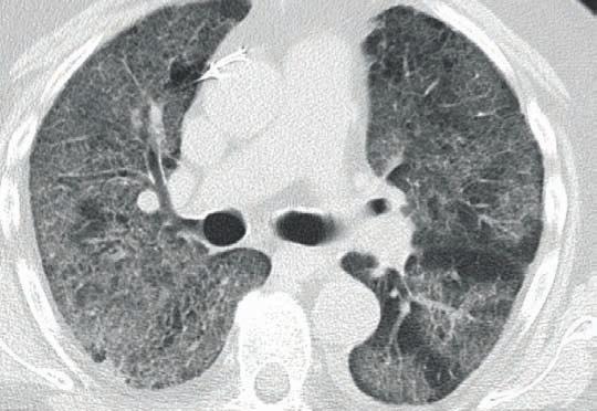
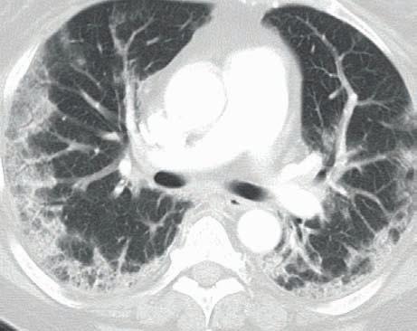
Ground glass opacification: Noncontrast CT shows diffuse ground glass opacification (GGO). The pulmonary architecture, including vasculature and bronchi, can be still seen, which is characteristic for GGO. Although these imaging findings are nonspecific, this was a case of acute respiratory distress syndrome (ARDS).
• Ground glass opacification is histologically due to either partial filling of the alveoli (by blood, pus, water, or cells), alveolar wall thickening, or reduced aeration of alveoli (atelectasis).
• Ground glass is usually a term reserved for CT. CT shows a hazy, gauze-like opacity, through which pulmonary vessels are still visible.
The term ground glass was originally described for unenhanced CT as enhanced vessels are visible in consolidation as well; however, in common practice ground glass is used for any type of CT.
• As with consolidation, air bronchograms may be present.
• Acute ground glass opacification has a similar differential to acute consolidation, since many of the entities that initially cause partial airspace filling can progress to completely fill the airspaces later in the disease. The differential of acute ground glass includes:
Pulmonary edema, which is usually dependent.
Pneumonia. Unlike consolidation, ground glass is more commonly seen in atypical pneumonia such as viral or Pneumocystis jiroveci pneumonia.
Pulmonary hemorrhage
Acute respiratory distress syndrome (ARDS)
• Chronic ground glass opacification has a similar but broader differential diagnosis compared to chronic consolidation. In addition to all of the entities which may cause chronic consolidation, the differential diagnosis of chronic ground glass also includes:
Bronchioloalveolar carcinoma, which tends to be focal or multifocal.
Organizing pneumonia, typically presenting as rounded, peripheral chronic consolidation.
Chronic eosinophilic pneumonia, usually with an upper-lobe predominance.
Idiopathic pneumonias, which are a diverse group of inflammatory responses to pulmonary injury.
Hypersensitivity pneumonitis (HSP), especially the subacute phase. HSP is a type III hypersensitivity reaction to inhaled organic antigens. In the subacute phase there is ground glass, centrilobular nodules, and mosaic attenuation.
Alveolar proteinosis, an idiopathic disease characterized by alveolar filling by a proteinaceous substance. The distribution is typically central, with sparing of the periphery.
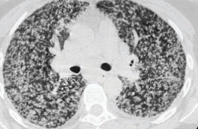
Coronal CT demonstrates central-predominant ground glass and associated septal thickening (the crazy paving pattern) in a case of alveolar proteinosis.
• The differential diagnosis for ground glass in a central distribution includes:
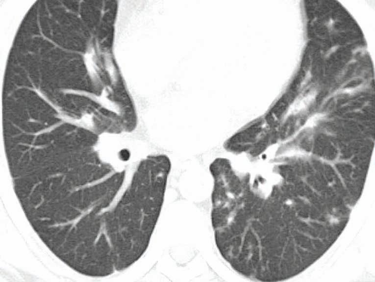
Axial CT shows peripheral and subpleural ground glass attenuation. This was a case of organizing pneumonia.
• The differential diagnosis for peripheral consolidation or ground glass includes:
Organizing pneumonia.
Chronic eosinophilic pneumonia, typically with an upper lobe predominance.
Atypical or viral pneumonia.
Pulmonary edema. Peripheral pulmonary edema tends to be noncardiogenic in etiology, such as edema triggered by drug reaction. Peripheral consolidation/ground glass is unusual for cardiogenic pulmonary edema.
Interlobular septal thickening – smooth
Schematic demonstrates smooth interlobular septal thickening. Smooth interlobular septal thickening: CT demonstrates smooth thickening of the interlobular septa (arrows) in pulmonary edema.
Courtesy Ritu R. Gill, MD, MPH, Brigham and Women’s Hospital.
• Conditions that dilate the pulmonary veins cause smooth interlobular septal thickening.
• By far the most common cause of smooth interlobular septal thickening is pulmonary edema; however, the differential diagnosis for smooth interlobular septal thickening is identical to the differential for central ground glass:
Pulmonary edema (by far the most common cause of smooth interlobular septal thickening).
Pulmonary alveolar proteinosis
Pulmonary hemorrhage
Atypical pneumonia, especially Pneumocystis jiroveci pneumonia.
Interlobular septal thickening – nodular, irregular, or asymmetric
Schematic demonstrates irregular and nodular interlobular septal thickening.
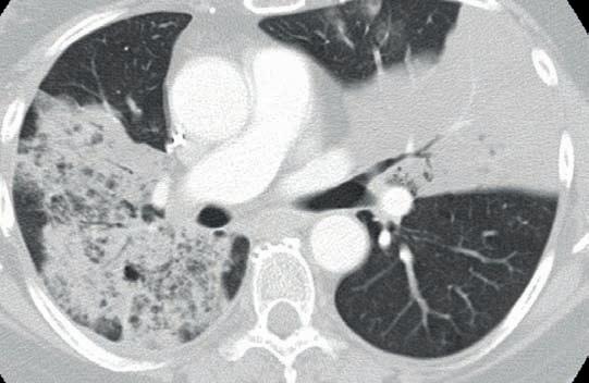
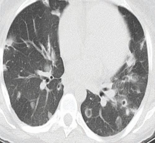
Nodular septal thickening: CT shows a dominant mass in the right lung (yellow arrows) with peripheral nodularity and septal thickening. This was a case of lymphangitic carcinomatosis.
• Nodular, irregular, or asymmetric septal thickening tends to be caused by processes that infiltrate the peripheral lymphatics, most commonly lymphangitic carcinomatosis and sarcoidosis:
Lymphangitic carcinomatosis is tumor spread through the lymphatics.
Sarcoidosis is an idiopathic, multi-organ disease characterized by noncaseating granulomas, which form nodules and masses primarily in a lymphatic distribution.
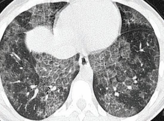
Schematic demonstrates interlobular septal thickening and ground glass opacification.
Axial CT shows interlobular septal thickening in regions of ground glass opacification, representing crazy paving. This was a case of alveolar proteinosis, the entity in which crazy paving was first described.
• Crazy paving describes interlobular septal thickening with superimposed ground glass opacification, which is thought to resemble the appearance of broken pieces of stone.
• Although nonspecific, this pattern was first described for alveolar proteinosis, where the ground glass opacification is caused by filling of alveoli by proteinaceous material and the interlobular septal thickening is caused by lymphatics taking up the same material.
• The differential diagnosis for crazy paving includes:
Alveolar proteinosis
Pneumocystis jiroveci pneumonia
Organizing pneumonia
Bronchioloalveolar carcinoma, mucinous subtype.
Lipoid pneumonia, an inflammatory pneumonia caused by a reaction to aspirated lipids.
Acute respiratory distress syndrome
Pulmonary hemorrhage.
Approach to multiple nodules
Centrilobular nodules
Schematic demonstrates a centrilobular nodule, located at the center of the pulmonary lobule.
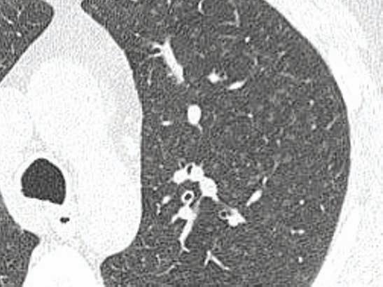
Axial CT demonstrates innumerable subcentimeter centrilobular nodules of ground glass attenuation (arrows). None of the nodules extends to the pleural surface, which is typical of a centrilobular distribution. This was a case of respiratory bronchiolitis interstitial lung disease (RB-ILD).
Courtesy Ritu R. Gill, MD, MPH, Brigham and Women’s Hospital.
• Centrilobular nodules represent opacification of the centrilobular bronchiole (or less commonly the centrilobular artery) at the center of each secondary pulmonary lobule.
• On CT, multiple small nodules are seen in the centers of secondary pulmonary lobules. Centrilobular nodules never extend to the pleural surface. The nodules may be solid or of ground glass attenuation, and range in size from tiny up to a centimeter.
• Centrilobular nodules may be caused by infectious or inflammatory conditions.
• Infectious causes of centrilobular nodules include:
Endobronchial spread of tuberculosis or atypical mycobacteria. Atypical mycobacteria are a diverse spectrum of acid-fast mycobacteria that do not cause tuberculosis. The typical pulmonary manifestation of atypical mycobacteria is a low-grade infection typically seen in elderly women, most commonly caused by Mycobacterium avium-intracellulare.
Bronchopneumonia, which is spread of infectious pneumonia via the airways.
Atypical pneumonia, especially mycoplasma pneumonia.
• The two most common inflammatory causes of centrilobular nodules include hypersensitivity pneumonitis (HSP) and respiratory bronchiolitis interstitial lung disease (RB-ILD), both exposure-related lung diseases. More prominent centrilobular nodules are suggestive of HSP.
HSP is a type III hypersensitivity reaction to an inhaled organic antigen. The subacute phase of HSP is primarily characterized by centrilobular nodules.
Hot tub lung is a hypersensitivity reaction to inhaled atypical mycobacteria, with similar imaging to HSP.
RB-ILD is an inflammatory reaction to inhaled cigarette smoke mediated by pigmented macrophages. Diffuse panbronchiolitis is a chronic inflammatory disorder characterized by lymphoid hyperplasia in the walls of the respiratory bronchioles resulting in bronchiolectasis. It typically affects patients of Asian descent.
Silicosis, an inhalation lung disease that develops in response to inhaled silica particles, is characterized by upper lobe predominant centrilobular and perilymphatic nodules.
peribronchovascular septal/subpleural
Perilymphatic nodules:
Schematic of the secondary pulmonary lobule (top left image) demonstrates two of the three distributions of perilymphatic nodules. The gray nodules are located along the bronchovascular bundle and the white nodules are located along the interlobular septa.
Schematic of the lungs (left image) demonstrates the peribronchovascular and subpleural/fissural distribution.
Axial CT (top right image) demonstrates multiple subpleural/ fissural nodules (yellow arrows) and nodules along the bronchovascular bundles (red arrows). This was a case of sarcoidosis.
• Perilymphatic nodules follow the anatomic locations of pulmonary lymphatics, which can be seen in three locations in the lung:
1) Subpleural.
2) Peribronchovascular.
3) Septal (within the interlobular septa separating the hexagonal secondary pulmonary lobules).
• Sarcoidosis is by far the most common cause of perilymphatic nodules, typically with an upper-lobe distribution. The nodules may become confluent creating the galaxy sign. The differential of perilymphatic nodules includes:
Sarcoidosis
Pneumoconioses (silicosis and coal workers pneumoconiosis) are reactions to inorganic dust inhalation. The imaging may look identical to sarcoidosis with perilymphatic nodules, but there is usually a history of exposure (e.g. a sandblaster who develops silicosis).
Lymphangitic carcinomatosis
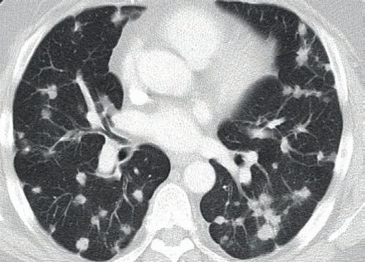
Random nodules:
Schematic of the secondary pulmonary lobule (top left image) demonstrates nodules distributed randomly throughout the SPL.
Schematic of the lungs (bottom left image) demonstrates nodules scattered randomly. Some of the nodules are in close contact with the pleural surface.
Axial CT (top right image) demonstrates multiple random nodules. Some of the nodules abut the pleural surface. This was a case of metastatic colon cancer.
• Randomly distributed nodules usually occur via hematogenous spread and have an angiocentric distribution. The differential of random nodules includes:
Hematogenous metastases
Septic emboli. Embolic infection has a propensity to cavitate but early emboli may be irregular or solid.
Pulmonary Langerhans's cell histiocytosis (PLCH), a smoking-related lung disease that progresses from airway-associated and random nodules to irregular cysts. PLCH is usually distinguishable from other causes of random nodules due to the presence of cysts and non-angiocentric distribution.
• A miliary pattern is innumerable tiny random nodules disseminated hematogenously, suggestive of the appearance of millet seeds. The differential of miliary nodules includes:
Miliary nodules: Axial CT shows innumerable tiny nodules distributed randomly throughout both lungs in a miliary pattern. This was a case of miliary tuberculosis.
Case courtesy Ritu R. Gill, MD, MPH, Brigham and Women’s Hospital.
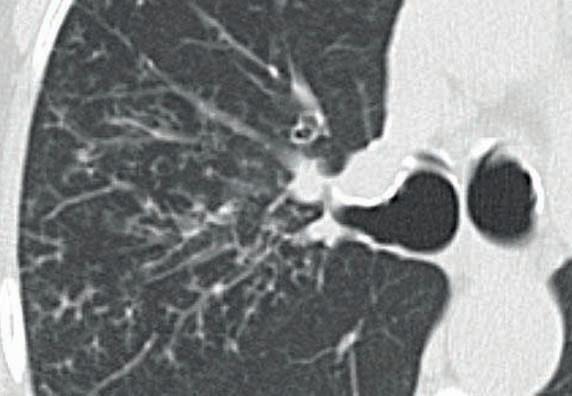
Schematic shows several nodules centered on an opacified small airway.
Tree-in-bud nodularity: Axial CT show numerous small nodules (arrows) “budding” off of linear branching structures in the right middle lobe. This case was secondary to atypical mycobacteria.
• Tree-in-bud nodules are multiple small nodules connected to linear branching structures, which resembles a budding tree branch in springtime as seen on CT. The linear branching structures represent the impacted bronchioles, which are normally invisible on CT, and the nodules represent impacted terminal bronchioles. Tree-in-bud nodules are due to mucus, pus, or fluid impacting bronchioles and terminal bronchioles.
• Tree-in-bud nodules are almost always associated with small airways infection, such as endobronchial spread of tuberculosis. The differential of tree-in-bud nodules includes:
Mycobacteria tuberculosis and atypical mycobacteria
Bacterial pneumonia
Aspiration pneumonia
Airway-invasive aspergillus. Aspergillus is an opportunistic fungus with several patterns of disease. The airway-invasive pattern is seen in immunocompromised patients and may present either as bronchopneumonia or small airways infection.
Cavitary and cystic lung disease
Solitary cavitary nodule/mass
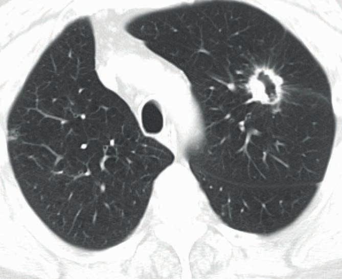
Coronal schematic demonstrates a single cavitary lesion.
Axial CT shows a single spiculated cavitary lesion in the left upper lobe (arrow). This was a case of squamous cell carcinoma.
• A cavitary lesion has a thick, irregular wall, often with a solid mural component. Although the findings of benign and malignant cavitary nodules overlap, a maximum wall thickness of ≤4 mm is usually benign and a wall thickness >15 mm is usually malignant. Spiculated margins also suggest malignancy.
• A solitary cavitary lesion is most likely cancer or infection.
Primary bronchogenic carcinoma. While both squamous cell and adenocarcinoma can cavitate, squamous cell cavitates more frequently. Small cell carcinoma is never known to cavitate. Tuberculosis classically produces an upper-lobe cavitation.
Axial CT shows numerous cavitary and noncavitary lesions bilaterally, in a random distribution. This was a case of tricuspid endocarditis and septic emboli.
Case courtesy Michael Hanley, MD, University of Virginia Health System.
• Multiple cavitary lesions are typically vascular or spread through the vascular system: Septic emboli.
Vasculitis, including Wegener granulomatosis, which is especially prone to cavitate.
Metastases, of which squamous cell carcinoma and uterine carcinosarcoma are known to cavitate.
Axial CT shows bilateral thin-walled cysts that are of varying sizes but are predominantly regular in shape. There is a small left pleural effusion. This was a case of lymphangioleiomyomatosis.
• A cyst is an air-containing lucency with a thin, nearly imperceptible wall. In general, cystic lung disease is usually due to a primary airway abnormality.
• The differential diagnosis for multiple lung cysts includes:
Lymphangioleiomyomatosis (LAM), a diffuse cystic lung disease caused by smooth muscle proliferation of the distal airways. LAM causes uniformly distributed, thin-walled cysts in a diffuse distribution. It is classically associated with chylous effusion, as demonstrated in the above right case.
Emphysema, which tends to be upper-lobe predominant in a smoker.
Pulmonary Langerhans cell histiocytosis, which features irregular cysts and nodules predominantly in the upper lungs.
Diffuse cystic bronchiectasis. Bronchiectasis is dilation of the bronchioles. Although cystic fibrosis is the most common cause of bronchiectasis and has an upper-lobe predominance, congenital or post-infectious causes can have a diffuse or lower-lobe distribution.
Pneumocystis jiroveci pneumonia, which features cysts in late-stage disease.
Lymphoid interstitial pneumonia (LIP), an exceptionally rare disease usually associated with Sjögren syndrome and characterized by alveolar distortion from lymphocytic infiltrate and multiple cysts.
• The differential for a single cyst includes:
Bulla. A bulla is an air-filled cyst measuring >1 cm. A giant bulla occupies at least 30% of the volume of the thorax.
Bleb. A bleb is a air-filled cystic structure contiguous with the pleura measuring <1 cm. Rupture of a bleb is the most common cause of spontaneous pneumothorax.
Pneumatocele, which is an air-filled space caused by prior lung trauma or infection.
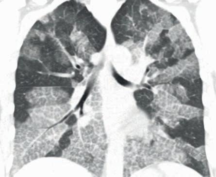
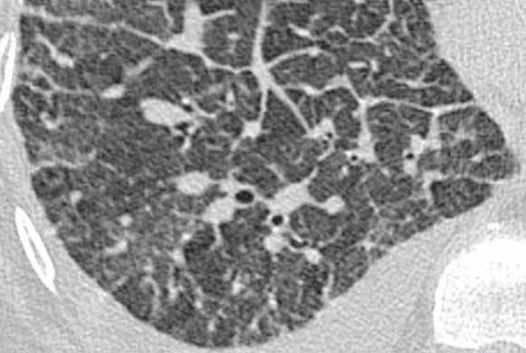
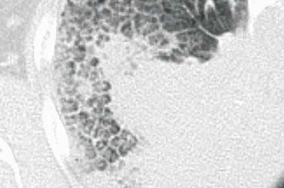
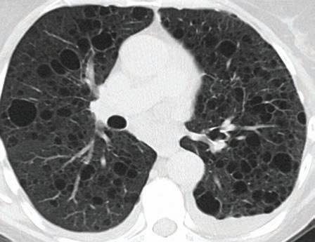
Coronal CT shows bibasilar fibrosis and honeycombing (arrows), with relative sparing of the upper lobes. This was a case of idiopathic pulmonary fibrosis.
• The differential diagnosis of basal-predominant fibrotic change includes:
Idiopathic pulmonary fibrosis (IPF), which is a clinical syndrome of progressive pulmonary fibrosis of unknown etiology and is most common cause of basilar fibrosis. It almost always features basilar honeycombing.
End-stage asbestosis. Asbestosis is an asbestos-induced inflammatory process ultimately producing pulmonary fibrosis. Usually other signs of asbestos exposure are present, such as pleural plaques.
Nonspecific interstitial pneumonia (NSIP), fibrotic form. NSIP is an idiopathic pneumonia. It is a lung response to injury commonly associated with collagen vascular disease and drug reaction. The two histologic subtypes are cellular and fibrotic forms, of which the latter may produce basalpredominant fibrosis. In contrast to IPF, honeycombing is usually absent.
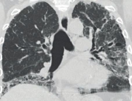
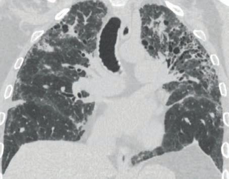
Upper lobe fibrotic changes
Coronal schematic shows fibrotic changes in the upper lobes.
Coronal CT shows upper-lobe predominant subpleural fibrosis and traction bronchiectasis. A pathologic diagnosis was not established in this case.
• Although IPF is the most common cause of pulmonary fibrosis, fibrosis primarily affecting the upper lobes should raise concern for an alternative diagnoses, such as:
End-stage sarcoidosis. Sarcoidosis is a disease that primarily affects the upper lobes. The late stage of sarcoidosis leads to upper-lobe predominant fibrosis.
Chronic hypersensitivity pneumonitis may cause upper-lobe fibrosis in long-standing disease.
End-stage silicosis. The late stage of silicosis may lead to fibrosis with an upper lobe predominance.
Pulmonary infection
Clinical classification of pneumonia
Community-acquired pneumonia (CAP)
• S. pneumoniae is the most common cause of community-acquired pneumonia (CAP).
• Atypical pneumonia, including Mycoplasma, viral, and Chlamydia, typically infects young and otherwise healthy patients.
Mycoplasma has a varied appearance and can produce consolidation, areas of ground glass attenuation, centrilobular nodules, and tree-in-bud nodules.
• Legionella most commonly occurs in elderly smokers. Infections tend to be severe. Peripheral consolidation often progresses to lobar and multifocal pneumonia.
• Infection by Klebsiella and other gram-negatives occurs in alcoholics and aspirators. Klebsiella classically leads to voluminous inflammatory exudates causing the bulging fissure sign.
Hospital acquired pneumonia (HAP)
• Hospital acquired pneumonia (HAP) occurs in hospitalized patients and is due to aspiration of colonized secretions. HAP is caused by a wide variety of organisms, but the most important pathogens include MRSA and resistant gram-negatives including Pseudomonas
Health care associated pneumonia (HCAP)
• Health care associated pneumonia is defined as pneumonia in a nursing home resident or in a patient with a >2 day hospitalization over the past 90 days. Pathogens are similar to HAP.
Ventilator associated pneumonia (VAP)
• Ventilator associated pneumonia is caused by infectious agents not present at the time mechanical ventilation was started. Most infections are polymicrobial and primarily involve gram-negative rods such as Pseudomonas and Acinetobacter.
Pneumonia in the immunocompromised patient
• Any of the above pathogens, plus opportunistic infections including Pneumocystis, fungi such as Aspergillus, Nocardia, CMV, etc., can be seen in immunocompromised patients.
Radiographic patterns of infection
Lobar pneumonia
• Lobar pneumonia is consolidation of a single lobe. It is usually bacterial in origin and is the most common presentation of community acquired pneumonia.
• The larger bronchi remain patent, causing air bronchograms.
Lobular pneumonia (bronchopneumonia)
• Lobular pneumonia manifests as patchy consolidation with poorly defined airspace opacities, usually involving several lobes, and most commonly due to S. aureus
Interstitial pneumonia
• Interstitial pneumonia is caused by inflammatory cells located predominantly in the interstitial tissue of the alveolar septa causing diffuse or patchy ground glass opacification. It can be caused by viral pneumonia, Mycoplasma, Chlamydia, or Pneumocystis
Round pneumonia
• Round pneumonia is an infectious mass-like opacity seen only in children, most commonly due to Streptococcus pneumoniae.
• Infection remains somewhat confined due to incomplete formation of pores of Kohn.
Complications of pneumonia
Pulmonary abscess
• Pulmonary abscess is necrosis of the lung parenchyma typically due to Staphylococcus aureus, Pseudomonas, or anaerobic bacteria.
• An air–fluid level is almost always present.
• An abscess is usually spherical, with equal dimensions on frontal and lateral views.
Pulmonary gangrene
• Pulmonary gangrene is a very rare complication of pneumonia where there is extensive necrosis or sloughing of a pulmonary segment or lobe. Pulmonary gangrene is a severe manifestation of pulmonary abscess.
Empyema
Pneumatocele
• Empyema is infection within the pleural space.
• There are three stages in the development of an empyema:
1) Free-flowing exudative effusion: Can be treated with needle aspiration or simple drain.
2) Development of fibrous strands: Requires large-bore chest tube and fibrinolytic therapy.
3) Fluid becomes solid and jelly-like: Usually requires surgery.
• Although pneumonia is often associated with a parapneumonic effusion, most pleural effusions associated with pneumonia are not empyema, but are instead a sterile effusion caused by increased capillary permeability.
• An empyema conforms to the shape of the pleural space, causing a longer air–fluid level on the lateral radiograph. This is in contrast to an abscess, discussed above, which typically is spherical and has the same dimensions on the frontal and lateral radiographs.
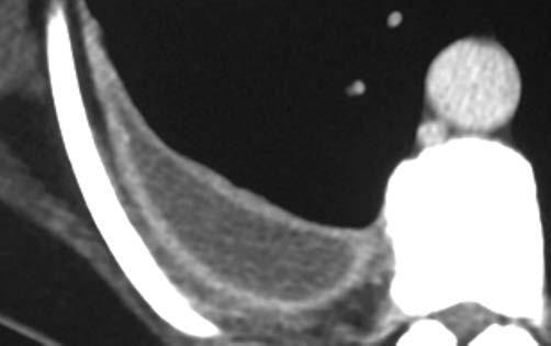
• The split pleura sign describes enhancing parietal and visceral pleura of an empyema seen on contrast-enhanced study.
Split pleura sign: Contrast-enhanced CT shows enhancement of the thickened visceral and parietal pleural layers (arrows), which encase a pleural fluid collection.
The split pleura sign is seen in the majority of exudative effusions, although it is not specific. Similar findings can be seen in malignant effusion, mesothelioma, fibrothorax, and after talc pleurodesis.
Case courtesy Ritu R. Gill, MD, MPH, Brigham and Women’s Hospital.
• A pneumatocele is a thin-walled, gas-filled cyst that may be post-traumatic or develop as a sequela of pneumonia, typically from Staphylococcus aureus or Pneumocystis.
• Pneumatoceles almost always resolve.
Bronchopleural fistula (BPF)
• Bronchopleural fistula (BPF) is an abnormal communication between the airway and the pleural space. It is caused by rupture of the visceral pleura. By far the most common cause of BPF is surgery; however, other etiologies include lung abscess, empyema, and trauma.
• On imaging, new or increasing gas is present in a pleural effusion. A connection between the bronchial tree and the pleura is not always apparent, but is helpful when seen.
• The treatment of BPF is controversial and highly individualized.
Empyema necessitans
• Empyema necessitans is extension of an empyema to the chest wall, most commonly secondary to tuberculosis. Other causative organisms include Nocardia and Actinomyces
Tuberculosis (TB)
• Tuberculosis (TB), caused by Mycobacterium tuberculosis, remains an important disease despite remarkable progress in public health and antituberculous therapy over the past century. Tuberculosis remains a significant problem in developing countries. In the United States, TB is seen primarily in the immigrant population and immunocompromised individuals.
• Initial exposure to TB can lead to two clinical outcomes:
1) Contained disease (90%) results in calcified granulomas and/or calcified hilar lymph nodes. In a patient with normal immunity, the tuberculous bacilli are sequestered with a caseating granulomatous response.
2) Primary tuberculosis results when the host cannot contain the organism. Primary tuberculosis is seen more commonly in children and immunocompromised patients.
• Reactivation (post-primary) TB is reactivation of a previously latent infection.
Primary tuberculosis
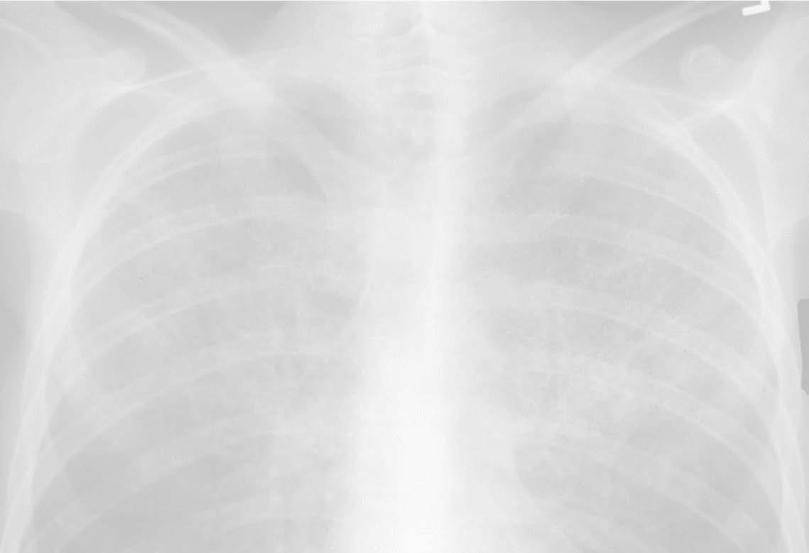
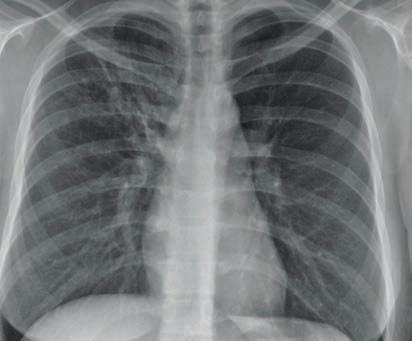
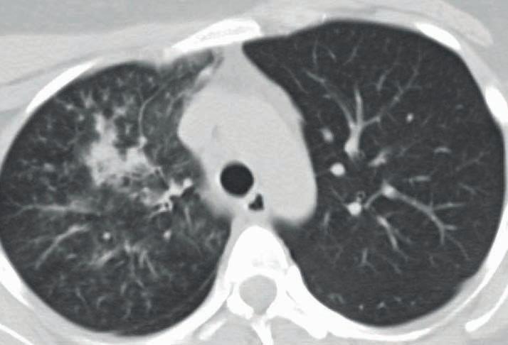
tuberculosis: Chest radiograph (left image)
a vague right upper lung opacity
CT shows a patchy opacification (arrow) in the lower portion of the right upper lobe with adjacent tree-inbud nodularity. The patient's sputum grew Mycobacterium tuberculosis. Case courtesy Ritu R. Gill, MD, MPH, Brigham and Women’s Hospital.
• Primary tuberculosis represents infection from the first exposure to TB. Primary TB may involve the pulmonary parenchyma, the airways, and the pleura. Primary TB often causes adenopathy.
• As many as 15% of patients infected with primary TB have no radiographic changes and the imaging appearance of primary tuberculosis is nonspecific.
• The four imaging manifestations of primary TB (of which any, none, or all may be present) are ill-defined consolidation, pleural effusion, lymphadenopathy, and miliary disease. Primary TB may occur in any lobe, but the most typical locations are the lower lobes or right middle lobe. It can be difficult to distinguish between primary and postprimary TB, and in clinical practice, the treatment (antituberculous therapy) is the same.
• Classic imaging findings are not always seen, but include:
Ghon focus: Initial focus of parenchymal infection, usually located in the upper part of the lower lobe or the lower part of the upper lobe.
Ranke complex: Ghon focus and lymphadenopathy.
• Cavitation is rare in primary TB, in contrast to reactivation TB.