Acknowledgements
I am both delighted and humbled by the most enthusiastic reception of the first and second editions of this book and am immensely grateful to my students for all their valuable insights and suggestions, which I have tried to incorporate in this edition. Their overwhelming response keeps me going and makes the entire effort worthwhile.
I would not have been able to complete this all encompassing project without the tremendous patience and endurance of my daughter, the valued support and guidance of my husband, the blessings of my parents, and the constant encouragement and motivation of my brother.
I would like to offer my sincere appreciation to my friends, particularly Dr Sonal Sharma, who has always very generously helped me whenever required. I am also grateful to my departmental staff for their unquestioning support and cooperation.
I am extremely thankful to my publisher, RELX India Pvt. Ltd., particularly Shabina Nasim, Sr Manager–Education Solutions; and Goldy Bhatnagar, Sr Content Development Specialist, for overseeing this project with great commitment and efficiency while conceding to my requests for changes and amendments till the last minute. It has been an absolute pleasure working with them.
Last but not the least I continue to be indebted to the authors of the various publications and reference books that I have consulted during the compilation of this book and regret being unable to acknowledge them all, individually.
This page intentionally left blank
Preface to the Third Edition vii
Preface to the First Edition ix
Acknowledgments xi
SECTION I
General Pathology 1
1 Cell Injury and Cell Death 2
2 Acute and Chronic Inflammation 31
3 Healing and Repair 53
4 Haemodynamic Disorders, Thrombosis and Shock 67
5 Diseases of Immunity 89
6 Neoplasia 123
7 Infections 150
8 Genetic and Paediatric Disorders 189
9 Environmental and Nutritional Pathology 211
SECTION II Diseases of Organ Systems 227
10 Blood Vessels 228
11 Disorders of the Heart 254
12 Haematology 285
13 The Lung 356
14 The Oral Cavity and Gastrointestinal Tract 384
15 Diseases of the Hepatobiliary System and Pancreas 420
16 Diseases of the Kidney and Lower Urinary Tract 453
17 Male Genital Tract 489
18 Female Genital System 502
19 The Breast 522
20 Endocrinology 534
21 Musculoskeletal System 569
22 The Skin 603
23 The Central Nervous System 613
Index 625
Online University and PGMEE Patterned MCQs and SAQs SAQ
Chapterwise Important Images
Excessive physiologic stress
Cell in homeostasis
Pathologic stimuli
Cellular adaptation
FLOWCHART 1.2. Cellular adaptation.
Q. Define cell injury.
Ans. When the cell cannot adapt anymore or when the limits of adaptive response to a stimulus are exceeded, a sequence of events labelled cell injury follows.
Q. Enumerate the various cellular responses to injury.
Ans. Cellular responses to injury may manifest as
1. Cellular adaptations: Include atrophy, hypertrophy, hyperplasia and metaplasia.
2. Cell injury: Sublethal or chronic injurious stimuli can cause (a) ‘reversible and irreversible injury’ (the latter may lead to cell death by necrosis or apoptosis) and (b) ‘subcellular alterations’ (residual effects of cell injury).
3. Intracellular accumulations: Sublethal or chronic injurious stimuli as well as metabolic derangements can cause intracellular accumulation of normal cellular constituents, abnormal cellular constituents or pigments (Flowchart 1.3).
Intracellular accumulations
Normal cellular constituents
Abnormal cellular constituents Pigments
FLOWCHART 1.3. Intracellular accumulations.
4. Cell ageing: Represents progressive accumulation over the years of sublethal injury that manifests with either cell death or inadequate response of the cell to injury. Ageing is influenced by genetic factors, diet and social environment as well as diseases like atherosclerosis, diabetes and osteoarthritis.
Q. What are the different types of cell injuries?
Ans. Types of cell injuries:
1. Reversible : If the structural and functional changes, induced by an injurious stimulus, can revert to normal on removal of the same, it is called reversible injury ( Fig. 1.1 ).
2. Irreversible: If the structural and functional changes, induced by an injurious stimulus, cannot be reversed even after removal of the same, it is called irreversible injury (Fig. 1.1).
Q. Enumerate and describe in brief different types of cellular adaptations.
Ans. Adaptive response may be in the form of
1. Hyperplasia
2. Hypertrophy
3. Atrophy
4. Metaplasia
FIGURE 1.1. Cell injury.
Hyperplasia
Definition
Increase in number of cells in an organ or tissue leading to increased size/mass of the tissue or organ. Hyperplasia takes place in cells, which are capable of synthesizing DNA. In nondividing cells, only hypertrophy occurs.
Mechanism
• Production of transcription factors that induce genes encoding growth factors, receptors for growth factors and cell-cycle regulators.
• In hormonal hyperplasia, hormones themselves act as growth factors and trigger transcription of genes.
• In compensatory hyperplasia, there is proliferation of remaining cells and development of new cells from stem cells.
Types
1. Physiologic hyperplasia:
(a) Hormonal hyperplasia: Hormonal stimulation increases the functional capacity of the tissue when needed, eg, breast and uterus in puberty, pregnancy and lactation.
(b) Compensatory hyperplasia: Increase in tissue mass after damage or partial resection, eg, regeneration of liver after partial hepatectomy.
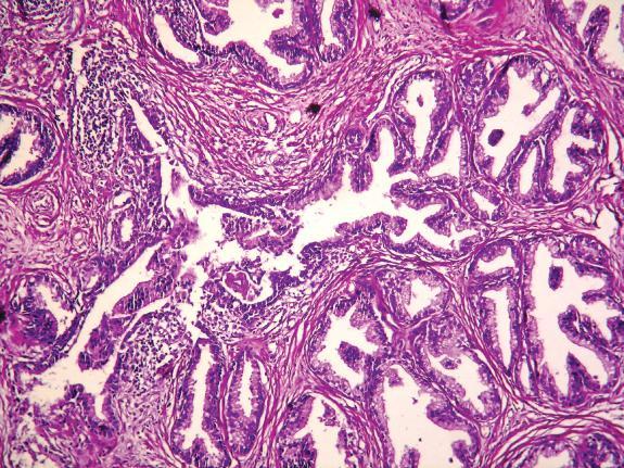
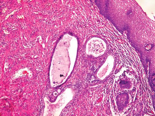
2. Pathologic hyperplasia: Hyperplasia due to excessive hormonal stimulation or excessive effects of growth factors on target cells, eg, endometrial hyperplasia (occurs when balance between progesterone and oestrogen is disturbed) and benign nodular prostatic hyperplasia or NHP (occurs due to androgen excess; Fig. 1.2).
Hypertrophy
Definition
Increase in size of the cell due to increased synthesis of structural components and not due to cellular swelling is known as hypertrophy. Nondividing cells, eg, myocardial fibres, undergo hypertrophy only. Dividing cells (stable cells, quiescent cells) undergo both hyperplasia and hypertrophy.
Papillary projection Stroma
Proliferating glands
FIGURE 1.2. NHP prostate showing hyperplastic glands lying back to back. The glands are lined by two distinct layers of epithelium indicating benign nature of the lesion (H&E; 1003).
Mechanism
Induction of genes stimulates synthesis of cellular proteins, eg, genes encoding transcription factors, growth factors and vasoactive agents. In the heart, increased workload (mechanical stretch), growth factors (transforming growth factor-beta and Insulin-like growth factor-1) and a-adrenergic hormones activate signal transduction pathways (phosphoinositide-3-kinase/AKT pathway and downstream signalling of G-protein coupled receptors), which in turn activate transcription factors like GATA4 (critical transcription factor for proper mammalian cardiac development and essential for survival of the embryo), NFAT (nuclear factor of activated T cells) and MEF 2 (myocyte enhancer 2). They work together to increase synthesis of proteins responsible for cardiac hypertrophy.
Types
1. Physiological hypertrophy: This occurs due to increased functional demand and stimulation by growth factors and hormones, eg, uterine enlargement in pregnancy and breast hypertrophy during lactation.
2. Pathological hypertrophy:
(a) Hypertrophy of cardiac muscle in systemic hypertension and aortic valve stenosis (chronic haemodynamic overload) leading to left ventricular hypertrophy (Fig. 1.3).
(b) Compensatory hypertrophy, which occurs when an organ or tissue is called upon to do additional work or to perform the work of destroyed tissue or of a paired organ.
Atrophy
Definition
A decrease in size of a body organ, tissue or cell along with decreased function, owing to disease, injury or lack of use.
Mechanism
Atrophy is the result of decreased protein synthesis or increased protein degradation. Protein degradation is mediated by
• Lysosomal acid hydrolases, which degrade endocytosed proteins (taken up from extracellular environment, cell surface as well as some cellular components).
• Ubiquitin-proteasome pathway, which causes degradation of many cytosolic and nuclear proteins.
FIGURE 1.3. Pathological hypertrophy, left ventricle.
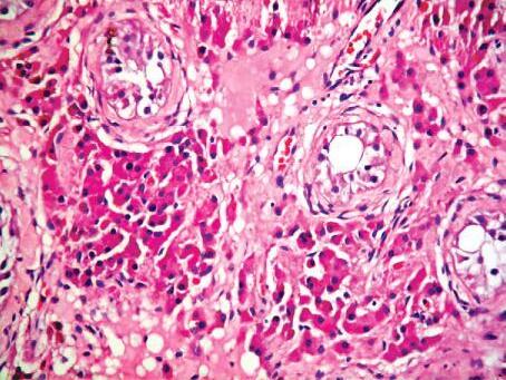
FIGURE 1.4. Atrophic testis showing marked loss of germ cells within the tubules, with peritubular and interstitial fibrosis and proliferation of interstitial cells of Leydig (H&E; 1003).
Types
1. Physiological atrophy: Common during early development, eg, atrophy of notochord or thyroglossal duct during fetal development and uterus after parturition.
2. Pathological atrophy:
(a) Decreased workload due to immobilization and prolonged functional inactivity leads to disuse atrophy.
(b) In denervation atrophy, there is loss of innervation of muscle which induces its wasting, as in polio and motor neuron disease.
(c) Atherosclerosis can cause ischaemic atrophy.
(d) Nutritional deficiency, eg, marasmus and cancer cachexia are associated with the use of skeletal muscle as a source of energy and lead to nutritional atrophy.
(e) Loss of endocrine stimulation after menopause induces atrophy of reproductive organs.
(f) Senile atrophy is an ageing-associated cell loss which is typically seen in tissues containing permanent cells, eg, brain and heart or testes (Fig. 1.4).
Metaplasia
Definition
Reversible change in which there is replacement of one adult/differentiated cell type (epithelial or mesenchymal) by another adult/differentiated cell type.
Mechanism
Occurs owing to altered/aberrant differentiation of stem cells due to their reprogramming. Examples
• Columnar to squamous metaplasia in respiratory tract, in response to chronic irritation (cigarette smoking) and vitamin-A deficiency. Stones in excretory ducts of salivary glands, pancreas and gall bladder may also result in squamous metaplasia. Squamous metaplasia in cervix is usually associated with chronic infection (Fig. 1.5).
• Connective tissue metaplasia (formation of cartilage, bone or adipose tissue in tissues that normally do not contain these elements), eg, bone formation in muscle (myositis ossificans), which occurs after bone fracture.
Note: The factors that predispose to metaplasia, if persistent, may eventually lead to induction of cancer in metaplastic epithelium, eg, metaplasia from squamous to columnar epithelium in Barrett’s oesophagus may progress to adenocarcinoma oesophagus.
Q. Define dysplasia.
Squamous metaplastic lining
Endocervical glands
Ans. Dysplasia indicates disordered cellular development characterized by
• Loss of orientation of cells with respect to one another, eg, disorderly arrangement of the cells from basal to surface layer as in stratified squamous epithelium (architectural disorientation).
• Lack of uniformity of individual cells (cellular pleomorphism).
• Causes of dysplasia include diverse cellular insults, including physical, chemical and biological.
• It is typically seen in epithelial cells and may be reversible (at least in its early stage). More severe dysplasia is known to progress to carcinoma in situ and invasive carcinoma.
• Dysplastic cells are characterized by the following cellular features:
• Accelerated cell proliferation (increased mitoses);
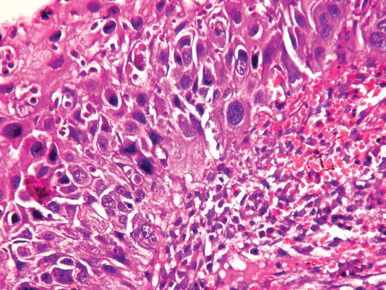
• Nuclear abnormalities such as hyperchromasia (increased basophilia on staining with haematoxylin) and pleomorphism (altered nuclear size and nuclear shape; Fig. 1.6);
• Increased nuclear-cytoplasmic ratio.
FIGURE 1.5. Section from cervix showing squamous metaplasia of the endocervical mucosa (H&E; 1003).
Atypia involving entire thickness of surface epithelium
Intact basement membrane Stroma
FIGURE 1.6. Stratified squamous epithelium showing severe dysplastic changes (diffuse atypia and loss of maturation) (H&E; 2003).
Q. Differentiate between metaplasia and dysplasia.
Ans. Differences between metaplasia and dysplasia are shown in Table 1.1
TABLE 1.1.
Features
Definition
Differences between metaplasia and dysplasia
Metaplasia
Replacement of one adult epithelial or mesenchymal cell type by another
Types
Cellular pleomorphism
Natural history
Squamous, columnar (epithelial) and osseous, cartilaginous (mesenchymal)
Mature cellular development; no pleomorphism
Reversible on withdrawal of stimulus
Dysplasia
Disordered cellular development characterized by
(a) Loss of orientation of cells with respect to one another
(b) Lack of uniformity of individual cells
Epithelial only
Disordered cellular development due to aberrant/delayed maturation or differentiation; pleomorphism present
May regress on withdrawal of inciting stimulus or progress to higher grades of dysplasia or carcinoma in situ
Q. Write briefly on aetiopathogenesis and biochemical basis of cell injury.
Ans. Sublethal or chronic injurious stimuli can cause ‘reversible and irreversible cell injury’.
Causes of Cell Injury
• Genetic
• Development defects (errors in morphogenesis)
• Cytogenetic defects (chromosomal abnormalities)
• Single gene defects (Mendelian disorders)
• Multifactorial inheritance disorders
• Acquired
• Hypoxia (ischaemia, anemia, carbon monoxide poisoning, cardiorespiratory failure).
• Physical agents (trauma, thermal injury, radiation, electric shock, pressure changes)
• Chemical agents/drugs (heavy metals, acids/alkalies, insecticides/herbicides, alcohol, smoking)
• Microbial agents (bacteria, viruses, fungi, rickettsiae, parasites)
• Immunological agents (autoimmunity, hypersensitivity)
• Nutritional imbalance (deficiency of protein, calories, trace elements, vitamins, excess cholesterol).
• Psychological factors
The cellular responses to pathological stimuli depend on
(a) Type, duration and severity of the injury.
(b) Type, status and adaptability of the target cell.
The most important targets of injurious stimuli are
(a) Aerobic respiration (involving mitochondrial oxidative phosphorylation and production of ATP)
(b) Cell membrane
(c) Protein synthesis
(d) Cytoskeleton
(e) Genetic apparatus
TABLE 1.2.
Biochemical Basis of Cell Injury
Cell injury occurs due to the following mechanisms:
• ATP depletion: ATP is required for
• Membrane transport
• Protein synthesis
• Lipogenesis
• Phospholipid turnover
ATP depletion results in dysfunction in the above functions/mechanisms.
• Damage due to oxygen and oxygen-derived free radicals (See page 11)
• Loss of calcium homeostasis:
• Normal cytosolic-free calcium levels are very low
• Most intracellular calcium sequestered in endoplasmic reticulum and mitochondria
• Injury causes influx of calcium across cell membrane and its release into cytosol from mitochondria and endoplasmic reticulum.
• Increase in cytosolic calcium leads to activation of enzymes initiating cell injury
Q. Differentiate between reversible and irreversible cell injury.
Ans. Differences between reversible and irreversible cell injury are shown in Table 1.2
Differences between reversible and irreversible cell injury
Features Reversible injury
Definition If the structural and functional changes, induced by an injurious stimulus, can revert to normal on removal of the same, it is called reversible injury
Cell membrane
(a) Blebbing, blunting, distortion Present
Irreversible injury
If the structural and functional changes, induced by an injurious stimulus, cannot be reversed even after removal of the same, it is called irreversible injury
Present; more prominent than reversible injury
(b) Defect Absent Present
Endoplasmic reticulum Shows swelling only
Ribosomes Dispersed
Lysosomes
Mitochondria
Nucleus
Autophagy of organelles by lysosomes, no rupture
Swelling, small densities present
Clumping of nuclear chromatin
Shows swelling and lysis
Dispersed and destroyed
Rupture of lysosomes and autolysis of cell
Swelling, large densities present
Pyknosis, karyolysis or karyorrhexis
Calcification Absent Dystrophic calcification may be seen
Q. Describe the sequence of events occurring in reversible and irreversible injury.
Ans. Sequence of events occurring in
• Reversible injury (Flowchart 1.4)
Ischaemia
Decreased mitochondrial oxidative phosphorylation
Decreased ATP
Increased glycolysis
Decreased Na+ K + ATPase (anaerobic respiration)pump activity
↓ pH
↓ Glycogen stores
• Influx of Ca2+, H2O, Na+
• Efflux of K+
Clumping of nuclear chromatin
Cellular swelling
Detachment of ribosomes
Decreased protein synthesis
Lipid deposition/fatty change
Loss of microvilliBlebsER swellingMyelin figures
FLOWCHART 1.4. Sequence of events in reversible injury.
• Irreversible injury (Flowchart 1.5)
↓ pH Membrane injury Ischaemia
Intracellular release of lysosomal enzymes
↓ Ribonucleic protein, nuclear changes and loss of cell shape
• Loss of membrane phospholipids due to phospholipases
• Cytoskeletal alterations due to proteases
• Lipid peroxidation and DNA damage due to free radicals
FLOWCHART 1.5. Sequence of events in irreversible injury.
Q. Write briefly on free radical-mediated cell injury.
Ans. Free radicals are chemical species with an unpaired electron in their outer orbit. They react with inorganic and organic molecules (proteins, lipids and carbohydrates), which are mainly present in membranes and nucleic acids.
Free radical production is induced by
• Absorption of radiant energy: UV rays, X-rays.
• Enzymatic metabolism of exogenous chemicals/drugs: CCl4 to CCl3
• Reduction–oxidation reaction processes that occur during normal metabolism: Formation of superoxide anion (O2–), hydrogen peroxide (H2O2), hydroxyl ion ( OH).
• Reactions involving transition metals: iron (Fenton reaction), copper, etc.
• Reactions involving nitric oxide (NO): acts as a free radical and can be converted to highly reactive peroxynitrite anion (ONOO–) as well as NO2 and NO3–
Effects of free radicals:
• Lipid peroxidation: Lipid and free radical interactions produce peroxides (initiation). Peroxides are reactive and unstable species, which start a chain reaction of lipid peroxidation (propagation). In some cases, chain reaction may be terminated by antioxidants
• Modification of proteins by oxidation: Oxidation of amino acid residue side chain leads to formation of protein–protein cross-linkage and disruption of the protein backbone resulting in protein fragmentation
• DNA lesions: Attack thymine and other nucleotides of nuclear and mitochondrial DNA to produce single- or double-stranded breaks in DNA as well as cross-linking of DNA strands.