Associate Professor, Department of Surgery and Department of Human Anatomy and Cell Sciences, Max Rady College of Medicine, Rady Faculty of Health Sciences, University of Manitoba Vice-Provost (Teaching and Learning), University of Manitoba Winnipeg, Manitoba, Canada
Professor Emeritus and Former Head, Department of Human Anatomy and Cell Science Professor of Pediatrics and Child Health Associate Professor of Obstetrics, Gynecology, and Reproductive Sciences
Max Rady College of Medicine, Rady Faculty of Health Sciences, University of Manitoba, Winnipeg, Manitoba, Canada
Elsevier
1600 John F. Kennedy Blvd. Ste 1800 Philadelphia, PA 19103-2899
CONCISE CLINICAL EMBRYOLOGY: AN INTEGRATED, CASE-BASED APPROACH
No part of this publication may be reproduced or transmitted in any form or by any means, electronic or mechanical, including photocopying, recording, or any information storage and retrieval system, without permission in writing from the publisher. Details on how to seek permission, further information about the Publisher’s permissions policies and our arrangements with organizations such as the Copyright Clearance Center and the Copyright Licensing Agency, can be found at our website: www.elsevier.com/permissions
This book and the individual contributions contained in it are protected under copyright by the Publisher (other than as may be noted herein).
Notice
Practitioners and researchers must always rely on their own experience and knowledge in evaluating and using any information, methods, compounds or experiments described herein. Because of rapid advances in the medical sciences, in particular, independent verification of diagnoses and drug dosages should be made. To the fullest extent of the law, no responsibility is assumed by Elsevier, authors, editors or contributors for any injury and/or damage to persons or property as a matter of products liability, negligence or otherwise, or from any use or operation of any methods, products, instructions, or ideas contained in the material herein.
Library of Congress Control Number: 2020951836
Content Strategist: Jeremy Bowes
Content Development Specialist: Erika Ninsin
Publishing Services Manager: Deepthi Unni
Project Manager: Srividhya Vidhyashankar
Design Direction: Margaret Reid
Mark:
For Eddie James Torchia
Our dear grandson and little ray of sunshine.
To my wife, Barbara, our children Erik and Muriel, and their spouses Sarah and Caleb - thank you for your love and support.
Vid:
For Gisela
My lovely wife and best friend, for her endless support and patience.
Preface
This comprehensive yet concise textbook is designed for students in all health fields learning human embryology, as well as for the review of human embryology in clinical practice. The text is copiously illustrated to provide visual cues and resources for better understanding. Accompanying this book, in online format, are 18 exceptional colour animations, with narrations, which will assist the student in learning the various stages of human embryo and fetal development.
A clinical case scenario is provided at the beginning of each chapter. These cases are not straightforward and many of the concepts or considerations will require the reader to seek information outside the direct field of clinical embryology; this helps to place the knowledge and details of embryology within the larger concept of clinical care. Follow-on scenarios for each case are found at the end of each chapter and provide the reader with an opportunity to further expand the ability to problem solve, think broadly and search for answers beyond this textbook. Answers are not provided to these cases so that they can be used for
ongoing discussion amongst peers, advanced learning and knowledge testing.
Each chapter also provides fundamental molecular biology considerations. This information is derived from the extant literature and is based mainly on experiments with animal models including mice because the human cells or tissues required to examine such science are not generally available. As such, keep in mind that as knowledge of molecular genetics and biology progresses, the specific genes and their products that are identified may change or be expanded upon.
The section on Clinical Issues in each chapter provides a description of the common congenital anomalies and other clinical information related to the embryology details contained in the chapter. Finally, each chapter has a brief reference list that can be used to find additional details about the clinical cases, molecular biology and clinical embryology.
Learners wanting to test their knowledge or prepare for examinations will also benefit from the multiple choice questions we have provided through the website.
We are indebted to Mr. Jeremy Bowes, Senior Content Strategist, for his invaluable insights and unstinting support in the preparation of Concise Clinical Embryology. We are particularly grateful to Ms. Erika Ninsin, Content Development Specialist, Ms. Meghan Andress, Content Development Manager, and Ms. Sri Vidhya Vidhyashankar,
Acknowledgements
Project Manager/Health Content Manager for their helpful suggestions. Finally, we would like to Dr. Brad Smith, University of Michigan, for graciously providing the image (Carnegie Stage 18 human embryo) which is on the cover of this book (Imaging performed at the Center for In-Vivo Microscopy, Duke University).
SECTION 1 GENERAL DEVELOPMENT OF THE EMBRYO AND FETUS
1 Introduction 2
2 Reproductive Organs and Gametogenesis 4
3 Fertilisation and Reproductive Technologies 12
4 Implantation and Week 2 18
5 Weeks 3 to 8—General Organogenesis 23
6 Placentation and Membranes 31
7 Fetal and Neonatal Period 37
SECTION 2 DEVELOPMENT OF ORGAN SYSTEMS
8 Development of the Cardiovascular, Haematopoietic and Lymphatic Systems 44
9 Development of the Body Cavities, Diaphragm, Respiratory System, and Head and Neck 58
10 Development of the Nervous System, Eyes and Ears 71
11 Development of the Alimentary System 82
12 Development of the Urogenital System 89
13 Development of Skeletal, Muscular and Integumentary Systems 98
14 Teratogenesis and Birth Defects 107 Multiple Choice Question Answers 112 Index 115
Video 3.1 Fertilisation 12
Video 3.2 Blastocyst 12
Video 4.1 Implantation 18
Video 5.1 Gastrulation 23
Video 5.2 Folding 28
Video 8.1 Heart 44
Video 8.2 Vascular 49
Video 9.1 Body Cavities 58
Video 9.2 Respiratory System 61
Video Table of Contents
Video 9.3 Pharyngeal Apparatus 64
Video 9.4 Face and Palate 66
Video 10.1 Nervous System 71
Video 10.2 Eyes 76
Video 10.3 Ears 78
Video 11.1 Gastrointestinal 82
Video 12.1 Urinary 89
Video 12.2 Reproductive System 91
Video 13.1 Limbs 101
GENERAL DEVELOPMENT OF THE EMBRYO AND FETUS
Introduction 1
Case Scenario
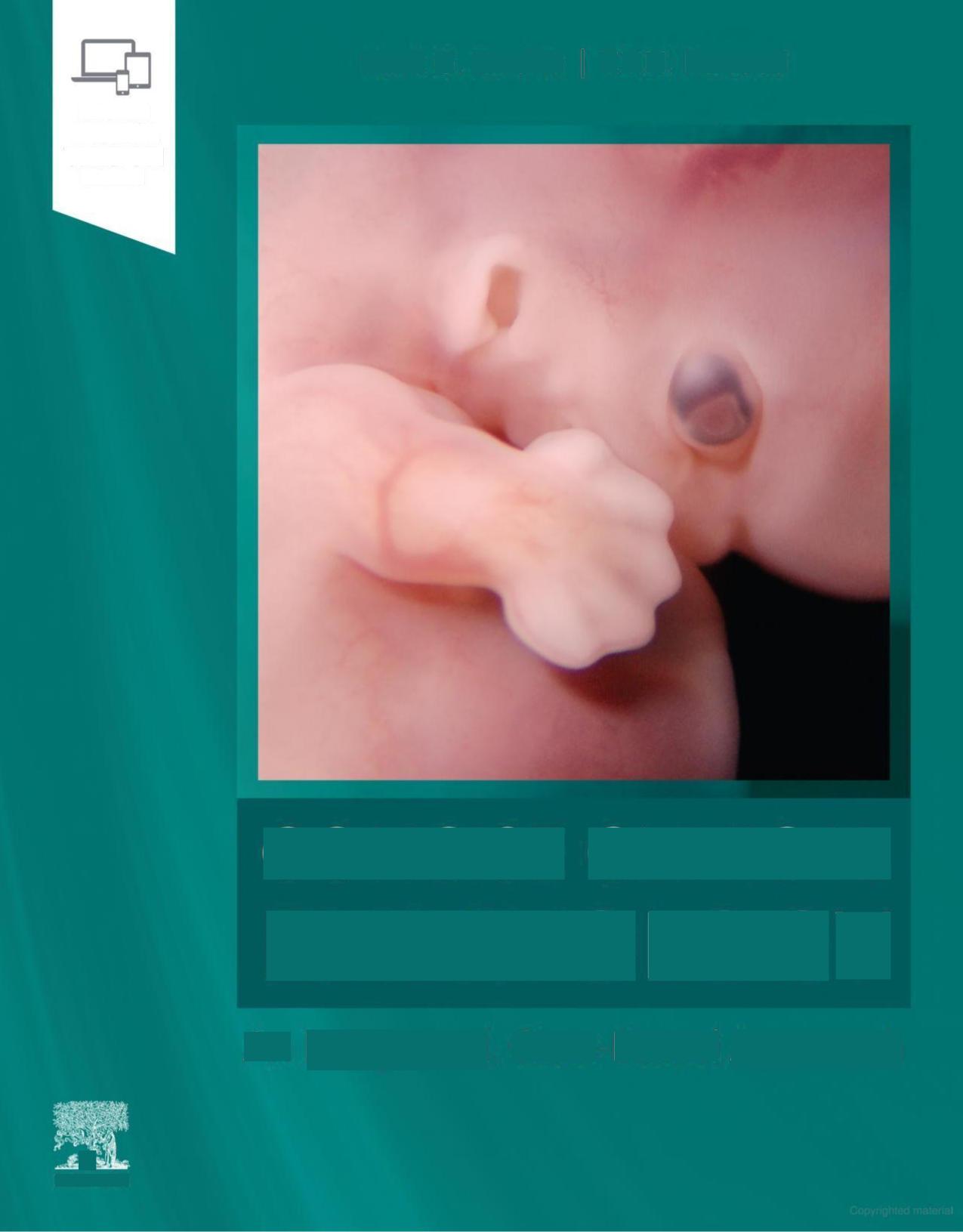
A 26-year-old woman (GW) presents to you, a nurse practitioner at a public health clinic, with severe odynophagia. The history and physical examination leads you to strongly believe she has a streptococcal pharyngitis; a swab is taken for rapid strep test (which is positive) and you prescribe amoxicillin. When you inquire about her obvious pregnancy, GW reports being approximately 5 months pregnant based on when she remembered having had her last menstrual period. She said that the father is a 55-year-old companion. GW has been living on the street and in shelters for the past year since discharge from an inpatient facility for treatment of a crystal methamphetamine addiction. She vehemently denies use of methamphetamine since that treatment. She has not sought any other medical care in the interim. GW has had only one previous pregnancy which resulted in the birth of a son, now 5 years old, and currently living with the maternal grandparents. Her son was born with a bilateral cleft palate. You recommend a fetal ultrasound as soon as possible, to which she agrees.
Questions for reflection: Why might an ultrasound fetal assessment be warranted? What concerns might you have related to the health of GW? What impact, if any, might these issues have on the health of her fetus, including risk for anomalies? Is the father’s age or the fact that her 5-year-old son had a cleft palate relevant to the current pregnancy? Why?
The study of embryology is essential for the understanding of both normal anatomy and congenital anomalies. Moreover, the practice of obstetrics and neonatal–perinatal medicine involves clinical embryology. Although infant mortality rates have been decreasing steadily in North America for the past 50 years, the 2018 rate in the United States remains at 5.6 per 1000 live births, 4.3 per 1000 in Canada and 11 per 1000 in Mexico. Given that congenital anomalies are the second leading cause of infant mortality (behind premature birth), the need to better understand the mechanisms of normal embryo and fetal development and the factors that impact this development, leading to congenital anomalies remains very high. The growing field of molecular biology and the development of many novel laboratory techniques have led to a significant improvement of our knowledge of the temporal and regional expression of genes and their products to control such processes as morphogenesis.
USING THIS TEXTBOOK
This textbook is designed to offer a concise knowledge base for the study or review of clinical embryology. The accompanying illustrations (drawing and medical imaging) provide a
visual resource to further enhance the textual explanations and development paths.
A clinical case scenario is provided for each chapter. As you will discover, the cases are not straightforward, and many of the words, concepts or considerations will require the reader to seek information outside the direct field of clinical embryology—this helps to situate the knowledge and details of embryology within the larger concept of clinical care. The clinical case in this chapter is a good example. You will need to consider, for example, infectious disease, genetics, pharmacology and neonatal cardiology and combine that knowledge to answer the question. The follow-on scenarios to the original case, found at the end of each chapter, will further expand your need to problem solve, think broadly and search for answers beyond this textbook. Answers are not provided to the cases so that they can be used for ongoing discussion amongst peers, advanced learning and knowledge testing.
Each chapter also provides molecular biology considerations. This information is based mainly on experiments with animal models including mice because the human cells or tissues required to examine such science are not generally available. As such, keep in mind that as knowledge of molecular genetics and biology progresses, the specific genes and their products that are identified may change or be expanded upon.
The section Clinical Issues in each chapter provides a description of the common congenital anomalies and other clinical information, related to the embryology details contained in the chapter.
Finally, each chapter provides a brief reference list that can be used to find additional details about the clinical cases, molecular biology and details of clinical embryology. We encourage you to seek additional information during your studies as the timing of book printing, relative to the constant gain of knowledge and reporting, negates the possibility of including the very most recent literature, although the authors have tried their utmost to provide citations that are as current as possible.
OTHER IMPORTANT INFORMATION
Throughout this textbook, the specified age of embryos and fetuses as it relates to specific structures and other
developments, has been quoted as fertilisation age—length of time from the date of fertilisation.
In the clinical context, gestational age is indicated as the time from the date of the start of the last menstrual period (LMP). Given that ovulation (and shortly thereafter, fertilisation) occurs typically around 14 days after the start of the menstrual period, gestational age LMP is approximately 2 weeks or 14 days greater than fertilisation age.
It is important to specifically describe the method used for indicating ‘gestational age’, so that confusion does not arise, especially when ordering or interpreting ultrasound images or comparing between times within a patient history.
Because the Federative International Committee on Anatomical Terminology does not recommend the use of eponyms, for the most part, this book follows suit (there are few exceptions to this when the clinical eponym is most commonly used).
There will be a number of terms in this book that may not be familiar to the reader, not limited to just those of embryology. It is recommended that the reader search for those definitions from a reliable source of such medical information.
Anatomical position and direction terms are used throughout this book. In adults, the terms anterior and posterior are used to describe the front and back of the body or limbs or relative positions of one structure to another. In the fetus or embryo, the terms ventral and dorsal are used, respectively. In addition, the terms caudal or rostral are used to denote a relationship to the head, whereas caudal is used to denote relationship to the caudal eminence or tail.
CLINICAL ISSUES
CLEFT PALATE
Palate clefts arise from failure of the lateral palatine process to fuse with:
• The primary palate (anterior palate cleft)
• Each other and the nasal septum (posterior palate cleft)
• The primary palate, with each other, and the nasal septum (secondary palate cleft).
Some clefts appear as part of single mutant gene or chromosomal syndromes or following the effects of teratogenic substances.
Case Outcome
Fetal ultrasound showed a male fetus of approximately 22 weeks of age (based on femur length, biparietal diameter, head circumference and abdominal circumference), which would approximately align with the predicted age based on the patient’s last menstrual period. The ultrasound also detected an isolated membranous ventricular septal defect (VSD). The remainder of the examination was normal. Sixteen weeks later, GW had a vaginal delivery. The neonate had good Apgar scores (7/8 at 1/5 minutes). The birth weight was at the 4th percentile. Otherwise the infant appeared normal.
Additional reflection: What is the error rate for estimating delivery dates from a single ultrasound examination at 22 weeks? Was it likely that the ultrasound was in error or that GW delivered early or both or neither? Why? What is the likely cause of the VSD? How common are these anomalies and what treatment is required and when, if any? What might be the causes for the baby to be born at such a low percentile birth weight? What other concerns might you have regarding the health of the neonate or GW?
BIBLIOGRAPHY
Methods for estimating the due date. Committee Opinion No. 700. American College of Obstetricians and Gynecologists. Obstet Gynecol 2017;129:e150–154.
Deshpande AS, Goudy SL. Cellular and molecular mechanisms of cleft palate development. Laryngoscope Investig Otolaryngol 2019;4(1):160–4.
Reproductive Organs and Gametogenesis 2
Case Scenario
A 24-year-old woman (KR) presents at her new family physician with difficulty conceiving. She and her husband have been trying to have a child for almost 5 years. Her husband recently had his sperm count and morphology tested, and this has proven to be normal. KR is now seeking additional advice and investigation for herself. KR describes her menstrual cycle as varying in length, and occasionally she has missed her period entirely. Otherwise, she has been healthy. KR also has severe acne. It had been previously controlled after a course of antibiotics and topical gel treatment when she was 21 years old, but the acne has now returned. KR mentioned that she has had acne since she was 13 years old. Recently, she began to notice more dark hair growth on her chin and areolas, and that her leg hair has had a noticeable regrowth after shaving. Her body mass index (BMI) is 20.6.
Questions for reflection: Are there other fertility considerations for KR’s husband beyond the semen analysis that might be investigated? What might be the connection, if any, between KR’s recurrent acne and potential fertility concerns? What further testing or consultation might be appropriate for KR?
PUBERTY
The reproductive organs (or primary sex characteristics) develop in utero. Maturation of the reproductive organs and the appearance of secondary sex characteristics (such as breast growth, presence of axillary and public hair) occur after puberty—the transitional process from childhood to adulthood. The exact biological trigger that starts the process of puberty is unclear; however the initiation of gonadotropin-releasing hormone (GnRH) pulsing leads to the secretion of luteinising hormone (LH) and follicle-stimulating hormone (FSH) by the pituitary. LH and FSH, in turn, stimulate the secretion of androgens and oestrogens from the gonads (the hypothalamic–pituitary–gonadal axis). The Tanner scale or sexual maturity rating (SMR; 1 = preadolescence to 5 = sexual maturity) is used as a framework on which to objectively classify the development of secondary sexual characteristics.
In females, the appearance of breast buds is the start of SMR 2, the first indication of the onset of puberty, and typically occurs between the ages of 8 and 12 years. Simultaneously, the labia, uterus and ovaries increase in size, and the
tissues of the uterus and vagina (endometrium and mucosa, respectively) increase in thickness. It is not until approximately 30 months later that menstruation begins, although the regularity of menstruation may be variable for a number of months as anovulatory cycles are common. In general, the age at which puberty begins in females has been decreasing since the mid-1940s; the reasons for this are not known, but may be related to the increase in child obesity or other environmental factors.
In males, the enlargement of the testicles and development of pubic hair are the early signs of the onset of puberty, typically occurring at approximately 10 years of age (SMR 2). The testes and the penis continue to enlarge until late adolescence under the influence of both LH and testosterone secretion as do the prostate and seminal vesicles. Sperm appear approximately 3 to 4 years following the onset of puberty. Although males undergo some degree of breast enlargement, gynaecomastia, during puberty, this tends to resolve spontaneously in later stages of adolescence.
MALE REPRODUCTIVE ORGANS
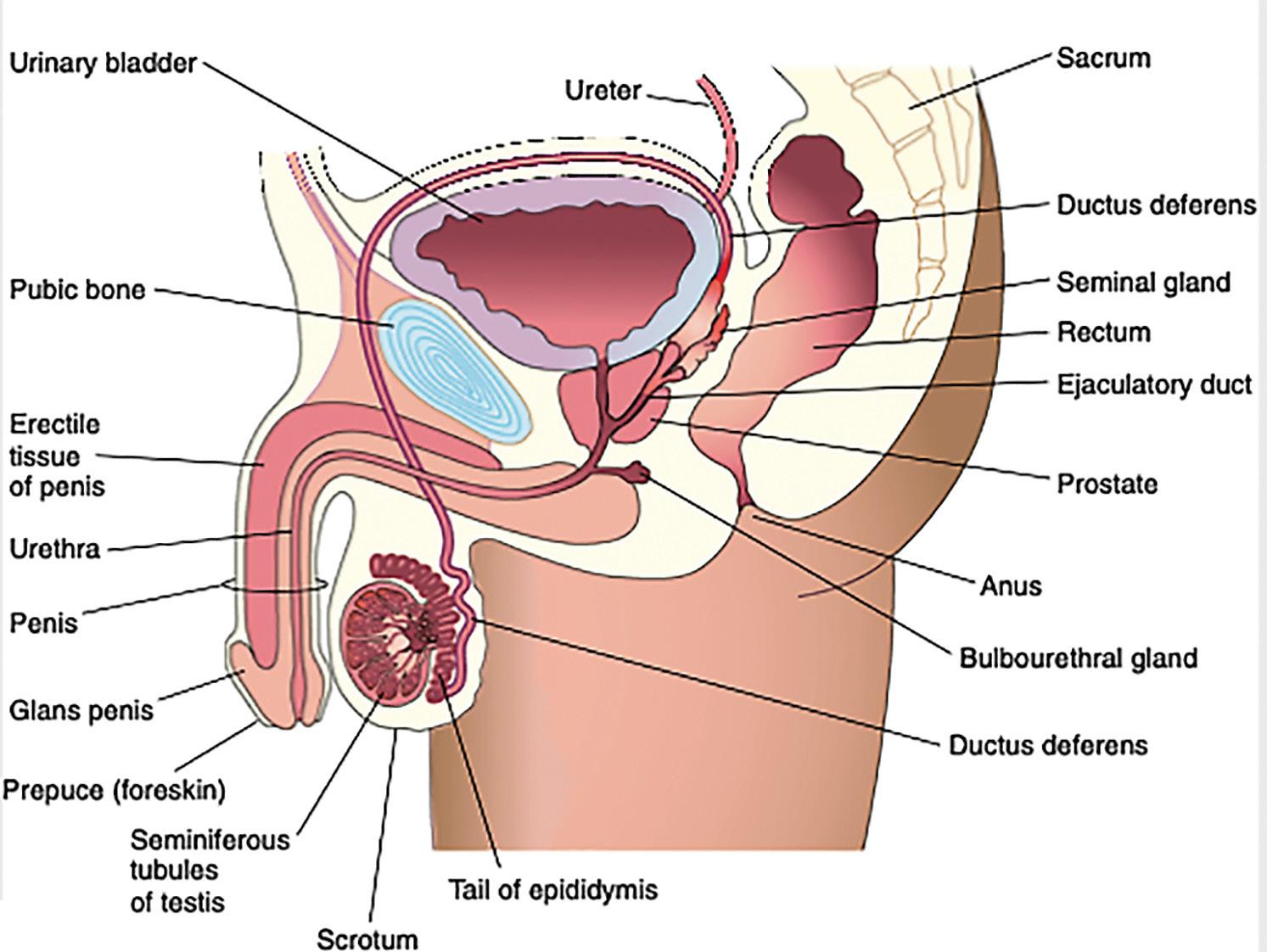
The penis (Fig. 2.1) acts as the conduit for both urine and ejaculate to exit the body. It consists of the glans or head, which in uncircumcised men is covered by the prepuce or foreskin. The urethral opening is found at the tip of glans penis which forms from the expanded distal end of the corpus spongiosum. The vascular corpus cavernous surrounds the corpus spongiosum, which when expanded by blood, provide the erectile function of the penis. The erectile tissue of the corpus spongiosum supports the urethra and maintains its patency during an erection.
The testes are the oval-shaped, sperm- and testosteroneproducing organs found within the scrotum. The testes are covered with a thick fibrous capsule, the tunica albuginea, and contain a series of coiled seminiferous tubules within which sperm development occurs. The seminiferous tubules are connected to the tubuli recti. The rete testes are connected to the epididymis. The duct of the epididymis (ductus deferens) passes from the epididymis through the inguinal canal into the pelvic cavity. The ductus deferens traverses the prostate gland where it joins the urethra. The prostate gland secretes prostatic fluid into the semen, which supports transportation and nutrition of the sperm. Paired seminal vesicles and the bulbourethral glands provide additional secretion to the semen.
FEMALE REPRODUCTIVE ORGANS
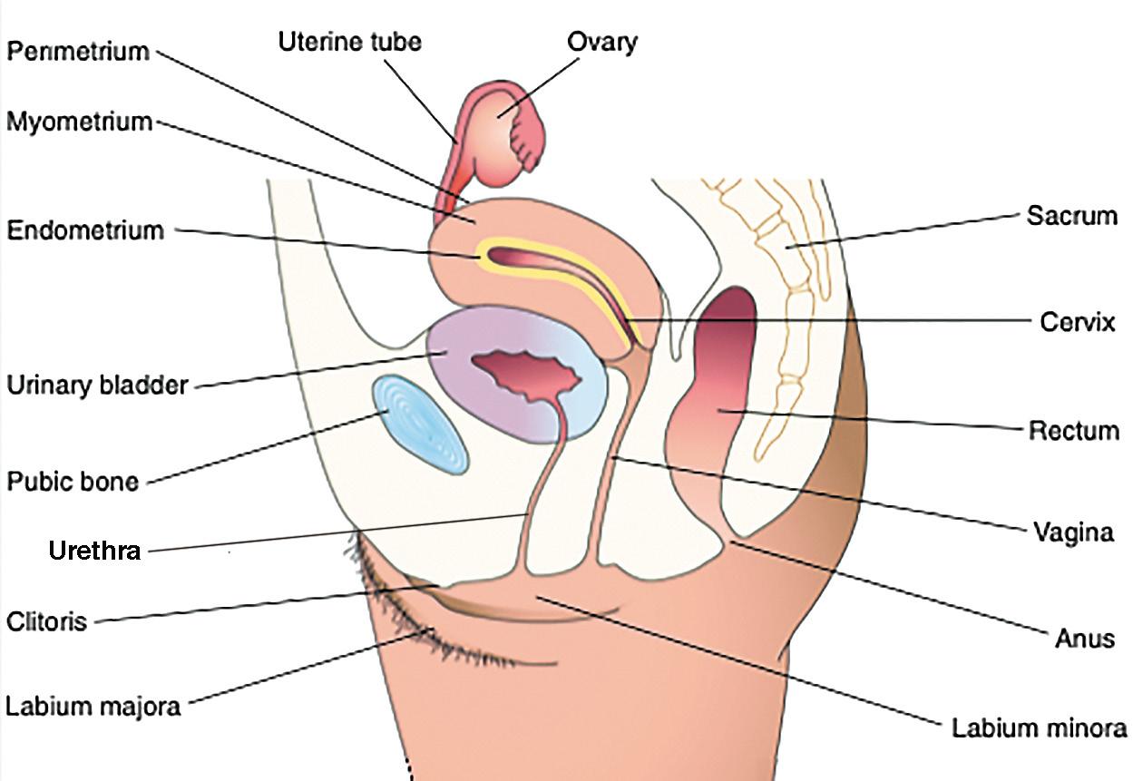
The vagina (Fig. 2.2) is a fibromuscular organ that extends from the external genitalia (vulvar structures) to the cervix of the uterus. The opening of the vagina is situated posterior to the opening of the urethra and is covered by the labia minora. The uterus is a thick-walled muscular organ consisting of the body (upper two-thirds) and the cervix (lower one-third). The cervix is cylindrical with constricted opening at both ends, the internal and external os. The body of the uterus is comprised of three tissue layers, endometrium
(internal), myometrium (middle muscular) and perimetrium (external). The endometrium can be further distinguished into the compact, spongy and basal layers, and varies in thickness according to stages of the menstrual cycle.
The uterine tubes are continuous with the uterine horns found at the superior end of the uterus, the fundus. The uterine tubes are approximately 10-cm long, and consist of four parts: infundibulum, ampulla, isthmus and uterine part. The tubes are lined with cilia that help to propel the ovum and sperm, first to the site of fertilisation (ampulla) and then to assist in moving the cleaving zygote to the uterus for implantation.
Fig. 2.1 Sagittal section of the male pelvic region. (From Moore KL, Persaud TVN, & Torchia MG. The Developing Human: Clinically Oriented Embryology. 10th ed. Philadelphia: Elsevier; 2015.)
Fig. 2.2 Sagittal section of the female pelvic region. (From Moore KL, Persaud TVN, & Torchia, MG. Before We Are Born: Essentials of Embryology and Birth Defects. 9th ed. Philadelphia: Elsevier; 2016.)
The ovaries are oval-shaped glands adjacent to the uterus and the uterine tube infundibulum, with its finger-like fimbriae. The ovaries produce the oocytes, as well as hormones (oestrogen and progesterone) that regulate the process of sexual development, menstruation and pregnancy. The external female genitalia consist of the labia minora, labia majora and the clitoris.
GAMETOGENESIS
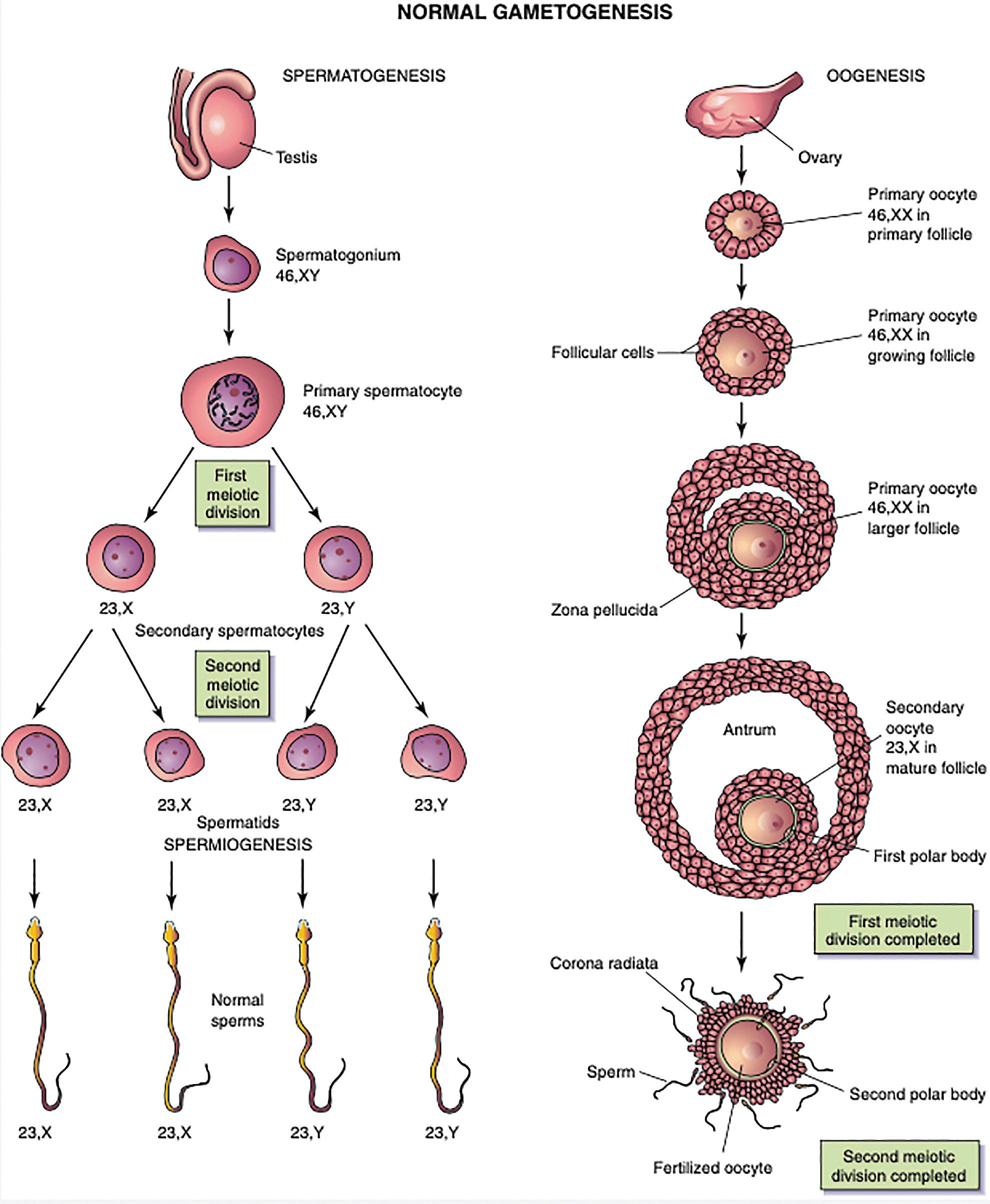
Gametogenesis (oogenesis and spermatogenesis) (Fig. 2.3) is the process that produces oocytes and sperms from bipotential primordial germ cells and prepares these gametes for fertilisation. The sperm and oocyte are highly specialised
Fig. 2.3 Simplified diagram showing normal gametogenesis. (From Moore KL, Persaud TVN, & Torchia MG. The Developing Human: Clinically Oriented Embryology. 10th ed. Philadelphia: Elsevier; 2015.)
sex cells, each of which contains the haploid number of chromosomes that are present in somatic cells. The number of chromosomes is reduced during meiosis, a special type of cell division that occurs only during gametogenesis. Meiosis involves two meiotic cell divisions resulting in diploid germ cells giving rise to haploid gametes.
The first meiotic division is a reduction division because the chromosome number is reduced to haploid by pairing of homologous chromosomes in prophase and their segregation at anaphase with one representative of each pair randomly going to each pole of the meiotic spindle. At this stage, the chromosomes are double-chromatid chromosomes. (The X and Y chromosomes are not homologues, but they have homologous segments at the tips of their short arms and pair in these regions only.) This disjunction of paired homologous chromosomes is the physical basis of segregation, the separation of allelic genes during meiosis. The second meiotic division does not have an interphase, but each double-chromatid chromosome divides, and each half, or chromatid, is drawn to a different pole. Thus the haploid number of chromosomes remains, and each daughter cell has one representative of each chromosome pair (now a single-chromatid chromosome). The process of meiosis provides constancy of the chromosome number from generation to generation, allows random assortment of maternal and paternal chromosomes between the gametes and relocates segments of maternal and paternal chromosomes by crossing over of chromosome segments, which produces a recombination of genetic material.
SPERM CHARACTERISTICS AND DEVELOPMENT
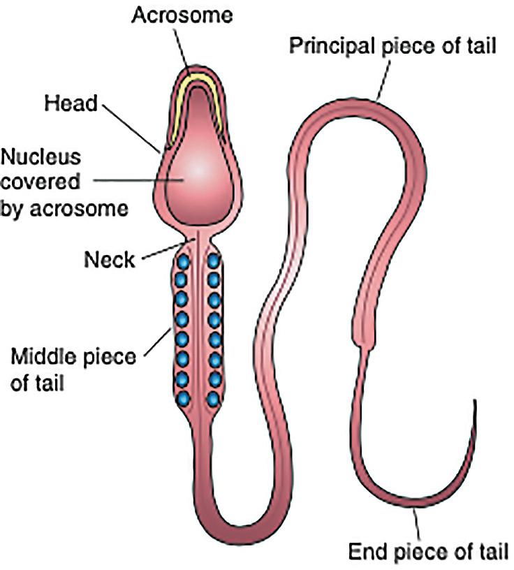
Sperms are highly differentiated, actively motile cells consisting of a head and a tail (Fig. 2.4) and approximately 4 µm in length. The head forms most of the bulk of the sperm and contains the nucleus. The anterior two-thirds of the head is covered by the acrosome, a saccular organelle
containing several enzymes and other factors which, when released, facilitate dispersion of the follicular cells of the corona radiata and sperm penetration of the zona pellucida during fertilisation. The tail has four segments: the connecting, middle, principal and end pieces, and it provides the motility of the sperm for transport to the site of fertilisation. The axoneme is the motility machinery of the sperm and is comprised of cytoskeleton and dyneins (ATPase molecular motors). The helically arranged mitochondria in the middle piece provide the energy required for motility. Sperm travel at approximately 3 mm/min.
Spermatogonia (primordial male germ cells) are dormant in the seminiferous tubules of the testes during the fetal and postnatal periods. At puberty, spermatogenesis begins, a 2-month highly complex process that transforms spermatogonia into mature sperms. More than one dozen different subtypes of male germ cells have been identified. There are also a number of cells and factors within the testes involved in sperm development. Peritubular myoid cells are found surrounding and supporting the seminiferous tubules and are thought to regulate Sertoli cells, assist in managing the blood–testis barrier (an important controller of the germ cell microenvironment) and push testicular fluid with sperm towards the rete testis. Leydig cells (LCs) are found clustered near seminiferous tubules and the adjacent blood vessels. LCs produce testosterone, which is released into the systemic circulation. LCs ensure a much higher local concentration of testosterone, which is required for normal sperm production. LCs also produce oestradiol from testosterone, which appears to be required for successful spermatogenesis. Sertoli cells (SCs) make up approximately 20% of the epithelial cells of the seminiferous tubules. The role of the SCs is complex and broad. Their unique structure allows each SC to shepherd up to 50 germ cells during differentiation; this is accomplished by sophisticated cytoskeletal elements. SCs produce anti-Müllerian hormone, critical to the normal embryological develop of male and female reproductive organs. SCs also act as macrophages, and produce inhibin B (regulating FSH production) and androgen-binding protein.
The male germ cells are arranged in the seminiferous tubules in a specific manner with least-mature cells in the basal compartment and more-mature cells found adjacent to the lumen.
The earliest germ cells in the testes (gonocytes) remain in G0 phase of the cell cycle until after birth. In the first few neonatal months, they are transformed into inactive spermatogonia which begin to undergo rapid mitosis at approximately 6 years of age. Later, at puberty, the spermatogonia undergo the process of spermatogenesis. Briefly, spermatogonia first develop into primary spermatocytes, the largest germ cells in the seminiferous tubules of the testes. Each primary spermatocyte subsequently undergoes the first meiotic division to form two haploid secondary spermatocytes. These secondary spermatocytes undergo the second meiotic division and form four haploid spermatids. The spermatids are gradually transformed into four mature sperms by a process known as spermiogenesis. When spermiogenesis is complete, the sperms enter the lumina of the seminiferous tubules. Sperms are transported passively from the seminiferous tubules to the epididymis, where they are stored.
Fig. 2.4 Main parts of the human sperm. (From Moore KL, Persaud TVN, & Torchia MG. The Developing Human: Clinically Oriented Embryology. 10th ed. Philadelphia: Elsevier; 2015; Fig. 2.5.)
OOCYTE CHARACTERISTICS AND DEVELOPMENT
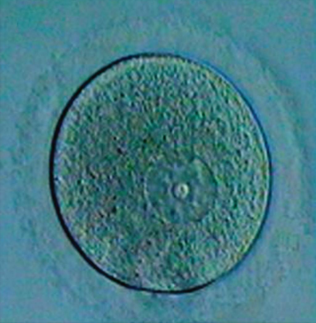
The mature (secondary) oocyte (Fig. 2.5) is an immotile cell with a diameter of approximately 100 µm, making it one of the largest cells in the female human body, and just visible to the unaided eye. It typically contains a transparent moderately granular cytoplasm with refractile structures such as lipid, lipofuscin bodies, and autophagic vacuoles. A single polar body is also associated with the secondary oocyte (see later).
Oogenesis transforms oogonia (primordial female germ cells) into mature oocytes. All oogonia develop prenatally, and the process of oogenesis ceases following menopause. In early fetal life, oogonia proliferate by mitosis and enlarge to form primary oocytes, each surrounded by a single layer of flattened, connective tissue follicular cells. The primary oocyte enclosed by this layer of follicular cells constitutes a primordial follicle. As the primary oocyte enlarges during puberty, the follicular epithelial cells become columnar shaped and the oocyte becomes covered with the glycoproteinaceous zona pellucida. Primary oocytes begin the first meiotic divisions before birth, but completion of prophase does not occur until puberty. The follicular cells surrounding the primary oocytes secrete a substance, oocyte maturation inhibitor, which keeps the meiotic process of the oocyte arrested. With puberty, the ovarian follicle (typically only one) matures each month. As a follicle matures, the primary oocyte increases in size and shortly before ovulation it completes the first meiotic division to give rise to a secondary oocyte and the first polar body. The secondary oocyte receives almost all the cytoplasm; the polar body is destined for degeneration. At ovulation, the nucleus of the secondary oocyte begins the second meiotic division, but it
progresses only to metaphase. If a sperm penetrates the secondary oocyte at fertilisation, the second meiotic division is completed, and most cytoplasm is again retained by one cell, the fertilised oocyte. The second polar body is formed and will degenerate.
There are approximately 2 million primary oocytes in the ovaries of a neonate, but most of them regress during childhood so that by adolescence no more than about 40,000 primary oocytes remain. Of these, only approximately 400 become secondary oocytes and are expelled at ovulation during the reproductive period. Very few of these oocytes, if any, are fertilised. The long duration of the first meiotic division (up to 45 years) may account in part for the relatively high frequency of meiotic errors that occur with increasing maternal age.
FEMALE REPRODUCTIVE CYCLE
The female reproductive cycle (Fig. 2.6) is highly complex and involves activities of the hypothalamus, pituitary gland, ovaries, uterus, uterine tubes, vagina and mammary glands, all towards preparation of the reproductive system for pregnancy.
GnRH secreted by the hypothalamus stimulates the anterior lobe of the pituitary gland to release FSH, which stimulates the development of ovarian follicles and the production of oestrogen by the follicular cells, and LH, which triggers ovulation and stimulates follicular cells and corpus luteum to produce progesterone and causes growth of the follicles and endometrium.
As the primary follicle increases in size, the adjacent connective tissue organises into a capsule, the theca folliculi. This theca soon differentiates an internal vascular and glandular layer (theca interna) and a capsule-like layer (theca externa). The follicular cells produce a stratified layer around the oocyte. Fluid-filled spaces appear around the follicular cells, which coalesce to form the antrum, containing follicular fluid at this stage; the ovarian follicle is then called a secondary follicle. The primary oocyte is pushed to one side of the follicle. At approximately the midpoint of the cycle, FHS and LH stimulation cause rapid follicle growth leading to the formation of a small avascular spot, follicular stigma, on the surface of the ovary. Rupture of the stigma and expulsion of the secondary oocyte (ovulation) occurs 12 to 24 hours after this surge of LH production. The expelled secondary oocyte is surrounded by the zona pellucida and one or more layers of radially arranged follicular cells (corona radiata). Shortly after ovulation, the walls of the ovarian follicle and theca folliculi collapse and develop into the corpus luteum. The corpus luteum secretes progesterone and some oestrogen, causing the endometrial glands to secrete and prepare the endometrium for implantation of the blastocyst. If the oocyte is fertilised, the corpus luteum enlarges to form a corpus luteum of pregnancy and increases its hormone production. The corpus luteum of pregnancy remains functionally active throughout about the first 20 weeks of pregnancy. By this time, the placenta has assumed the production of oestrogen and progesterone necessary for the maintenance of pregnancy. If the oocyte is not fertilised, the corpus luteum involutes and degenerates 10 to 12 days after ovulation.
Fig. 2.5 Photomicrograph of a human oocyte. (From Zhang P, Zucchelli M, Bruce S, et al. Transcriptome profiling of human pre-implantation development. PLoS One 2009; 4(11): e7844. With permission.)
MENSTRUAL CYCLE
Changes in the oestrogen and progesterone levels cause cyclic changes in the structure of the female reproductive tract, notably the uterine endometrium (Fig. 2.6). The menstrual cycle is a continuous process lasting on average 28 days, with each phase gradually passing into the next. Day 1 is designated as the day menstrual flow begins. In the menstrual phase (4–5 days), the functional layer of the endometrium is sloughed off as the menses (blood discharged from the vagina combined with small pieces of endometrial tissue). After menstruation, the remaining endometrium is thin.
During the proliferative phase (9 days), the ovarian follicles grow and the uterine surface epithelium reforms and covers the endometrium. The uterine glands increase in number and length and endometrial spiral arteries elongate. The luteal (secretory) phase lasts approximately 13 days and coincides with the formation, functioning and growth of the corpus luteum. The progesterone produced
by the corpus luteum stimulates the glandular epithelium to secrete a glycogen-rich material. The glands become wide, tortuous and saccular, and the endometrium thickens because of the influence of progesterone and oestrogen from the corpus luteum. As the spiral arteries grow into the superficial compact layer, they become increasingly coiled. The venous network becomes complex, and large lacunae (venous spaces) develop in the endometrium. If fertilisation does not occur the corpus luteum degenerates, oestrogen and progesterone levels fall, and the secretory endometrium enters an ischemic phase. The spiral arteries constrict, glandular secretion stops, interstitial fluid is reduced, endometrium shrinks and venous stasis occurs. This leads to patchy ischaemic necrosis of the functional layer of the endometrium. Rupture of damaged vessel walls allows blood to leak into the surrounding connective tissue, resulting in bleeding (typical loss of 20–80 mL).
If fertilisation occurs, the zygote undergoes cleavage and blastogenesis, and the blastocyst begins to implant in the
Fig. 2.6 Schematic drawing of the ovarian and menstrual cycles. (From Moore KL, Persaud TVN, & Torchia MG. The Developing Human: Clinically Oriented Embryology. 10th ed. Philadelphia: Elsevier; 2015.)
endometrium (sixth day of the luteal phase). The embryo syncytiotrophoblast produces chorionic gonadotropin, which keeps the corpora luteum secreting oestrogens and progesterone; the luteal phase continues and menstruation does not occur.
Molecular Biology Considerations
• PI3K/PTEN and TSC/mTOR pathways—activation of primordial follicles
• GDF9 and BMP15—development of secondary and preovulatory follicle
• cAMP meiotic arrest
• MAPK3/1—ovulation control
• bFGF—maintenance of blood–testis barrier
• TGF-a/b and GNDF—maintenance of spermatogenesis microenvironment
• PModS—regulates Sertoli cell function
• HOX—shaping of sperm head
CLINICAL ISSUES
FERTILITY
In 85% to 90% of cases, heterosexual couples are able to achieve pregnancy through sexual intercourse. In the remaining couples, fertility issues for both the male and female require investigation; these male/female concerns often coexist. In men, the most common causes of reduced fertility are a blockage of sperm delivery, altered sperm morphology, motility and function and reduced sperm numbers. Previous infection, retrograde ejaculation, prior trauma and tumours are examples of causes of blocked semen flow. Abnormal sperm morphology includes large or double heads and bent or double tails; causes include genetic disorders, exposure to environmental toxins or high testicular temperatures. Men with fewer than 10 million sperms per millilitre of semen are less likely to be fertile, especially when the specimen contains immotile and abnormal sperms. Environmental factors (drug or alcohol abuse, exposure to environmental toxins), medication and hormone imbalance are only a few of the reasons that low sperm counts may occur. In women, the most common causes of infertility are blockage of oocyte transportation attributed to tubal scarring or endometriosis, reduced production of oocytes because of increased age, and hormonal imbalances such as from polycystic ovarian syndrome (PCOS) and obesity.
CONTRACEPTION
The use of hormonal methods of female contraception can result in some or all of thickened cervical mucus, alteration of the endometrium, prevention of ovulation or blockage of sperm. These contraceptives include progestin-only pills, combined oestrogen–progestin pills, emergency contraceptive pills, vaginal rings, contraceptive patches and injectable long-acting medications including drug-integrated implants. Intrauterine devices may contain either hormones or copper and prevent sperm from reaching the ovum or
implantation. No hormone-based contraceptive is available for men. Barrier methods, including condoms (male or female) and contraceptive diaphragms, prevent sperm from entering the vagina or uterus, respectively. Sterilisation (implant, vasectomy or tubal ligation) are permanent forms of birth control.
NONDISJUNCTION
Nondisjunction is an error in cell division in which there is failure of a chromosomal pair (autosome or sex chromosome) to separate during mitosis or meiosis, resulting in numeric aberrations of chromosomes. Nondisjunction may occur during maternal or paternal gametogenesis, resulting in some gametes having 24 chromosomes while others have only 22. If these gametes should become fertilised with a normal gamete, a zygote with either trisomy (three copies of a chromosome) or monosomy (one copy of a chromosome) results. Clinical conditions resulting from such nondisjunction include trisomy 21 (Down syndrome), trisomy 18 (Edwards syndrome), XXY trisomy (Klinefelter syndrome) and monosomy X (Turner syndrome).
Case Outcome
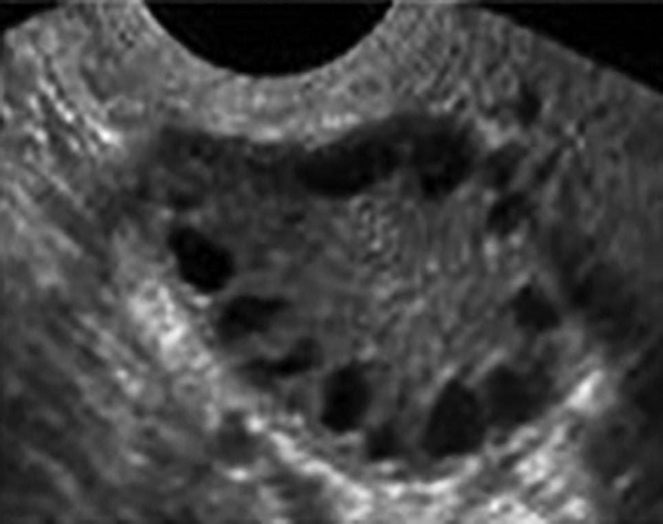
KR was sent for blood tests as well as endocrine and gynaecological consultations. Higher than normal levels of androgens were detected in her blood; there were no other abnormal findings. A pelvic examination was normal. A pelvic ultrasound demonstrated multiple cysts on her ovaries (see Fig. 2.7). She was diagnosed with polycystic ovarian syndrome (PCOS) and returned to her family physician for discussion of treatment options and follow-up.
Additional reflection: Did KR present with the typical signs for PCOS? Given KR’s desire for children, how might this be a consideration for her long-term treatment of PCOS? What is the likelihood of KR conceiving a child, should her husband’s fertility prove to be normal? What might be some psychological implications of PCOS?
Fig. 2.7
Pelvic ultrasound demonstrating cystic structures on the oval in polycystic ovarium syndrome. (From Karakas SE. New biomarkers for diagnosis and management of polycystic ovary syndrome. Clin Chim Acta 2017; 471: 248–253.With permission.)
QUESTIONS
1. Which of the following types of germ cell does not undergo cell division?
a. spermatogonia
b. primary oocytes
c. spermatids
d. secondary spermatocytes
e. oogonia
2. An infant is diagnosed as having 47 chromosomes instead of 46. This abnormal condition (trisomy) results from:
a. gene mutation
b. nondisjunction
c. disturbances in spermiogenesis
d. disturbances in mitosis
e. abnormal spermatogonia
BIBLIOGRAPHY
Datta J, Palmer MJ, Tanton C, et al. Prevalence of infertility and help seeking among 15,000 women and men. Hum Reprod 2016;31:2108–18. Neto FTL, Bach PV, Najari BB, Li PS, Goldstein M. Spermatogenesis in humans and its affecting factors. Sem Cell Dev Biol 2016;59:10–26. Pasquali R. Contemporary approaches to the management of polycystic ovary syndrome. Ther Adv Endocrinol Metb 2018;9(4):123–34.
Fertilisation and Reproductive Technologies 3
Case Scenario
A 49-year-old woman (PG) presents to her family physician reporting a positive home pregnancy test and claims to be approximately 2-months pregnant based on timing of her last normal menstrual period. She is concerned because this is her first pregnancy; she and her partner have not used birth control for the past three months because her menstrual periods have been very irregular for the past year and she believed that she was ‘in menopause and not infertile’.
A second pregnancy test was ordered which was positive. An ultrasound (endovaginal sonogram) showed a live embryo with crown–rump length of approximately 9 mm, aged between 35 and 38 days.
Given PG’s age, she was counselled regarding the options for (or no) prenatal screening for fetal aneuploidies.
Questions for reflection: Why is PG’s age a concern and how may age impact normal gametogenesis. What types of prenatal screening are available and at what gestational age? Which of the tests is considered diagnostic? What are the most common aneuploidies?
FERTILISATION
During ovulation, the fimbriated end of the uterine tube becomes closely applied to the surface of the ovary. The sweeping action of the tube and of fluid currents produced by the ciliated mucosal cells of the fimbriae, causes the extruded oocyte to enter the infundibulum of the uterine tube. The oocyte then passes into the ampulla of the tube, mainly as the result of tube peristalsis. During sexual intercourse and ejaculation, sperms are rapidly transported from the epididymis to the urethra by peristaltic contractions of the thick muscular coat of the ductus deferens. Between 200 and 600 million sperms are deposited in the vagina, around the external os and the fornix, and then some pass through the cervical canal. The cervical mucus increases in amount and becomes less viscid during ovulation, making it more favourable for sperm passage. Approximately 200 sperms reach the ampulla of the uterine tube; the remainder degenerate and are absorbed in the female genital tract. Sperms must undergo capacitation, lasting approximately 7 hours, before they are able to fertilise the oocyte. During this process, a glycoprotein coat and seminal proteins are removed from the surface of the sperm acrosome and the membrane components of the sperms are extensively altered. Sperms are usually capacitated while they are in the uterus or uterine tubes by substances secreted by these parts of the female
genital tract. Oocytes are usually fertilised within 12 hours of ovulation, and it appears that they cannot be fertilised after 24 hours (see Video 3.1).
The usual site of fertilisation is in the ampulla of the uterine tube. If the oocyte is not fertilised, it slowly passes along the tube to the body of the uterus, where it degenerates and is resorbed.
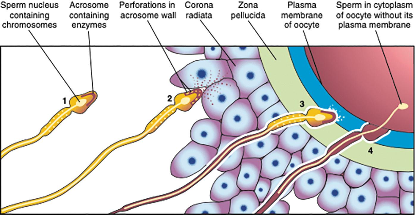
Fertilisation is a sequence of coordinated events (Fig. 3.2), beginning with the passage of a sperm through the corona radiata. Hyaluronidase released from the sperm acrosome, tubal mucosa enzymes and sperm motion appear to cause dispersal of the follicular cells of the corona radiata. Passage of a sperm through the zona pellucida is the next phase and also results from the action of enzymes released from the acrosome, including acrosin, esterase and neuraminidase. Once a sperm penetrates the zona pellucida, a change in the properties of the zona pellucida (zona reaction) occurs that makes it impermeable to other sperms. The zona reaction is believed to result from the action of lysosomal enzymes released by cortical granules near the plasma membrane of the oocyte. The contents of these granules also cause changes in the plasma membrane that make it impermeable to other sperms. Fusion and localised breakdown of cell membranes of the oocyte and sperm occurs next, resulting in the head and tail of the sperm entering the cytoplasm of the oocyte (the cell membrane and mitochondria of the sperm remain behind). Penetration of the oocyte by a sperm activates the oocyte into completing the second meiotic division and forming a mature oocyte and a second polar body. The maternal chromosomes decondense and the nucleus of the mature oocyte becomes the female pronucleus.
The nucleus of the sperm enlarges to form the male pronucleus and the tail of the sperm degenerates. Both pronuclei duplicate their DNA and the oocyte becomes an ootid. When the pronuclei fuse into a single diploid aggregation of chromosomes, the ootid becomes a zygote. The chromosomes in the zygote become arranged on a cleavage spindle in preparation for cleavage of the zygote. The zygote is genetically unique.
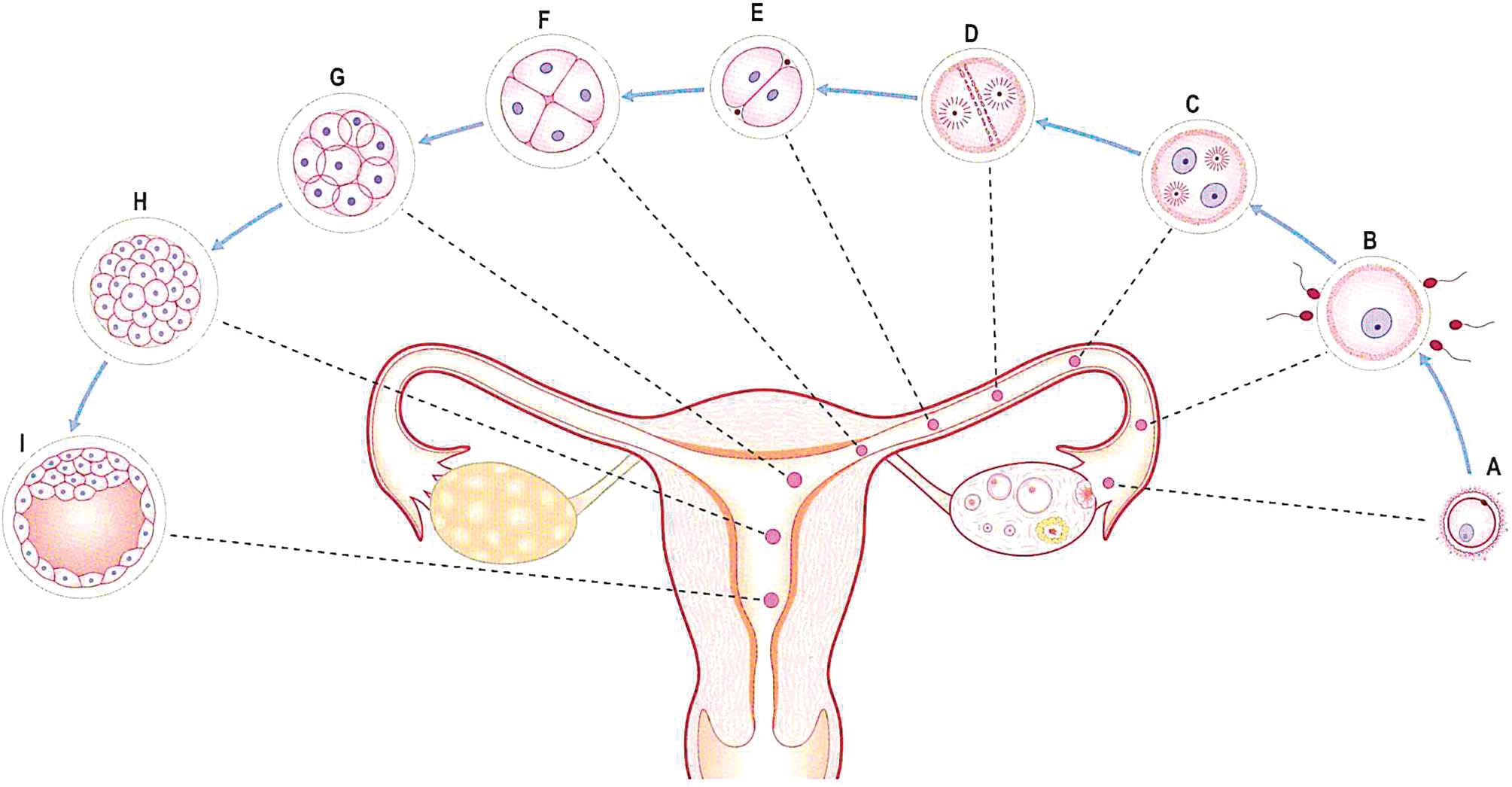
CLEAVAGE OF THE ZYGOTE AND FORMATION OF THE BLASTOCYST
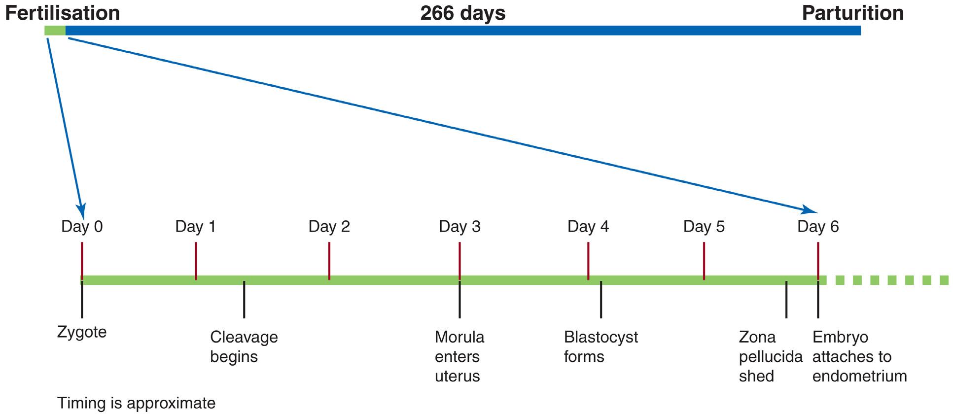
Cleavage occurs approximately 30 hours after fertilisation as the zygote, within the zona pellucida, passes along the uterine tube towards the uterus. Cleavage consists of repeated mitotic divisions of the zygote, resulting in a rapid increase in the number of cells (blastomeres) and decrease
in the size of subsequent blastomeres with each successive cleavage division. After the nine-cell stage, the blastomeres undergo compaction, changing their shape and tightly aligning themselves against each other to form a compact ball of cells. Compaction changes the cell cytoskeleton, permitting greater cell-to-cell interaction. Polarisation of the blastomeres into apical and basolateral domains also takes place. Compaction is necessary for segregation of the internal cells that will form the embryoblast (inner cell mass) of the blastocyst from surrounding cells that form the trophoblast (Fig. 3.3). At the 12- to 32-blastomeres stage, the developing embryo is called a morula. Shortly after the morula enters the uterus (approximately 4 days postfertilisation), the fluid-filled blastocystic cavity appears inside the morula separating the blastomeres into the trophoblast (thin outer cell layer giving rise to the embryonic part of the placenta) and the embryoblast (centrally located blastomeres which form the embryo). Early pregnancy factor (EPF), an immunosuppressant protein, is secreted by the trophoblastic cells and aids in the prevention of early maternal immune attack of the embryo (see Video 3.2).
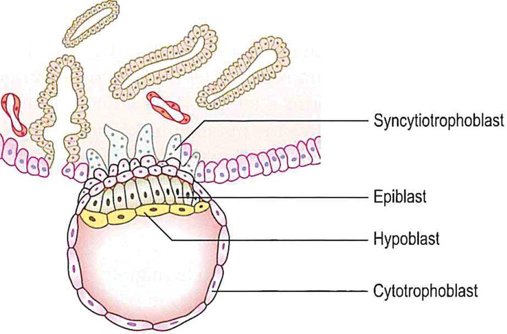
After the blastocyst has floated in the uterine secretions for approximately 2 days, shedding of the zona pellucida occurs, permitting the blastocyst to increase rapidly in size. While in the uterus, the embryo derives nourishment from secretions of the uterine glands. At approximately 6 days, the blastocyst (usually adjacent to the embryonic pole) attaches to the endometrial epithelium. The trophoblast proliferates rapidly and differentiates into two layers—an inner layer of cytotrophoblast that is mitotically active and forms new mononuclear cells that migrate into the increasing mass of syncytiotrophoblast, and an outer layer of syncytiotrophoblast (multinucleated protoplasmic mass) (Fig. 3.4). The syncytiotrophoblast begins to invade the uterine connective tissue so that the blastocyst can now derive its nourishment from the eroded maternal tissues. Endometrial cells assist to control the depth of penetration of the syncytiotrophoblast. At approximately 7 days, a layer of cells, the hypoblast (primary endoderm), appears on the surface of the embryoblast facing the blastocystic cavity. Comparative embryological data suggest that the hypoblast arises by delamination of blastomeres from the embryoblast.
Fig. 3.2 Events taking place in fertilisation. (From Moore KL, Persaud TVN, & Torchia MG The Developing Human: Clinically Oriented Embryology. 10th ed. Philadelphia: Elsevier; 2015.)
Fig. 3.1 Timeline of development related to fertilisation.
Fig. 3.3 Stages of development during the first week. (A) Ovulated oocyte; (B) fertilisation; (C) pronuclei formation; (D) first cleavage spindle; (E–G) cleavage of zygote; (H) morula; (I) blastocyst. (From Mitchell B, Sharma R. Embryology: An Illustrated Colour Text. 2nd ed. London: Elsevier; 2009.)
Fig. 3.4 A 7-day blastocyst beginning to implant. (From Mitchell B, Sharma R. Embryology: An Illustrated Colour Text. 2nd ed. London: Elsevier; 2009.)
Molecular Biology Considerations
• SPAM1, HYAL5, ACE3, ADAMS1–3—gamete fusion
• Hippo—segregation of embryoblast from trophoblast
• TGF-b—proliferation and differentiation of the trophoblast
CLINICAL ISSUES
ASSISTED REPRODUCTIVE TECHNOLOGIES
IN VITRO FERTILISATION AND EMBRYO TRANSFER
In vitro fertilisation (IVF) of oocytes and transfer of cleaving zygotes into the uterus have provided an opportunity for
many women who are sterile to have children. Since 1978, when Robert G. Edwards and Patrick Steptoe pioneered IVF, several million children have been born following an IVF procedure. The steps involved during IVF and embryo transfer are briefly noted. Beginning on day 1 of the menstrual cycle, ovarian follicles are stimulated to grow and mature (superovulation), typically by the administration of a drug that increases follicle-stimulating hormone (FSH) and/or luteinising hormone (LH) secretion by the pituitary. At the optimal time (often determined by ultrasound), another medication (synthetic human chorionic gonadotropin [hCG]) is given to trigger ovulation. Using an ultrasonically guided, minimally invasive procedure, several mature oocytes (typically 8–15) are aspirated from mature ovarian follicles. The oocytes are then placed in a Petri dish containing a special culture medium and capacitated sperms. Fertilisation of the oocytes and cleavage of the zygotes are monitored by microscope for 3 to 5 days. Depending on the mother’s age, one to three of the resulting embryos (four-cell to eight-cell stage, or early blastocysts) are transferred by introducing a catheter through the vagina and cervical canal into the uterus. Any remaining embryos are frozen for later use. Approximately 2 weeks later, a pregnancy test is performed.
CRYOPRESERVATION OF EMBRYOS
Early embryos resulting from IVF can be preserved for long periods by freezing them in liquid nitrogen with a cryoprotectant (e.g., glycerol or dimethyl sulfoxide). Successful transfer of four- to eight-cell embryos and blastocysts to the uterus after thawing is now a common practice. The longest period of sperm cryopreservation that resulted in a live birth was reported to be 21 years.
INTRACYTOPLASMIC SPERM INJECTION
A sperm can be injected directly into the cytoplasm of a mature oocyte. This technique has been successfully used for
the treatment of couples in whom typical IVF has failed, or in cases where there are too few sperms available.
ASSISTED IN VIVO FERTILISATION
Gamete intrafallopian (intratubal) transfer enables fertilisation to occur in the uterine tube. It involves superovulation (similar to that used for IVF), oocyte retrieval, sperm collection and laparoscopic placement of several oocytes and sperms into the uterine tubes. Using this technique, fertilisation occurs in the ampulla, its usual location.
SURROGATE MOTHERS
Some women produce mature oocytes but are unable to become pregnant, for example, a woman who has had a hysterectomy. In these cases, IVF may be performed, and the embryos transferred to another woman’s uterus for fetal development and delivery.
PREGNANCY TESTING
Most pregnancy tests are based on the detection or measurement of human chorionic gonadotropin (hCG) produced by the syncytiotrophoblast. hCG can be measured in urine or in blood. hCG rapidly increases in concentration from the time of early implantation (approximately day 6–12). The blood testing performed by clinical laboratories uses a more sensitive assay, and it also measures hCG concentration which can be followed over time depending on clinical needs. There are a number of biological and pharmacological factors that can produce false positive and negative hCG test results including heterotrophic antibodies, rheumatoid factors and ectopic pregnancies.
Transvaginal ultrasonography can detect the gestational sac at approximately 2.5 to 3 weeks following conception.
ANEUPLOIDY
Aneuploidy, usually resulting from nondisjunction, is any deviation from the diploid number of 46 chromosomes and is the most common (3%–4% of pregnancies) and clinically significant numeric chromosomal abnormalities.
MONOSOMY X
The incidence of Turner syndrome (45,X) is about 1:8000 live births. Only 1% of monosomy X female embryos survive, with 45,X being the most common abnormality detected in all spontaneous abortions. When it is possible to trace, it is the paternal X chromosome that is missing in approximately 75% of cases. In some cases, mosaicism occurs (XX/X and XY/X mosaics) and in these cases there is a lesser degree of abnormalities. The abnormalities typically seen with 45,X include small stature, ovarian dysgenesis, broad chest with wide-spaced nipples, congenital lymphedema and a short and/or webbed neck.
AUTOSOMAL TRISOMY
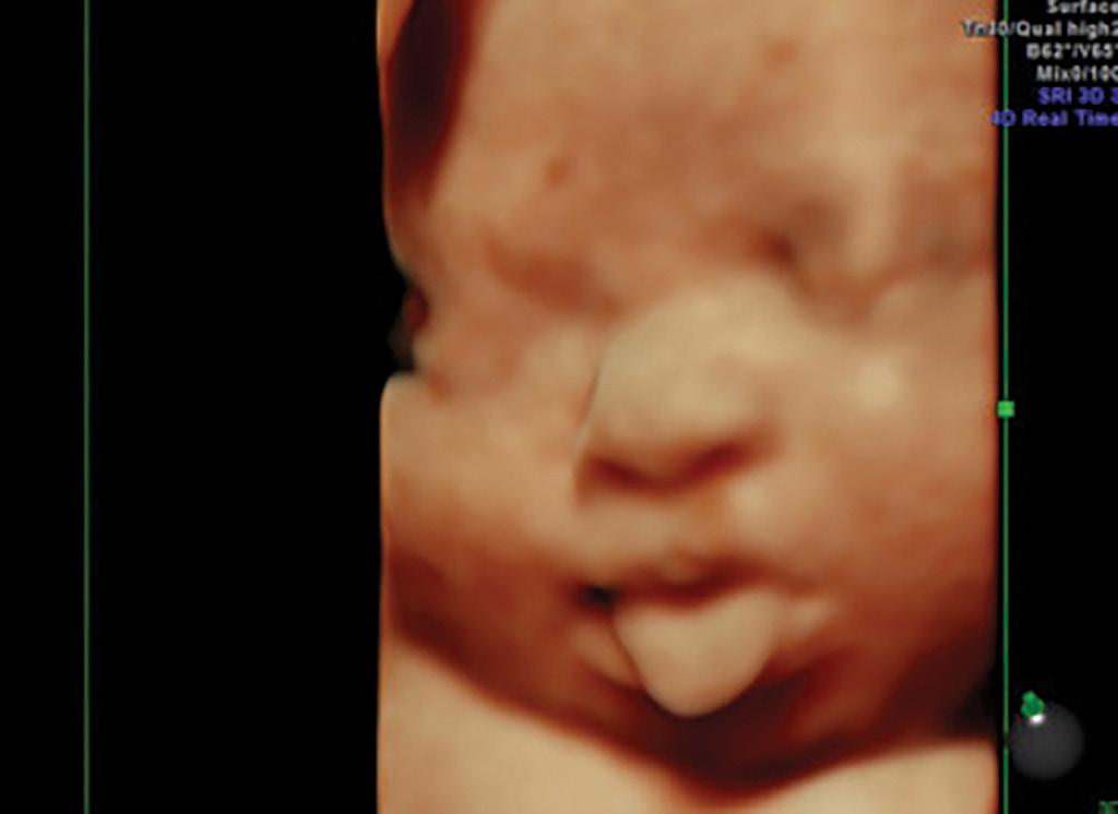
Trisomy is the most common aneuploidy. Trisomy of autosomes is mainly associated with three syndromes: trisomy 18 (Edwards syndrome, 1.3:10,000 live births), trisomy 13 (Patau syndrome, 0.8:10,000) and trisomy 21 (Down syndrome, 12:10,000). Infants with trisomy 13 and trisomy 18 are severely malformed (trisomy 13—includes defects of the lip,
nose and eyes, holoprosencephaly, polydactyly, skin defects; trisomy 18—includes clenched hands, prominent occiput, short sternum, small pelvis, cryptorchidism) and have major neurodevelopmental disabilities. Infants with these lifelimiting disorders (trisomy 18 and 23) have a 1-year survival rate of approximately 6% to 12%. Infants with trisomy 21 (Fig. 3.5) have abnormalities that include cognitive deficiency, hypotonia, bradycephaly, upward slanting palpebral fissures, protruding tongue, small ears, heart defects such as endocardial defects, ventricular septal defect or atrial septal defect, and a transverse palmar flexion crease. More than one-half of trisomic embryos spontaneously abort early. Trisomy of the autosomes occurs with increasing frequency as maternal age increases. For example, trisomy 21 occurs once in approximately 1400 births among mothers between the ages of 20 and 24 years but once in approximately 25 births among mothers 45 years and older. Because of the current trend of increasing maternal age, it has been estimated that children born to women older than 34 years will account for 39% of infants with trisomy 21.
TRISOMY OF SEX CHROMOSOMES
These disorders are not usually detected until puberty because there are no characteristic physical findings in infants or children. These disorders include XYY syndrome (1:1000; tall stature, cognitive disabilities, severe acne, autism spectrum disorder, normal fertility), XXX (1:1000; normal puberty, normal fertility, some degree of cognitive deficiency can occur); and XXY syndrome (Klinefelter syndrome) (1:500; most common cause of hypogonadism and infertility, gynaecomastia, inadequate virilisation, long limbs and possible developmental delay).
MOSAICISM
A person with at least two cell lines with two or more genotypes is considered a mosaic. The autosomes or sex chromosomes may be involved. The defects usually are less serious than in persons with monosomy or trisomy. For instance, the features of Turner syndrome are not as evident in 45,X/46,XX mosaic females as in the usual 45,X females.
Fig. 3.5 Three-dimensional ultrasound image of a fetus with trisomy 21, showing characteristic features including a protruding tongue (macroglossia).
Although mosaicism usually results from nondisjunction, it can also occur through the loss of a chromosome by anaphase lagging; chromosomes separate normally, but one of them is delayed in its migration and is eventually lost.
MULTIPLE GESTATIONS
In North America, twins naturally occur approximately once in every 85 pregnancies, triplets approximately once in 902 pregnancies, quadruplets once in 903 pregnancies and quintuplets approximately once in every 904 pregnancies. Twins that originate from two zygotes are dizygotic (DZ) twins whereas twins that originate from one zygote are monozygotic (MZ) twins. Two-thirds of twins are DZ, with marked racial differences whereas the incidence of MZ twinning is approximately the same in all populations. DZ twins may be of the same sex or different sexes and are no more alike genetically than brothers or sisters born at different times. The fetal membranes and placentas vary according to the origin of the twins. DZ twins always have two amnions and two chorions, but the chorions and placentas may be fused. Anastomoses between blood vessels of fused placentas of DZ twins may result in erythrocyte mosaicism. MZ twins are genetically identical; physical differences between MZ twins are caused by environmental differences, chance variation and uneven X-chromosome activation. MZ twinning usually results from division of the embryoblast into two embryonic primordia, with each embryo in its own amniotic sac but sharing the same chorionic sac and placenta (monochorionic–diamniotic twin). Uncommonly, early separation of embryonic blastomeres (e.g., during the two-cell to eightcell stages) results in MZ twins with two amnions, two chorions and two placentas that may or may not be fused. Twin transfusion syndrome occurs in as many as 10% to 15% of monochorionic–diamniotic MZ twins. There is shunting of arterial blood from one twin through unidirectional umbilical–placental arteriovenous anastomoses into the venous circulation of the other twin. The donor twin is small, pale and anaemic whereas the recipient twin is large and has polycythaemia. In lethal cases, death results from anaemia in the donor twin and congestive heart failure in the recipient twin. Late division of early embryonic cells, such as division of the embryonic disc during the second week, results in MZ twins that are in one amniotic sac and one chorionic sac. A monochorionic–monoamniotic twin placenta is associated with fetal mortality rates that are higher by up to 10%, with the cause being cord entanglement. Because ultrasonographic studies are a common part of prenatal care, it is known that early death and resorption of one member of a twin pair is common. Triplets may be derived from one zygote and be identical, two zygotes and consist of identical twins and a singleton or three zygotes and be of the same sex or of different sexes. The determination of twin zygosity is done by molecular diagnosis.
PREIMPLANTATION GENETICS
In couples with inherited genetic disorders and using IVF, preimplantation genetic diagnosis can determine the
genotype of the embryo and allow selection of a chromosomally healthy embryo for transfer. Preimplantation genetic diagnosis can be carried out 3 to 5 days after IVF of the oocyte. One or two cells (blastomeres) are removed from the embryo and these cells are then analysed before transfer into the uterus. The sex of the embryo can also be determined from one blastomere taken from a six- to eight-cell dividing zygote, and analysed by polymerase chain reaction and fluorescence in situ hybridisation techniques. This procedure has been used to detect female embryos during IVF in cases in which a male embryo would be at risk of a serious X-linked disorder. The polar body may also be tested for diseases where the mother is the carrier (Fig. 2.15A).
Case Outcome
Patient PG opted for noninvasive prenatal testing (NIPT) through cell-free DNA (cfDNA) screening. This testing was conducted approximately 3 weeks after her previous visit (8 weeks post conception—10 weeks gestational age). The test results demonstrated a high risk for trisomy 21 (Down syndrome).
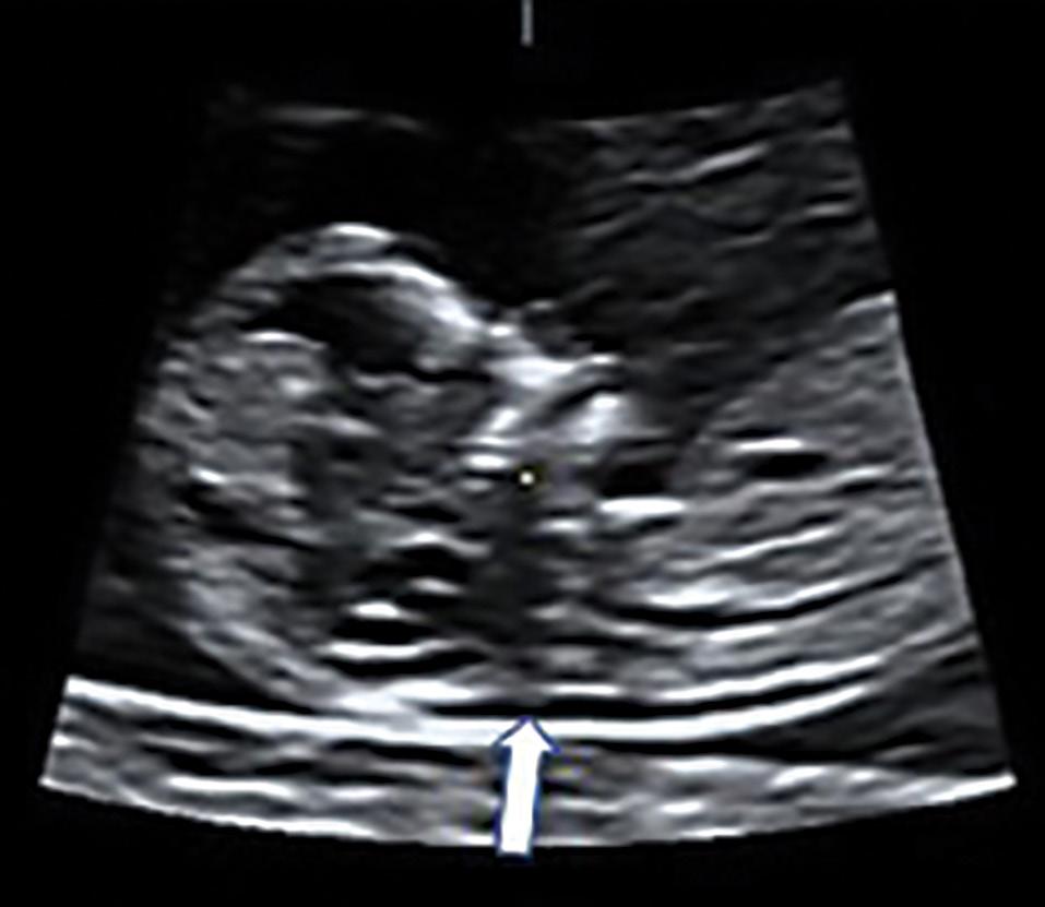
A diagnostic chorionic villus sampling (CVS) was then performed that confirmed the diagnosis of trisomy 21. PG opted to continue the pregnancy. A later fetal ultrasound demonstrated enhanced nuchal translucency (Fig. 3.6), but no cardiovascular anomalies. The remainder of the pregnancy was uneventful, and PG delivered a baby girl at 38 weeks.
Additional reflection: What is the difference between a screening test and a diagnostic test? How is CVS performed, at what gestational age, and with what possible risks to fetus and the mother? What is a nuchal translucency, and why was there a concern about cardiovascular anomalies?
Fig. 3.6 Ultrasound of a fetus demonstrating an enhanced nuchal translucency (arrow)
QUESTIONS
1. How many sperms would probably be deposited by a normal young adult male in the vagina during intercourse:
a. 300,000
b. 3 million
c. 30 million
d. 300 million
e. 3 billion
2. The secondary oocyte completes the second meiotic division:
a. before ovulation
b. during ovulation
c. at fertilisation
d. before birth
e. at puberty
3. The sperm penetrates the zona pellucida, partially assisted by enzymes that are released from which portion of the sperm:
a. middle piece
b. acrosome
c. neck
d. main piece
e. head
4. Morphologically abnormal sperm may cause:
a. monosomy
b. congenital anomalies
c. trisomy
d. abnormal embryos
e. infertility
BIBLIOGRAPHY
Jelin AC, Sagasser KG, Wilkins L. Prenatal genetic testing options. Pediatr Clin North Am 2019;66:281–93. Bamberg C, Hecher K. Update on twin-to-twin transfusion syndrome. Best Pract Res Clin Obstet Gynaecol 2019;58:55–65. Katz DJ, Teloken P, Shoshany O. Male infertility – The other side of the equation. Aust Fam Physician 2017;46:641–6.