Activate the eBook version of this title at no additional charge.
Elsevier eBooks for Practicing Clinicians gives you the power to browse and search content, view enhanced images, highlight and take notes—both online and o ine.
Unlock your eBook today.
1. Visit expertconsult.inkling.com/redeem
2. Scratch box below to reveal your code
3. Type code into “Enter Code” box
4. Click “Redeem”
5. Log in or Sign up
6. Go to “My Library” It’s that easy!
For technical assistance: email expertconsult.help@elsevier.com call 1-800-401-9962 (inside the US) call +1-314-447-8300 (outside the US) Place Peel Off Sticker Here
Use of the current edition of the electronic version of this book (eBook) is subject to the terms of the nontransferable, limited license granted on expertconsult.inkling.com. Access to the eBook is limited to the first individual who redeems the PIN, located on the inside cover of this book,
No part of this publication may be reproduced or transmitted in any form or by any means, electronic or mechanical, including photocopying, recording, or any information storage and retrieval system, without permission in writing from the publisher. Details on how to seek permission, further information about the Publisher’s permissions policies and our arrangements with organizations such as the Copyright Clearance Center and the Copyright Licensing Agency, can be found at our website: www.elsevier.com/permissions.
This book and the individual contributions contained in it are protected under copyright by the Publisher (other than as may be noted herein).
Notice
Practitioners and researchers must always rely on their own experience and knowledge in evaluating and using any information, methods, compounds or experiments described herein. Because of rapid advances in the medical sciences, in particular, independent veri cation of diagnoses and drug dosages should be made. To the fullest extent of the law, no responsibility is assumed by Elsevier, authors, editors or contributors for any injury and/or damage to persons or property as a matter of products liability, negligence or otherwise, or from any use or operation of any methods, products, instructions, or ideas contained in the material herein.
Chief of Gynecologic Specialty Surgery, Sloane Hospital for Women Columbia University New York, New York
Jamie N. Bakkum-Gamez, MD Professor Obstetrics and Gynecology Mayo Clinic Rochester, Minnesota
Genevieve Bouchard-Fortier, MD, FRCSC, MSc
Assistant Professor University Health Network Division of Gynecologic Oncology
Obstetrics and Gynecology University of Toronto Toronto, Ontario, Canada
Anne Burke, MD, MPH
Associate Professor Gynecology and Obstetrics Johns Hopkins University Baltimore, Maryland
Leslie H. Clark, MD Assistant Professor
Obstetrics and Gynecology, Division of Gynecologic Oncology University of North Carolina at Chapel Hill, Chapel Hill, North Carolina
Robert L. Coleman, MD Professor & Deputy Chair Department of Gynecologic Oncology & Reproductive Medicine University of Texas MD Anderson Cancer Center Houston, Texas
Allan Covens, MD, FRCSC Head Sunnybrook Health Science Center; Professor & Chair Division of Gynecologic Oncology
Obstetrics and Gynecology University of Toronto Toronto, Ontario, Canada
Deborah S. Cowley, MD Professor Psychiatry and Behavioral Sciences University of Washington Seattle, Washington
Anne R. Davis, MD, MPH
Wyeth Ayerst Professor
Obstetrics and Gynecology
Columbia University Irving Medical Center New York, New York
Mary Segars Dolan, MD, MPH
Associate Professor
Gynecology and Obstetrics
Emory University Atlanta, Georgia
Sarah K. Dotters-Katz, MD, MMHPE
Assistant Professor
Obstetrics and Gynecology
Duke University Durham, North Carolina
Nataki C. Douglas, MD, PhD
Associate Professor
Department of Obstetrics, Gynecology and Women’s Health
Rutgers–New Jersey Medical School Newark, New Jersey
Sean C. Dowdy, MD Professor
Obstetrics and Gynecology Mayo Clinic Rochester, Minnesota
Linda O. Eckert, MD Professor
Department of Obstetrics and Gynecology; Adjunct Professor Department of Global Health University of Washington Seattle, Washington
Michael Fialkow, MD, MPH Professor
Obstetrics and Gynecology University of Washington School of Medicine Seattle, Washington
Eric J. Forman, MD, HCLD Medical and Laboratory Director Department of Obstetrics and Gynecology Columbia University Irving Medical Center
Division of Reproductive Endocrinology & Infertility
New York, New York
Michael Frumovitz, MD, MPH Professor and Associate Chief Patient Experience Of cer
Gynecologic Oncology and Reproductive Medicine
University of Texas MD Anderson Cancer Center Houston, Texas
Paola Alvarez Gehrig, MD
Professor & Chief
Division of Gynecologic Oncology
University of North Carolina Chapel Hill, North Carolina
David M. Gershenson, MD Professor
Gynecologic Oncology and Reproductive Medicine
University of Texas MD Anderson Cancer Center Houston, Texas
Jennifer Bushman Gilner, MD, PhD
Assistant Professor
Obstetrics and Gynecology
Duke University Durham, North Carolina
Laura J. Havrilesky, MD, MHSc Professor, Division of Gynecologic Oncology
Obstetrics and Gynecology
Duke University
Durham, North Carolina
Cherie C. Hill, MD
Assistant Professor Gynecology and Obstetrics
Emory University School of Medicine
Atlanta, Georgia
Hye-Chun Hur, MD, MPH
Associate Professor
Minimally Invasive Gynecologic Surgery Department of Obstetrics and Gynecology
Columbia University Irving Medical Center
New York, New York
Anuja Jhingran, MD Professor Radiation Oncology
University of Texas MD Anderson Cancer Center Houston, Texas
James M. Kelley III, BA, JD Managing Partner Medical Malpractice
Elk & Elk Co Ltd Cleveland, Ohio
Anna C. Kirby, MD, MAS
Assistant Professor
Obstetrics and Gynecology
University of Washington Seattle, Washington
Jeffrey A. Kuller, MD
Professor of Obstetrics and Gynecology Division of Maternal-Fetal Medicine
Duke University Medical Center Durham, North Carolina
Eduardo Lara-Torre, MD, FACOG Vice Chair, Department of OBGYN Section Chief, Academic Specialists in General OBGYN Carilion Clinic
Professor Department of OBGYN and Pediatrics Virginia Tech-Carilion School of Medicine Roanoke, Virginia
Gretchen M. Lentz, MD, FACOG Professor, Obstetrics and Gynecology Adjunct Professor, Urology Division Director, Urogynecology University of Washington Medical Center Seattle, Washington
Roger A. Lobo, MD Professor, Obstetrics and Gynecology Division of Reproductive Endocrinology Columbia University New York, New York
Karen H. Lu, MD Chair and Professor Gynecologic Oncology and Reproductive Medicine
University of Texas MD Anderson Cancer Center Houston, Texas
Vicki Mendiratta, MD
Associate Professor Obstetrics and Gynecology University of Washington Seattle, Washington
Larissa A. Meyer, MD, MPH
Associate Professor Gynecologic Oncology and Reproductive Medicine University of Texas MD Anderson Cancer Center Houston, Texas
Jane L. Miller, MD
Associate Professor Urology University of Washington Seattle, Washington
Andra Nica, MD, MSc, FRCSC Clinical Fellow
Obstetrics and Gynaecology
Division of Gynecologic Oncology University of Toronto Toronto, Ontario, Canada
Jaclyn D. Nunziato, MD, MS
Assistant Professor of Obstetrics and Gynecology
Department Obstetrics and Gynecology
Virginia Tech Carilion School of Medicine Roanoke, Virginia Roanoke, Virginia
James W. Orr, Jr., MD, FACS, FACOG
Clinical Professor, Florida State College of Medicine
Medical Director, Regional Cancer Center Lee Health
Chief of Surgical Oncology, GenesisCare Tallahassee, Florida
Amanda Padro, MS, CGC
Prenatal Genetic Counselor
MFM OB/GYN
Duke University Raleigh, North Carolina
Natacha Phoolcharoen, MD Lecturer
Obstetrics and Gynecology
Faculty of Medicine, Chulalongkorn University Bangkok Thailand;
Visiting Scientist
Gynecologic Oncology and Reproductive Medicine
University of Texas MD Anderson Cancer Center Houston, Texas
Thomas M. Price, MD
Professor
Obstetrics and Gynecology
Duke University Durham, North Carolina
Beth W. Rackow, MD
Associate Professor
Obstetrics & Gynecology and Pediatrics
Columbia University Medical Center
New York, New York
Pedro T. Ramirez, MD
Professor
Gynecologic Oncology & Reproductive Medicine
University of Texas MD Anderson Cancer Center; Director
Minimally Invasive Surgical Research & Education
University of Texas MD Anderson Cancer Center Houston, Texas; Editor in Chief
International Journal of Gynecological Cancer
Licia Raymond, MD
Clinical Assistant Professor
Obstetrics-Gynecology
University of Washington
Seattle, Washington
Eleanor H. J. Rhee, MD
Assistant Professor
Division of Maternal Fetal Medicine
Obstetrics and Gynecology
Duke University
Katherine Rivlin, MD, MSc
Assistant Professor
Obstetrics and Gynecology
The Ohio State University Wexner School of Medicine
Columbus, Ohio
David T. Rock, MD
Director of Breast Surgery Fellowship
21st Century Oncology;
Breast Surgeon
Regional Breast Care
Fort Myers, Florida
Timothy Ryntz, MD
Assistant Professor
Obstetrics and Gynecology
Columbia University School of Medicine
New York, New York
Mila Pontremoli Salcedo, MD, PhD
Associate Professor
The Department of Obstetrics & Gynecology
Federal University of Health Sciences/ Irmandade Santa Casa de Misericordia de Porto Alegre, Porto Alegre, Brazil; Visiting Assistant Professor
The Department of Gynecologic Oncology and Reproductive Medicine
University of Texas MD Anderson Cancer Center
Houston, Texas
Gloria Salvo, MD
Medical Research
Gynecologic Oncology and Reproductive Medicine
University of Texas MD Anderson Cancer Center
Houston, Texas
Samith Sandadi, MD, MSc
Gynecologic Oncologist
Breast Surgeon
Clinical Assistant Professor
Florida State School of Medicine
Florida Gynecologic Oncology
21st Century Oncology
Fort Myers, Florida
Kathleen M. Schmeler, MD Professor
Department of Gynecologic Oncology & Reproductive Medicine
The University of Texas MD Anderson Cancer Center
Houston, Texas
Judith A. Smith, BS, PharmD
Associate Professor
Obstetrics, Gynecology and Reproductive Sciences
UTHealth-McGovern Medical School
Houston , Texas;
Oncology Clinical Pharmacy Specialist
Pharmacy
Memorial Hermann Hospital Cancer Center
Houston, Texas
Pamela T. Soliman, MD, MPH Professor
Gynecologic Oncology and Reproductive Medicine
University of Texas MD Anderson Cancer Center
Houston, Texas
Anil K. Sood, MD
Professor and Vice Chair Gynecologic Oncology & Reproductive Medicine
University of Texas MD Anderson Cancer Center
Houston, Texas
Premal H. Thaker, MD, MSc
Professor and Director of Gynecologic Oncology Clinical Research Department of Obstetrics and Gynecology
Washington University School of Medicine St. Louis, Missouri
Mireille Truong, MD
Assistant Professor Program Director, Fellowship in Minimally Invasive Gynecologic Surgery
Cedars-Sinai Medical Center
Jenna Turocy, MD
Reproductive Endocrinology and Infertility Fellow
Obstetrics and Gynecology
Columbia University
New York, New York
Fidel A. Valea, MD Professor and Chair
Department of Obstetrics and Gynecology
Division of Gynecologic Oncology
Virginia Tech Carilion School of Medicine Roanoke, Virginia
Catherine H. Watson, MD Gynecologic Oncology Fellow Obstetrics and Gynecology
Duke University Durham, North Carolina
Shannon N. Westin, MD, MPH
Associate Professor
Gynecologic Oncology and Reproductive Medicine
University of Texas MD Anderson Cancer Center
Houston, Texas
Zev Williams, MD, PhD
Associate Professor and Division Chief
Department of Obstetrics and Gynecology
Division of Reproductive Endocrinology & Infertility
Columbia University Irving Medical Center
New York, New York
Preface
“Wisdom is not a product of schooling but of the lifelong attempt to acquire it.”
Albert Einstein
Having rst been published in 1987, Comprehensive Gynecology is now in its eighth edition. And once again, it is appropriate to pay tribute to the legacy of the original editors—Drs. William Droegmueller, Arthur L. Herbst, Daniel R. Mishell, Jr., and Morton A. Stenchever—each of whom was a giant within our discipline and who had the wisdom and foresight to create a textbook that has guided generations of gynecologists to make a difference in the lives of women.
At this writing, we are in the midst of the 2020 COVID-19 pandemic. “The Great In uenza” pandemic occurred a little over a century ago, and one of the scientists in that ght was Simon Flexner, rst Director of the Rockefeller Institute and brother of Abraham Flexner, author of the 1910 Flexner Report, which examined the state of American medical education. Thinking about Flexner’s emphasis on medical education reform, Einstein’s advice about the acquisition of wisdom, and the current attention to lifelong learning and self-assessment by the American Board of Obstetrics and Gynecology, it is appropriate to introduce this latest edition of Comprehensive Gynecology, with the hope that it will be of value to practicing gynecologists, trainees in obstetrics and gynecology, and subspecialists alike
The doubling time of medical knowledge was estimated to be 50 years in 1950, 7 years in 1980, and 3.5 years in 2010. In 2020, it is projected to be 0.2 years (73 days). Certainly, the eld of gynecology is no exception. Mastering complex surgical procedures, keeping abreast of the latest medical therapies for gynecologic conditions, grasping the advances and nuances of the electronic medical record, and understanding the rapidly expanding eld of molecular biology and genetics as it relates to our specialty is challenging.
Despite the doubling time of medical information, the contributors and editors have made every effort to deliver the most updated and relevant content. In this edition, we have maintained
the same chapters, although in two instances we have consolidated chapters, combining vulvar and vaginal cancers, as well as combining fallopian tube and peritoneal cancers with ovarian cancer. As in the previous two editions, we have added several new coauthors to continue to enhance the expertise necessary to maintain the book’s high quality. Importantly, each of the chapters has been signi cantly updated.
We have provided the most important references in the body of the chapter, allowing the reader to have immediate access to the source, rather than having to search for the reference. In addition, we have maintained a limited number of Key References at the end of each chapter and Suggested Readings, which are available online.
As in the prior edition, we have provided video content to provide a more visual experience for the reader. New and better illustrations have also been added to assist in visual learning.
Nearly every chapter has key points, which have been bundled together in an online synopsis of the entire book. This will allow rapid assessment of the content of each chapter for more indepth reading of areas of greater interest, as well as provide key learning facts in all areas of gynecology.
We hope readers will enjoy this edition and learn as much as they can from this ever-evolving eld in order to provide better health care for women.
We would like to extend our gratitude to the Elsevier staff— Sarah Barth, Senior Content Strategist; and Melissa Rawe, Content Development Specialist—who have shepherded this entire process with extraordinary professionalism.
We would also like to thank our families, without whose support, patience, and encouragement this project could not have been accomplished.
David M. Gershenson, MD
Gretchen M. Lentz, MD
Fidel A. Valea, MD
Roger A. Lobo, MD
PART I
Basic Science
1 Fertilization and Embryogenesis, 1
Thomas M. Price, Fidel A. Valea
2 Reproductive Genetics, 21
Jennifer Bushman Gilner, Eleanor H. J. Rhee, Amanda Padro, Jeffrey A. Kuller
3 Reproductive Anatomy, 47
Jaclyn D. Nunziato, Fidel A. Valea
4 Reproductive Endocrinology, 76
Nataki C. Douglas, Roger A. Lobo
5 Evidence-Based Medicine and Clinical Epidemiology, 106
Catherine H. Watson, Fidel A. Valea, Laura J. Havrilesky
6 Medical-Legal Risk Management, 116
James M. Kelley III, Gretchen M. Lentz
PART II
Comprehensive Evaluation of the Women
7 History, Physical Examination, and Preventive Health Care, 127
Vicki Mendiratta, Gretchen M. Lentz
8 Interaction of Medical Diseases and Female Physiology, 140
Sarah K. Dotters-Katz, Fidel A. Valea
9 Additional Considerations in Gynecologic Care, 148
Deborah S. Cowley, Anne Burke, Gretchen M. Lentz
10 Endoscopy in Minimally Invasive Gynecologic Surgery, 188
Licia Raymond, Gretchen M. Lentz
PART III
General Gynecology
11 Congenital Abnormalities of the Female Reproductive Tract, 207
Beth W. Rackow, Roger A. Lobo, Gretchen M. Lentz
12 Pediatric and Adolescent Gynecology, 221
Eduardo Lara-Torre, Fidel A. Valea
13 Contraception and Abortion, 238
Katherine Rivlin, Anne R. Davis
14 Menopause and Care of the Mature Woman, 255
Roger A. Lobo
15 Breast Diseases, 289
Samith Sandadi, David T. Rock, James W. Orr Jr., Fidel A. Valea
16 Early and Recurrent Pregnancy Loss, 323
Jenna Turocy, Zev Williams
17 Ectopic Pregnancy, 342
Hye-Chun Hur, Roger A. Lobo
18 Benign Gynecologic Lesions, 362
19
Mary Segars Dolan, Cherie C. Hill, Fidel A. Valea
Endometriosis, 409
Arnold P. Advincula, Mireille Truong, Roger A. Lobo
20 Pelvic Organ Prolapse, Abdominal Hernias, and Inguinal Hernias, 428
Anna C. Kirby, Gretchen M. Lentz
21 Lower Urinary Tract Function and Disorders, 461
Gretchen M. Lentz, Jane L. Miller
22 Anal Incontinence, 495
23
24
25
26
Gretchen M. Lentz, Michael Fialkow
Genital Tract Infections, 515
Linda O. Eckert, Gretchen M. Lentz
Preoperative Counseling and Management, 543
Jamie N. Bakkum-Gamez, Sean C. Dowdy, Fidel A. Valea
Perioperative Management of Complications, 559
Leslie H. Clark, Paola Alvarez Gehrig, Fidel A. Valea
Abnormal Uterine Bleeding, 594
Timothy Ryntz, Roger A. Lobo
PART IV
Gynecologic Oncology
27 Molecular Oncology in Gynecologic Cancer, 606
Premal H. Thaker, Anil K. Sood
28 Principles of Radiation Therapy and Chemotherapy in Gynecologic Cancer, 618
29
Judith A. Smith, Anuja Jhingran
Intraepithelial Neoplasia of the Lower Genital Tract (Cervix, Vagina, Vulva), 637
Mila Pontremoli Salcedo, Natacha Phoolcharoen, Kathleen M. Schmeler
30 Neoplastic Diseases of the Vulva and Vagina, 648
Michael Frumovitz
31 Malignant Diseases of the Cervix, 674
32
33
Anuja Jhingran, Larissa A. Meyer
Malignant Diseases of the Uterus, 691
Pamela T. Soliman, Karen H. Lu
Malignant Diseases of the Ovary, Fallopian Tube, and Peritoneum, 707
Robert L. Coleman, Shannon N. Westin, Pedro T. Ramirez, Gloria Salvo, David M. Gershenson
Gestational Trophoblastic Disease, 754
Andra Nica, Geneviève Bouchard-Fortier, Allan Covens
V
Endocrinology and Infertility
35 Primary and Secondary Dysmenorrhea, Premenstrual Syndrome, and Premenstrual Dysphoric Disorder, 768
Vicki Mendiratta, Gretchen M. Lentz
36 Primary and Secondary Amenorrhea and Precocious Puberty, 781
Roger A. Lobo
37 Hyperprolactinemia: Evaluation and Management, 801
Roger A. Lobo
38 Androgen Excess in Women, 810
Roger A. Lobo
39 Polycystic Ovary Syndrome, 824
Roger A. Lobo
40 Infertility, 838
Roger A. Lobo
41 In Vitro Fertilization, 861
Eric J. Forman, Roger A. Lobo
Index, 873
Video Contents
1 Fertilization and Embryogenesis
Thomas M. Price, Fidel A. Valea
1.1 Embryo Biopsy and Cell Extrusion
3 Reproductive Anatomy
Jaclyn D. Nunziato, Fidel A. Valea
3.1 Uterine Artery Dissection
3.2 Anatomy of Uterosacral Ligaments
3.3 Identi cation of the Course of the Ureter
10 Endoscopy in Minimally Invasive Gynecologic Surgery
Licia Raymond, Gretchen M. Lentz
10.1 Transection of the Round Ligament and Dissection of the Broad Ligament
Fertilization and Embryogenesis
Thomas M. Price, Fidel A. Valea
KEY POINTS
• Oocyte meiosis is arrested in prophase I from the fetal period until a luteinizing surge (LH) preceding ovulation. With the LH surge, the oocyte completes meiosis I associated with a decrease to 23 chromosomes with diploid (2N) DNA quantity and extrusion of the frst polar body. With fertilization, meiosis II is completed with separation of sister chromatids resulting in 23 chromosomes with haploid (1N) DNA content and extrusion of the second polar body.
• Implantation is a complex process necessitating hormones of estrogen and progesterone, cytokines such as growth factors and interleukins along with prostaglandins. During implantation extravillous trophoblast invade the endometrium to anchor the pregnancy and to remodel the spiral arteries to make the placenta a high-fow, low-resistance organ. Villous trophoblast are in contact with maternal blood in the intervillous space for gas and nutrient transfer.
• Human chorionic gonadotropin (hCG) is secreted by syncytiotrophoblast and functions to maintain steroid production by the corpus luteum through interaction with the LH receptor. Other functions may include promotion of angiogenesis in the uterus, myometrial relaxation, inhibition of immune interaction at the uteroplacental interface, stimulation of fetal testosterone production and mediation of hyperemesis through receptors in the brain.
• Genetic sex is determined at the time of conception. Male differentiation is determined by expression of the SRY
Accompanying video for this chapter is available on ExpertConsult.com.
MEIOSIS, FERTILIZATION, IMPLANTATION, EMBRYONIC DEVELOPMENT, AND SEXUAL DIFFERENTIATION
Several areas of medical investigation have brought increased attention to the processes of fertilization and embryonic development, including teratology, stem cell research, immunogenetics, and assisted reproductive technology (ART). The preimplantation, implantation, and embryonic stages of development in the human can now be studied because of the development of newer techniques and areas of research. This chapter considers the processes of oocyte meiosis, fertilization and early cleavage, implantation, development of the genitourinary system, and sex differentiation.
THE OOCYTE AND MEIOSIS
The oocyte is a unique and extremely specialized cell. The primordial germ cells in both males and females are large eosinophilic cells derived from endoderm in the wall of the yolk sac.
(sex-determining region Y) gene found on the short arm of the Y chromosome. SRY protein is a transcription factor and expression is unique to the Sertoli cell of the developing testis. SRY induces expression of another transcription factor, SOX9, which is also obligatory for male sex differentiation. A loss of function mutation of either SRY or SOX9 results in XY sex reversal in which genetic men are phenotypic women. Several genes regulate SRY/SOX9 expression including WT1 (Wilms’ tumor suppressor 1) and SF1 (steroidogenic factor 1). Although ovarian formation can only occur in the absence of SRY/SOX9, there are unique genes necessary for development. FOXL2 encodes a transcription factor necessary for granulosa cell expansion. BMP15, located on the X chromosome, and GDF9 on chromosome 5 encode growth factors expressed in oocytes required for granulosa cell proliferation.
• Renal and internal genital development are closely related. Under the infuence of testosterone, the primordial renal mesonephros (wolffan ducts) differentiate into the vas deferens, epididymis, and seminal vesicles, while the paramesonephric ducts (müllerian ducts) are suppressed because of the secretion and action of antimüllerian hormone (AMH), also known as müllerian Inhibitory Substance (MIS), by Sertoli cells. In the absence of MIS, the wolffan ducts regress and the müllerian ducts differentiate into the fallopian tubes, uterus, and cervix.
These 700 to 1300 cells migrate to the germinal ridge by way of the dorsal mesentery of the hindgut by ameboid action by 5 to 6 weeks. Oogenesis begins with the replication of the diploid oogonia through mitosis to produce primary oocytes, reaching a peak number of 600,000 (confdence interval [CI]: 70,000 to 5,000,000) at 18 to 22 weeks of gestation. Through apoptosis, the numbers decline to about 360,000 (CI: 42,000 to 3,000,000) at menarche (Wallace, 2010). As can be seen, there is a large variance among individuals and a direct correlation between the number of fetal oocytes and the age of menopause. The maximum rate of fetal apoptosis occurs between 14 and 28 weeks gestation. Accelerated apoptosis is seen in Turner syndrome resulting in few oocytes at birth.
The meiotic process actually begins at 10 to 12 weeks gestation and is the mechanism by which diploid organisms reduce their gametes to a haploid state so that they can recombine again during fertilization to become diploid organisms. In humans this process reduces 46 chromosomes to 23 chromosome structures in the gamete. The haploid gamete contains only one chromosome for each homologous pair of chromosomes, so that it has either the maternal or paternal chromosome for each pair, but not both. Meiosis is also the mechanism by which genetic exchange is completed through chiasma
Oogonia Oocytes undergoing meiosis
Oocytes at diplotene
1.1 Diagram of the different meiotic cell types and their proportions in the ovaries during fetal life. (Courtesy Edith Cheng, MD.)
formation and crossing over (recombination) between homologous chromosome pairs. Two meiotic cell divisions are required to produce haploid gametes. In the human female, oogonia enter meiosis in “waves” (Fig. 1.1), that is, not all oogonia enter meiosis at the same time.
Meiosis initiation is dependent on mesonephric-produced retinoic acid (Childs, 2011). Oocytes in the frst substage of prophase, leptotene, are found in the human fetal ovaries as early as 10 weeks’ gestation. With increasing gestational age, greater proportions of oocytes in later stages of meiosis may be observed,
and by the end of the second trimester of pregnancy, the majority of oocytes in the fetal ovaries have cytologic characteristics that are consistent with the diplotene/dictyotene substages of prophase I of meiosis I (the stage at which the oocytes are arrested until ovulation) (Fig. 1.2).
Meiosis is preceded by interphase I during which DNA replication occurs, thus transforming the diploid oogonia with a DNA content of 2N to an oocyte with a DNA content of 4N. Meiosis is defned in two stages. The frst, known as the reduction division (division I, or meiosis I), initiates in the fetal ovaries but is then arrested and completed at the time of ovulation.
Meiosis I starts with prophase I (prophase includes leptotene, zygotene, pachytene, and diplotene), which occurs exclusively during fetal life and sets the stage for genetic exchange that ensures genetic variation in our species (Fig. 1.3). More oocytes are found in the leptotene stage of prophase then in the other three stages of zygotene, pachytene, and diplotene in the fetal ovary. Leptotene is proportionately the most abundant of all the prophase I substages in early gestation. Cells in this meiotic phase are characterized by a large nucleus with fne, diffuse, string-like chromatin evenly distributed within the nucleus (Fig. 1.3A). Chromatin of homologous pairs occupies “domains” and does not occur as distinct linear strands of chromosomes. The zygotene substage is defned by the initiation of pairing, which is characterized by the striking appearance of the synaptonemal complex formation in some of the chromosomes (Fig. 1.3B). There is cytologic evidence of chromosome condensation and linearization, and the chromatin is seen as a fne, stringlike structure. The
(resting stage)
1st polar body formation
Fertilization
I
mature each cycle Meiosis continues
bodies
2nd polar body formation
Meiosis complete
Fig. 1.2 Diagram of oocyte meiosis. For simplicity, only one pair of chromosomes is depicted. Prophase stages of the first meiotic division occur in the female during fetal life. The meiotic process is arrested at the diplotene stage (“first meiotic arrest”), and the oocyte enters the dictyotene stages Meiosis I resumes at puberty and is completed at the time of ovulation The second meiotic division takes place over several hours in the oviduct only after sperm penetration. (Courtesy Edith Cheng, MD.)
Fig.
Dictyotene
Puberty
Oocytes
Anaphase
A
C D B
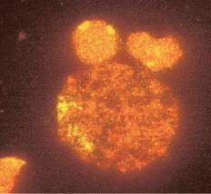
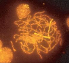
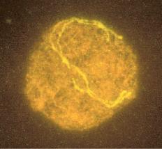
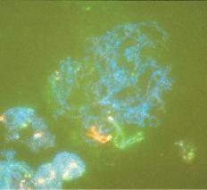
Fig. 1.3 Fetal ovary with fluorescent in situ hybridization. The first three images are meiotic cells from a 21-week fetal ovary. A, Fluorescent in situ hybridization (FISH) with a whole chromosome probe for chromosome X was completed to visualize the pairing characteristics of the X chromosome during leptotene. B, Zygotene. C, Pachytene. D, Image of a meiotic cell from a 34-week fetal ovary that underwent dual FISH with probes for chromosomes 13 (green signal) and 21 (red signal) to illustrate the pairing characteristics of this substage of prophase in meiosis I. (Courtesy Edith Cheng, MD.)
pachytene substage is the most easily recognizable period of the prophase and is characterized by clearly defned chromosomes that appear as continuous ribbons of thick beadlike chromatin (Fig. 1.3C). By defnition, this is the substage in which all homologues have paired. In this substage the paired homologues are structurally composed of four closely opposed chromatids and are known as a tetrad. The frequency of oocytes in pachytene increases with gestational age and peaks in the mid-second trimester of pregnancy (about 20 to 25 weeks’ gestation). The diplotene substage is a stage of desynapsis that occurs as the synaptonemal complex dissolves and the two homologous chromosomes pull away from each other. However, these bivalents, which are composed of a maternally and a paternally derived chromosome, are held together at the centromere and at sites of chiasma formation that represent sites where crossing over has occurred (Fig. 1.3D). In general, chiasma formation occurs only between chromatids of homologous pairs and not between sister chromatids. Usually, one to three chiasma occur for each chromosome arm. Oocytes at this stage of prophase I constitute the majority of third-trimester fetal and newborn ovaries. Diplotene merges with diakinesis, the last substage of meiosis I, and is a stage of transition to metaphase, lasting many years in the humans.
During puberty, folliculogenesis includes progression of the follicle, consisting of the oocyte and granulosa cells from primordial to antral, which is characterized by granulosa cell proliferation, development of gonadotropin receptors, and expression of enzymes for sex steroid production (Baerwald, 2012). It takes approximate 85 days for a follicle to mature to the point of ovulation. There is no change in the chromosome stage during folliculogenesis.
Meiosis I resumes with the surge of luteinizing hormone before ovulation completing metaphase, anaphase, and telophase. The result is two daughter cells, which are diploid (2N) in DNA
content but contain 23 chromosome structures, each containing two closely held sister chromatids. One daughter cell, the oocyte, receives the majority of the cytoplasm, and the other becomes the frst polar body. The polar body is located in the perivitelline space between the surface of the oocyte (oolemma) and the zona pellucida (ZP).
Meiosis II is rapid, with the oocyte advancing immediately to metaphase II, where the sister chromatids for each chromosome are aligned at the equatorial plate, held together by spindle fbers at the centromere. With sperm penetration, meiosis II is completed with extrusion of the second polar body yielding a haploid oocyte (1N) that is entered by a haploid (1N) sperm (Fig. 1.4).
Crossover and Female Aneuploidy
Aneuploidy in embryos is the most common cause of miscarriage and certain chromosomal abnormalitie s in live births, including Down syndrome (trisomy 21). The majority of the time these originate from an abnormal oocyte, increasing with age, and are more likely to affect chromosomes with short p arms (acrocentric). These chromosome segregation errors occur predominantly during meiosis I and are more common in the oocyte compared with the sperm. This is associated with defcient formation of chiasma between homologous chromosomes associated with DNA crossover (recombinati on) sites. Defective sites lead to less tension between homologous chromosomes, making segregation errors more likely as the spindles (microtubules) attached to the kinetochore protein complex adjacent to the centromere pull chromosomes toward the centrioles ( Wang, 2017 ).
Oocyte Cryopreservation
The clinical importance of meiotic spindle integrity was evident during the development of oocyte cryopreservation. Oocyte freezing is becoming more common for fertility preservation in women with medical conditions, such as cancer, for which chemotherapy and/or radiation therapy may result in ovarian failure, and in women of increasing reproductive age. The original technique for oocyte freezing was referred to as slow freezing, which was subsequently replaced by vitrification. Freezing involves removal of intracellular water so that ice crystals will not form during freezing, which may disrupt organelles. With slow freezing, cryoprotectants such as dimethyl sulfoxide (DMSO) and ethylene glycol are allowed to permeate the cell, replacing the water, as the oocyte is slowly cooled at 1°C to 2°C/min to –196°C and stored in liquid nitrogen. In contrast, vitrification involves the use of higher concentrations of cryoprotectant and very rapid cooling at 15,000°C to 30,000°C/min. With slow freezing there is a slow change from liquid to solid, whereas vitrification consists of immediate solidification of the cryoprotectant into a glasslike consistency. With human oocytes, vitrification causes much less spindle damage, resulting in higher oocyte survival rates.
FERTILIZATION AND EARLY CLEAVAGE
In most mammals, including humans, the egg is released from an ovary in the metaphase II stage (Fig. 1.5). When the egg enters the fallopian tube, it is surrounded by a cumulus of granulosa cells (cumulus oophorus) and intimately surrounded by a clear ZP. Within the ZP are both the egg and the frst polar body. Meanwhile, spermatozoa are transported through the cervical mucus and the uterus and into the fallopian tubes.
Although 20 to 200 million sperm may enter the vagina during intercourse, only 1 in 25,000 will make it to the fallopian tubes (Williams, 1993). This journey involves processes of capacitation, chemotaxis, hyperactivated motility, and acrosome reaction
Fig. 1.4 Diagram of oocyte meiosis. For simplicity, only three pairs of chromosomes are depicted (1 to 4). Prophase stages of the first meiotic division, which occur in most mammals during fetal life. The meiotic process is arrested at the diplotene stage (“first meiotic arrest”), and the oocyte enters the dictyate stages (5 to 6). When meiosis is resumed, the first maturation division is completed (7 to 11). Ovulation occurs usually at the metaphase II stage (11), and the second meiotic division (12 to 14) takes place in the oviduct only after sperm penetration. (From Tsafriri A. Oocyte maturation in mammals. In Jones RE, ed. The Vertebrate Ovary. New York: Plenum; 1978. With permission of Springer Science and Business Media.)
(Fig. 1.6). Capacitation precedes all other changes and involves initial removal of cholesterol from the plasma membrane altering the permeability and fuidity. This allows infux of calcium and bicarbonate, with many downstream effects, such as increased cyclic adenosine monophosphate (cAMP), protein tyrosine phosphorylation, and activation of protein kinases. A function of capacitation is to allow localization of protein complexes in the head of the sperm that will subsequently bind the ZP. Chemotaxis is shown by a greater number of sperm in the ampullary portion of the fallopian tube containing a cumulus-oocyte complex (COC) compared with the side lacking a COC. In vitro, follicular fuid acts as a chemoattractant, possibly because of progesterone, but the exact responsible constituents of the fuid continue to be debated (Eisenbach, 1999). Hyperactivated motility involves increased vigorous movement of the sperm to penetrate the cumulus (granulosa) cells surrounding the oocyte and is most likely caused by progesterone. A major action of progesterone is to increase calcium infux into the sperm, with multiple downstream effects. Likely the progesterone concentration increases as the sperm approaches the egg, resulting in more aggressive motility. When the egg is reached, receptor complexes on the outer
most plasma membrane bind to speci c ZP glycoprotein receptors (primarily ZP 3). These interactions are very species specifc. Human sperm can only bind to the ZP of human, baboon, and gibbon oocytes. Binding results in fenestrations forming between the plasma membrane and the underlying acrosome membrane, releasing enzymes, including acrosin (a serine protease), to locally degrade the ZP.
Because many sperm may initially bind the ZP, a mechanism must be in place to prevent fertilization by more than one sperm (polyspermia). With initial binding of the sperm membrane to the oolemma, a calcium-dependent release of cortical granules occurs. Cortical granules are vesicles containing protein made during oogenesis and located in the periphery of the cell. Contents are released into the perivitelline space and modify ZP proteins and enlarge the perivitelline space to prevent sperm entry. With sperm entry, the oocyte completes its second meiotic division, casting off the second polar body into the perivitelline space.
The majority of a single sperm enters the oocyte, and this is indeed the case during intracytoplasmic sperm injection (ICSI) for infertility. Only the centrioles and the nucleus survive, whereas
Posterior wall of uterus
Oocyte penetrated by sperm
Early primary follicle
Blood vessels
Epithelium
Corpus albicans
Mature corpus luteum
Atretic (degenerating) follicle
Endometrium
Released oocyte
Ruptured follicle
Connective tissue Coagulated blood
Developing corpus luteum
Fig. 1.5 Summary of the ovarian cycle, fertilization, and human development during the first week Stage 1 of development begins with fertilization in the uterine tube and ends when the zygote forms. Stage 2 (days 2 to 3) comprises the early stages of cleavage (from 2 to approximately 32 cells, the morula). Stage 3 (days 4 to 5) consists of the free (unattached) blastocyst. Stage 4 (days 5 to 6) is represented by the blastocyst attaching to the posterior wall of the uterus, the usual site of implantation. The blastocysts have been sectioned to show their internal structure. (From Moore KL, Persaud TVN. The Developing Human: Clinically Oriented Embryology. 7th ed. Philadelphia: WB Saunders; 2003.)
mitochondria in the midpiece and tail are destroyed. The sperm centrioles interact with -tubulin from the oocyte to form a microtubule network for migration of pronuclei and subsequent separation of chromosomes during the frst mitosis (Schatten, 2009). Thus mitochondria are of maternal origin and centrioles are paternal. Early cell division (cleavage) is not synchronous and varies in time (Fig. 1.7). Time intervals from two pronuclei to two cells, two cells to three cells, three cells to four cells, and four cells to fve cells are 26 hours, 12 hours, 0.8 hours, and 14 hours, respectively, as determined with time-lapse photography during in vitro fertilization (IVF) (Meseguer, 2011). A signifcant number of fertilized oocytes do not complete cleavage for a number of reasons, including failure of appropriate chromosome arrangement on the spindle, specifc gene defects that prevent the formation of the spindle, and environmental
factors. Importantly, teratogens acting at this point are usually either completely destructive or cause little or no effect. Twinning may occur by the separation of the two cells produced by cleavage, each of which has the potential to develop into a separate embryo. Twinning may occur at any stage until the formation of the blastocyst (blast) because each cell is totipotent. Both genetic and environmental factors are probably involved in the causation of twinning.
Morula and Blastula Stage: Early Differentiation
After fertilization the zygote (term for a fertilized egg) has a diameter of 83 to 105 m and undergoes rapid mitotic division to reach the next stage of approximately 16 cells called a morula. The cells of the zygote and early cleavage embryo are considered totipotent
Perivitelline space
Cytoplasm of oocyte
Sperm nucleus containing chromosomes
Acrosome containing enzymes
Plasma membrane of sperm
Perforations in acrosome wall
Plasma membrane of oocyte
Zona pellucida
Corona radiata
Second meiotic metaphase
First polar body
Plasma membrane of oocyte
Enzymes breaking down zona pellucida
Sperm in cytoplasm of oocyte without its plasma membrane
Fig. 1.6 Acrosome reaction and a sperm penetrating an oocyte. The detail of the area outlined in A is given in B 1, Sperm during capacitation, a period of conditioning that occurs in the female reproductive tract. 2, Sperm undergoing the acrosome reaction, during which perforations form in the acrosome. 3, Sperm digesting a path through the zona pellucida by the action of enzymes released from the acrosome. 4, Sperm after entering the cytoplasm of the oocyte. Note that the plasma membranes of the sperm and oocyte have fused and that the head and tail of the sperm enter the oocyte, leaving the sperm’s plasma membrane attached to the oocyte’s plasma membrane. (From Moore KL, Persaud TVN: The Developing Human: Clinically Oriented Embryology, 7th ed. Philadelphia, WB Saunders, 2003.)
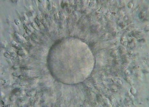
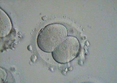
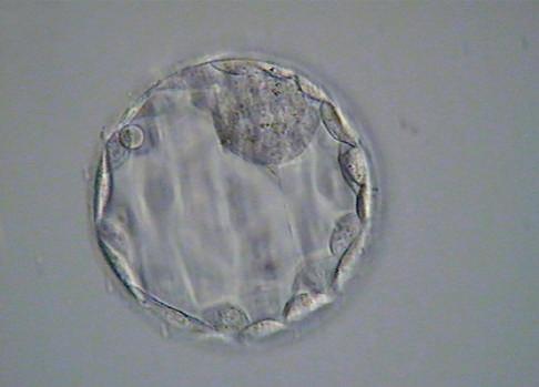
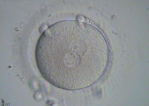
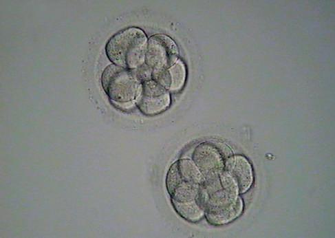
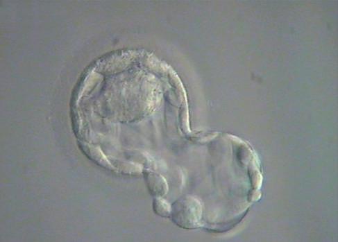
Fig. 1.7 Six photomicrographs of fresh, unmounted human eggs and embryos. A, Recently retrieved human oocyte surrounded by cumulus cells. B, Fertilized oocyte demonstrating male and female pronuclei and both polar bodies at approximately 11 and 12 o’clock position. C, Two-cell zygote with scattered cumulus cells remaining attached to the zone pellucida. D, Eight-cell zygotes. E, Blastocyst with the inner cell mass seen at 12 o’clock. F, A hatching blastocyst in which a portion of the trophectoderm has extruded from the zona pellucida at the 4 o’clock position. (Photos Courtesy Douglas Raburn, PhD.)
because they are capable of producing all human tissue types (embryonic and extraembryonic). After 4 to 5 days traversing the fallopian tube, the embryo arrives in the uterine cavity at the blastocyst (blast) stage. The blast is characterized by a cavity (blastocoele) and differentiation of cells into the trophectoderm (TE), which will ultimately produce the fetal membranes and placenta, and the inner cell mass (ICM) that will produce the fetus. The cells in the blastocyst are referred to as pluripotent, meaning cells have differentiated into a group that can only yield embryonic cells and a group that can only yield extraembryonic cells During IVF the blast forms 5 days after fertilization with a diameter of 155 to 265 m consisting of about 40 TE cells and 20 ICM cells. In the human, implantation generally takes place 3 days after the embryo enters the uterus. The development of the blast with the separation of the ICM from the developing TE together make up the frst stage of differentiation in the embryo. Differentiation within the ICM proceeds fairly rapidly, and if separation of cells and twinning occur at this point, the twins may be conjoined in some fashion.
Advances in ART and genet ics now provi de pract itioners assess to the early embry o for preimplantation genetic
Fertilization
testing (PGT). This includes PGT for monog enic/singlegene disorders (PGT- M), testi ng for aneup loidy (PGT- A), and testing for chrom osome struc tural rearr angements (PGT-SR) such as trans locations ( Fig. 1.8 ). This technique involves removal of up to 10 (typi cally 5 to 10) TE cells from the day 5 blast for analy sis. For PGT-M of singl e-gene disorders, DNA is extra cted from the cells and the mutat ion analyzed by polym erase chain react ion (PCR) ampli fication or single nucle otide polym orphism (SNP) micro array. For PGT-A and PGT-S R, analy sis of DNA is perfo rmed with comparative genom ic hybri dization (CGH) array or parti al genomic sequencing (next gener ation seque ncing).
IMPLANTATION
Implantation consists of apposition, attachment, and invasion. This very complex process has redundancy and involves multiple factors, including ovarian hormones, cytokines, transcription factors, growth factors, and extracellular matrix proteins (ECM) (Table 1.1). These factors are produced by both the endometrium and the embryo. Communication between the embryo and the
Aspirating TE cells
D Preimplantation Genetic Testing – Aneuploidy
Next generation sequencing or Single nucleotide polymorphism array
Fig. 1.8 Schematic of preimplantation testing. A, Commonly the oocyte is fertilized with a single sperm using the technique of intracytoplasmic sperm injection (ICSI). This precludes the possibility of contamination from sperm remaining attached to the outside of the embryo during embryo biopsy. B, On day 3 of culture, when the embryo has cleaved to about eight cells, a small opening is made in the zona pellucida (ZP) with either a laser or through brief exposure to an acid solution. C, By day 5 of culture the embryo has progressed to the blastocyst stage and a portion of the trophectoderm (TE) cells have prolapsed out the opening in the ZP. These cells are removed for subsequent DNA isolation. D, DNA from TE cells is used to determine chromosome number and insertions and deletions for preimplantation genetic screening using techniques of array comparative genomic hybridization or next generation sequencing. DNA may also be used to detect single-gene abnormalities for different diseases using single nucleotide polymorphism microarray or polymerase chain reaction amplification in the process of preimplantation genetic diagnosis.
Sperm Needle
Inner cell mass
Acidic Tyrode’s Laser
Pipet
Trophectoderm (IL)
Zona pellucida (ZP)
Egg cell (ovum)
ICSI
Opening the Zona pellucida
TABLE 1.1 Events of Implantation
Event
Days After Ovulation
Zona pellucida disappears 4-5
Blastocyst attaches to epithelial surface of endometrium 6
Trophoblast erodes into endometrial stroma 7
Trophoblast differentiates into cytotrophoblastic and syncytial trophoblastic layers 7-8
endometrium is key. Implantation occurs 7 to 10 days after ovulation, corresponding to cycle days 21 to 24 of an idyllic 28-day cycle with ovulation on day 14. During apposition the human embryo is oriented with the ICM and polar TE (the TE next to the ICM) adjacent to the endometrium.
For attachment to the endometrium, the embryonic cells must frst be expelled from the surrounding ZP during the process of “hatching.” Hatching involves the rupture of the ZP in one small area as opposed to a general dissolution of the entire ZP. This may involve both hydrostatic pressure from inside the ZP and from zonalytic proteases produced by the TE and endometrium. These cysteine proteases, named cathepsins, are essential for hatching. Attachment of the embryonic cells to the endometrial cells involves cell adhesion proteins, integrins, and ECM proteins such as fbronectin, laminin, and collagen. Integrins are cell surface proteins that bind extracellular matrix proteins and are expressed on both the luminal epithelium and TE.
Next, invasion of TE cells occurs by penetration between the luminal epithelial cells, through the basement membrane and into the stroma of the endometrium. These initial TE cells form the extravillous trophoblasts (EVTs), which invade the inner third of the myometrium for anchoring and into the spiral arteries for remodeling. During spiral artery remodeling, endovascular EVT disorganize and partially replace the smooth muscle wall and the vascular endothelial cells. Proliferation of endovascular EVT leads to plugging and obstruction of the decidual spiral arteries, resulting in a decrease in blood fow and oxygen tension. A low oxygen setting promotes proliferation and transformation of cytotrophoblast to syncytiotrophoblast. Before 8 weeks’ gestation, nutrition to the embryo is derived from endometrial gland secretion and plasma seeping through the obstructed spiral arteries into the intervillous space. With continued remodeling of the spiral arteries, patency is reestablished and maternal blood cells enter the intervillous space around 9 weeks’ gestation with a rise in oxygen tension. Lack of adequate EVT invasion and spiral artery remodeling is a key feature in preeclampsia, intrauterine growth restriction, and stillbirths.
The idea of low oxygen tension during early embryo development has been explore d with IVF. With a limited number of trials, culturing embryos in 5% oxygen as opposed to 20% oxygen results in a modest increas e in implant ation rate ( Bontekoe, 2012 ).
Villous trophoblast form ngerlike projections extending into the intervillous space and that are surrounded by maternal blood. Syncytiotrophoblast form the outer layer with an underlying layer of precursor cytotrophoblast surrounding matrix containing capillaries, fbroblasts, and macrophages (Hofbauer cells). Cytotrophoblast become less numerous as pregnancy progresses.
Blood levels of the pregnancy hormone human chorionic gonadotropin (hCG) can be detected within 48 hours of implantation. Regular hCG is produced by the syncytiotrophoblast of placental villi. Blood levels peak at 56 to 68 days, reach a nadir at 18 weeks, and then remain fairly consistent until delivery. Gonadotropin-releasing hormone (GnRH) produced in the cytotrophoblast induces expression of hCG. In spontaneous pregnancies hCG can be detected 9 days after follicle rupture observed by ultrasound. In IVF pregnancies the hormone can be found 8 days after embryo transfer. The hCG levels rise exponentially up to 8 weeks from the last menstrual period (LMP), but the doubling time increases as the level increases. For example, in a conception cycle with ovulation on cycle day 14, the doubling time from cycle days 25 to 37 for hCG is 1.6 days and from days 38 to 44 is 2.3 days (Zegers-Hochschild, 1994). The doubling time is independent of the number of gestations, although the absolute hCG level is higher for multiple pregnancies.
The classic action of regular hCG is maintenance of the corpus luteum (CL) by binding the luteinizing hormone (LH) receptor for continued estrogen and progesterone production. Yet other identifed actions include promotion of angiogenesis in the uterus, myometrial relaxation, inhibition of immune interaction at the uteroplacental interface, stimulation of fetal testosterone production, and mediation of hyperemesis through receptors in the brain.
Hyperglycosylated hCG (H-hCG) is produced by EVT. H-hCG is key in promoting angiogenesis and cell invasion and correspondingly is found in the early frst trimester. The protein does not activate the LH receptor and does not preserve CL function. Instead it appears to function via the transforming growth factor beta (TGF- ) receptor. Low levels of H-hCG indicate poor EVT development and are associated with spontaneous abortion and early preeclampsia (Fournier, 2015).
Decidualization
Progesterone is responsible for “decidualization” of the endometrium. This refers to morphologic and functional changes in stromal cells. In humans, stromal cells close to the spiral arteries undergo progesterone-induced decidual changes in the late secretory phase and this process progresses throughout the stroma with implantation and hCG production. HCG derived from a pregnancy within the uterus is not required, as decidualization is a common fnding with ectopic pregnancies. Decidual cells show morphologic changes of increased size with increased glycogen and lipid accumulation. During pregnancy the endometrium is now referred to as the decidua, separated into areas of the decidua basalis or placentalis, which interact with the TE (area of mature placenta), the decidua vera or parietalis (decidua distant from implantation site), and the decidua capsularis (surrounding the embryo on the side opposite the placenta).
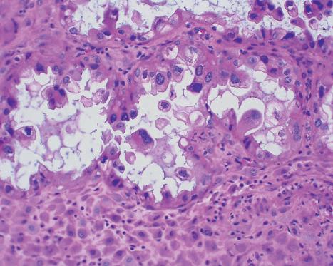
Another classic histologic change seen in early pregnancy is the Arias-Stella reaction (Fig. 1.9). This occurs in the glandular cells with a hallmark of nuclear enlargement. These cells may be misinterpreted as atypical or malignant. In the presence of hCG the Arias-Stella reaction may be seen in extrauterine tissues such as endometriosis, vaginal adenosis, paraovarian cysts, and mucinous cystadenomas (Arias-Stella, 2002).
Morphologically, luminal epithelial cells develop extensions of the plasma membrane called pinopods (also called uterodomes)
Fig. 1.9 Photomicrograph of the Arias-Stella reaction. The hCG action results in nuclear enlargement in endometrial glandular cells (arrows) resulting in visual characteristics of malignant cells. Magnification 200. (Courtesy Rex Bentley, MD, and Stanley Robboy, MD, Duke University.)
during the window of receptivity. Pinopods release key proteins, including leukemia inhibitory factor (LIF), through exocytosis and apocrine secretion (Kabir-Salmani, 2005).
Downstream effects of progesterone-dependent decidualization have not been completely elucidated, but loss of function studies show the necessity of transcription factors including CCAAT/enhancer binding protein beta (C/EBP ), homeobox A10 (Hoxa10), forkhead/winged helix protein (Fox01) and chicken ovalbumin upstream-promoter (COUP-TFII). A functional progesterone receptor requires interaction with chaperone proteins. In mice one of these proteins, named FK506 binding protein 52 (FKBP52), is expressed in the endometrium during the window of receptivity, and a loss of function mutation disrupts decidualization.
LIF is a cytokine produced by endometrial glandular cells around the time of implantation. LIF acts on EVT to increase fbronectin production necessary for embryo attachment and invasion. Mice lacking expression of LIF (knockout mice) have both failure of decidualization and implantation.
Indian hedgehog (Ihh) protein is a morphogen produced by luminal epithelial cells under the control of progesterone. Morphogens are signaling proteins that diffuse throughout the decidua yielding a concentration gradient. Signaling is dependent on the concentration in a given area. Ihh knockout mice fail to decidualize or implant (Ramathal, 2010).
Embryo-Endometrial Communication
Implantation involves molecular interactions between the embryo and the adjacent endometrium. For example, the embryo produces heparin-binding epidermal growth factor-like growth factor (HB-EGF), which is both found on the cell membrane and is released from the cell (soluble). HB-EGF induces expression of itself in the adjacent endometrial cells (auto-induction loop). HB-EGF on the endometrial cells then acts to attach the embryo via EGF receptors expressed on the embryo (Lim, 2009). Additionally, the soluble HB-EGF from the embryo induces expression of cyclooxygenase to increase prostacyclin (PGI2) in the endometrium, resulting in enhanced endometrial vascular permeability to help with embryo invasion.
Immunology of Implantation
The paternal contribution to the embryo results in the mother being exposed to allogenic cells. Although villous trophoblast do not express major human leukocyte antigens (HLAs), the EVT express HLA-C, -E, and -G, which may be recognized by the maternal immune system. Thus the maternal immune system must be locally suppressed to prevent rejection.
The majority of immune cells in the decidua are uterine natural killer (uNK) cells. These cells are present in the secretory endometrium, under the control of progesterone, and increase in number with pregnancy to form an infltrate around the invading EVT. These cells start to dissipate in the second trimester. uNK cells are not cytotoxic to trophoblast cells and in fact appear to be supportive. A low number of uNK cells in the decidua of early pregnancy is associated with poor invasion of the EVT. Cytokines such as interferon gamma and angiogenic factors secreted by uNK cells are key to proper EVT development and function.
T-helper (Th) cells are also found in the decidua and are functionally classifed as Th1 (cellular immunity), Th2 (humoral immunity), Th3 (production of transforming growth factor beta for immunosuppression), and Tr1 (production of interleukin 10 for immunosuppression). In early pregnancy there is an increase in the percentage of decidual Th2 and Th3 cells.
T-regulatory (Treg) cells function in antigen recognition for future immune tolerance. Mice lacking Treg cells experience abortion when mated with an allogenic male but not when mated with a syngenic male (Darasse-Jèze, 2006). These cells are key in developing tolerance to male antigens. Development of immunity to specifc paternal antigens may explain observations including lower preeclampsia rates in women exposed to their partner’s semen before pregnancy compared with women conceiving with donor insemination (Salha, 1999) and the lower preeclampsia rate in the second pregnancy with the same partner as opposed to a new partner.
Early Organogenesis in the Embryonic Period
During the third week after fertilization, the primitive streak forms in the caudal portion of the embryonic disk, and the embryonic disk begins to grow and change from a circular to a pear-shaped confguration. At that point the epithelium superiorly is considered ectoderm and will eventually give rise to the developing central nervous system, and the epithelium facing downward toward the yolk sac is endoderm. During this week the neuroplate develops with its associated notochordal process. By the sixteenth day after conception the third primitive germ layer, the intraembryonic mesoderm, begins to form between the ectoderm and endoderm. Early mesoderm migrates cranially, passing on either side of the notochordal process to meet in front in the formation of the cardiogenic area. The heart soon develops from this area. Later in the third week extraembryonic mesoderm joins with the yolk sac and the developing amnion to contribute to the developing membranes.
An intraembryonic mesoderm develops on each side of the notochord and neural tube to form longitudinal columns, the paraxial mesoderm. Each paraxial column thins laterally into the lateral plate mesoderm, which is continuous with the extraembryonic mesoderm of the yolk sac and the amnion. The lateral plate mesoderm is separated from the paraxial mesoderm by a continuous tract of mesoderm called the intermediate mesoderm. By the twentieth day, paraxial mesoderm begins to divide into paired linear bodies known as somites. About 38 pairs of somites form during the next 10 days. Eventually a total of 42 to 44 pairs will develop, and these will give rise to body musculature.
Angiogenesis, or blood vessel formation, can be seen in the extraembryonic mesoderm of the yolk sac by day 15 or 16. Embryonic vessels can be seen about 2 days later and develop when
mesenchymal cells known as angioblasts aggregate to form masses and cords called blood islands. Spaces then appear within these islands, and the angioblasts arrange themselves around these spaces to form primitive endothelium. Isolated vessels form channels and then grow into adjacent areas by endothelial budding. Primitive blood cells develop from endothelial cells as the vessels develop on the yolk sac and allantois. However, blood formation does not begin within the embryo until the second month of gestation, occurring frst in the developing liver and later in the spleen, bone marrow, and lymph nodes. Separate mesenchymal cells surrounding the primitive endothelial vessels differentiate into muscular and connective tissue elements. The primitive heart forms in a similar manner from mesenchymal cells in the cardiogenic area. Paired endothelial channels, called heart tubes, develop by the end of the third week and fuse to form the primitive heart. By the twenty-frst day, this primitive heart has linked up with blood vessels of the embryo, forming a primitive cardiovascular system. Blood circulation starts about this time, and the cardiovascular system becomes the frst functioning organ system within the embryo (Clark, 1987). All the organ systems form between the fourth week and seventh week of gestation.
A teratogenic event that takes place during the embryonic period gives rise to a constellation of malformations related to the organ systems that are actively developing at that particular time. Thus cardiovascular malformations tend to occur because of teratogenic events early in the embryonic period, whereas genitourinary abnormalities tend to result from later events Teratogenic effects before implantation often cause loss of the embryo but not malformations. The effects of a particular teratogen depend on the individual’s genetic makeup, other environmental factors in play at the time, the embryonic developmental stage during which the teratogenic exposure occurred, and in some cases the dose of the teratogen and the duration of exposure. Some teratogens in and of themselves are actually harmless, but their metabolites cause the damage. Teratogens may be chemical substances and their by-products, or they may be physical phenomena, such as temperature elevation and irradiation. The embryo is most sensitive to teratogens during organogenesis of the embryonic period from 18 to 56 days after conception. Before day 18, exposure is most likely to either result in embryo death with miscarriage or no effect because the majority of cells are pluripotent. Teratogen exposure after the embryonic period of development may injure or kill the embryo or cause developmental and growth retardation but usually will not be responsible for specifc malformations. The period of embryonic development is said to be complete at 56 days (8 weeks) from fertilization or 70 days (10 weeks) from the LMP followed by the fetal stage.
DEVELOPMENT OF THE GENITOURINARY SYSTEM
The development of the genital organs is intimately involved with the development of the renal system.
Renal Development
Nephrogenic cords develop from the intermediate mesoderm as early as the 2-mm embryo stage, beginning in the more cephalad portions of the embryo Three sets of excretory ducts and tubules develop bilaterally (Little, 2010). The frst, the pronephros, with its pronephric ducts, forms in the most cranial portion of the embryo at about the beginning of the fourth week after conception. The tubules associated with the duct probably have no excretory function in the human, but the caudal end will form the adrenal gland. Late in the fourth week, a second set of tubules, the mesonephric tubules, and their accompanying mesonephric ducts begin to develop. These are associated with tufts of capillaries, or glomeruli, and tubules for excretory purposes. Thus the
mesonephros functions as a fetal kidney, producing urine for about 2 or 3 weeks. As new tubules develop, those derived from the more cephalad tubules degenerate. Usually about 40 mesonephric tubules function on either side of the embryo at any given time. The gonads arise from the central region of the mesonephros. The metanephros, or permanent kidney, begins its development early in the ffth week of gestation and starts to function late in the seventh or early in the eighth week. The metanephros develops both from the metanephrogenic mass of mesoderm, which is the most caudal portion of the nephrogenic cord, and from its duct system, which is derived from the metanephric diverticulum (ureteric bud). It is a cranially growing outpouching of the mesonephric duct close to where it enters the cloaca. The metanephric duct system gives rise to the ureter, the renal pelvis, the calyces, and the collecting tubules of the adult kidney A critical process in the development of the kidney requires that the cranially growing metanephric diverticulum meets and fuses with the metanephrogenic mass of mesoderm so that formation of the kidney can take place. Originally the metanephric kidney is a pelvic organ, but by differential growth it becomes located in the lumbar region
The fetus produces urine starting at 8 weeks’ gestation (Underwood, 2005). Starting in the second trimester, fetal urine is a major contributor to amniotic fuid volume. The fetus may swallow the amniotic fuid and recirculate it through the digestive system. Congenital abnormalities that impair normal development or function of the fetal kidneys generally result in little or no amniotic fuid (oligohydramnios or anhydramnios), whereas structural abnormalities of the gastrointestinal tract or neuromuscular conditions that prevent the fetus from swallowing can lead to excess amniotic fuid (polyhydramnios).
Bladder and Urethra
The embryonic cloaca is divided by the urorectal septum into a dorsal rectum and a ventral urogenital sinus. The urogenital sinus, in turn, is divided into three parts: the cranial portion (the vesicourethral canal), which is continuous with the allantois; a middle pelvic portion; and a caudal urogenital sinus portion, which is covered externally by the urogenital membrane. The epithelium of the developing bladder is derived from the endoderm of the vesicourethral canal. The muscular layers and serosa of the bladder develop from adjacent splanchnic mesenchyme. As the bladder develops, the caudal portion of the mesonephric ducts is incorporated into its dorsal wall. The portion of the mesonephric duct distal to the points where the metanephric duct is taken up into the bladder becomes the trigone of the bladder. Although this portion is from the mesoderm, it probably is epithelialized by endodermal epithelium from the urogenital sinus. In this way the ureters, derived from the metanephric duct, come to open directly into the bladder.
In the male the mesonephric ducts open into the urethra as the ejaculatory ducts. Also in the male, mesenchymal tissue surrounding the developing urethra where it exits the bladder develops into the prostate gland, through which the ejaculatory ducts traverse. Fig. 1.10 demonstrates graphically the development of the male and female urinary systems.
The epithelium of the female urethra is derived from endoderm of the vesicourethral canal. The urethral sphincter develops from a mesenchymal condensation around the urethra after the division of the cloaca in the 12- to 15-mm embryo. After the opening of the anal membrane at the 20- to 30-mm stage, the puborectalis muscle appears. At 15 weeks’ gestation, striated muscle can be seen and a smooth muscle layer thickens at the level of the developing bladder neck, forming the inner part of the urethral musculature. Thus the urethral sphincter is composed of both central smooth muscle and peripheral striated muscle. The sphincter develops primarily in the anterior wall of the urethra in a horseshoe or omega shape.
Urogenital sinus
Mesonephros
Mesonephric duct
Metanephric diverticulum
Urorectal septum
Cloacal membrane
Allantois
Vesical part
Pelvic part
Phallic part
Rectum
Urogenital sinus
Mesonephric duct
Mesonephros
Metanephros
Urinary bladder
Ureter
Rectum
Urorectal septum
Urachus
Uterine tube
Kidney
Ovary
Uterus
Vagina
Mesonephros
Metanephric diverticulum (ureteric bud)
Mesonephric duct
Mesonephros
Mesonephric duct
Metanephros (primordium of permanent kidney)
Ureter
Mesonephros Gonad
Metanephros
Ureter
Mesonephric duct
Pelvic part of urogenital sinus
Urinary bladder
Penis
Spongy urethra
Fig. 1.10 Diagrams showing division of the cloaca into the urogenital sinus and rectum; absorption of the mesonephric ducts; development of the urinary bladder, urethra, and urachus; and changes in the location of the ureters. A, Lateral view of the caudal half of a 5-week embryo. B, D, and F, Dorsal views. C, E, G, and H, Lateral views. The stages shown in G and H are reached by the twelfth week. (From Moore KL, Persaud TVN. The Developing Human: Clinically Oriented Embryology. 7th ed. Philadelphia: WB Saunders; 2003.)
Kidney
Testis
Ureter
Ductus deferens
Clitoris
Genital tubercle
MOLECULAR BASIS OF SEX DIFFERENTIATION
Genetic sex is determined at the time of conception. A Y chromosome is necessary for the development of the testes, and the testes are responsible for the organization of the sexual duct system into a male confguration and for the suppression of the paramesonephric (müllerian) system of the female. In the absence of a Y chromosome or in the absence of a gonad, development will be female in nature. Male differentiation is determined by expression of the SRY gene found on the short arm of the Y chromosome. SRY protein is a transcription factor and expression is unique to the Sertoli cell of the developing testis. SRY induces expression of another transcription factor, SOX9, which is also obligatory for male sex differentiation. A loss of function mutation of either SRY or SOX9 results in XY sex reversal in which genetic males are phenotypic females. Several genes regulate SRY/SOX9 expression, including Wilms’ tumor suppressor 1 (WT1) and steroidogenic factor 1 (SF1). WT1 is a transcription factor expressed in both urinary tract and gonadal tissue. A loss of function mutation results in glomerulosclerosis and gonadal dysgenesis. SF1 encodes a nuclear receptor necessary for steroidogenesis, gonadal differentiation and adrenal formation. A loss of function mutation is associated with adrenal failure and XY sex reversal (Ozisik, 2003).
Although ovarian formation can only occur in the absence of SRY/SOX9, there are unique genes necessary for development. FOXL2 encodes a transcription factor necessary for granulosa cell expansion. A loss of function mutation causes ovarian failure with other associated abnormalities found in blepharophimosisptosis-epicanthus inversus syndrome (BPES) (De Baere, 2001). BMP15, located on the X chromosome, and GDF9 on chromosome 5 encode growth factors expressed in oocytes required for granulosa cell proliferation. A heterozygous loss of function mutation results in ovarian failure (Di Pasquale, 2004).
The understanding of the molecular basis of sex determination continues to expand with more than 25 genes so far identifed in the process (Wilhelm, 2007).
Genital Development
Male gonadal development precedes female development (Fig. 1.11). During the ffth week after conception, coelomic epithelium, later known as germinal epithelium, thickens in the area of the medial aspect of the mesonephros. As germinal epithelial cells proliferate, they invade the underlying mesenchyme, producing a prominence known as the gonadal ridge. In the sixth week the primordial germ cells (PGCs), which have formed at about week 4 in the wall of the yolk sac, migrate up the dorsal mesentery of the hindgut and enter the undifferentiated gonad. The somatic cells of the primitive gonadal ridge then differentiate into interstitial cells (Leydig cells) and Sertoli cells. As they do the PGCs and Sertoli cells become enclosed within seminiferous tubules, and the interstitial cells remain outside these tubules. Sertoli cells are encased in the seminiferous tubules in the seventh and eighth weeks. In the eighth week Leydig cells differentiate and begin to produce testosterone. At this point the mesonephric (wolffan) duct differentiates into the vas deferens, epididymis, and seminal vesicles, whereas the paramesonephric duct (müllerian duct) is suppressed because of the secretion and action of antimüllerian hormone (AMH), also known as müllerian inhibitory substance (MIS), by Sertoli cells.
Primary sex cords, meanwhile, have condensed and extended to the medullary portion of the developing testes. They branch and join to form the rete testis. The testes therefore is primarily a medullary organ, and eventually the rete testis connects with the tubules of the mesonephric system and joins the developing epididymal duct.
Development of the ovaries occurs at about the eleventh or twelfth week, although the PGCs have migrated several weeks
HUMAN SEX DIFFERENTIATION
Bipotential gonad
Seeded with primordial germ cells
Fig. 1.11 Development of sexual differentiation in the human. Note the lag from male to female development. (Modified from Grumbach MM, Hughes IA, Conte FA. Disorders of sex differentiation. In Larsen PR, Kronenberg HM, Melmed S, Polonsky KS, eds. Williams Textbook of Endocrinology. 10th ed. Philadelphia: WB Saunders; 2003:870.)
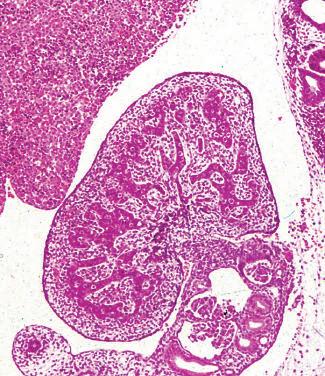
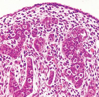
Fig. 1.12 Ovary in embryo. A, A developing ovary (O) in a 9-week-old embryo is shown close to the developing kidney (K). B, At this stage of development, the columns of primordial germ cells (G) are embedded in a mesenchymal stroma (S) covered by a layer of cuboidal surface cells (E). (From Stevens A, Lowe J. Human Histology. 3rd ed. Philadelphia: Elsevier Mosby; 2005:357.)
earlier to the germinal ridge (Fig. 1.12). Two functional X chromosomes are necessary for optimal development of the ovaries. Deletion of either the short arm or the long arm of a single X chromosome precludes normal ovarian function, with the former being associated with Turner syndrome. The processes of gonadal development are schematically summarized in Fig. 1.13.
Genital Duct System
Early in embryonic life, two sets of paired genital ducts develop in each sex: the mesonephric (wolffan) ducts and the paramesonephric (müllerian) ducts. The mesonephric duct development precedes the paramesonephric duct development. The paramesonephric ducts develop on each side of the mesonephric ducts from the evaginations of the coelomic epithelium. The more cephalad ends of the ducts open directly into the peritoneal cavity, and the distal ends grow caudally, fusing in the lower midline to form the uterovaginal primordium. This tubular structure joins the dorsal wall of the urogenital sinus and produces an elevation, the müllerian tubercle. The mesonephric ducts enter the urogenital sinus on either side of the tubercle.
Male Genital Ducts
Seminiferous tubules are produced in the fetal testes during the seventh and eighth weeks after conception. During the eighth week, interstitial (Leydig) cells differentiate and begin to produce testosterone. Male internal genital development is mainly dependent on testosterone, whereas external genitalia development is dependent on 5 -dihydrotestosterone (DHT). Testosterone produced by the Leydig cells stimulates growth and development of the wolffan duct structures of the vas deferens, epididymis, and seminal vesicles. DHT formed in target tissues by the enzyme type 2 5 -reductase is responsible for formation of the prostate, scrotum, and penis.
Maternal hCG production may be key to male genital development. The maximum serum level of hCG at approximately 8 weeks after conception or 10 menstrual weeks correlates with the timing of male genital formation, and the highest fetal testosterone levels are seen at 11 to 17 weeks with a subsequent
decline. hCG acting via the LH receptor is responsible for stimulating Leydig cell testosterone production.
The bulbourethral glands, which are small structures that develop from outgrowths of endodermal tissue from the membranous portion of the urethra, incorporate stroma from the adjacent mesenchyme. The most distal portion of the paramesonephric duct remains, in the male, as the appendix of the testes. The most proximal end of the paramesonephric duct remains as a small outpouching within the body of the prostate gland, known as the prostatic utricle. Rarely, the prostatic utricle is developed to the point where it will excrete a small amount of blood and cause hematuria in adult life (Schuhrke, 1978).
Female Genital Ducts
In the absence of AMH, the mesonephric ducts regress and the paramesonephric ducts develop into the female genital tract. This process begins at about 6 weeks and proceeds in a cephalad to caudal fashion. The more cephalad portions of the paramesonephric ducts, which open directly into the peritoneal cavity, form the fallopian tubes. The fused portion, or uterovaginal primordium, gives rise to the epithelium and glands of the uterus and cervix. Endometrial stroma and myometrium are derived from adjacent mesenchyme. Failure of development of the paramesonephric ducts leads to agenesis of the cervix and the uterus, referred to as müllerian agenesis or Mayer-RokitanskyKuster-Hauser syndrome (Langman, 1982). Failure of fusion of the caudal portion of these ducts may lead to a variety of uterine anomalies, including complete duplication of the uterus and cervix or partial duplication of a variety of types, which are outlined in Chapter 11 (Congenital Anomalies of the Female Reproductive Tract). Peritoneal refections in the area adjacent to the fusion of the two paramesonephric ducts give rise to the formation of the broad ligaments. Mesenchymal tissue here develops into the parametrium.
Development of the uterine ligaments includes the round ligament at the eighth week, the cardinal ligaments at the tenth week, and the broad ligament at week 19. The round ligament (ligament teres) is analogous to the male gubernaculum. It extends from the uterine body through the broad ligament and
Development of Testes
Aggregation of neural crest cells
Mesonephric duct
Primary sex cord
Primordial germ cells
Indifferent gonad
Gonadal ridge
5-week embryo
Development of Ovaries
Neural tube
Sympathetic ganglion
Aorta
Paramesonephric duct
Primordium of suprarenal medulla
Primordium of suprarenal cortex
12 weeks
Surface epithelium
Former gonadal cords
Seminiferous cord
Hindgut
Primordial germ cells
Duct of epididymis
Mesonephric duct
Cortical cords
Paramesonephric duct
Degenerating rete ovarii
Rete testis
Septum of testis
Seminiferous cord
Mesonephric duct and tubule
Primordial ovarian follicle
Surface epithelium
Hindgut
Paramesonephric duct
Primordial germ cells
Uterine tube
Stromal (connective tissue) cells
Spermatogonium
cell
Section of seminiferous tubule
Section of ovarian cortex
Fig. 1.13 Schematic illustration showing differentiation of the indifferent gonads of a 5-week embryo (top) into ovaries or testes. Left side shows the development of testes resulting from the effects of the testisdetermining factor (TDF), also called the SRY gene, located on the Y chromosome. Note that the gonadal cords become seminiferous cords, the primordium of the seminiferous tubules. The parts of the gonadal cords that enter the medulla of the testis form the rete testis. In the section of the testis at the bottom left, observe that there are two kinds of cells: spermatogonia derived from the primordial germ cells and sustentacular (Sertoli) cells derived from mesenchyme. Right side shows the development of ovaries in the absence of TDF. Cortical cords have extended from the surface epithelium of the gonad, and primordial cells have entered them. They are the primordia of the oogonia. Follicular cells are derived from the surface epithelium of the ovaries. (From Moore KL, Persaud TVN. The Developing Human: Clinically Oriented Embryology. 7th ed. Philadelphia: WB Saunders; 2003.)
Suprarenal medulla
Suprarenal cortex
Mesorchium
Mesovarium
Sertoli
Oogonium
Follicular cell
Tunica albuginea
Hindgut
TABLE 1.2
Male and Female Derivatives of Embryonic Urogenital Structures
Embryonic Structure
Labioscrotal swellings
Urogenital folds
Male
Scrotum
Ventral portion of penis
Phallus Penis
Urogenital sinus
Derivatives
Glans, corpora cavernosa penis, and corpus spongiosum
Urinary bladder
Prostate gland
Prostatic utricle
Bulbourethral glands
Seminal colliculus
Paramesonephric duct Appendix of testes
Mesonephric duct Appendix of epididymis
Ductus of epididymis
Ductus deferens
Ejaculatory duct and seminal vesicle
Metanephric duct
Ureters, renal pelvis, calyces, and collecting system
Mesonephric tubules Ductuli efferentes
Paradidymis
Undifferentiated gonad Testis
Cortex Seminiferous tubules
Medulla
Rete testis
Gubernaculum Gubernaculum testis
peritoneum exists the pelvis via the internal ring through the inguinal canal ending in the labia majora.
The vagina develops from paired solid outgrowths of endoderm of the urogenital sinus—the sinovaginal bulbs. These grow caudally as a solid core toward the end of the uterovaginal primordium. This core constitutes the fbromuscular portion of the vagina. The sinovaginal bulbs then canalize to form the vagina. Abnormalities in this process may lead to either transverse or horizontal vaginal septa. The junction of the sinovaginal bulbs with the urogenital sinus remains as the vaginal plate, which forms the hymen. This remains imperforate until late in embryonic life, although occasionally perforation does not take place completely (imperforate hymen). Failure of the sinovaginal bulbs to form leads to agenesis of the vagina. Auxiliary genital glands in the female form from buds that grow out of the urethra. The buds derive contributions from the surrounding mesenchyme and form the urethral glands and the paraurethral glands (Skene glands). These glands correspond to the prostate gland in males. Similar outgrowths of the urogenital sinus form the vestibular glands (Bartholin glands), which are homologous to the bulbourethral glands in the male. The remnants of the mesonephric duct in the female include a small structure called the appendix vesiculosa, a few blind tubules in the broad ligaments (the epoophoron), and a few blind tubules adjacent to the uterus (collectively called the paroöphoron). Remnants of the mesonephric duct system are often present in the broad ligaments or may be
Female
Labia majora
Labia minora
Clitoris
Glans, corpora cavernosa, bulb of the vestibule
Urinary bladder
Urethral and paraurethral glands
Vagina
Greater vestibular glands
Hymen
Hydatid of Morgagni
Uterus and cervix
Fallopian tubes
Appendix vesiculosa
Duct of epoophoron
Gartner duct
Ureters, renal pelvis, calyces, and collecting system
Epoophoron
Paroöphoron
Ovaries
Ovarian follicles
Medulla
Rete ovarii
Round ligament of uterus
present adjacent to the uterus or the vagina as Gartner duct cysts (Deppisch, 1975). The epoophoron or paroöphoron may develop into cysts. Cysts of the epoophoron are known as paraovarian cysts (Chapter 18, Benign Gynecologic Lesions). Remnants of the paramesonephric duct in the female may be seen as a small, blind cystic structure attached by a pedicle to the distal end of the fallopian tube: the hydatid of Morgagni. Table 1.2 categorizes the adult derivatives and residual remnants of the urogenital structures in both the male and the female. Fig. 1.14 outlines schematically the development of the internal sexual organs in both sexes.
External Genitalia
In the fourth week after fertilization, the genital tubercle develops at the ventral tip of the cloacal membrane. Two sets of lateral bodies, the labioscrotal swellings and urogenital folds, develop soon after on either side of the cloacal membrane. The genital tubercle then elongates to form a phallus in both males and females. By the end of the sixth week, the cloacal membrane is joined by the urorectal septum. The septum separates the cloaca into the urogenital sinus ventrally and the anal canal and rectum dorsally. The point on the cloacal membrane where the urorectal septum fuses becomes the location of the perineal body in later development. The cloacal membrane is then divided into the ventral urogenital membrane and the dorsal anal membrane.