CONTRIBUTORS
James L. Achord, MD
Professor Emeritus
University of Mississippi Medical Center
Jackson, Misssissippi
1: The History of Gastrointestinal Endoscopy
Michelle J. Alfa, BSc, MSc, PhD
Principal Investigator
St. Boniface Research Centre; Professor
Department of Medical Microbiology
University of Manitoba Winnipeg, Manitoba, Canada
4: Cleaning and Disinfecting Gastrointestinal Endoscopy Equipment
Mohammad Al-Haddad, MD, MSc, FASGE, FACG, AGAF
Associate Professor of Medicine
Division of Gastroenterology and Hepatology
Indiana University School Medicine Indianapolis, Indiana
62: Evaluation and Staging of Pancreaticobiliary Malignancy
Andrea Anderloni, MD, PhD
Digestive Endoscopy Unit
Division of Gastroenterology
Humanitas Research Hospital
Milan, Italy
28: Palliation of Malignant Dysphagia and Esophageal Fistulas
Joseph C. Anderson, MD
Associate Professor of Medicine
Department of Veterans Affairs Medical Center
White River Junction, Vermont; The Geisel School of Medicine at Dartmouth Hanover, New Hampshire; Division of Gastroenterology and Hepatology
University of Connecticut School of Medicine
Farmington, Connecticut
36: Colorectal Cancer Screening and Surveillance
Anna Baiges, MD
Hepatic Hemodynamic Laboratory
Liver Unit, Hospital Clínic
Barcelona, Spain
15: Portal Hypertensive Bleeding
John Baillie, MD
Professor
Division of Gastroenterology and Hepatology
Department of Medicine
Virginia Commonwealth University School of Medicine
Richmond, Virginia
3: How Endoscopes Work
Alan N. Barkun, MD, MSc
Division of Gastroenterology
McGill University Health Center
Montreal, Québec, Canada
14: Nonvariceal Upper Gastrointestinal Bleeding
Todd H. Baron, MD, FASGE Professor of Medicine
Division of Gastroenterology and Hepatology
University of North Carolina Chapel Hill, North Carolina
20: Endoscopic Diagnosis and Management of Zenker’s Diverticula
Omer Basar, MD
Pancreas Biliary Center, Gastrointestinal Unit
Massachusetts General Hospital Boston, Massachusetts; Professor of Medicine
Department of Gastroenterology
Hacettepe University
Ankara, Turkey
61: Pancreatic Cystic Lesions
Mark Benson, MD
Assistant Professor
Division of Gastroenterology and Hepatology
University of Wisconsin School of Medicine and Public Health
Madison, Wisconsin
22: Ingested Foreign Objects and Food Bolus Impactions
Lyz Bezerra Silva, MD, MSC
Associate Professor of Surgery Department of Surgery
Federal University of Pernambuco
Recife, Brazil
45: Intramural and Transmural Endoscopy
Stas Bezobchuk, MD
Institute of Gastroenterology, Hepatology, and Nutrition
Emek Medical Center
Afula, Israel
17: Middle Gastrointestinal Bleeding
Kenneth F. Binmoeller, MD
Director, Interventional Endoscopy Services
Paul May and Frank Stein Interventional Endoscopy Center
California Pacific Medical Center
San Francisco, California
58: Pancreatic Fluid Collections and Leaks
Sarah Blankstein, AB, JD
Boston, Massachusetts
10: Legal Concepts for Gastroenterologists
Daniel Blero, MD, PhD
Department of Gastroenterology
Chu Charleroi
Charleroi, Belgium; Hôpital Erasme
Brussels, Belgium
43: Endoscopic Techniques for Weight Loss
Michael J. Bourke, BSc, MD
Department of Gastroenterology and Hepatology
Westmead Hospital
Sydney, Australia
34: Duodenal and Papillary Adenomas
William R. Brugge, MD
Chief
Division of Gastroenterology
Mount Auburn Hospital
Cambridge, Massachusetts
61: Pancreatic Cystic Lesions
Marco J. Bruno, MD, PhD
Department of Gastroenterology and Hepatology
Erasmus Medical Center
University of Rotterdam
Rotterdam, The Netherlands
63: Palliation of Malignant Pancreaticobiliary Obstruction
Anna M. Buchner, MD, PhD
Assistant Professor of Medicine
Division of Gastroenterology
University of Pennsylvania
Philadelphia, Pennsylvania
38: Endoscopic Diagnosis and Staging of Inflammatory Bowel Disease
Kyle J. Fortinsky, MD, BSc
Division of Gastroenterology
University of Toronto
Toronto, Ontario, Canada
14: Nonvariceal Upper Gastrointestinal Bleeding
Martin L. Freeman, MD
Professor of Medicine
Division of Gastroenterology, Hepatology, and Nutrition
University of Minnesota Minneapolis, Minnesota
57: Recurrent Acute Pancreatitis
Juan Carlos García-Pagán, MD, PhD Head
Barcelona Hepatic Hemodynamic Lab; Senior Consultant in Hepatology
Associate Professor
University of Barcelona; Liver Unit, Hospital Clínic Barcelona, Spain
15: Portal Hypertensive Bleeding
Hans Gerdes, MD
Attending Physician Department of Medicine
Memorial Sloan Kettering Cancer Center; Professor of Clinical Medicine
Weill Cornell Medical College of Cornell University
New York, New York
30: Gastric Polyps and Thickened Gastric Folds
Joanna A. Gibson, MD, PhD
Assistant Professor of Pathology
Yale University School of Medicine
New Haven, Connecticut
5: Tissue Sampling, Specimen Handling, and Laboratory Processing
Gregory G. Ginsberg, MD Professor of Medicine
Department of Medicine, Division of Gastroenterology
Hospital of the University of Pennsylvania Philadelphia, Pennsylvania
50: Difficult Cannulation and Sphincterotomy
Marc Giovannini, MD
Head, Gastroenterology and Endoscopy Department
Paoli-Calmettes Institute
Marseille, France
52: Endoscopic Ultrasound-Guided Access and Drainage of the Pancreaticobiliary Ductal Systems
Ian M. Gralnek, MD, MSHS, FASGE
Clinical Associate Professor of Medicine/ Gastroenterology
Rappaport Faculty of Medicine Technion
Israel Institute of Technology; Chief, Institute of Gastroenterology, Hepatology and Nutrition
Emek Medical Center
Afula, Israel
17: Middle Gastrointestinal Bleeding
Frank G. Gress, MD Professor of Medicine
Chief, Interventional Endoscopy Division of Digestive & Liver Diseases
Columbia University Medical Center
New York, New York
47: Extraintestinal Endosonography
Robert H. Hawes, MD Professor
Department of Medicine
University of Central Florida College of Medicine;
Medical Director
Florida Hospital Institute for Minimally Invasive Therapy
Florida Hospital Orlando
Orlando, Florida
59: Chronic Pancreatitis
Virginia Hernández-Gea, MD, PhD
Hepatic Hemodynamic Laboratory
Liver Unit, Hospital Clínic
Barcelona, Spain
15: Portal Hypertensive Bleeding
Ikuo Hirano, MD
Professor of Medicine
Department of Medicine, Division of Gastroenterology
Northwestern University Feinberg School of Medicine; Director, Northwestern Esophageal Center
Northwestern Medicine
Chicago, Illinois
23: Eosinophilic Esophagitis
Juergen Hochberger, MD, PhD
Chairman
Department of Gastroenterology
Vivantes Klinikum im Friedrichshain
Berlin, Germany
50: Difficult Cannulation and Sphincterotomy
Douglas A. Howell, MD
Director, Advanced Interventional Endoscopy Fellowship
Director, Pancreaticobiliary Center
Maine Medical Center
Portland, Maine;
Associate Clinical Professor
Tufts University School of Medicine
Boston, Massachusetts
60: The Indeterminate Biliary Stricture
Chin Hur, MD, MPH
Associate Director, Institute for Technology Assessment
Director, GI Health Outcomes Research
Massachusetts General Hospital; Associate Professor of Medicine
Harvard Medical School
Boston, Massachusetts
26: Screening for Esophageal Squamous Cell Carcinoma
Joo Ha Hwang, MD, PhD
Professor of Medicine
Department of Medicine
Division of Gastroenterology and Hepatology
Stanford University
Stanford, California
6: Electrosurgery in Therapeutic Endoscopy
Maite Betés Ibáñez, PhD, MD
Department of Gastroenterology
University Clinic of Navarra
Pamplona, Navarra, Spain
18: Occult and Unexplained Chronic Gastrointestinal Bleeding
Takao Itoi, MD, PhD, FASGE, FACG
Chair and Professor
Department of Gastroenterology and Hepatology
Tokyo Medical University
Tokyo, Japan
52: Endoscopic Ultrasound-Guided Access and Drainage of the Pancreaticobiliary Ductal Systems
Prasad G. Iyer, MD, MS
Professor and Consultant
Department of Gastroenterology and Hepatology
Mayo Clinic
Rochester, Minnesota
27: Endoscopic Treatment of Early Esophageal Neoplasia
David A. Johnson, MD, MACG, FASGE, FACP
Professor of Medicine and Chief
Division of Gastroenterology and Hepatology
Department of Internal Medicine
Eastern Virginia Medical School
Norfolk, Virginia
9: Bowel Preparation for Colonoscopy
Sreeni Jonnalagadda, MD
Professor of Medicine
Director of Therapeutic and Biliary Endoscopy
Saint Luke’s Hospital
University of Missouri—Kansas City
Kansas City, Missouri
12: Postsurgical Endoscopic Anatomy
Charles J. Kahi, MD, MS, FACP, FACG, AGAF, FASGE
Professor of Clinical Medicine
Indiana University School of Medicine; Gastroenterology Section Chief
Richard L. Roudebush VA Medical Center
Indianapolis, Indiana
36: Colorectal Cancer Screening and Surveillance
Tonya Kaltenbach, MD, MAS
Associate Professor of Clinical Medicine
Division of Gastroenterology, Department of Medicine
University California San Francisco; Director of Advanced Endoscopy
San Francisco Veterans Affair Medical Center
San Francisco, California
37: Colonoscopic Polypectomy, Mucosal Resection, and Submucosal Dissection
Leila Kia, MD
Assistant Professor of Medicine
Department of Medicine, Division of Gastroenterology
Northwestern University Feinberg School of Medicine
Chicago, Illinois
23: Eosinophilic Esophagitis
Michael B. Kimmey, MD
Franciscan Digestive Care Associates
Gig Harbor, Washington
35: Acute Colonic Pseudo-Obstruction
Amir Klein, MD
Department of Gastroenterology and Hepatology
Rambam Health Care Campus
Haifa, Israel
34: Duodenal and Papillary Adenomas
Michael L. Kochman, MD
Wilmott Family Professor of Medicine
Division of Gastroenterology, Department of Medicine
Perelman School of Medicine
University of Pennsylvania
Philadelphia, Pennsylvania
21: Benign Esophageal Strictures
Divyanshoo R. Kohli, MD
Division of Gastroenterology and Hepatology
Department of Medicine
Virginia Commonwealth University School of Medicine
Richmond, Virginia
3: How Endoscopes Work
Andrew Korman
Division of Gastroenterology and Hepatology
Saint Peter’s University Hospital
New Brunswick, New Jersey
55: Infections of the Biliary Tract
Wilson T. Kwong, MD, MS
Assistant Professor
Department of Gastroenterology
University of California San Diego
La Jolla, California
16: Lower Gastrointestinal Bleeding
Ryan Law, DO
Clinical Lecturer
Division of Gastroenterology and Hepatology
University of Michigan
Ann Arbor, Michigan
20: Endoscopic Diagnosis and Management of Zenker’s Diverticula
David A. Leiman, MD, MSHP
Assistant Professor of Medicine
Division of Gastroenterology
Duke University School of Medicine
Durham, North Carolina
24: Gastroesophageal Reflux Disease
Anne Marie Lennon, MB, PhD, FRCPI
Benjamin Baker Scholar
Associate Professor of Medicine and Surgery
The Johns Hopkins Hospital
Baltimore, Maryland
61: Pancreatic Cystic Lesions
Michael Levy, MD
Professor of Medicine
Division of Gastroenterology and Hepatology
Mayo Clinic
Rochester, Minnesota
62: Evaluation and Staging of Pancreaticobiliary Malignancy
David Lichtenstein, MD
Director of Endoscopy
Department of Gastroenterology
Boston Medical Center
Boston University School of Medicine
Boston, Massachusetts
4: Cleaning and Disinfecting Gastrointestinal Endoscopy Equipment
Gary R. Lichtenstein, MD
Professor of Medicine
Director, Center for Inflammatory Bowel Disease
Division of Gastroenterology
University of Pennsylvania
Philadelphia, Pennsylvania
38: Endoscopic Diagnosis and Staging of Inflammatory Bowel Disease
Alisa Likhitsup, MD
Gastroenterology Fellow
Department of Gastroenterology
University of Missouri—Kansas City
Kansas City, Missouri
12: Postsurgical Endoscopic Anatomy
Jimmy K. Limdi, MBBS, FRCP, FRCPE, FACG
Consultant Gastroenterologist
Department of Gastroenterology
The Pennine Acute Hospitals NHS Trust; Honorary Senior Lecturer
Institute of Inflammation and Repair
University of Manchester
Manchester, United Kingdom
39: Dysplasia Surveillance in Inflammatory Bowel Disease
Gianluca Lollo, MD
Department of Surgical Oncology and Gastroenterological Sciences
University of Padua
Padua, Italy
28: Palliation of Malignant Dysphagia and Esophageal Fistulas
Fauze Maluf-Filho, MD, PhD, FASGE Professor
Department of Gastroenterology
Medical School of University of São Paulo; Chief Endoscopy Unit
Institute of Cancer of Univeristy of São Paulo
63: Palliation of Malignant Pancreaticobiliary Obstruction
Jennifer Maranki, MD, MSc
Associate Professor of Medicine Director of Endoscopy Division of Gastroenterology and Hepatology
Penn State Hershey Medical Center Hershey, Pennsylvania
46: Endoscopic Full-Thickness Resection of Subepithelial Lesions of the GI Tract
Richard W. McCallum, MD, FACP, FRACP (Aust), FACG, AGAF
Professor of Medicine and Founding Chair Department of Internal Medicine
Texas Tech University El Paso, Texas; Honorary Professor University of Queensland Queensland, Australia
29: Endoscopic Approaches for Gastroparesis
Stephen A. McClave, MD Professor of Medicine
Department of Medicine
University of Louisville School of Medicine Louisville, Kentucky
42: Techniques in Enteral Access
Klaus Mergener, MD, PhD, MBA Partner
Digestive Health Specialists Tacoma, Washington
2: Setting Up an Endoscopy Facility
David C. Metz, MD Professor of Medicine Division of Gastroenterology Perelman School of Medicine
University of Pennsylvania Philadelphia, Pennsylvania
24: Gastroesophageal Reflux Disease
Volker Meves, MD
Department of Gastroenterology
Vivantes Klinikum im Friedrichshain Berlin, Germany
50: Difficult Cannulation and Sphincterotomy
Marcia L. Morris, MS
Electrosurgery Consultant
St. Paul, Minnesota
6: Electrosurgery in Therapeutic Endoscopy
Daniel K. Mullady, MD
Associate Professor of Medicine
Director, Interventional Endoscopy
Department of Gastroenterology
Washington University in St. Louis School of Medicine
St. Louis, Missouri
53: Gallstone Disease: Choledocholithiasis, Cholecystitis, and Gallstone Pancreatitis
Miguel Muñoz-Navas, PhD, MD Professor of Medicine
University of Navarra School of Medicine; Director
Department of Gastroenterology
University of Navarra Clinic
Pamplona, Navarra, Spain
18: Occult and Unexplained Chronic Gastrointestinal Bleeding
V. Raman Muthusamy, MD, MAS
Director of Endoscopy, UCLA Health System Professor of Clinical Medicine
Vatche and Tamar Manoukian Division of Digestive Diseases
David Geffen School of Medicine at UCLA
Los Angeles, California
1: The History of Gastrointestinal Endoscopy
Zaheer Nabi, MD, DNB Consultant Gastoenterologist
Asian Institute of Gastroenterology
Hyderabad, India
55: Infections of the Biliary Tract
Andrew Nett, MD
Paul May and Frank Stein Interventional Endoscopy Center
California Pacific Medical Center; Department of Medicine University of California San Francisco San Francisco, California
58: Pancreatic Fluid Collections and Leaks
Nam Q. Nguyen, MBBS (Hons), FRACP, PhD
Associate Professor Head, Education and Research Department of Gastroenterology and Hepatology
Royal Adelaide Hospital
University of Adelaide
Adelaide, South Australia, Australia
8: Patient Preparation and Pharmacotherapeutic Considerations
Nicholas Nickl, MD
Professor of Medicine
University of Kentucky Medical Center
Lexington, Kentucky
31: Subepithelial Tumors of the Esophagus and Stomach
Satoru Nonaka, MD, PhD
Endoscopy Division
National Cancer Center Hospital
Tokyo, Japan
32: Diagnosis and Treatment of Superficial Gastric Neoplasms
Ichiro Oda, MD
Endoscopy Division
National Cancer Center Hospital
Tokyo, Japan
32: Diagnosis and Treatment of Superficial Gastric Neoplasms
Robert D. Odze, MD, FRCPC Professor of Pathology
Department of Pathology
Brigham and Women’s Hospital
Boston, Massachusetts
5: Tissue Sampling, Specimen Handling, and Laboratory Processing
Edward C. Oldfield IV, MD
Department of Internal Medicine
Eastern Virginia Medical School
Norfolk, Virginia
9: Bowel Preparation for Colonoscopy
Parth J. Parekh, MD
Department of Internal Medicine
Division of Gastroenterology and Hepatology
Tulane University
New Orleans, Louisiana
9: Bowel Preparation for Colonoscopy
Patrick R. Pfau, MD
Professor of Medicine, Chief of Clinical Gastroenterology
Division of Gastroenterology and Hepatology
University of Wisconsin School of Medicine and Public Health
Madison, Wisconsin
22: Ingested Foreign Objects and Food Bolus Impactions
Mathieu Pioche, MD, PhD
Department of Hepatogastroenterology and Digestive Endoscopy
Edouard Herriot Hospital Lyon, France
11: Small-Caliber Endoscopy
Heiko Pohl, MD
Associate Professor of Medicine
Geisel School of Medicine at Dartmouth
Hanover New Hampshire; Department of Gastroenterology
Veterans Affair Medical Center
White River Junction, Vermont
37: Colonoscopic Polypectomy, Mucosal Resection, and Submucosal Dissection
Thierry Ponchon, MD, PhD
Department of Hepatogastroenterology and Digestive Endoscopy
Edouard Herriot Hospital
Lyon, France
11: Small-Caliber Endoscopy
Robert J. Ponec, MD
Consulting Gastroenterologist and Therapeutic Endoscopist
Department of Gastroenterology and Hepatology
Salem Gastroenterology Consultants
Salem, Oregon
35: Acute Colonic Pseudo-Obstruction
Michael W. Rajala, MD, PhD
Assistant Professor of Clinical Medicine
Division of Gastroenterology, Department of Medicine
Perelman School of Medicine
University of Pennsylvania Philadelphia, Pennsylvania
21: Benign Esophageal Strictures
Nageshwar Reddy, MBBS, MD, DM
Chairman and Chief of Gastroenterology
Asian Institute of Gastroenterology
Hyderabad, India
55: Infections of the Biliary Tract
Alessandro Repici, MD
Professor of Gastroenterology
Director of Endoscopy
Humanitas Research Hospital & Humanitas University
Milan, Italy
28: Palliation of Malignant Dysphagia and Esophageal Fistulas
Jérôme Rivory, MD
Department of Hepatogastroenterology and Digestive Endoscopy
Edouard Herriot Hospital
Lyon, France
11: Small-Caliber Endoscopy
Marvin Ryou, MD
Division of Gastroenterology, Hepatology, and Endoscopy
Brigham and Womens’ Hospital; Instructor
Harvard Medical School
Boston, Massachusetts
44: Management of Post-Bariatric Complications
Yutaka Saito, MD, PhD, FASGE, FACG
Chief, Director Endoscopy Division
National Cancer Center Hospital
Tokyo, Japan
32: Diagnosis and Treatment of Superficial Gastric Neoplasms
Jason B. Samarasena, MD
Associate Clinical Professor of Medicine
Division of Gastroenterology and Hepatology
University of California—Irvine
Orange, California
51: Endoscopic Ultrasound and Fine-Needle Aspiration for Pancreatic and Biliary Disorders
Thomas J. Savides, MD
Professor of Clinical Medicine
Division of Gastroenterology
University of California San Diego
La Jolla, California
16: Lower Gastrointestinal Bleeding
Mark Schoeman, MBBS, PhD, FRACP
Head, Gastrointestinal Investigation Unit
Department of Gastroenterology and Hepatology
Royal Adelaide Hospital
Adelaide, South Australia, Australia
8: Patient Preparation and Pharmacotherapeutic Considerations
Allison R. Schulman, MD, MPH
Physician
Division of Gastroenterology, Hepatology, and Endoscopy
Brigham and Women’s Hospital; Harvard Medical School
Boston, Massachusetts
44: Management of Post-Bariatric Complications
Amrita Sethi, MD, MSc
Associate Professor of Medicine
Director of Pancreaticobiliary Endoscopy Services
Columbia University Medical Center
New York, New York
60: The Indeterminate Biliary Stricture
Pari M. Shah, MD, MSCE
Assistant Attending Physician
Department of Medicine
Memorial Sloan Kettering Cancer Center;
Assistant Professor of Clinical Medicine
Weill Cornell Medical College of Cornell University
New York, New York
30: Gastric Polyps and Thickened Gastric Folds
Stuart Sherman, MD
Glen A. Lehman Professor of Gastroenterology
Professor of Medicine
Division of Gastroenterology and Hepatology
Indiana University School of Medicine
Indianapolis, Indiana
49: Cholangiography and Pancreatography
Uzma D. Siddiqui, MD
Center for Endoscopic Research and Therapeutics
University of Chicago School of Medicine
Chicago, Illinois
59: Chronic Pancreatitis
Vikesh K. Singh, MD, MSc
Director, Pancreatitis Center
Associate Professor of Medicine
John Hopkins University School of Medicine
Baltimore, Maryland
48: Preparation for Pancreaticobiliary Endoscopy
Roy Soetikno, MD, MS
Veterans Affairs Palo Alto Health Care System
Stanford University School of Medicine
Palo Alto, California
37: Colonoscopic Polypectomy, Mucosal Resection, and Submucosal Dissection
Stavros N. Stavropoulos, MD, FASGE
Chief, GI Endoscopy
Director, Program in Advanced GI Endoscopy (P.A.G.E.)
Winthrop University Hospital
Mineola, New York; Adjunct Professor of Clinical Medicine
Columbia University
New York, New York
46: Endoscopic Full-Thickness Resection of Subepithelial Lesions of the GI Tract
To my parents Bina and Kota and my sister Sheila, who provided a nurturing environment and encouraged me to dream big. The values that you instilled from an early age will forever remain with me.
To my wife Meghana and our children Siddhant and Adya, who have allowed me to pursue my dreams even if it meant being away from home. Every professional accomplishment is only possible because of your love and support.
To my colleagues, friends, trainees, and professional acquaintances: I appreciate everything you have taught me over the years. I am especially ever grateful to Drs. Gregory Ginsberg and Michael Kochman for providing me with unbelievable opportunities, including serving as an editor for this textbook.
—Vinay Chandrasekhara
To my parents, Carol and Hadi, for showing me the right path and to my wife, Alli, for taking it with me. To our patients, without whom there would be no progress.
—B. Joseph Elmunzer
This book is dedicated to my family, trainees, nurses, colleagues and mentors. It took a tremendous effort and commitment to put this comprehensive endoscopy book together. I am grateful to both my personal family and my work family who allowed me to have the focus, dedication, and time to be a coeditor of this book.
—Mouen
A. Khashab
I dedicate this book to my teachers, colleagues, and trainees who continue to challenge me to question what is felt to already be known. To my patients for their inspiration in motivating me to continually improve on the care we deliver. To my entire family, I thank you for your constant love and support. Specifically, to my mother, who has always encouraged me to follow my own path, and to my father, who left a medical school faculty position in India 45 years ago to start over as a resident in the USA with nothing other than $20 in his pocket and the American Dream, for the many opportunities I have had in my life and to whom I owe everything. Finally, to my wife Nanda and daughter Sonali for your substantial patience, compassion, warmth, and most importantly for bringing so much joy and laughter into my life.
—V. Raman Muthusamy
SECTION I Equipment and General Principles of Endoscopy
3 How Endoscopes Work
Video 3.1 Distinguishing Colonic Pathology
11 Small-Caliber Endoscopy
Video 11.1 Transnasal Endoscopy
SECTION II Luminal Gastrointestinal Disorders
14 Nonvariceal Upper Gastrointestinal Bleeding
Video 14.1 Endoscopic Clipping of Actively Bleeding Peptic Ulcer
15 Portal Hypertensive Bleeding
Video 15.1 Endoscopic Band Ligation
16 Lower Gastrointestinal Bleeding
Video 16.1 Contact Thermal Therapy for a Colonic Arteriovenous Malformation
Video 16.2 Combination Therapy for Delayed Postpolypectomy Bleeding I
Video 16.3 Combination Therapy for Delayed Postpolypectomy Bleeding II
17 Middle Gastrointestinal Bleeding
Video 17.1 VCE With Fresh Blood
Video 17.2 VCE With Suspected Celiac Disease
Video 17.3 VCE With Angioectasia
Video 17.4 VCE With Suspected Crohn’s Disease (1)
Video 17.5 VCE With Suspected Crohn’s Disease (2)
Video 17.6 VCE With Ulcerated Small Bowel Mass Lesion
Video 17.7 VCE With GIST
Video 17.8 VCE With Large Submucosal Mass Lesion
Video 17.9 VCE With NSAID Enteropathy (1)
Video 17.10 VCE With NSAID Enteropathy (2)
Video 17.11 Double-Balloon Enteroscopy With Ulcerated Jejunal GIST
Video 17.12 Double-Balloon Enteroscopy With Ileal Hemangioma
Video 17.13 Double-Balloon Enteroscopy With Small Bowel Angioectasia
Video 17.14 Double-Balloon Enteroscopy With Metastatic Melanoma
Video 17.15 Double-Balloon Enteroscopy With Polypectomy in Peutz-Jehger’s Disease
Video 17.16 Double-Balloon Enteroscopy With Balloon Dilatation of Crohn’s Stricture
Video 17.17 Spiral Enteroscopy
19 Esophageal Motility Disorders
Video 19.1 Peroral Endoscopic Myotomy in Achalasia (1)
Video 19.2 Peroral Endoscopic Myotomy in Achalasia (2)
Video 19.3 Peroral Endoscopic Myotomy in Achalasia (3)
Video 19.4 Peroral Endoscopic Myotomy in Achalasia (4)
20 Endoscopic Diagnosis and Management of Zenker’s Diverticula
Video 20.1 Endoscopic Management of Zenker’s Diverticulum
VIDEO CONTENTS
23 Eosinophilic Esophagitis
Video 23.1 Felinization
Video 23.2 Fixed Rings and Stenoses Characteristic of EoE
Video 23.3 Salient Endoscopic Features of EoE
24 Gastroesophageal Reflux Disease
Video 24.1 Endoscopic Evaluation of a Surgical Nissen Fundoplication
25 Barrett’s Esophagus: Diagnosis, Surveillance, and Medical Management
Video 25.1 Barrett’s Esophagus Inspection Technique
26 Screening for Esophageal Squamous Cell Carcinoma
Video 26.1 Esophageal Squamous Cell Carcinoma With Verrucous Features
Video 26.2 Long Segment Esophageal Squamous Cell Carcinoma In Situ
27 Endoscopic Treatment of Early Esophageal Neoplasia
Video 27.1 Cap-Assisted Endoscopic Mucosal Resection
Video 27.2 Band Endoscopic Mucosal Resection
29 Endoscopic Approaches for Gastroparesis
Video 29.1 Gastric Peroral Endoscopic Pyloromyotomy
31 Subepithelial Tumors of the Esophagus and Stomach
Video 31.1 Endoscopic Ultrasonography of Subepithelial Lesion: Gastrointestinal Stromal Tumor
Video 31.2 Endoscopic Ultrasonography of Submucosal Lesion: Lipoma
Video 31.3 Endoscopic Ultrasonography of Extramural Lesion: Hepatic Hemangioma
32 Diagnosis and Treatment of Superficial Gastric Neoplasms
Video 32.1 Systematic Examination of Endoscopic Images
Video 32.2 Endoscopic Submucosal Dissection Using IT Knife for Early Gastric Cancer—Greater Curvature of Lower Gastric Body
Video 32.3 Endoscopic Submucossal Dissection Using IT Knife for Early Gastric Cancer—Lesser Curvature of the Lower Gastric Body
33 Palliation of Gastric Outlet Obstruction
Video 33.1 Self-Expandable Metal Stent Placement for Malignant Gastric Outlet Obstruction
34 Duodenal and Papillary Adenomas
Video 34.1 Endoscopic Mucosal Resection of a Duodenal Adenoma
Video 34.2 En-Bloc Papillectomy
Video 34.3 Endoscopic Resection of a Lateral Spreading Lesion of the Papilla
37 Colonoscopic Polypectomy, Mucosal Resection, and Submucosal Dissection
Video 37.1 Cold Snare Polypectomy
Video 37.2 Hot Snare Polypectomy
Video 37.3 Endoscopic Mucosal Resection of a Laterally Spreading Tumore-Granular Type Lesion in the Ascending Colon
Abstract
The development of endoscopy is a testimony to human ingenuity. Instruments have evolved from dangerous straight tubes, illuminated by light reflected from candles, to more flexible and safer instruments with an image transmitted through a series of prism lenses and illumination by an electric light bulb, to images transmitted through fiberoptic bundles with illumination transmitted by fiber bundles from an external source, to our present remarkably safe electronic instruments with digital images transmitted to a video screen through wires and processed by computers. Most recently, we can visualize the lumen of the gut without touching the patient. Now we not only can visualize, biopsy tissue, and perform procedures within the hidden cavities of the body, but also directly and indirectly see beneath the mucosa and into immediately adjacent organs. The evolution of gastrointestinal endoscopy is a truly remarkable story, and advances in the diagnostic and therapeutic capabilities of these instruments continue to be made at a rapid pace. To know and understand what has occurred previously lends strength to efforts toward achieving what is to come.
Keywords gastrointestinal endoscopes
fiberoptics
videoendoscopy
capsule endoscopy
gastroscopy
sigmoidoscopy
colonoscopy
endoscopic retrograde cholangiopancreatography (ERCP)
endoscopic ultrasonography (EUS)
enteroscopy
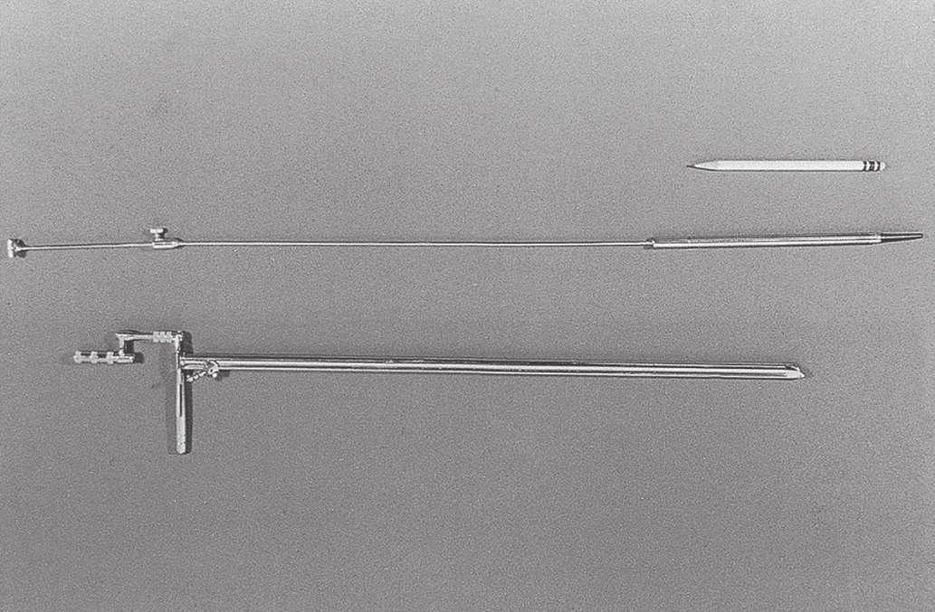
his talents in the GI tract. Under his influence, esophagoscopy was considered the exclusive province of ear, nose, and throat (ENT) departments in many community hospitals in the United States as late as the 1950s. The design of the esophagoscope remained a straight rigid tube, usually with a rubber finger-tipped obturator to make insertion safer. With the later addition of a 4 × power lens on the proximal end and a distal incandescent bulb, various models were popular until the introduction of fiberoptics in 1961. The Eder-Hufford rigid esophagoscope (Fig. 1.4), introduced in 1949, was popular and still in use in the early 1960s.
It was not until after 1900 that persistent efforts to develop a usable gastroscope were successful. All attempts to build a flexible instrument using a multiplicity of lenses were designed to be straightened after introduction and were fragile, easily damaged, and cumbersome. Straight tubes with simpler optics were useful, but perforations were still a problem.1 In 1911, Elsner introduced a rigid gastroscope with an outer tube through which a separate inner optical tube with a flexible rubber tip and sideviewing portal could be passed (Fig. 1.5). The rubber tip, previously used in the esophagoscope obturator, was more crucial than it might appear, for it seemed to be, along with the later
addition of a flexible metal coil proximal to it, the single feature that reduced the rate of perforation. Elsner’s instrument worked as designed and was widely used, especially by Schindler, then in his native Germany, who called it the“mother of all instruments until 1932.”5
In 1922, Schindler introduced his own version of the Elsner gastroscope, the major innovation of which was the important addition of an air channel to clear the lens of secretions. With the Elsner gastroscope, Schindler examined the stomachs of several hundred patients and meticulously recorded his findings in each procedure. He published Lehrbuch und Atlas der Gastreoskopie in 1923, with descriptions and remarkably accurate drawings. He trained others in the technique and was responsible for wide acceptance of gastroscopy. The procedure began with emptying the stomach using a nasogastric tube, followed by sedation. The patient was placed on the left side, and an assistant held the head rigidly extended to produce a straight path into the esophagus and the stomach (the “sword swallower’s technique”). The role of the assistant was crucial. Schindler’s effort was impressive and convinced many of the value of an expert examination of the stomach.
Semiflexible Gastroscopes
It became apparent that straight, rigid tubes were not ideal for examination of the stomach. Fatal perforations continued to the detriment of acceptance of the procedure. Visualization of the surface of the stomach was incomplete at best, with many consistent blind spots. These problems stimulated investigation of methods to manufacture safer, “flexible” instruments. The use of the term flexible here is problematic in view of what we think of today as flexible instruments. Although these early instruments were not flexible by our standards, they were more flexible than the straight, rigid instruments that came before. Semiflexible, with passive angulation of the distal portion of 34 degrees and sometimes more, was a more appropriate term.
In 1911, Hoffman showed that an image could be transmitted through a curved line by linking several short-focus prisms. Using this principle, several instruments were constructed, but these were unsatisfactory or were not widely accepted. Schindler, working with Wolf, the renowned instrument maker, constructed a semiflexible instrument with a rigid proximal portion and a distal portion made elastic by coiled copper wire and terminating with first a rubber finger and later a small rubber ball. Illumination was with a distal incandescent light bulb. Air insufflation was made possible with a rubber bulb, expanding the stomach wall to beyond the focal length of the prisms, which were manufactured by Zeiss. In 1932, the sixth and final version was patented. This instrument, known as the Wolf-Schindler gastroscope, greatly improved the safety and efficacy of gastroscopy and was used throughout the world (Fig. 1.6).
Thanks to the published meticulous work and enthusiasm of Schindler, whose designation as the “father of gastroscopy” is well deserved, the procedure was finally widely accepted as a valuable extension of the physical examination. The era of the semiflexible gastroscope from 1932 to 1957 has been called the Schindler era. Schindler was chiefly responsible for transforming gastroscopy from a dangerous and seldom used procedure to one that was relatively safe and indispensable for evaluation of known or suspected disease of the stomach. He insisted that all clinicians who planned to use the instrument be properly trained and that “… no manipulation inside of the body is without danger; therefore no endoscopic examination should be done
FIG 1.4 Eder-Hufford esophagoscope, the result of multiple attempts to develop a clinically useful instrument, 1949.
FIG 1.5 Elsner’s gastroscope, 1911. (From Edmonson JM: History of the instruments for gastrointestinal endoscopy. Gastrointest Endosc 37[Suppl 2]:S27–S56, 1991.)
Many clinicians did not believe the additional expense of replacing the older, beloved instruments with which they had been successful for many years was warranted. Even ACMI officials did not see the fiberscope as totally replacing the instruments with a lens system.2 Despite reservations, comparison and experiential studies showed the advantages of the new fiberscopes.14–17 Following the flagship ACMI model 4990, several models of the fiberscope were introduced by ACMI and other companies, each with significant improvements, including the controllable tip in the side-viewing ACMI model 5004. Visualization of the gastric pouch, including retroflexed views of the cardia, was now complete. The major objection to these instruments was the inability to pass the instrument under direct vision and examine the esophagus; in addition, the area beyond the pylorus could not be consistently examined.
Most clinicians were already fully trained in use of the EderHufford esophagoscope, and in the absence of a forward-viewing fiberscope, use of the Eder-Hufford esophagoscope continued. A forward-viewing scope was mandatory. LoPresti modified the tip of the fiberscope to create the foroblique fiberoptic esophagoscope in 1964.18 Passing the instrument under direct vision was possible, and clinicians immediately discovered that they could examine not only the esophagus, but also a large portion of the proximal stomach. At a length of 90 cm, however, one could not reach the duodenum. Working with ACMI, LoPresti produced the longer Panview Mark “87” gastroesophageal endoscope in 1970. By about 1971, the instrument had been lengthened to 105 cm with a four-way controllable tip capable of 180 degrees of deflection (Fig. 1.11).
The aptly named panendoscope was now a reality. Japanese and American manufacturers began to produce new models with such rapidity that endoscopists hardly had time to become thoroughly familiar with one before another, significantly improved (and more expensive) model was on the market. Patient comfort was greatly improved, and the relative safety of the fiberoptic endoscopes rapidly became apparent. By 1970, most gastroscopic examinations were done with fiberscopes. The development of a “teaching head” fiberoptic bundle with a light splitter and attached eyepiece and attachment to the eyepiece of the scope allowed two people to visualize the image. Dividing the light from the endoscope considerably diminished the brightness of the image, however, to both the operator and the observer. This device saw limited use and was utilized primarily in teaching institutions.
Endoscopic Retrograde Cholangiopancreatography (ERCP)
With access to the duodenum, the ampulla of Vater became visible. It followed that one should be able to inject contrast material into the bile and pancreatic ducts and increase diagnostic capabilities. Initial attempts in 1968 by McCune et al19 to modify an existing scope were only partially successful, but did show that endoscopic visualization by injection of radiologic contrast agents into ducts was possible. In 1970, Machida and Olympus in Japan produced usable, side-viewing scopes with controllable tips and elevators to move the injection tube to the ampulla. Japanese endoscopists20 developed the technique of endoscopic retrograde cholangiopancreatography (ERCP) with an 80% success rate.Vennes and Silvis21 showed the utility of ERCP in the United States and taught many physicians to use it.4 It was immediately apparent that if clinicians could visualize the biliary and pancreatic ducts endoscopically (i.e., nonsurgically), they should be able to apply by some means long-established surgical techniques for treatment of choledocholithiasis and pancreatitis, such as sphincterotomy and stone removal. In 1974, just 4 years after the demonstration of the diagnostic utility of the new ERCP
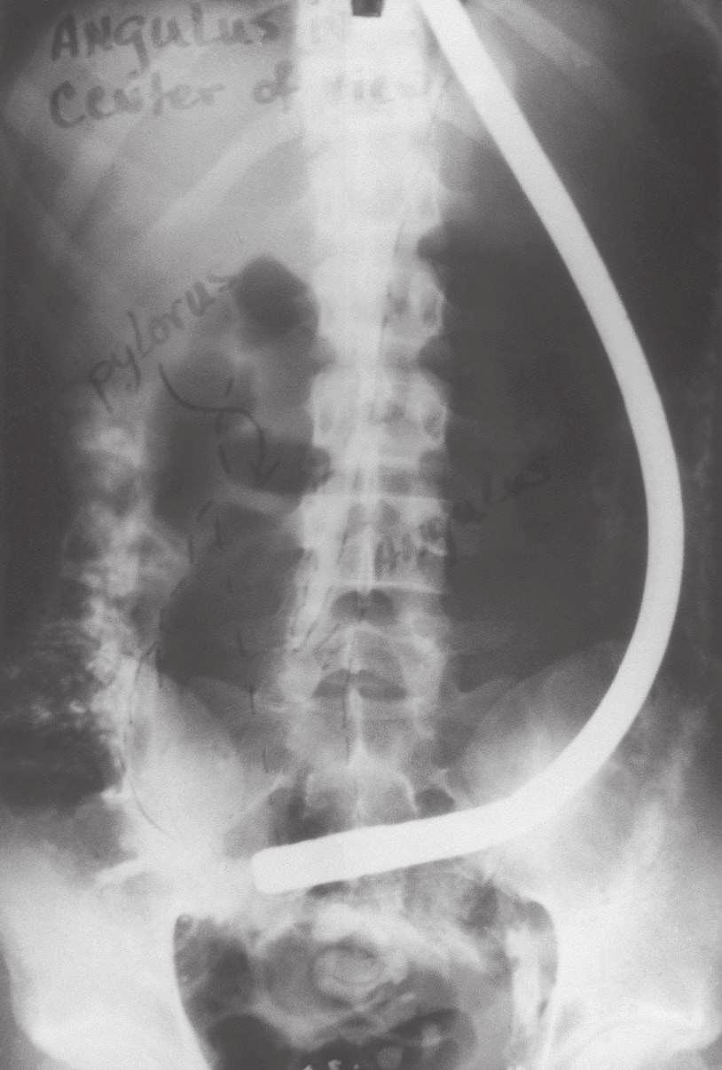
FIG 1.10 Visualization of duodenum was sometimes obtained by overinflating the stomach.
FIG 1.11 LoPresti forward-viewing esophagogastroscope. (From advertisement in Gastrointest Endosc 16:79, 1970.)
of diagnosis and recurrence of neoplasia, especially in the pancreas; portal hypertension; the colon and rectum; and bile ducts.41 In 1991, Wiersema et al42,43 showed that EUS could be used to obtain fine-needle aspiration cytology of mediastinal nodes and of nodes and lesions of the upper and lower GI tract. The addition of Doppler technology has now made possible the study of the flow through various structures, including the thoracic duct and blood vessels. EUS is increasingly being used to provide therapy, leading to the development of “interventional EUS.” EUS-guided interventions include celiac plexus block/ neurolysis, placement of fiducial markers to facilitate radiotherapy, direct injection of alcohol or chemotherapeutic agents for the treatment of tumors or cystic lesions, drainage of the pancreatic or biliary ductal systems, and the creation of gastrojejunal anastomoses using lumen-apposing metal stents. The techniques of using EUS instruments differ only slightly from using videoendoscopes, but dedicated training is necessary to interpret the sonographic images obtained accurately. EUS is not amenable to self-instruction. EUS training centers have been established in academic centers, but retraining of practicing physicians is challenging due to the duration of training necessary to achieve competence.44
Capsule Endoscopy (Wireless Endoscopy)
In 2000, Iddan et al45 reported the development of a capsule containing a tiny CMOS camera that could be swallowed, obtain images (at 2 frames per second), and transmit the images over 7 hours to a receiving digital storage unit worn by the patient as he or she goes about his or her normal activities. These frames are downloaded to a computer from which they are projected onto a monitor at a rate that can be controlled by the observer. Pictures can be printed of areas of interest. Gastroenterologists in Israel conducted randomized trials comparing the efficacy of the wireless capsule with push enteroscopy and obtained superior results with the capsule.46–48
Wireless capsule endoscopy caught the imagination of gastroenterologists over the world, and capsule endoscopy has been adopted as a part of standard practice for small bowel imaging. The findings are virtually unanimous in demonstrating better results in identifying lesions in the small bowel with capsule endoscopy when compared to push enteroscopy.49 The capsule avoids the discomfort and need for sedation inherent with push enteroscopy. In addition to lack of biopsy capability, an additional disadvantage is the time needed to review the study, but this has been overcome by a variety of methods including software advancements, improved training techniques, and utilizing non-physician personnel to initially review the obtained images. The major use of the capsule to date has been in elucidating the cause of occult bleeding from small bowel sources, where it seems to be superior to other methods. Future applications, such as in the colon, are continuing to be investigated in large, multicenter comparative studies. The future of wireless capsule endoscopy is bright. It will be interesting to see how the principle of wireless endoscopy is incorporated into videoendoscopes, such as the potential for a wireless connection between the endoscope and the image processor.
Enteroscopy
The small intestine has traditionally been regarded as the final frontier of GI endoscopy. Although capsule endoscopy provides remarkable images of the small bowel mucosa, tissue acquisition
and therapy with a capsule-based instrument is many years away. Surgically assisted small bowel enteroscopy may be performed via either the transoral or anal route or via a mid–small bowel enterotomy incision. The disadvantage of this technique is its invasive nature.50 Endoscopic examination of the small intestine has remained technically difficult. The many loops of the small intestine prevent progression of the instrument tip by simple pushing. This problem was overcome initially with the use of the Sonde enteroscope,51 which is a very fine, floppy instrument with a balloon at the tip. The Sonde enteroscope progressed through much of the small bowel under peristalsis, and then the proceduralist would slowly withdraw the instrument, assessing the mucosa while pulling back. This technique was thought to visualize 50% to 70% of the mucosal surface.52 However, the procedure was uncomfortable, time-consuming, and did not permit therapeutics, all of which limited its use.
The concept of small bowel enteroscopy was revolutionized by Yamamoto with the introduction of the double-balloon enteroscope in 2001.53 This technique uses traction between a balloon at the tip of the enteroscope and another balloon on a flexible overtube to fix the loops of small bowel and provide traction for forward movement. The procedure requires peroral and anal procedures to examine the entire small intestine, and even then only in a minority of Western patients is the whole small bowel visualized. Nonetheless, double-balloon–assisted enteroscopy permits endoscopic therapeutics to most of the small bowel without the need for surgical assistance. A single balloon version is also available.
Natural Orifice Transluminal Endoscopic Surgery (NOTES) and Peroral Endoscopy Myotomy (POEM)
A new development in endoscopy is natural orifice transluminal endoscopic surgery (NOTES), in which the endoscope is inserted into the abdominal cavity via an incision in an accessible organ. The first report appeared in 2002. Incisions have been made in the stomach, vagina, and colon with successful tubal ligation, liver biopsies, biopsy of peritoneal metastases, oophorectomy, cholecystectomy, and nephrectomy procedures having been performed. Most published articles report experimental use in animals, but more recent reports have described the simultaneous use of NOTES with laparoscopic techniques. Comparative studies are ongoing. A difficulty with the technique has been overcoming the lack of instrument “triangulation”; that is, approaching a surgical site from two or more directions to create countertraction, tie sutures, and so forth. Although NOTES is an exciting development, its remarkable potential will have to await the development of new instruments and the acquisition of additional expertise. At a minimum, it appears the development of NOTES will result in marked improvements in mucosal and transmural closure devices. Recently, flexible endoscopes have also been used to tunnel into the submucosal space of the esophagus and perform a myotomy, resulting in a treatment for achalasia termed peroral endoscopy myotomy, or POEM. First performed by Inoue in 2008 and reported by Inoue in 2010, this procedure has gained widespread popularity worldwide and has been performed thousands of times to date with impressive short- and long-term results and an excellent safety profile.54,55 Additional applications of “submucosal” endoscopy include performing a similar procedure in the antrum to treat gastroparesis (G-POEM) and to perform resection of intramural lesions of the GI tract.56,57