CONTRIBUTORS
James L. Achord, MD
Professor Emeritus
University of Mississippi Medical Center
Jackson, Misssissippi
1: The History of Gastrointestinal Endoscopy
Michelle J. Alfa, BSc, MSc, PhD
Principal Investigator
St. Boniface Research Centre; Professor
Department of Medical Microbiology
University of Manitoba Winnipeg, Manitoba, Canada
4: Cleaning and Disinfecting Gastrointestinal Endoscopy Equipment
Mohammad Al-Haddad, MD, MSc, FASGE, FACG, AGAF
Associate Professor of Medicine
Division of Gastroenterology and Hepatology
Indiana University School Medicine Indianapolis, Indiana
62: Evaluation and Staging of Pancreaticobiliary Malignancy
Andrea Anderloni, MD, PhD
Digestive Endoscopy Unit
Division of Gastroenterology
Humanitas Research Hospital
Milan, Italy
28: Palliation of Malignant Dysphagia and Esophageal Fistulas
Joseph C. Anderson, MD
Associate Professor of Medicine
Department of Veterans Affairs Medical Center
White River Junction, Vermont; The Geisel School of Medicine at Dartmouth Hanover, New Hampshire; Division of Gastroenterology and Hepatology
University of Connecticut School of Medicine
Farmington, Connecticut
36: Colorectal Cancer Screening and Surveillance
Anna Baiges, MD
Hepatic Hemodynamic Laboratory
Liver Unit, Hospital Clínic
Barcelona, Spain
15: Portal Hypertensive Bleeding
John Baillie, MD
Professor
Division of Gastroenterology and Hepatology
Department of Medicine
Virginia Commonwealth University School of Medicine
Richmond, Virginia
3: How Endoscopes Work
Alan N. Barkun, MD, MSc
Division of Gastroenterology
McGill University Health Center
Montreal, Québec, Canada
14: Nonvariceal Upper Gastrointestinal Bleeding
Todd H. Baron, MD, FASGE Professor of Medicine
Division of Gastroenterology and Hepatology
University of North Carolina Chapel Hill, North Carolina
20: Endoscopic Diagnosis and Management of Zenker’s Diverticula
Omer Basar, MD
Pancreas Biliary Center, Gastrointestinal Unit
Massachusetts General Hospital Boston, Massachusetts; Professor of Medicine
Department of Gastroenterology
Hacettepe University
Ankara, Turkey
61: Pancreatic Cystic Lesions
Mark Benson, MD
Assistant Professor
Division of Gastroenterology and Hepatology
University of Wisconsin School of Medicine and Public Health
Madison, Wisconsin
22: Ingested Foreign Objects and Food Bolus Impactions
Lyz Bezerra Silva, MD, MSC
Associate Professor of Surgery Department of Surgery
Federal University of Pernambuco
Recife, Brazil
45: Intramural and Transmural Endoscopy
Stas Bezobchuk, MD
Institute of Gastroenterology, Hepatology, and Nutrition
Emek Medical Center
Afula, Israel
17: Middle Gastrointestinal Bleeding
Kenneth F. Binmoeller, MD
Director, Interventional Endoscopy Services
Paul May and Frank Stein Interventional Endoscopy Center
California Pacific Medical Center
San Francisco, California
58: Pancreatic Fluid Collections and Leaks
Sarah Blankstein, AB, JD
Boston, Massachusetts
10: Legal Concepts for Gastroenterologists
Daniel Blero, MD, PhD
Department of Gastroenterology
Chu Charleroi
Charleroi, Belgium; Hôpital Erasme
Brussels, Belgium
43: Endoscopic Techniques for Weight Loss
Michael J. Bourke, BSc, MD
Department of Gastroenterology and Hepatology
Westmead Hospital
Sydney, Australia
34: Duodenal and Papillary Adenomas
William R. Brugge, MD
Chief
Division of Gastroenterology
Mount Auburn Hospital
Cambridge, Massachusetts
61: Pancreatic Cystic Lesions
Marco J. Bruno, MD, PhD
Department of Gastroenterology and Hepatology
Erasmus Medical Center
University of Rotterdam
Rotterdam, The Netherlands
63: Palliation of Malignant Pancreaticobiliary Obstruction
Anna M. Buchner, MD, PhD
Assistant Professor of Medicine
Division of Gastroenterology
University of Pennsylvania
Philadelphia, Pennsylvania
38: Endoscopic Diagnosis and Staging of Inflammatory Bowel Disease
Andrés Cárdenas, MD, MMSc, PhD, AGAF, FAASLD
Faculty Member/Consultant
Institute of Digestive Diseases and Metabolism Hospital Clinic
University of Barcelona
Barcelona, Spain
15: Portal Hypertensive Bleeding
54: Postoperative Biliary Strictures and Leaks
David Carr-Locke, MD, FRCP, FASGE, AGAF, NYSGEF
Clinical Director
Center for Advanced Digestive Care
Gastroenterology & Hepatology
Weill Cornell Medical College
Cornell University
New York, New York
55: Infections of the Biliary Tract
Kenneth Chang, MD
Professor and Chief
Division of Gastroenterology and Hepatology
University of California—Irvine
Orange, California
51: Endoscopic Ultrasound and Fine-Needle Aspiration for Pancreatic and Biliary Disorders
Saurabh Chawla, MD, FACG
Director of Endoscopy
Grady Memorial Hospital;
Assistant Professor of Medicine
Emory University School of Medicine
Atlanta, Georgia
48: Preparation for Pancreaticobiliary Endoscopy
John O. Clarke, MD
Clinical Associate Professor
Department of Medicine
Stanford University
Stanford, California
19: Esophageal Motility Disorders
29: Endoscopic Approaches for Gastroparesis
Jonathan Cohen, MD
Clinical Professor
Department of Medicine
New York University Langone School of Medicine
New York, New York
13: Endoscopic Simulators
Andrew P. Copland, MD
Assistant Professor of Medicine
Division of Gastroenterology and Hepatology
University of Virginia Health Systems
Charlottesville, Virginia
40: Colonic Strictures
Guido Costamagna, MD, FACG
Digestive Endoscopy Unit
Catholic University
Gemelli University Hospital
Rome, Italy
54: Postoperative Biliary Strictures and Leaks
Peter B. Cotton, MD, FRCS, FRCP
Professor of Medicine
Digestive Disease Center
Medical University of South Carolina
Charleston, South Carolina
56: Sphincter of Oddi Disorders
Amit P. Desai, MD
Texas Digestive Diseases Consultants
Texas Health Presbyterian Hospital
Dallas, Texas
47: Extraintestinal Endosonography
Jacques Devière, MD, PhD
Professor of Medicine
Chairman, Department of Gastroenterology, Hepatopancreatology, and Digestive Oncology
Erasme Hospital
Université Libre de Bruxelles
Brussels, Belgium
43: Endoscopic Techniques for Weight Loss
Christopher J. DiMaio, MD
Director of Therapeutic Endoscopy
Associate Professor of Medicine
Division of Gastroenterology
Icahn School of Medicine at Mount Sinai
New York, New York
53: Gallstone Disease: Choledocholithiasis, Cholecystitis, and Gallstone Pancreatitis
Peter Draganov, MD
Professor of Medicine
Department of Internal Medicine
University of Florida
Gainesville, Florida
37: Colonoscopic Polypectomy, Mucosal Resection, and Submucosal Dissection
Jérôme Dumortier, MD
Department of Hepatogastroenterology and Digestive Endoscopy
Edouard Herriot Hospital
Lyon, France
11: Small-Caliber Endoscopy
Jeffrey J. Easler, MD
Assistant Professor of Medicine
Division of Gastroenterology and Hepatology
Indiana University School of Medicine; Richard L. Roudebush VA Medical Center
Indianapolis, Indiana
49: Cholangiography and Pancreatography
Gary W. Falk, MD, MS
Professor of Medicine
Department of Medicine, Division of Gastroenterology
University of Pennsylvania Perelman School of Medicine
Philadelphia, Pennsylvania
25: Barrett’s Esophagus: Diagnosis, Surveillance, and Medical Management
Francis A. Farraye, MD, MSc
Clinical Director
Section of Gastroenterology
Boston Medical Center; Professor of Medicine
Department of Medicine
Boston University School of Medicine
Boston, Massachusetts
39: Dysplasia Surveillance in Inflammatory Bowel Disease
Andrew Feld, MD, JD
Program Chief, Group Health Cooperative
Clinical Professor
University of Washington
Seattle, Washington
10: Legal Concepts for Gastroenterologists
Kayla Feld, JD
Quinn Emanuel Urquhart & Sullivan
Washington, D.C.
10: Legal Concepts for Gastroenterologists
Paul Fockens, MD, PhD, FASGE Professor and Chair
Department of Gastroenterology and Hepatology
Academic Medical Center
Amsterdam, The Netherlands
33: Palliation of Gastric Outlet Obstruction
Evan L. Fogel, MD, MSc, FRCP(C) Professor of Medicine
Department of Gastroenterology and Hepatology
Indiana University School of Medicine
Indianapolis, Indiana
49: Cholangiography and Pancreatography
Kyle J. Fortinsky, MD, BSc
Division of Gastroenterology
University of Toronto
Toronto, Ontario, Canada
14: Nonvariceal Upper Gastrointestinal Bleeding
Martin L. Freeman, MD
Professor of Medicine
Division of Gastroenterology, Hepatology, and Nutrition
University of Minnesota Minneapolis, Minnesota
57: Recurrent Acute Pancreatitis
Juan Carlos García-Pagán, MD, PhD Head
Barcelona Hepatic Hemodynamic Lab; Senior Consultant in Hepatology
Associate Professor
University of Barcelona; Liver Unit, Hospital Clínic Barcelona, Spain
15: Portal Hypertensive Bleeding
Hans Gerdes, MD
Attending Physician Department of Medicine
Memorial Sloan Kettering Cancer Center; Professor of Clinical Medicine
Weill Cornell Medical College of Cornell University
New York, New York
30: Gastric Polyps and Thickened Gastric Folds
Joanna A. Gibson, MD, PhD
Assistant Professor of Pathology
Yale University School of Medicine
New Haven, Connecticut
5: Tissue Sampling, Specimen Handling, and Laboratory Processing
Gregory G. Ginsberg, MD Professor of Medicine
Department of Medicine, Division of Gastroenterology
Hospital of the University of Pennsylvania Philadelphia, Pennsylvania
50: Difficult Cannulation and Sphincterotomy
Marc Giovannini, MD
Head, Gastroenterology and Endoscopy Department
Paoli-Calmettes Institute
Marseille, France
52: Endoscopic Ultrasound-Guided Access and Drainage of the Pancreaticobiliary Ductal Systems
Ian M. Gralnek, MD, MSHS, FASGE
Clinical Associate Professor of Medicine/ Gastroenterology
Rappaport Faculty of Medicine Technion
Israel Institute of Technology; Chief, Institute of Gastroenterology, Hepatology and Nutrition
Emek Medical Center
Afula, Israel
17: Middle Gastrointestinal Bleeding
Frank G. Gress, MD Professor of Medicine
Chief, Interventional Endoscopy Division of Digestive & Liver Diseases
Columbia University Medical Center
New York, New York
47: Extraintestinal Endosonography
Robert H. Hawes, MD Professor
Department of Medicine
University of Central Florida College of Medicine;
Medical Director
Florida Hospital Institute for Minimally Invasive Therapy
Florida Hospital Orlando
Orlando, Florida
59: Chronic Pancreatitis
Virginia Hernández-Gea, MD, PhD
Hepatic Hemodynamic Laboratory
Liver Unit, Hospital Clínic
Barcelona, Spain
15: Portal Hypertensive Bleeding
Ikuo Hirano, MD
Professor of Medicine
Department of Medicine, Division of Gastroenterology
Northwestern University Feinberg School of Medicine; Director, Northwestern Esophageal Center
Northwestern Medicine
Chicago, Illinois
23: Eosinophilic Esophagitis
Juergen Hochberger, MD, PhD
Chairman
Department of Gastroenterology
Vivantes Klinikum im Friedrichshain
Berlin, Germany
50: Difficult Cannulation and Sphincterotomy
Douglas A. Howell, MD
Director, Advanced Interventional Endoscopy Fellowship
Director, Pancreaticobiliary Center
Maine Medical Center
Portland, Maine;
Associate Clinical Professor
Tufts University School of Medicine
Boston, Massachusetts
60: The Indeterminate Biliary Stricture
Chin Hur, MD, MPH
Associate Director, Institute for Technology Assessment
Director, GI Health Outcomes Research
Massachusetts General Hospital; Associate Professor of Medicine
Harvard Medical School
Boston, Massachusetts
26: Screening for Esophageal Squamous Cell Carcinoma
Joo Ha Hwang, MD, PhD
Professor of Medicine
Department of Medicine
Division of Gastroenterology and Hepatology
Stanford University
Stanford, California
6: Electrosurgery in Therapeutic Endoscopy
Maite Betés Ibáñez, PhD, MD
Department of Gastroenterology
University Clinic of Navarra
Pamplona, Navarra, Spain
18: Occult and Unexplained Chronic Gastrointestinal Bleeding
Takao Itoi, MD, PhD, FASGE, FACG
Chair and Professor
Department of Gastroenterology and Hepatology
Tokyo Medical University
Tokyo, Japan
52: Endoscopic Ultrasound-Guided Access and Drainage of the Pancreaticobiliary Ductal Systems
Prasad G. Iyer, MD, MS
Professor and Consultant
Department of Gastroenterology and Hepatology
Mayo Clinic
Rochester, Minnesota
27: Endoscopic Treatment of Early Esophageal Neoplasia
David A. Johnson, MD, MACG, FASGE, FACP
Professor of Medicine and Chief
Division of Gastroenterology and Hepatology
Department of Internal Medicine
Eastern Virginia Medical School
Norfolk, Virginia
9: Bowel Preparation for Colonoscopy
Sreeni Jonnalagadda, MD
Professor of Medicine
Director of Therapeutic and Biliary Endoscopy
Saint Luke’s Hospital
University of Missouri—Kansas City
Kansas City, Missouri
12: Postsurgical Endoscopic Anatomy
Charles J. Kahi, MD, MS, FACP, FACG, AGAF, FASGE
Professor of Clinical Medicine
Indiana University School of Medicine; Gastroenterology Section Chief
Richard L. Roudebush VA Medical Center
Indianapolis, Indiana
36: Colorectal Cancer Screening and Surveillance
Tonya Kaltenbach, MD, MAS
Associate Professor of Clinical Medicine
Division of Gastroenterology, Department of Medicine
University California San Francisco; Director of Advanced Endoscopy
San Francisco Veterans Affair Medical Center
San Francisco, California
37: Colonoscopic Polypectomy, Mucosal Resection, and Submucosal Dissection
Leila Kia, MD
Assistant Professor of Medicine
Department of Medicine, Division of Gastroenterology
Northwestern University Feinberg School of Medicine
Chicago, Illinois
23: Eosinophilic Esophagitis
Michael B. Kimmey, MD
Franciscan Digestive Care Associates
Gig Harbor, Washington
35: Acute Colonic Pseudo-Obstruction
Amir Klein, MD
Department of Gastroenterology and Hepatology
Rambam Health Care Campus
Haifa, Israel
34: Duodenal and Papillary Adenomas
Michael L. Kochman, MD
Wilmott Family Professor of Medicine
Division of Gastroenterology, Department of Medicine
Perelman School of Medicine
University of Pennsylvania
Philadelphia, Pennsylvania
21: Benign Esophageal Strictures
Divyanshoo R. Kohli, MD
Division of Gastroenterology and Hepatology
Department of Medicine
Virginia Commonwealth University School of Medicine
Richmond, Virginia
3: How Endoscopes Work
Andrew Korman
Division of Gastroenterology and Hepatology
Saint Peter’s University Hospital
New Brunswick, New Jersey
55: Infections of the Biliary Tract
Wilson T. Kwong, MD, MS
Assistant Professor
Department of Gastroenterology
University of California San Diego
La Jolla, California
16: Lower Gastrointestinal Bleeding
Ryan Law, DO
Clinical Lecturer
Division of Gastroenterology and Hepatology
University of Michigan
Ann Arbor, Michigan
20: Endoscopic Diagnosis and Management of Zenker’s Diverticula
David A. Leiman, MD, MSHP
Assistant Professor of Medicine
Division of Gastroenterology
Duke University School of Medicine
Durham, North Carolina
24: Gastroesophageal Reflux Disease
Anne Marie Lennon, MB, PhD, FRCPI
Benjamin Baker Scholar
Associate Professor of Medicine and Surgery
The Johns Hopkins Hospital
Baltimore, Maryland
61: Pancreatic Cystic Lesions
Michael Levy, MD
Professor of Medicine
Division of Gastroenterology and Hepatology
Mayo Clinic
Rochester, Minnesota
62: Evaluation and Staging of Pancreaticobiliary Malignancy
David Lichtenstein, MD
Director of Endoscopy
Department of Gastroenterology
Boston Medical Center
Boston University School of Medicine
Boston, Massachusetts
4: Cleaning and Disinfecting Gastrointestinal Endoscopy Equipment
Gary R. Lichtenstein, MD
Professor of Medicine
Director, Center for Inflammatory Bowel Disease
Division of Gastroenterology
University of Pennsylvania
Philadelphia, Pennsylvania
38: Endoscopic Diagnosis and Staging of Inflammatory Bowel Disease
Alisa Likhitsup, MD
Gastroenterology Fellow
Department of Gastroenterology
University of Missouri—Kansas City
Kansas City, Missouri
12: Postsurgical Endoscopic Anatomy
Jimmy K. Limdi, MBBS, FRCP, FRCPE, FACG
Consultant Gastroenterologist
Department of Gastroenterology
The Pennine Acute Hospitals NHS Trust; Honorary Senior Lecturer
Institute of Inflammation and Repair
University of Manchester
Manchester, United Kingdom
39: Dysplasia Surveillance in Inflammatory Bowel Disease
Gianluca Lollo, MD
Department of Surgical Oncology and Gastroenterological Sciences
University of Padua
Padua, Italy
28: Palliation of Malignant Dysphagia and Esophageal Fistulas
Fauze Maluf-Filho, MD, PhD, FASGE Professor
Department of Gastroenterology
Medical School of University of São Paulo; Chief Endoscopy Unit
Institute of Cancer of Univeristy of São Paulo
63: Palliation of Malignant Pancreaticobiliary Obstruction
Jennifer Maranki, MD, MSc
Associate Professor of Medicine Director of Endoscopy Division of Gastroenterology and Hepatology
Penn State Hershey Medical Center Hershey, Pennsylvania
46: Endoscopic Full-Thickness Resection of Subepithelial Lesions of the GI Tract
Richard W. McCallum, MD, FACP, FRACP (Aust), FACG, AGAF
Professor of Medicine and Founding Chair Department of Internal Medicine
Texas Tech University El Paso, Texas; Honorary Professor University of Queensland Queensland, Australia
29: Endoscopic Approaches for Gastroparesis
Stephen A. McClave, MD Professor of Medicine
Department of Medicine
University of Louisville School of Medicine Louisville, Kentucky
42: Techniques in Enteral Access
Klaus Mergener, MD, PhD, MBA Partner
Digestive Health Specialists Tacoma, Washington
2: Setting Up an Endoscopy Facility
David C. Metz, MD Professor of Medicine Division of Gastroenterology Perelman School of Medicine
University of Pennsylvania Philadelphia, Pennsylvania
24: Gastroesophageal Reflux Disease
Volker Meves, MD
Department of Gastroenterology
Vivantes Klinikum im Friedrichshain Berlin, Germany
50: Difficult Cannulation and Sphincterotomy
Marcia L. Morris, MS
Electrosurgery Consultant
St. Paul, Minnesota
6: Electrosurgery in Therapeutic Endoscopy
Daniel K. Mullady, MD
Associate Professor of Medicine
Director, Interventional Endoscopy
Department of Gastroenterology
Washington University in St. Louis School of Medicine
St. Louis, Missouri
53: Gallstone Disease: Choledocholithiasis, Cholecystitis, and Gallstone Pancreatitis
Miguel Muñoz-Navas, PhD, MD Professor of Medicine
University of Navarra School of Medicine; Director
Department of Gastroenterology
University of Navarra Clinic
Pamplona, Navarra, Spain
18: Occult and Unexplained Chronic Gastrointestinal Bleeding
V. Raman Muthusamy, MD, MAS
Director of Endoscopy, UCLA Health System Professor of Clinical Medicine
Vatche and Tamar Manoukian Division of Digestive Diseases
David Geffen School of Medicine at UCLA
Los Angeles, California
1: The History of Gastrointestinal Endoscopy
Zaheer Nabi, MD, DNB Consultant Gastoenterologist
Asian Institute of Gastroenterology
Hyderabad, India
55: Infections of the Biliary Tract
Andrew Nett, MD
Paul May and Frank Stein Interventional Endoscopy Center
California Pacific Medical Center; Department of Medicine University of California San Francisco San Francisco, California
58: Pancreatic Fluid Collections and Leaks
Nam Q. Nguyen, MBBS (Hons), FRACP, PhD
Associate Professor Head, Education and Research Department of Gastroenterology and Hepatology
Royal Adelaide Hospital
University of Adelaide
Adelaide, South Australia, Australia
8: Patient Preparation and Pharmacotherapeutic Considerations
Nicholas Nickl, MD
Professor of Medicine
University of Kentucky Medical Center
Lexington, Kentucky
31: Subepithelial Tumors of the Esophagus and Stomach
Satoru Nonaka, MD, PhD
Endoscopy Division
National Cancer Center Hospital
Tokyo, Japan
32: Diagnosis and Treatment of Superficial Gastric Neoplasms
Ichiro Oda, MD
Endoscopy Division
National Cancer Center Hospital
Tokyo, Japan
32: Diagnosis and Treatment of Superficial Gastric Neoplasms
Robert D. Odze, MD, FRCPC Professor of Pathology
Department of Pathology
Brigham and Women’s Hospital
Boston, Massachusetts
5: Tissue Sampling, Specimen Handling, and Laboratory Processing
Edward C. Oldfield IV, MD
Department of Internal Medicine
Eastern Virginia Medical School
Norfolk, Virginia
9: Bowel Preparation for Colonoscopy
Parth J. Parekh, MD
Department of Internal Medicine
Division of Gastroenterology and Hepatology
Tulane University
New Orleans, Louisiana
9: Bowel Preparation for Colonoscopy
Patrick R. Pfau, MD
Professor of Medicine, Chief of Clinical Gastroenterology
Division of Gastroenterology and Hepatology
University of Wisconsin School of Medicine and Public Health
Madison, Wisconsin
22: Ingested Foreign Objects and Food Bolus Impactions
Mathieu Pioche, MD, PhD
Department of Hepatogastroenterology and Digestive Endoscopy
Edouard Herriot Hospital Lyon, France
11: Small-Caliber Endoscopy
Heiko Pohl, MD
Associate Professor of Medicine
Geisel School of Medicine at Dartmouth
Hanover New Hampshire; Department of Gastroenterology
Veterans Affair Medical Center
White River Junction, Vermont
37: Colonoscopic Polypectomy, Mucosal Resection, and Submucosal Dissection
Thierry Ponchon, MD, PhD
Department of Hepatogastroenterology and Digestive Endoscopy
Edouard Herriot Hospital
Lyon, France
11: Small-Caliber Endoscopy
Robert J. Ponec, MD
Consulting Gastroenterologist and Therapeutic Endoscopist
Department of Gastroenterology and Hepatology
Salem Gastroenterology Consultants
Salem, Oregon
35: Acute Colonic Pseudo-Obstruction
Michael W. Rajala, MD, PhD
Assistant Professor of Clinical Medicine
Division of Gastroenterology, Department of Medicine
Perelman School of Medicine
University of Pennsylvania Philadelphia, Pennsylvania
21: Benign Esophageal Strictures
Nageshwar Reddy, MBBS, MD, DM
Chairman and Chief of Gastroenterology
Asian Institute of Gastroenterology
Hyderabad, India
55: Infections of the Biliary Tract
Alessandro Repici, MD
Professor of Gastroenterology
Director of Endoscopy
Humanitas Research Hospital & Humanitas University
Milan, Italy
28: Palliation of Malignant Dysphagia and Esophageal Fistulas
Jérôme Rivory, MD
Department of Hepatogastroenterology and Digestive Endoscopy
Edouard Herriot Hospital
Lyon, France
11: Small-Caliber Endoscopy
Marvin Ryou, MD
Division of Gastroenterology, Hepatology, and Endoscopy
Brigham and Womens’ Hospital; Instructor
Harvard Medical School
Boston, Massachusetts
44: Management of Post-Bariatric Complications
Yutaka Saito, MD, PhD, FASGE, FACG
Chief, Director Endoscopy Division
National Cancer Center Hospital
Tokyo, Japan
32: Diagnosis and Treatment of Superficial Gastric Neoplasms
Jason B. Samarasena, MD
Associate Clinical Professor of Medicine
Division of Gastroenterology and Hepatology
University of California—Irvine
Orange, California
51: Endoscopic Ultrasound and Fine-Needle Aspiration for Pancreatic and Biliary Disorders
Thomas J. Savides, MD
Professor of Clinical Medicine
Division of Gastroenterology
University of California San Diego
La Jolla, California
16: Lower Gastrointestinal Bleeding
Mark Schoeman, MBBS, PhD, FRACP
Head, Gastrointestinal Investigation Unit
Department of Gastroenterology and Hepatology
Royal Adelaide Hospital
Adelaide, South Australia, Australia
8: Patient Preparation and Pharmacotherapeutic Considerations
Allison R. Schulman, MD, MPH
Physician
Division of Gastroenterology, Hepatology, and Endoscopy
Brigham and Women’s Hospital; Harvard Medical School
Boston, Massachusetts
44: Management of Post-Bariatric Complications
Amrita Sethi, MD, MSc
Associate Professor of Medicine
Director of Pancreaticobiliary Endoscopy Services
Columbia University Medical Center
New York, New York
60: The Indeterminate Biliary Stricture
Pari M. Shah, MD, MSCE
Assistant Attending Physician
Department of Medicine
Memorial Sloan Kettering Cancer Center;
Assistant Professor of Clinical Medicine
Weill Cornell Medical College of Cornell University
New York, New York
30: Gastric Polyps and Thickened Gastric Folds
Stuart Sherman, MD
Glen A. Lehman Professor of Gastroenterology
Professor of Medicine
Division of Gastroenterology and Hepatology
Indiana University School of Medicine
Indianapolis, Indiana
49: Cholangiography and Pancreatography
Uzma D. Siddiqui, MD
Center for Endoscopic Research and Therapeutics
University of Chicago School of Medicine
Chicago, Illinois
59: Chronic Pancreatitis
Vikesh K. Singh, MD, MSc
Director, Pancreatitis Center
Associate Professor of Medicine
John Hopkins University School of Medicine
Baltimore, Maryland
48: Preparation for Pancreaticobiliary Endoscopy
Roy Soetikno, MD, MS
Veterans Affairs Palo Alto Health Care System
Stanford University School of Medicine
Palo Alto, California
37: Colonoscopic Polypectomy, Mucosal Resection, and Submucosal Dissection
Stavros N. Stavropoulos, MD, FASGE
Chief, GI Endoscopy
Director, Program in Advanced GI Endoscopy (P.A.G.E.)
Winthrop University Hospital
Mineola, New York; Adjunct Professor of Clinical Medicine
Columbia University
New York, New York
46: Endoscopic Full-Thickness Resection of Subepithelial Lesions of the GI Tract
Tyler Stevens, MD
Associate Professor
Department of Gastroenterology and Hepatology
Cleveland Clinic Cleveland, Ohio
57: Recurrent Acute Pancreatitis
Christina Surawicz, MD
Professor Division of Gastroenterology Department of Medicine University of Washington
Seattle, Washington
41: Infections of the Luminal Digestive Tract
Barry Tanner, CPA
Chief Executive Officer Physicians Endoscopy
Jamison, Pennsylvania
2: Setting Up an Endoscopy Facility
Paul Tarnasky, MD
Digestive Health Associates of Texas Dallas, Texas
56: Sphincter of Oddi Disorders
Christopher C. Thompson, MD, MSc, FACG, FASGE, AGAF
Director of Therapeutic Endoscopy
Division of Gastroenterology, Hepatology, and Endoscopy
Brigham and Women’s Hospital; Assistant Professor of Medicine
Harvard Medical School
Boston, Massachusetts
44: Management of Post-Bariatric Complications
Mark Topazian, MD
Professor of Medicine
Division of Gastroenterology & Hepatology
Mayo Clinic
Rochester, Minnesota
51: Endoscopic Ultrasound and Fine-Needle Aspiration for Pancreatic and Biliary Disorders
George Triadafilopoulos, MD, DSc
Clinical Professor of Medicine
Stanford Multidimensional Program for Innovation and Research in the Esophagus (S-MPIRE)
Division of Gastroenterology and Hepatology
Stanford University School of Medicine
Stanford, California
19: Esophageal Motility Disorders
Emo E. van Halsema, MD
Department of Gastroenterology and Hepatology
Academic Medical Center
Amsterdam, The Netherlands
33: Palliation of Gastric Outlet Obstruction
Jeanin E. van Hooft, MD, PhD, MBA
Department of Gastroenterology and Hepatology
Academic Medical Center
Amsterdam, The Netherlands
33: Palliation of Gastric Outlet Obstruction
John Joseph Vargo II, MD, MPH
Vice Chair, Digestive Disease Institute Chair Department of Gastroenterology and Hepatology
Cleveland Clinic
Cleveland, Ohio
7: Sedation and Monitoring in Endoscopy
Kavel Visrodia, MD Fellow
Department of Internal Medicine, Division of Gastroenterology and Hepatology
Mayo Clinic
Rochester, Minnesota
27: Endoscopic Treatment of Early Esophageal Neoplasia
Vaibhav Wadhwa, MD
Clinical Fellow
Department of Gastroenterology and Hepatology
Cleveland Clinic Florida Weston, Florida
7: Sedation and Monitoring in Endoscopy
Kristian Wall, MD Fellow
Division of Digestive Diseases and Nutrition University of Kentucky Lexington, Kentucky
31: Subepithelial Tumors of the Esophagus and Stomach
Catharine M. Walsh, MD, MEd, PhD, FAAP, FRCPC
Division of Gastroenterology, Hepatology, and Nutrition and the Learning and Research Institutes
Department of Paediatrics Hospital for Sick Children; The Wilson Centre
University of Toronto
Toronto, Ontario, Canada
13: Endoscopic Simulators
Andrew Y. Wang, MD, AGAF, FACG, FASGE
Associate Professor of Medicine
Chief, Section of Interventional Endoscopy
Division of Gastroenterology and Hepatology
University of Virginia Health System
Charlottesville, Virginia
40: Colonic Strictures
Kenneth K. Wang, MD
Kathy and Russ VanCleve Professor of Gastroenterology Research
Department of Gastroenterology and Hepatology
Mayo Clinic
Rochester, Minnesota
27: Endoscopic Treatment of Early Esophageal Neoplasia
Sachin Wani, MD
Associate Professor of Medicine
Department of Medicine, Division of Gastroenterology
University of Colorado School of Medicine
Aurora, Colorado
25: Barrett’s Esophagus: Diagnosis, Surveillance, and Medical Management
C. Mel Wilcox, MD, MSPH
Director
Division of Gastroenterology and Hepatology
University of Alabama at Birmingham Birmingham, Alabama
41: Infections of the Luminal Digestive Tract
Field F. Willingham, MD, MPH, FASGE Director of Endoscopy
Associate Professor of Medicine
Emory University School of Medicine
Atlanta, Georgia
48: Preparation for Pancreaticobiliary Endoscopy
Patrick S. Yachimski, MD, MPH, FASGE
Associate Professor of Medicine
Vanderbilt University School of Medicine
Nashville, Tennessee
26: Screening for Esophageal Squamous Cell Carcinoma
Ricardo Zorron, MD, PhD
Professor of Surgery, University UNIRIO, UENF;
Director, Center for Innovative Surgery-ZIC, Center for Bariatric and Metabolic Surgery;
Department of Surgery, Campus Charité Mitte/Campus Virchow-Klinikum
Charité-Universitätsmedizin Berlin Berlin, Germany
45: Intramural and Transmural Endoscopy
Welcome to the third edition of Clinical Gastrointestinal Endoscopy Gastrointestinal endoscopy is a continuously evolving field with the advent of new technologies, refined techniques, and new applications. The prior editions of this book have been universally regarded as a comprehensive guide to the latest endoscopic techniques. Understanding and adoption of such practices leads to optimal outcomes with endoscopy. This text is unique because of the breadth of topics covered by experts in every discipline of gastrointestinal endoscopy from across the globe. Clinical Gastrointestinal Endoscopy has been an essential resource for anyone interested in learning about endoscopic procedures, as one can access a variety of topics in succinct, easily understood chapters from content specialists.
This edition marks the transition to a new editorial team and builds on the success of the two prior editions. The previous editions achieved great accolade due to the efforts of the editorial board lead by Gregory Ginsberg and coedited by Michael Kochman, Ian Norton, and Christopher Gostout. The new editorial team was selected due to their expertise in gastrointestinal endoscopy, enthusiasm for disseminating best practices to a worldwide audience, and diverse background of training and experience from different premiere institutions. Commensurate with the change in the editors, we were excited to invite a new set of content experts who share their insights into recent advances in endoscopy and the impact these innovations have had on improving patient care. This has led to an exciting, comprehensive textbook from today’s most prestigious specialists.
Clinical Gastrointestinal Endoscopy, third edition, is divided into three main sections covering Equipment and General Principles of Endoscopy, Luminal Gastrointestinal Disorders,
and Pancreaticobiliary Disorders. Section I elegantly describes the history of gastrointestinal endoscopy and then provides primers on how endoscopes, endoscopic devices, and endoscopy units function. There are many applicable practice-changing pearls of wisdom in this section. Section II: Luminal Gastrointestinal Disorders covers both benign and malignant disorders as well as emerging endoscopic areas. Section III: Pancreaticobiliary Disorders details standard and advanced techniques in ERCP and EUS for the diagnosis and management of benign and malignant disorders of the pancreaticobiliary systems.
Each chapter has been meticulously crafted to present relevant updates to the topic in a manner that is easy to read and readily retained. These chapters are filled with tips that will help deliver optimal care for your patients. In addition, the content has been enhanced with new images and illustrations to highlight recent major advances in endoscopic techniques and applications for the latest technologies. These images and pictures can be downloaded from the book’s website so that you can use them in your presentations. Furthermore, most topics have accompanying videos demonstrating the diagnostic and therapeutic endoscopic procedures. This media platform allows the reader to experience endoscopic procedures firsthand when accessing the content from their handheld device or computer. Each video clip has been meticulously edited to maximize the educational value.
The authors and editors draw upon their collective experience to provide you with the most current, authoritative, and impactful content for the sole purpose of enhancing the education of gastrointestinal endoscopy for years to come.
Vinay Chandrasekhara, MD
To my parents Bina and Kota and my sister Sheila, who provided a nurturing environment and encouraged me to dream big. The values that you instilled from an early age will forever remain with me.
To my wife Meghana and our children Siddhant and Adya, who have allowed me to pursue my dreams even if it meant being away from home. Every professional accomplishment is only possible because of your love and support.
To my colleagues, friends, trainees, and professional acquaintances: I appreciate everything you have taught me over the years. I am especially ever grateful to Drs. Gregory Ginsberg and Michael Kochman for providing me with unbelievable opportunities, including serving as an editor for this textbook.
—Vinay Chandrasekhara
To my parents, Carol and Hadi, for showing me the right path and to my wife, Alli, for taking it with me. To our patients, without whom there would be no progress.
—B. Joseph Elmunzer
This book is dedicated to my family, trainees, nurses, colleagues and mentors. It took a tremendous effort and commitment to put this comprehensive endoscopy book together. I am grateful to both my personal family and my work family who allowed me to have the focus, dedication, and time to be a coeditor of this book.
—Mouen
A. Khashab
I dedicate this book to my teachers, colleagues, and trainees who continue to challenge me to question what is felt to already be known. To my patients for their inspiration in motivating me to continually improve on the care we deliver. To my entire family, I thank you for your constant love and support. Specifically, to my mother, who has always encouraged me to follow my own path, and to my father, who left a medical school faculty position in India 45 years ago to start over as a resident in the USA with nothing other than $20 in his pocket and the American Dream, for the many opportunities I have had in my life and to whom I owe everything. Finally, to my wife Nanda and daughter Sonali for your substantial patience, compassion, warmth, and most importantly for bringing so much joy and laughter into my life.
—V. Raman Muthusamy
SECTION I Equipment and General Principles of Endoscopy
3 How Endoscopes Work
Video 3.1 Distinguishing Colonic Pathology
11 Small-Caliber Endoscopy
Video 11.1 Transnasal Endoscopy
SECTION II Luminal Gastrointestinal Disorders
14 Nonvariceal Upper Gastrointestinal Bleeding
Video 14.1 Endoscopic Clipping of Actively Bleeding Peptic Ulcer
15 Portal Hypertensive Bleeding
Video 15.1 Endoscopic Band Ligation
16 Lower Gastrointestinal Bleeding
Video 16.1 Contact Thermal Therapy for a Colonic Arteriovenous Malformation
Video 16.2 Combination Therapy for Delayed Postpolypectomy Bleeding I
Video 16.3 Combination Therapy for Delayed Postpolypectomy Bleeding II
17 Middle Gastrointestinal Bleeding
Video 17.1 VCE With Fresh Blood
Video 17.2 VCE With Suspected Celiac Disease
Video 17.3 VCE With Angioectasia
Video 17.4 VCE With Suspected Crohn’s Disease (1)
Video 17.5 VCE With Suspected Crohn’s Disease (2)
Video 17.6 VCE With Ulcerated Small Bowel Mass Lesion
Video 17.7 VCE With GIST
Video 17.8 VCE With Large Submucosal Mass Lesion
Video 17.9 VCE With NSAID Enteropathy (1)
Video 17.10 VCE With NSAID Enteropathy (2)
Video 17.11 Double-Balloon Enteroscopy With Ulcerated Jejunal GIST
Video 17.12 Double-Balloon Enteroscopy With Ileal Hemangioma
Video 17.13 Double-Balloon Enteroscopy With Small Bowel Angioectasia
Video 17.14 Double-Balloon Enteroscopy With Metastatic Melanoma
Video 17.15 Double-Balloon Enteroscopy With Polypectomy in Peutz-Jehger’s Disease
Video 17.16 Double-Balloon Enteroscopy With Balloon Dilatation of Crohn’s Stricture
Video 17.17 Spiral Enteroscopy
19 Esophageal Motility Disorders
Video 19.1 Peroral Endoscopic Myotomy in Achalasia (1)
Video 19.2 Peroral Endoscopic Myotomy in Achalasia (2)
Video 19.3 Peroral Endoscopic Myotomy in Achalasia (3)
Video 19.4 Peroral Endoscopic Myotomy in Achalasia (4)
20 Endoscopic Diagnosis and Management of Zenker’s Diverticula
Video 20.1 Endoscopic Management of Zenker’s Diverticulum
The History of Gastrointestinal Endoscopy
James L. Achord and V. Raman Muthusamy
CHAPTER OUTLINE
Introduction, 2
Sequential History of Endoscopy, 2
Rigid Gastrointestinal Endoscopes, 2
Semiflexible Gastroscopes, 4
Biopsy, 5
Fiberoptics, 6
Endoscopic Retrograde Cholangiopancreatography (ERCP), 7
INTRODUCTION
Photography, 8
Sigmoidoscopy and Colonoscopy, 8
Digital Endoscopy (Videoendoscopy), 8
Endoscopic Ultrasonography (EUS), 9
Capsule Endoscopy (Wireless Endoscopy), 10
Enteroscopy, 10
The role of the physician is to observe, detect anatomic abnormalities or disease, and conceive ways and means by which discovered deficiencies in function can be corrected or ameliorated. To extend the physical examination to areas hidden from external view, such as within body orifices, presents a problem of safe and effective access. In insatiable attempts to accomplish these goals, there is no human orifice along with its recesses that has not been inspected, probed, prodded, and otherwise examined over the centuries. It was a compelling necessity to develop safe, nonsurgical methods to accomplish this purpose. Before the 20th century, numerous attempts to access these hidden cavities were plagued by instrumentation that was inadequate and dangerous. The history of every science or technical development is invariably a series of small discoveries or innovations, often in fields remote from those under investigation. Small improvements, each resulting in incremental gains, lead toward the idealized goal. Often, changes that appear to be an advance are found to be an impediment by further discoveries, and we recognize that a different way is better. Therefore, the task is never ending.
The term endoscopy comes from the Greek prefix endo(“within”) and the verb skopein (“to view or observe”). In this chapter, we summarize major developments over the years in gastrointestinal (GI) endoscopy to the present.As in any summary, the contributions of some individuals inevitably are not cited, and we offer our apologies to these individuals.
SEQUENTIAL HISTORY OF ENDOSCOPY
The visual exploration and examination of body orifices date to at least Egyptian and later Greco-Roman times, during which
Natural Orifice Transluminal Endoscopic Surgery (NOTES) and Peroral Endoscopy Myotomy (POEM), 10
Summary, 11
mechanical specula for viewing the vagina and anus were developed and used to a limited extent. Further progress was delayed by lack of sufficiently strong metals and the ability to form them into usable instruments, as well as the lack of adequate illumination. These initial efforts were directed at the genitourinary (GU) tract, with cavities that were only a short and relatively straight distance from the exterior.
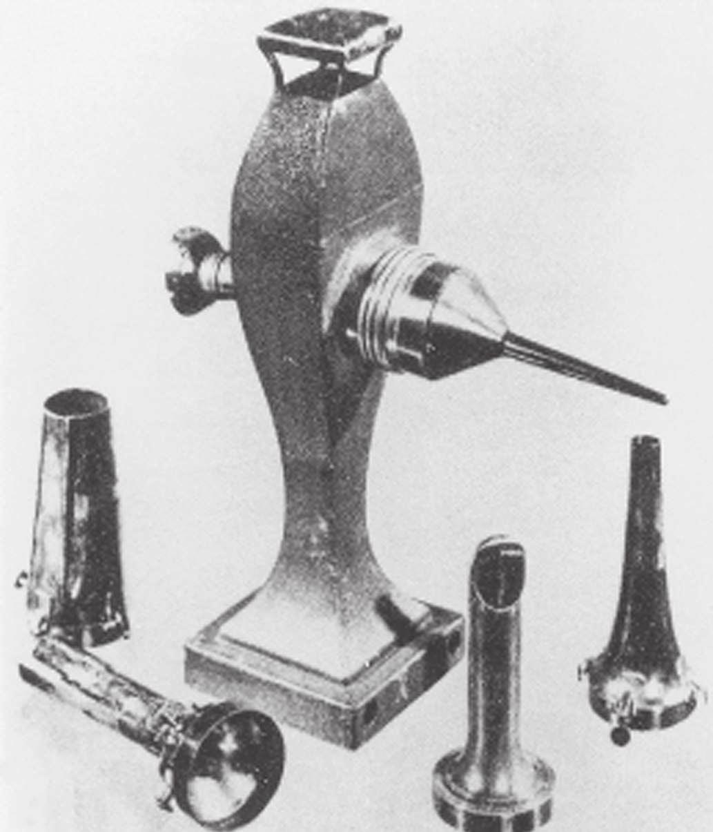
Bozini (1805) is credited with the earliest known attempt to visualize the interior of a body cavity with a primitive endoscope (Fig. 1.1).1–3 Bozini devised a tin tube illuminated by a candle from which light was reflected by a mirror; this was a device he called a lichtleiter (light conductor). He used this device to examine the urethra, urinary bladder, and vagina, but it was an impractical instrument that never gained wide acceptance. Although there were multiple attempts to develop more usable instruments, all directed toward the GU tract, none were widely used. The most notable efforts were by Segalas in France in 1826 and Fisher in Boston in 1827,2 both using straight metal tubes, but the lack of a satisfactory light source remained a major impediment.
The next significant development was the instrument of Desormeaux in France.2 Desormeaux’s contribution in 1855 was a better, although still inadequate, light source using a lamp fueled with alcohol and turpentine (“gazogene”) (Fig. 1.2). His instrument was based on that of Segalas. Others continued with efforts to improve the light source and the means to deliver it, but the devices were unsatisfactory for the more inaccessible areas of the GI tract.
Rigid Gastrointestinal Endoscopes
Kussmaul is credited as being the first to perform a gastroscopy in 1868, using a straight rigid metal tube passed over a flexible obturator and a cooperative sword swallower (Fig. 1.3).1–4
For a light source, he used a mirror reflecting light from the
Abstract
The development of endoscopy is a testimony to human ingenuity. Instruments have evolved from dangerous straight tubes, illuminated by light reflected from candles, to more flexible and safer instruments with an image transmitted through a series of prism lenses and illumination by an electric light bulb, to images transmitted through fiberoptic bundles with illumination transmitted by fiber bundles from an external source, to our present remarkably safe electronic instruments with digital images transmitted to a video screen through wires and processed by computers. Most recently, we can visualize the lumen of the gut without touching the patient. Now we not only can visualize, biopsy tissue, and perform procedures within the hidden cavities of the body, but also directly and indirectly see beneath the mucosa and into immediately adjacent organs. The evolution of gastrointestinal endoscopy is a truly remarkable story, and advances in the diagnostic and therapeutic capabilities of these instruments continue to be made at a rapid pace. To know and understand what has occurred previously lends strength to efforts toward achieving what is to come.
Keywords gastrointestinal endoscopes
fiberoptics
videoendoscopy
capsule endoscopy
gastroscopy
sigmoidoscopy
colonoscopy
endoscopic retrograde cholangiopancreatography (ERCP)
endoscopic ultrasonography (EUS)
enteroscopy
FIG 1.1 Bozzini’s lichtleiter, 1805. (From Edmonson JM: History of the instruments for gastrointestinal endoscopy. Gastrointest Endosc 37[Suppl 2]:S27–S56, 1991.)
FIG 1.2 Desormeaux’s endoscope, 1853. (From Edmonson JM: History of the instruments for gastrointestinal endoscopy. Gastrointest Endosc 37[Suppl 2]:S27–S56, 1991.)
Desormeaux device but found it inadequate. He also quickly discovered that gastric secretions were a problem, despite using a flexible tube he had developed earlier to empty the stomach before the procedure. The value of his efforts was the demonstration that the curves and bends of the esophagus and esophago-
1.3 Kussmaul’s gastroscope, 1868. (From Edmonson JM: History of the instruments for gastrointestinal endoscopy. Gastrointest Endosc 37[Suppl 2]:S27–S56, 1991.)
gastric junction could be traversed with careful manipulation and that the gastric pouch could be visualized. Kussmaul apparently demonstrated his “gastroscope” several times, but the illumination was too poor to allow a clinically useful image,4 and he abandoned his efforts.
Encouraged by the efforts of Kussmaul, others switched their attention to developing esophagoscopes because the esophagus is much easier to visualize, and a less complex design than the gastroscope was required. The problems of perforation, at that time usually fatal, and of illumination, remained major obstacles. Before the late 19th century, illumination of light reflected by a mirror into a straight metal tube continued to be used. As noted earlier, several light sources were developed, but the intensity left much to be desired. Several innovations were developed to solve this problem, including a burning magnesium wire, which produced a brilliant light but unacceptable heat and smoke. The most promising device seemed to be the brilliant light from a loop of platinum wire charged with direct current, introduced simultaneously by Bruck in Breslau and Milliot of Paris in 1882.2 Although the illumination was adequate, major difficulties were encountered with the considerable heat generated, necessitating a water cooling system and the cumbersome batteries used for a power source. Nevertheless, the platinum wire device was an encouraging development and was used in several instruments that saw relatively widespread use.
These instruments were made obsolete just a few years later by Edison’s incandescent electric light bulb, introduced in 1879. In 1886, Leiter, an instrument maker, was the first to use the electric incandescent light bulb in a cystoscope just 7 years after Edison introduced it. With a few short-lived exceptions, all instruments used Edison’s invention after 1886. Working with Leiter, von Mikulicz developed an unsuccessful gastroscope but a practical esophagoscope that he used extensively until distracted by his many other medical interests.
At the turn of the 20th century, Jackson, an otolaryngologist, also examined the esophagus and the stomach using a straight rigid tube and a distal electric light bulb, but few could match
FIG
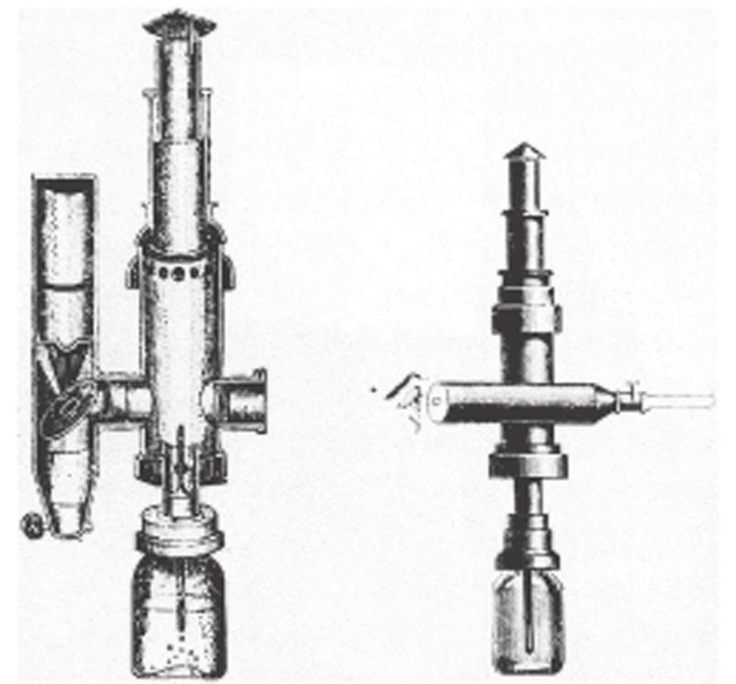
his talents in the GI tract. Under his influence, esophagoscopy was considered the exclusive province of ear, nose, and throat (ENT) departments in many community hospitals in the United States as late as the 1950s. The design of the esophagoscope remained a straight rigid tube, usually with a rubber finger-tipped obturator to make insertion safer. With the later addition of a 4 × power lens on the proximal end and a distal incandescent bulb, various models were popular until the introduction of fiberoptics in 1961. The Eder-Hufford rigid esophagoscope (Fig. 1.4), introduced in 1949, was popular and still in use in the early 1960s.
It was not until after 1900 that persistent efforts to develop a usable gastroscope were successful. All attempts to build a flexible instrument using a multiplicity of lenses were designed to be straightened after introduction and were fragile, easily damaged, and cumbersome. Straight tubes with simpler optics were useful, but perforations were still a problem.1 In 1911, Elsner introduced a rigid gastroscope with an outer tube through which a separate inner optical tube with a flexible rubber tip and sideviewing portal could be passed (Fig. 1.5). The rubber tip, previously used in the esophagoscope obturator, was more crucial than it might appear, for it seemed to be, along with the later
addition of a flexible metal coil proximal to it, the single feature that reduced the rate of perforation. Elsner’s instrument worked as designed and was widely used, especially by Schindler, then in his native Germany, who called it the“mother of all instruments until 1932.”5
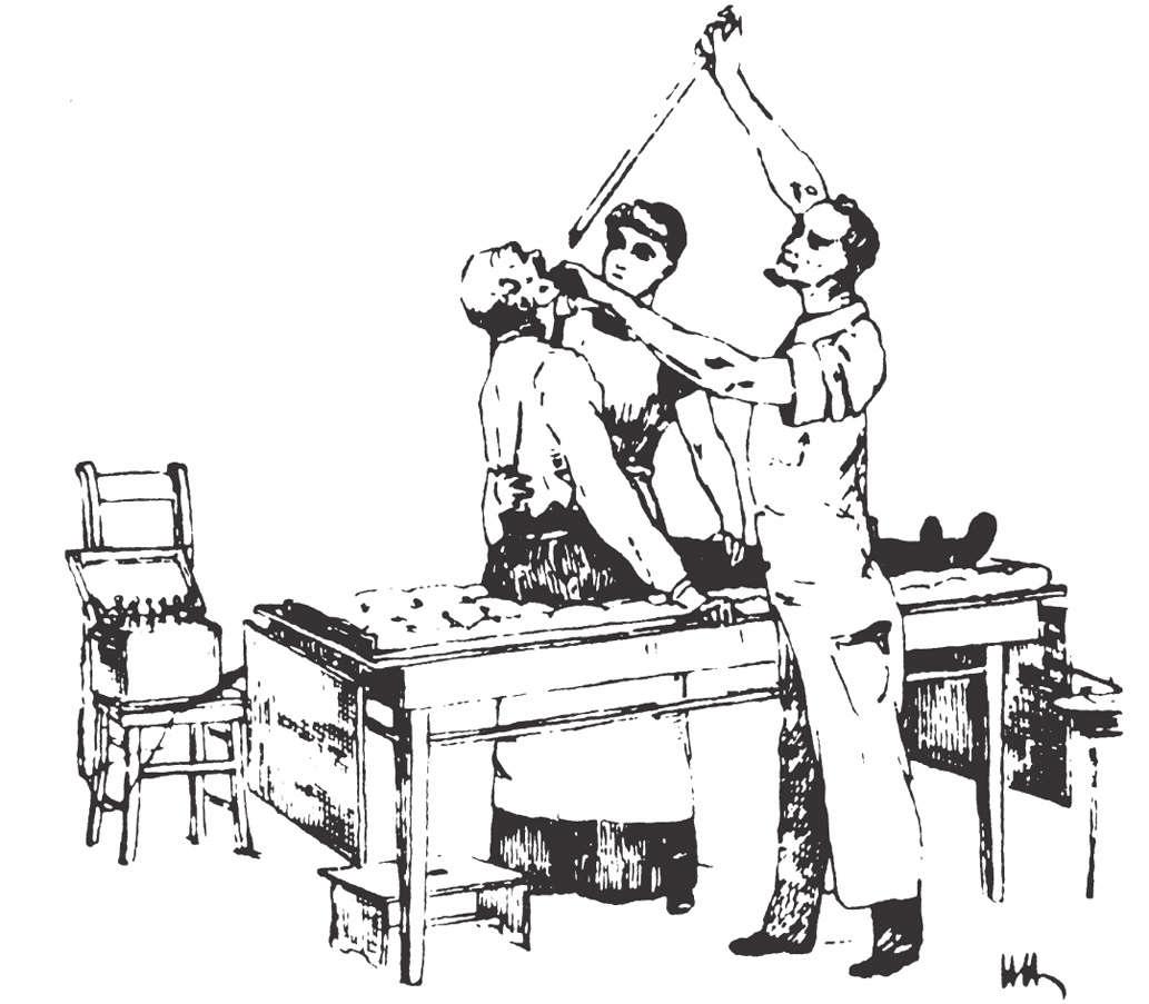
In 1922, Schindler introduced his own version of the Elsner gastroscope, the major innovation of which was the important addition of an air channel to clear the lens of secretions. With the Elsner gastroscope, Schindler examined the stomachs of several hundred patients and meticulously recorded his findings in each procedure. He published Lehrbuch und Atlas der Gastreoskopie in 1923, with descriptions and remarkably accurate drawings. He trained others in the technique and was responsible for wide acceptance of gastroscopy. The procedure began with emptying the stomach using a nasogastric tube, followed by sedation. The patient was placed on the left side, and an assistant held the head rigidly extended to produce a straight path into the esophagus and the stomach (the “sword swallower’s technique”). The role of the assistant was crucial. Schindler’s effort was impressive and convinced many of the value of an expert examination of the stomach.
Semiflexible Gastroscopes
It became apparent that straight, rigid tubes were not ideal for examination of the stomach. Fatal perforations continued to the detriment of acceptance of the procedure. Visualization of the surface of the stomach was incomplete at best, with many consistent blind spots. These problems stimulated investigation of methods to manufacture safer, “flexible” instruments. The use of the term flexible here is problematic in view of what we think of today as flexible instruments. Although these early instruments were not flexible by our standards, they were more flexible than the straight, rigid instruments that came before. Semiflexible, with passive angulation of the distal portion of 34 degrees and sometimes more, was a more appropriate term.
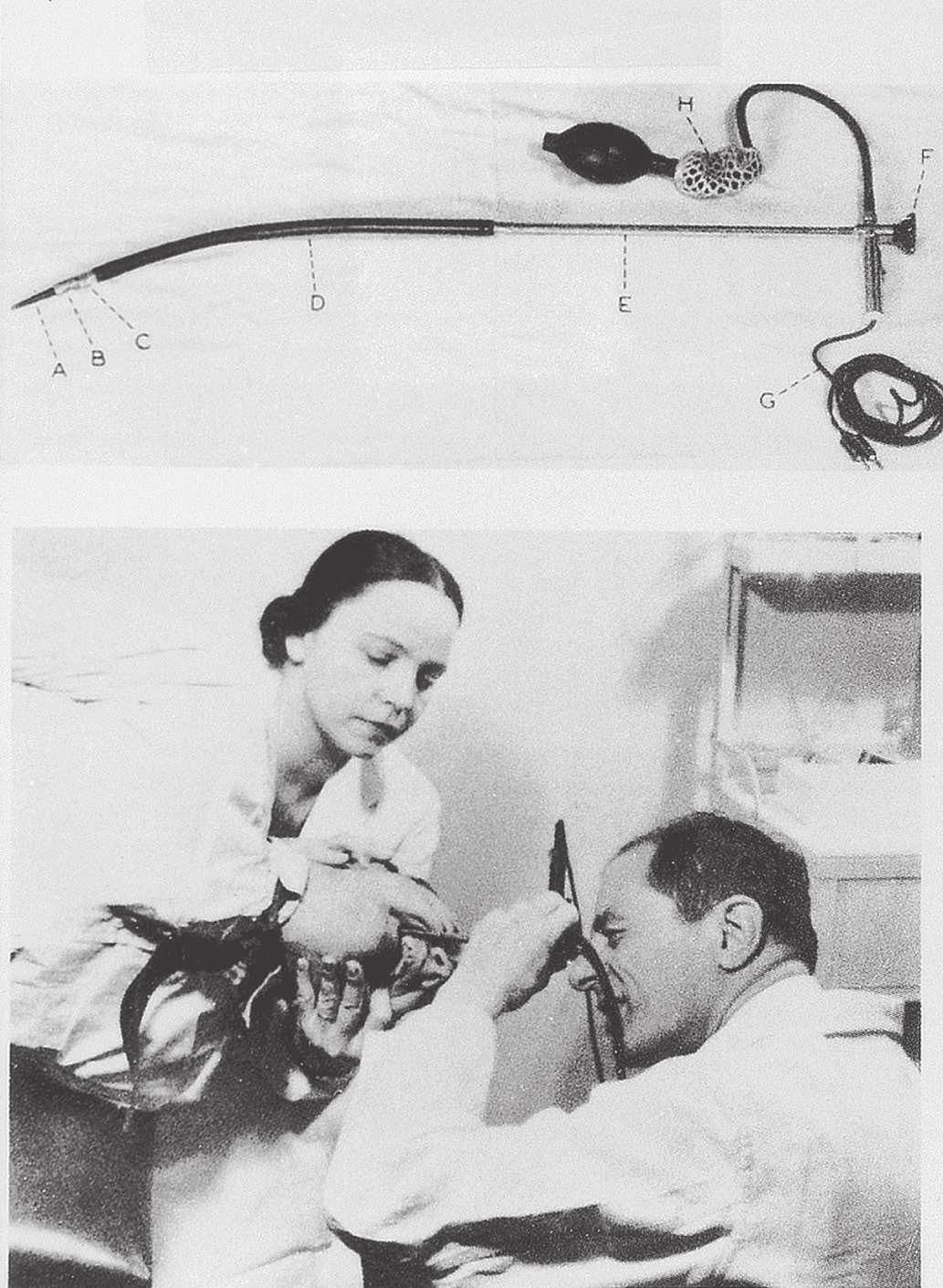
In 1911, Hoffman showed that an image could be transmitted through a curved line by linking several short-focus prisms. Using this principle, several instruments were constructed, but these were unsatisfactory or were not widely accepted. Schindler, working with Wolf, the renowned instrument maker, constructed a semiflexible instrument with a rigid proximal portion and a distal portion made elastic by coiled copper wire and terminating with first a rubber finger and later a small rubber ball. Illumination was with a distal incandescent light bulb. Air insufflation was made possible with a rubber bulb, expanding the stomach wall to beyond the focal length of the prisms, which were manufactured by Zeiss. In 1932, the sixth and final version was patented. This instrument, known as the Wolf-Schindler gastroscope, greatly improved the safety and efficacy of gastroscopy and was used throughout the world (Fig. 1.6).
Thanks to the published meticulous work and enthusiasm of Schindler, whose designation as the “father of gastroscopy” is well deserved, the procedure was finally widely accepted as a valuable extension of the physical examination. The era of the semiflexible gastroscope from 1932 to 1957 has been called the Schindler era. Schindler was chiefly responsible for transforming gastroscopy from a dangerous and seldom used procedure to one that was relatively safe and indispensable for evaluation of known or suspected disease of the stomach. He insisted that all clinicians who planned to use the instrument be properly trained and that “… no manipulation inside of the body is without danger; therefore no endoscopic examination should be done
FIG 1.4 Eder-Hufford esophagoscope, the result of multiple attempts to develop a clinically useful instrument, 1949.
FIG 1.5 Elsner’s gastroscope, 1911. (From Edmonson JM: History of the instruments for gastrointestinal endoscopy. Gastrointest Endosc 37[Suppl 2]:S27–S56, 1991.)
the instruments for gastrointestinal endoscopy. Gastrointest Endosc 37[Suppl 2]:S27–S56, 1991.)
without reasonable indication.”6 In today’s vernacular, the risk approaches infinity if the benefit approaches zero.
Schindler was born in Berlin in 1888. He gained considerable experience as an Army physician in World War I, where he became convinced that gastritis, then an often-disparaged cause of symptoms, was a bona fide disease. His interest in gastritis lasted throughout his career and undoubtedly stimulated his interest in gastroscopy. The Wolf-Schindler endoscope of 1932 and Schindler’s publications with drawings further enhanced what thereafter rapidly became a discipline. His enthusiasm for and talent in using the gastroscope led to what has been called his gospel of gastroscopy, which he and others spread throughout academia and to the community of practicing physicians. Because of his Jewish background, Schindler was put in “protective custody” by the Nazis, but with the help of the physicians Ortmeyer and Palmer and philanthropists in Chicago, he was able to immigrate to the United States in 1934.1–4,7
Chicago became the hub of GI endoscopy, and it was here, in Schindler’s home, that the first discussions were held about forming a new organization for GI endoscopy, now known, after several name changes, as the American Society of Gastrointestinal Endoscopy. In 1943, just 9 years after his arrival in the United States, Schindler left Chicago for Loma Linda University. In 1958, he accepted an appointment as Professor of Medicine at the
University of Minas Gerais in Belo Horizone, Brazil. He came back to the United States in 1960 because of an eventually fatal illness of his wife and returned to his native Berlin in 1964, where he died in 1968 at the age of 80.1 Despite his acclaim in endoscopy, Schindler insisted that one must be a physician first and an endoscopist second. He was very knowledgeable in the field of general gastroenterology and published, without coauthors, a synopsis of the entire field in 1957.6
The Wolf-Schindler endoscope was introduced into the United States by Benedict, Borland, and many others. Schindler’s immigration to Chicago inspired a surge of interest in the United States, but with the outbreak of war in Europe, the German source of instruments disappeared. Several US companies working with Schindler and others produced many popular gastroscopes that were significant variations on the Wolf-Schindler model, including Cameron Co., which produced its first instrument in 1940.8 The Eder-Hufford semiflexible gastroscope followed in 1946,9 and American Cystoscope Makers, Inc. (ACMI) produced a gastroscope in 1950. A combination of the Eder-Hufford esophagoscope with a semiflexible gastroscope to be passed through it was the Eder-Palmer transesophagoscopic flexible gastroscope produced by the Eder Company in 1953. Each gastroscope had its proponents.
Biopsy
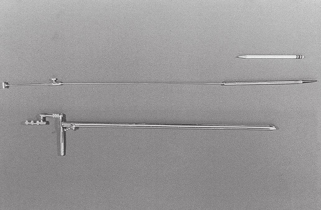
With the availability of instruments for visualization, it became apparent that tissue must be obtained to identify the nature of the observed abnormalities. Instruments for blind biopsies were used early on, but a device was needed that would allow the operator to obtain a biopsy specimen of abnormal tissue directly when seen at endoscopy. The Benedict Operating Gastroscope was produced in 1948 based on a 1940 model by Kenamore (Fig. 1.7).10 The Benedict instrument was a popular instrument that was widely used. In the debates about the necessity for biopsy, Benedict, a surgeon who switched entirely to endoscopy, stated that gastroscopy was not a routine procedure and should be reserved for those with a complex differential diagnosis, but “gastroscopic examination is not complete unless the gastroscopist has some means of biopsy readily available.”11 It soon became clear that the correlation between histology and a diagnosis based on visualization alone was often widely discrepant, and certain diagnoses could not be reliably made without tissue examination.
FIG 1.6 Wolf-Schindler “flexible” gastroscope (top) being used by Schindler (bottom) with his wife as the head holder. (From Edmonson JM: History of
FIG 1.7 Benedict operating gastroscope.
Efforts such as wash and brush cytology continued and have persisted in various forms to the present time.
Fiberoptics
By the 1950s, the ideal of a totally flexible GI endoscope with good visualization that could withstand the rigors of clinical use had not been realized, although the semi-flexible instruments with their biopsy capabilities were satisfactory for most clinical purposes. In fact, these instruments were not rapidly abandoned by all with the introduction of the remarkably flexible fiberscope. The development of the science of fiberoptics and its application to endoscopes truly revolutionized the diagnostic and, later, the therapeutic abilities of endoscopy. Its importance in the development of this field cannot be overstated.
The principle of internal reflection of light along a conduction pathway was used by Lamm in October 1930.1 The image was severely degraded by light escaping from the thin fibers of quartz he used, although the potential for total flexibility was present. Lamm could not interest Schindler or others in his efforts, and the experiment was discontinued. Almost 25 years later, in 1954, Hirschowitz, in fellowship training at the University of Michigan, visited Hopkins and Kapany in London to review their work12 with glass fibers, which totally confirmed the work of Lamm and his predecessors. Hirschowitz became convinced that application of this principle could be used to develop a totally new and superior endoscope. He began work with a graduate student, Curtiss, who developed a technique of coating glass fibers with glass of a different optical density, preventing the escape of light and degradation of the image. This was the critical discovery that made the principle of internal reflection through glass fibers workable.
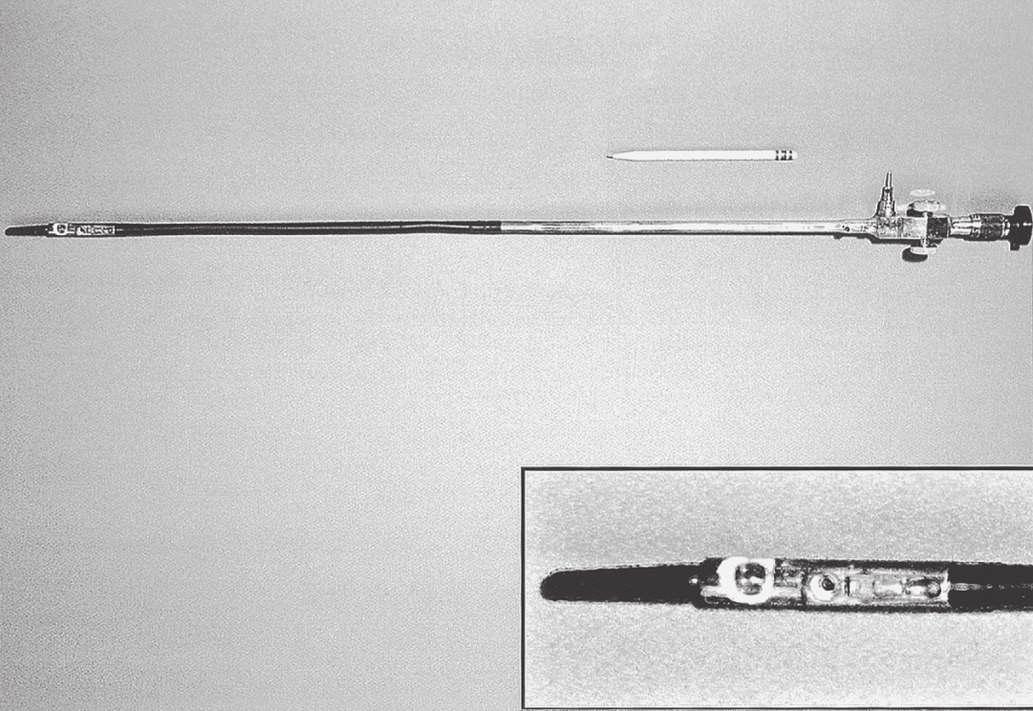
In 1957, Hirschowitz demonstrated his fiberscope, and he published his work in 1958 (Fig. 1.8).13 His audience was not impressed, and it took another 3 years, working with ACMI, to produce a marketable scope, which he called the Hirschowitz

Gastroduodenal Fiberscope. This was a very flexible side-viewing instrument with an electric light on its distal end, an air channel, and an adjustable focusing lens proximally. The tip lacked what was by then the “obligatory” rubber finger, and this omission was a source of criticism; one was added on a later model. Although some individuals criticized the quality of the image, most believed the size and brightness were superior to the semiflexible scopes. This model, the ACMI 4990, was introduced to the market late in 1960 after being tested by Hirschowitz on himself and numerous patients. In 1961, the senior author of this chapter was in a gastroenterology fellowship at the Emory University Clinic with Schroder. He vividly recalls Schroder’s reaction after the first use of the new fiberscope around March 1962 (Fig. 1.9). Upon finishing the initial examination using the new device, he turned to him and said, “Anybody want to buy a used Benedict operating scope?” The senior author does not recall it ever being used again, as the Hirschowitz Gastroduodenal Fiberscope was clearly superior in his view, and he finished his training with that instrument.
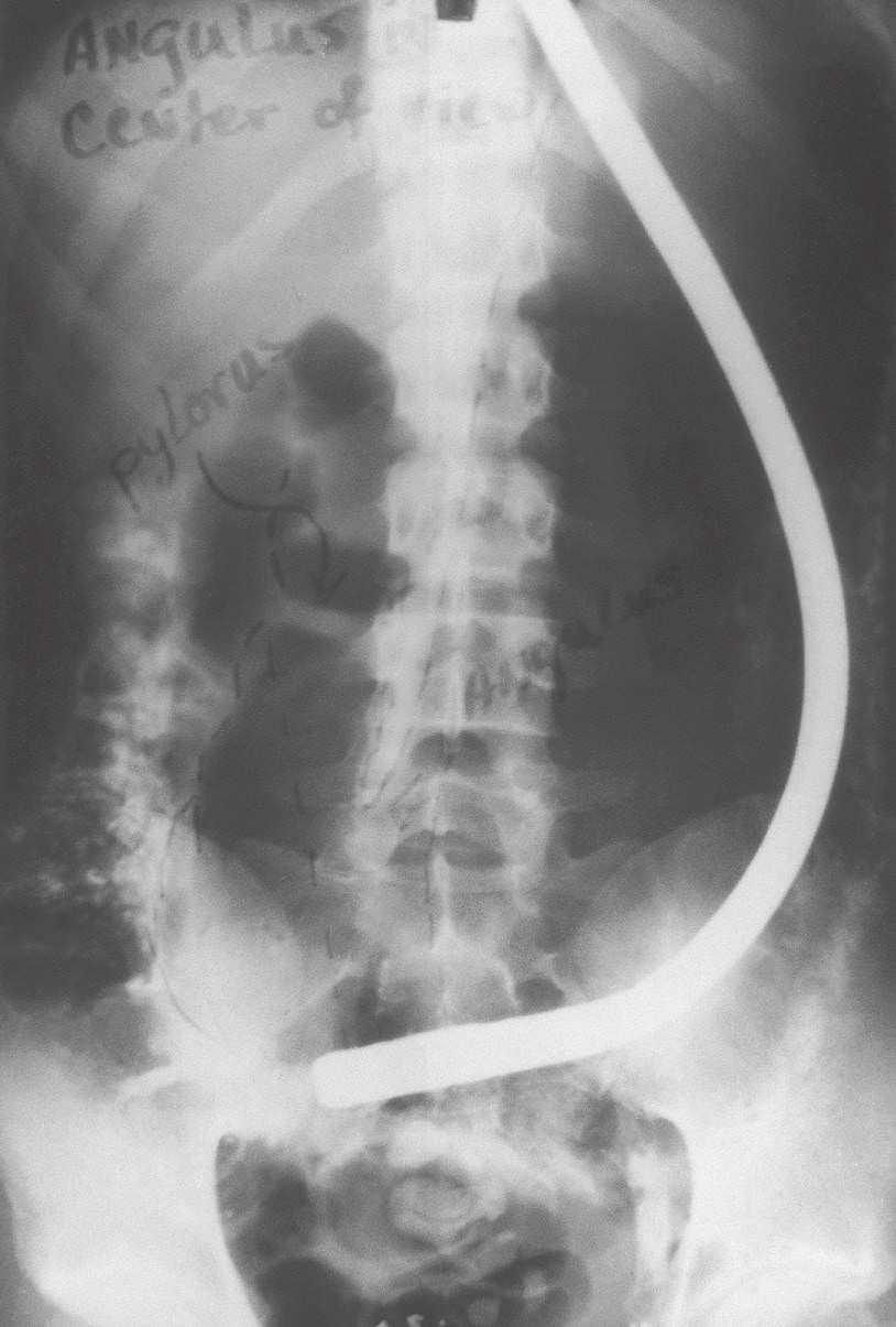
There were problems with the fiberscope noted by users. The distal light source would become so heated that thermal injury to the gastric mucosa was possible unless the tip was continuously moved. In prolonged procedures, protein in gastric secretions would coagulate on the bulb and the adjacent visualizing port, totally obscuring the lens. As the number of procedures with a single instrument increased, some glass fibers would break, producing small black dots in the visual field. This was a persistent problem with fiberscopes during their entire history and especially apparent in training programs where a single scope was used by several trainees on many patients. The side-viewing lens prevented visualization of the esophagus, and the scope had to be passed blindly through the pharyngeal orifice. The previous semiflexible scopes in use shared this problem, and it was not considered a defect at the time. The flexibility itself resulted in some difficulty in advancing because attempts to push the instrument through the pylorus and into the gut resulted in more bowing in the gastric pouch (Fig. 1.10). Although one could sometimes visualize the duodenum, this was done by overinflating the stomach and looking through the pylorus without actually entering it. If one managed to introduce the tip into the duodenum, as occasionally happened, the visual field was inside the focal length of the instrument, and only a “red-out” was observed.4

FIG 1.8 Hirschowitz examining the stomach of an outpatient. (From Hirschowitz BI: Endoscopic examination of the stomach and duodenal cap with the fiberscope. Lancet 277[7186]:1074–1078, 1961.)
FIG 1.9 ACMI fiberscope, 1962.
Many clinicians did not believe the additional expense of replacing the older, beloved instruments with which they had been successful for many years was warranted. Even ACMI officials did not see the fiberscope as totally replacing the instruments with a lens system.2 Despite reservations, comparison and experiential studies showed the advantages of the new fiberscopes.14–17 Following the flagship ACMI model 4990, several models of the fiberscope were introduced by ACMI and other companies, each with significant improvements, including the controllable tip in the side-viewing ACMI model 5004. Visualization of the gastric pouch, including retroflexed views of the cardia, was now complete. The major objection to these instruments was the inability to pass the instrument under direct vision and examine the esophagus; in addition, the area beyond the pylorus could not be consistently examined.
Most clinicians were already fully trained in use of the EderHufford esophagoscope, and in the absence of a forward-viewing fiberscope, use of the Eder-Hufford esophagoscope continued. A forward-viewing scope was mandatory. LoPresti modified the tip of the fiberscope to create the foroblique fiberoptic esophagoscope in 1964.18 Passing the instrument under direct vision was possible, and clinicians immediately discovered that they could examine not only the esophagus, but also a large portion of the proximal stomach. At a length of 90 cm, however, one could not reach the duodenum. Working with ACMI, LoPresti produced the longer Panview Mark “87” gastroesophageal endoscope in 1970. By about 1971, the instrument had been lengthened to 105 cm with a four-way controllable tip capable of 180 degrees of deflection (Fig. 1.11).
The aptly named panendoscope was now a reality. Japanese and American manufacturers began to produce new models with such rapidity that endoscopists hardly had time to become thoroughly familiar with one before another, significantly improved (and more expensive) model was on the market. Patient comfort was greatly improved, and the relative safety of the fiberoptic endoscopes rapidly became apparent. By 1970, most gastroscopic examinations were done with fiberscopes. The development of a “teaching head” fiberoptic bundle with a light splitter and attached eyepiece and attachment to the eyepiece of the scope allowed two people to visualize the image. Dividing the light from the endoscope considerably diminished the brightness of the image, however, to both the operator and the observer. This device saw limited use and was utilized primarily in teaching institutions.
Endoscopic Retrograde Cholangiopancreatography (ERCP)
With access to the duodenum, the ampulla of Vater became visible. It followed that one should be able to inject contrast material into the bile and pancreatic ducts and increase diagnostic capabilities. Initial attempts in 1968 by McCune et al19 to modify an existing scope were only partially successful, but did show that endoscopic visualization by injection of radiologic contrast agents into ducts was possible. In 1970, Machida and Olympus in Japan produced usable, side-viewing scopes with controllable tips and elevators to move the injection tube to the ampulla. Japanese endoscopists20 developed the technique of endoscopic retrograde cholangiopancreatography (ERCP) with an 80% success rate.Vennes and Silvis21 showed the utility of ERCP in the United States and taught many physicians to use it.4 It was immediately apparent that if clinicians could visualize the biliary and pancreatic ducts endoscopically (i.e., nonsurgically), they should be able to apply by some means long-established surgical techniques for treatment of choledocholithiasis and pancreatitis, such as sphincterotomy and stone removal. In 1974, just 4 years after the demonstration of the diagnostic utility of the new ERCP
FIG 1.10 Visualization of duodenum was sometimes obtained by overinflating the stomach.
FIG 1.11 LoPresti forward-viewing esophagogastroscope. (From advertisement in Gastrointest Endosc 16:79, 1970.)
scopes, Kawai et al in Japan22 and Classen and Demling in Germany23 independently developed methods of endoscopic electrosurgical sphincterotomy for extraction of biliary calculi in the common duct. This procedure requires great skill; in 1976, Geenen24 reported that only 62 operative procedures had been done by four endoscopists, and seven of the procedures were failures. In 1983, Schuman4 reported that several thousands of patients had undergone ERCP, and by now, hundreds of thousands of ERCP procedures have been done. Because of advances in radiologic techniques, ERCP is now seldom used for purely diagnostic purposes.
Photography
It is one thing to describe to others what one may see through any device and another to be able to show them. The large impact of Schindler’s early publications was related, in part, to the excellent color drawings he presented. Early on, neither cameras nor photographic films were advanced enough to allow good color reproduction or sharp, accurate images in relatively poor lighting. Such documentation is essential for widespread appreciation of endoscopy by individuals who do not perform the procedure. The first clinically useful photography came with improvements in film by Kodak and the construction of an external integrated camera by Segal and Watson in 1948.25,26 Although these authors reported that approximately 61% of the images were of good quality, this was not the experience of all clinicians.4
Although an intragastric camera was developed as early as 1848 by Lange and Meltzung, a clinically useful device was not available until 1950, when Uji, Sugiura, and Fukami, working with Olympus Corp. (Center Valley, PA),27 developed the Gastrocamera with synchronized flash, which took good intragastric pictures and had a controllable distal portion. By following a prescribed pattern of rotation and flexion, a series of pictures was obtained that included the entire surface of the stomach. The big disadvantage was that the operator could not see through the instrument and had to await development of the very narrow (5-mm) film before the results could be seen. Photographs for demonstration required additional time in the photo laboratory while enlargements were made.
After the introduction of fiberoptic scopes in 1961, Olympus introduced a combination Gastrocamera fiberscope (GTF-A) in 1964, but, as Schuman4 commented, “it was just a gastroscope” and never attained popularity. Simultaneously, rapid development and physician acceptance of fiberscopes with the ability to use technically advanced 35-mm cameras with an external adapter made the Gastrocamera obsolete, and it was abandoned.
Sigmoidoscopy and Colonoscopy
The problems presented by examination of the anus and rectum were relatively easy. Straight metal tubes were used and found in the ruins of Pompeii.2 The basic design of the anoscope has not changed in the past century or more except that it is now made of disposable plastic. It remains a tapering short tube with an obturator that is removed after introduction through the anal sphincter. Examination of the rectum and sigmoid required a longer tube, but no truly satisfactory device was available until 1894, when Kelly28 at Johns Hopkins developed a 30-cm rigid tube with light reflected down the tube from a head lamp. Tuttle29 incorporated a distal light source in his proctosigmoidoscope of 25 cm in 1903. These instruments have remained the basic design for the past 100 years. For the past 25 years or so, disposable
clear plastic tubes have been widely used. These are essentially a plastic version of the Kelly and Tuttle tubes with a distal electric light source, but visualization is possible through the clear plastic. With the application of fiberoptics to sigmoidoscopy in the late 1960s, examination of the sigmoid colon became not only satisfactory, but also much more comfortable for the patient.
Overholt,30 who later went on to be the principal developer of colonoscopy using similar technology, presented his results of flexible sigmoidoscopy in 250 patients in 1968. Although early flexible sigmoidoscopes were made in variable lengths, the current length of 60 cm came to be the preferred one. Examination of the colon above the sigmoid presents obvious additional problems of multiple curves and angulations amenable only to highly flexible instruments and trained operators. Attempts, all unsuccessful, were made using semiflexible instruments, and these are reviewed by Edmonson.2 Satisfactory examination of the length of the colon was impossible until the introduction of the flexible fiberscope. Attempts to use forward-viewing gastroscopes were not technically satisfactory, although several clinicians tried. Turell31 presented his attempts in 1967 using a modified gastroscope, but he concluded that the instrument was not ready for routine clinical use. By 1970, several manufacturers produced instruments specifically designed for colonoscopy, including ACMI working with Overholt in the United States and Olympus Corporation in Japan.
The primary problem with regularly completing examinations to the cecum was not the instruments so much as it was the techniques necessary for passage of the scopes into the more proximal portions of the colon. Earlier pioneers in developing successful techniques still in use include, among others, Overholt, Wolf, Shinya, and Waye in the United States; Niwa and colleagues in Japan; Salmon and Williams in England; and Dehyle in Germany.4 Many of these early efforts were accomplished with the guidance of fluoroscopy to negotiate the more difficult turns and to identify the actual area being observed, but, as experience was gained, fluoroscopy was no longer required. Learning under expert guidance and experience continues to be more necessary in colonoscopy (and ERCP) than in upper endoscopy. By 1971, the diagnostic advantage of fiberoptic colonoscopy over singlecontrast barium enema was firmly established,32 and the efficacy and safety of polypectomy were established by 1973.33
Digital Endoscopy (Videoendoscopy)
In 1984, barely 20 years after introduction of the endoscopic fiberscope, Welch Allyn, Inc. (Skaneateles Falls, NY), replaced the coherent fiberoptic image bundle in a colonoscope with a light-sensitive computer chip or charge-coupled device on which the image was focused by a small lens (see Chapter 3).34 The digital signal was fed to a video processor, which generated an image to a television monitor. The image did not occupy the entire screen, leaving space for information to be typed in by a keyboard. The resolution of the image was at least equal to that of the fiberscope.
It was unnecessary to change the basic mechanics of the fiberscope. The fiberoptic light bundle remained unchanged, as did water, suction, and biopsy channels; in addition, the deflection and locking mechanisms were the same. The basic elements of the videoendoscope have not changed, although a magnified image is now available. Since the original introduction of the videoendoscope by Welch Allyn, which no longer produces the Video Endoscope, the market has been supplied by Olympus, Pentax, and Fujinon. The technology was rapidly adapted to
all endoscopes, used not only in gastroenterology but also in other fields.
Advantages of the electronic instruments include an image that can be seen not only by the operator, but also by anyone with access to a connected monitor in the same or another room. This feature greatly enhanced the ability to teach others about the procedure and to inform other interested physicians about the findings in the individual patient. If desired, recording of procedures could be accomplished with videotape machines, and good-quality pictures of individual frames could be made immediately with externally integrated digital equipment. Individual endoscopists found that no adjustment of techniques was necessary when videoendoscopes were used, although they had to become accustomed to looking at the monitor screen rather than through an optical system with one eye (Fig. 1.12). This feature added to the useful length of the instrument because the whole scope could be held at the waist rather than being brought to eye level.
More recent innovations in colonoscopy instruments by Olympus include the ability to make a portion less flexible to facilitate navigation of difficult bends and turns. In addition, an enlarged image is now available that is an improvement in vision and ease of manipulation. A major disadvantage of videoendoscopes is cost. Fiberoptic endoscopes, when they were still in use, could be purchased for less than $6000 and did not require processors or monitors, whereas the latest videoendoscopes are priced at more than $20,000, and initial purchase of the entire package of endoscope, processing computer, monitors, and attachments may exceed $30,000. Initially, many questioned the wisdom of this added cost, which is passed on to the patient and their insurance companies.
Endoscopic Ultrasonography (EUS)
Although the improvements in GI endoscopy are remarkable in the synthesis of diverse but complementary technologies, the information gained remains confined to what one can see from
within the lumen of the gut. Simultaneous with these developments were those of computed tomography and external ultrasonographic tomograms. Conceptually, it was not only logical but also compelling to look beneath the mucosa of the gut by incorporating miniaturized models of ultrasonographic transducers already in use into GI endoscopes. The ability to noninvasively explore tissue and organs in proximity to the gut had exciting implications for diagnosis and therapy.
In Germany in 1976, working with Siemens Co., (Berlin, Germany) Lutz and Rosch35 reported the use of a 1-cm ultrasonographic 4-MHz probe that could be passed through the biopsy channel of an Olympus TGF. They used it in two patients to successfully differentiate between pancreatic pseudocysts and tumors.7 In 1980, Classen’s group in Germany36 and DiMagno et al37 at the Mayo Clinic reported EUS devices that were incorporated onto the tip of conventional fiberscopes, one using a 5-MHz transducer and the other using a 10-MHz transducer. These probes had good resolution at an acoustic focus depth of 3 cm. Others incorporated the transducer in the distal shaft of fiberoptic scopes and primarily explored the gut wall.33,38 By 1985, ultrasonic transducers with variable frequencies incorporated into videoendoscopes were readily available, although expensive (> $100,000 for initial setup) (Fig. 1.13). It was immediately apparent that this procedure could accurately evaluate known or suspected intramural lesions of the gut,39,40 and it was rapidly expanded to include the esophagus; problems
FIG 1.12 Fujinon fiberoptic panendoscope (top) and its successor, the Videopanendoscope (bottom), 1990, showing the two kinds of operating heads. (From advertisement in Gastrointest Endosc 36:240–241, 1990.) A
FIG 1.13 A to D, Ultrasonic endoscope system, model IV, made by Olympus Corp., 1986. (From Yasuda K, Mukai H, Fujimoto S, et al: The diagnosis of pancreatic cancer by endoscopic ultrasonography. Gastrointest Endosc 34:1–8, 1988.)
of diagnosis and recurrence of neoplasia, especially in the pancreas; portal hypertension; the colon and rectum; and bile ducts.41 In 1991, Wiersema et al42,43 showed that EUS could be used to obtain fine-needle aspiration cytology of mediastinal nodes and of nodes and lesions of the upper and lower GI tract. The addition of Doppler technology has now made possible the study of the flow through various structures, including the thoracic duct and blood vessels. EUS is increasingly being used to provide therapy, leading to the development of “interventional EUS.” EUS-guided interventions include celiac plexus block/ neurolysis, placement of fiducial markers to facilitate radiotherapy, direct injection of alcohol or chemotherapeutic agents for the treatment of tumors or cystic lesions, drainage of the pancreatic or biliary ductal systems, and the creation of gastrojejunal anastomoses using lumen-apposing metal stents. The techniques of using EUS instruments differ only slightly from using videoendoscopes, but dedicated training is necessary to interpret the sonographic images obtained accurately. EUS is not amenable to self-instruction. EUS training centers have been established in academic centers, but retraining of practicing physicians is challenging due to the duration of training necessary to achieve competence.44
Capsule Endoscopy (Wireless Endoscopy)
In 2000, Iddan et al45 reported the development of a capsule containing a tiny CMOS camera that could be swallowed, obtain images (at 2 frames per second), and transmit the images over 7 hours to a receiving digital storage unit worn by the patient as he or she goes about his or her normal activities. These frames are downloaded to a computer from which they are projected onto a monitor at a rate that can be controlled by the observer. Pictures can be printed of areas of interest. Gastroenterologists in Israel conducted randomized trials comparing the efficacy of the wireless capsule with push enteroscopy and obtained superior results with the capsule.46–48
Wireless capsule endoscopy caught the imagination of gastroenterologists over the world, and capsule endoscopy has been adopted as a part of standard practice for small bowel imaging. The findings are virtually unanimous in demonstrating better results in identifying lesions in the small bowel with capsule endoscopy when compared to push enteroscopy.49 The capsule avoids the discomfort and need for sedation inherent with push enteroscopy. In addition to lack of biopsy capability, an additional disadvantage is the time needed to review the study, but this has been overcome by a variety of methods including software advancements, improved training techniques, and utilizing non-physician personnel to initially review the obtained images. The major use of the capsule to date has been in elucidating the cause of occult bleeding from small bowel sources, where it seems to be superior to other methods. Future applications, such as in the colon, are continuing to be investigated in large, multicenter comparative studies. The future of wireless capsule endoscopy is bright. It will be interesting to see how the principle of wireless endoscopy is incorporated into videoendoscopes, such as the potential for a wireless connection between the endoscope and the image processor.
Enteroscopy
The small intestine has traditionally been regarded as the final frontier of GI endoscopy. Although capsule endoscopy provides remarkable images of the small bowel mucosa, tissue acquisition
and therapy with a capsule-based instrument is many years away. Surgically assisted small bowel enteroscopy may be performed via either the transoral or anal route or via a mid–small bowel enterotomy incision. The disadvantage of this technique is its invasive nature.50 Endoscopic examination of the small intestine has remained technically difficult. The many loops of the small intestine prevent progression of the instrument tip by simple pushing. This problem was overcome initially with the use of the Sonde enteroscope,51 which is a very fine, floppy instrument with a balloon at the tip. The Sonde enteroscope progressed through much of the small bowel under peristalsis, and then the proceduralist would slowly withdraw the instrument, assessing the mucosa while pulling back. This technique was thought to visualize 50% to 70% of the mucosal surface.52 However, the procedure was uncomfortable, time-consuming, and did not permit therapeutics, all of which limited its use.
The concept of small bowel enteroscopy was revolutionized by Yamamoto with the introduction of the double-balloon enteroscope in 2001.53 This technique uses traction between a balloon at the tip of the enteroscope and another balloon on a flexible overtube to fix the loops of small bowel and provide traction for forward movement. The procedure requires peroral and anal procedures to examine the entire small intestine, and even then only in a minority of Western patients is the whole small bowel visualized. Nonetheless, double-balloon–assisted enteroscopy permits endoscopic therapeutics to most of the small bowel without the need for surgical assistance. A single balloon version is also available.
Natural Orifice Transluminal Endoscopic Surgery (NOTES) and Peroral Endoscopy Myotomy (POEM)
A new development in endoscopy is natural orifice transluminal endoscopic surgery (NOTES), in which the endoscope is inserted into the abdominal cavity via an incision in an accessible organ. The first report appeared in 2002. Incisions have been made in the stomach, vagina, and colon with successful tubal ligation, liver biopsies, biopsy of peritoneal metastases, oophorectomy, cholecystectomy, and nephrectomy procedures having been performed. Most published articles report experimental use in animals, but more recent reports have described the simultaneous use of NOTES with laparoscopic techniques. Comparative studies are ongoing. A difficulty with the technique has been overcoming the lack of instrument “triangulation”; that is, approaching a surgical site from two or more directions to create countertraction, tie sutures, and so forth. Although NOTES is an exciting development, its remarkable potential will have to await the development of new instruments and the acquisition of additional expertise. At a minimum, it appears the development of NOTES will result in marked improvements in mucosal and transmural closure devices. Recently, flexible endoscopes have also been used to tunnel into the submucosal space of the esophagus and perform a myotomy, resulting in a treatment for achalasia termed peroral endoscopy myotomy, or POEM. First performed by Inoue in 2008 and reported by Inoue in 2010, this procedure has gained widespread popularity worldwide and has been performed thousands of times to date with impressive short- and long-term results and an excellent safety profile.54,55 Additional applications of “submucosal” endoscopy include performing a similar procedure in the antrum to treat gastroparesis (G-POEM) and to perform resection of intramural lesions of the GI tract.56,57
SUMMARY
The development of endoscopy is a testimony to human ingenuity. Instruments have evolved from dangerous straight tubes illuminated by light reflected from candles, to more flexible and safer instruments with an image transmitted through a series of prism lenses and illumination by an electric light bulb, to images transmitted through fiberoptic bundles with illumination transmitted by fiber bundles from an external source, to our present remarkably safe electronic instruments with digital images transmitted to a video screen through wires and processed by computers. Most recently, we can visualize the lumen of the gut without touching the patient. Now we can not only visualize, biopsy tissue, and perform surgical procedures within the hidden cavities of the body, but also directly and indirectly see beneath the mucosa and into immediately adjacent organs. The evolution of gastrointestinal endoscopy is a truly remarkable story, and advances in the diagnostic and therapeutic capabilities of these instruments continue to be made at a rapid pace. To know and understand what has occurred previously lends strength to efforts toward achieving what is to come.
KEY REFERENCES
1. Modlin IM: A brief history of endoscopy, Milano, 2000, MultiMed.
2. Edmonson JM: History of the instruments for gastrointestinal endoscopy. Gastrointest Endosc 37:S27–S56, 1991.
6. Schindler R: Synopsis of gastroenterology, Philadelphia, 1957, Grune & Stratton.
7. Kirsner JB: American gastroscopy—yesterday and today. Gastrointest Endosc 37:643–648, 1991.
11. Benedict EB: Gastroscopic biopsy. Gastroenterology 37:447–448, 1959.
12. Hopkins HH, Kapany NS: A flexible fiberscope using static scanning. Nature 173:39–41, 1954.
13. Hirschowitz BI, Curtiss LE, Pollard HM: Demonstration of the new gastroscope, the “fiberscope.” Gastroenterology 35:50–53, 1958.
15. Burnett W: An evaluation of the gastroduodenal fibrescope. Gut 3:361–365, 1962.
20. Takagi K, Ikeda S, Nakagawa Y, et al: Retrograde pancreatography and cholangiography by fiber duodenoscope. Gastroenterology 59:445–452, 1970.
21. Vennes JA, Silvis SE: Endoscopic visualization of bile and pancreatic ducts. Gastrointest Endosc 18:149–152, 1972.
22. Kawai K, Akasaka Y, Murakami K, et al: Endoscopic sphincterotomy of the ampulla of Vater. Gastrointest Endosc 20:148–151, 1974.
25. Segal HL, Watson JS: Color photography through the flexible gastroscope. Gastroenterology 10:575–585, 1948.
32. Wolff WI, Shinya H: Colonofiberoscopy. JAMA 217:1509–1512, 1971.
33. Wolff WI, Shinya H: Polypectomy via the fiberoptic colonoscope: Removal of neoplasms beyond the reach of the sigmoidoscope. N Engl J Med 288:329–332, 1973.
35. Lutz H, Rosch W: Transgastroscopic ultrasonography. Endoscopy 8:203–205, 1976.
37. DiMagno EP, Buxton JL, Regan PT, et al: Ultrasonic endoscope. Lancet 1:629–631, 1980.
42. Wiersema MJ, Hawes RH, Wiersema LM, et al: Endoscopic ultrasonography as an adjunct to fine needle aspiration cytology of the upper and lower gastrointestinal tract. Gastrointest Endosc 38:35–39, 1992.
43. Rex RK, Tarver RD, Wiersema M, et al: Endoscopic transesophageal fine needle aspiration of mediastinal masses. Gastrointest Endosc 37:465–468, 1991.
45. Iddan G, Meron G, Glukhovsky A, et al: Wireless capsule endoscopy. Nature 405:417, 2000.
47. Appleyard M, Glukhovsky A, Swain P, et al: Wireless-capsule diagnostic endoscopy for recurrent small-bowel bleeding. N Engl J Med 344: 232–233, 2001.
53. Yamamoto H, Sekine Y, Saito Y: Total enteroscopy with a non-surgical, steerable double-balloon method. Gastrointest Endosc 53:216–220, 2001.
54. Inoue H, Minami H, Kobayashi Y, et al: Peroral endoscopic myotomy (POEM) for esophageal achalasia. Endoscopy 42:265–271, 2010.
55. ASGE Technology Committee, Pannala R, Abu Dayyeh BK, et al: Per-oral endoscopic myotomy (with video). Gastrointest Endosc 83(6):1051–1060, 2016.
56. Khashab MA, Stein E, Clarke JO, et al: Gastric peroral endoscopic myotomy for refractory gastroparesis: first human endoscopic pyloromyotomy (with video). Gastrointest Endosc 78(5):764–768, 2013.
57. Xu MD, Cai MY, Zhou PH, et al: Submucosal tunneling endoscopic resection: a new technique for treating upper GI submucosal tumors originating from the muscularis propria layer (with videos). Gastrointest Endosc 75(1):195–199, 2012.
A complete reference list can be found online at ExpertConsult .com