adult. In the Lobata and Cestoidea there is, however, a definite larval stage, of the general appearance of a Mertensia, and during this stage fertile eggs and spermatozoa are formed and set free.
Distribution.—Ctenophora are found at the surface of nearly all seas, and many of the genera have a cosmopolitan distribution. Some of the Lobata, the Cestoidea, and the Platyctenea are more commonly found in the warmer regions of the world. Pleurobrachia pileus, Bolina infundibulum, Beroe ovata, and B. cucumis occur off the British coast.
Most of the Ctenophora are from 5 to 20 mm. in diameter, but Beroe reaches the length of 90 mm., Eucharis multicornis a height of 250 mm., and Cestus veneris has been found no less than 1½ metres from one extremity to the other.
Ctenophores usually go about in shoals, and in the case of Beroe cucumis and Eucharis multicornis the shoals may be of very great extent. Pleurobrachia pileus of the British coasts is often found at the end of the season (July) as a series of isolated individuals; but in June they occur in small shoals, swimming so close together that they will choke a tow-net in a very short space of time.
CLASS I. TENTACULATA
Ctenophora provided with a pair of tentacles in the larval stages only or in both larval and adult stages.
Order I. Cydippidea.
This order includes a number of spherical or oval Ctenophores, with a pair of tentacles retractile into deep tentacular pits in the adult stage.
Fam. 1. Mertensiidae.—The body is compressed in the transverse plane, and the ribs on the transverse areas are longer than those on the sagittal areas. The family includes the genus Euchlora, which occurs in the Mediterranean and in the northern part of the Atlantic Ocean. In Charistephane there are only two enormous ctenophoral plates in each of the longitudinal tracts. These plates are so broad that they almost meet laterally to form two continuous circlets round the body of the animal. This genus is found in the Mediterranean, but a few specimens have also been obtained in the Atlantic.
In Tinerfe the body is almost cylindrical, and there is a pair of kidneyshaped swellings at the sides of the aboral pole. It has a pale blue colour, and is found in the Guinea and south equatorial currents of the Atlantic Ocean.
The name Mertensia has been given to several forms that are undoubtedly the young stages of genera belonging to the Lobata, but Chun retains the name M. ovum for a species which is very abundant in the Arctic currents of the North Atlantic.
Fam. 2. Callianiridae.—Two or four wing-like processes, into which the longitudinal canals extend, are found at the aboral pole. Callianira has two of these processes arranged in the transverse plane, and Lophoctenia has four. Callianira is found in the Mediterranean and in the Atlantic from the Arctic to the Antarctic waters.
Fam. 3. Pleurobrachiidae.—The body is almost spherical in form, and the eight ribs are equal in length.
This family includes the genus Pleurobrachia, in which the ribs extend for a considerable distance along the lines of longitude of the spherical body, but do not reach either the oral or the aboral areas. P. pileus is the commonest British Ctenophore, and may be found in shoals in May, June, and July at the surface of the sea or cast up on
the sand as the tide ebbs. It is widely distributed in the North Atlantic waters. P. rhodopis of the Mediterranean has rather shorter ribs than P. pileus. Two new species have recently been described from the Malay Archipelago.[430] Hormiphora (Fig. 180, p. 413) differs from Pleurobrachia in having much shorter ribs, and in possessing two kinds of pinnae on the tentacles, those of the ordinary kind and others much larger and sometimes palmate in character. This genus has a world-wide distribution.
In Lampetia and Euplokamis the body is more cylindrical in shape than it is in the other genera, but the ribs and subjacent longitudinal canals extend up to the margin of the aboral field. Both these genera occur in the Mediterranean, but Lampetia is also found in the Malay Archipelago.
Order II. Lobata.
The body is considerably flattened in the transverse plane, and the sagittal areas are extended into the form of two wide peristomial lobes. The oral ends of the areas between the transverse and sagittal ribs are extended to form four flaps, called the "auricles." There are no tentacles nor tentacle-sheaths of the ordinary kind in the adult form; but numerous tentilla, similar in some respects to the pinnae of the tentacles of other Ctenophora, form a fringe round the margin of the auricles and the peristome. A single pair of long, filamentous, non-retractile tentacles arise from the sides of the peristomium in Eucharis multicornis. These tentacles have no sheaths, and do not bear pinnae. They are probably not homologous with those of other Ctenophora.
The characters that separate the families of Lobata are chiefly those of varying size, shape, and position of the peristomial lobes and auricles. In the Lesueuriidae the peristomial lobes are rudimentary; in the other families they are moderately or very large. In the Bolinidae the auricles are short, but in most of the other families they
are long and ribbon-like. In Eucharis they can be spirally twisted in repose.
The modifications of the external form seen in the Lobata are accompanied by some modifications of the internal structure. Among these, perhaps the most interesting is a communication between the transverse longitudinal and the paragastric canals, and the long convoluted tubes given off to the peristomial lobes by the sagittal longitudinal canals. Very little is known about the life-history and development of most of the Lobata, but Chun has shown that in Eucharis and Bolina there is a Cydippiform larval stage which produces ripe ova and spermatozoa. This is followed by a period of sterility, but when the adult characters are developed they become again sexually mature. To this series of sexual phenomena the name "Dissogony" is given.
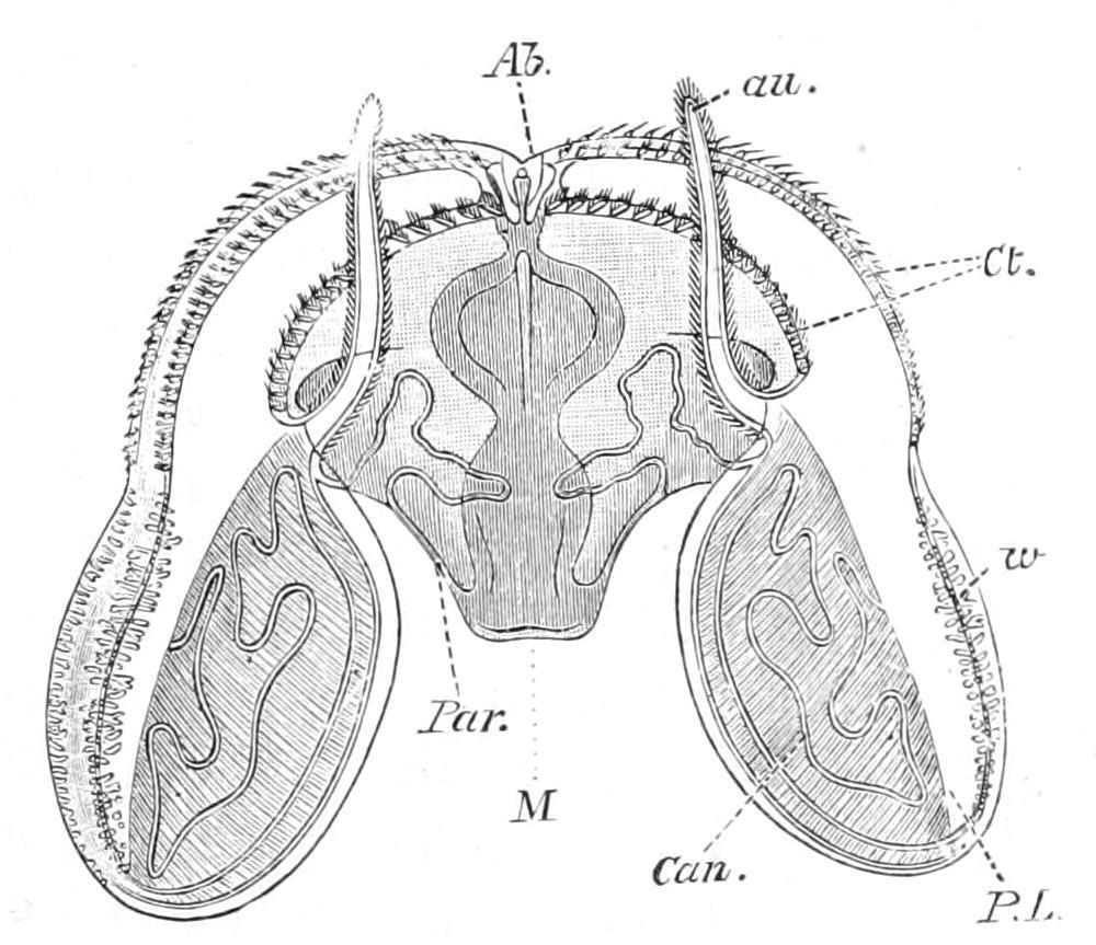
F��. 181. Ocyroe crystallina. Ab, aboral sense-organ; au, auricle; Can, diverticulum from the paragastric canal passing into peristomial lobe; Ct, costae; M, mouth; Par, paragastric canal passing outwards to join one of the transverse subcostal canals; P.L, peristomial lobe; w, wart-like tubercles on the lobe. (After Mayer.)
The order contains only fifteen genera, but they are usually arranged in the following eight families:—
1. Lesueuriidae. Lesueuria.
2. Bolinidae. Bolina, Bolinopsis.
3. Deiopeidae. Deiopea.
4. Eurhamphaeidae. Eurhamphaea.
5. Eucharidae. Eucharis.
6. Mnemiidae. Mnemia, Mnemiopsis.
7. Calymmidae. Calymma.
8. Ocyroidae. Ocyroe.
Most of these Ctenophores occur in the warm and tropical seas; but Bolina is found occasionally at Plymouth in the month of May, on the west coast of Ireland, and at other stations on the British coasts. Eucharis is regarded as one of the most beautiful of the Phylum. A swarm, some miles in length, of large specimens of E. multicornis was met by the Plankton Expedition in the south equatorial current of the Atlantic during the month of September
Order III. Cestoidea.
In this order the body is so much compressed in the transverse plane and elongated in the sagittal plane that it assumes the shape of a long narrow band or ribbon. The tentacular sheaths are present but the tentacles are degenerate in the adult. The tentacular functions are performed by numerous tentilla situated in long grooves extending along the whole length of the oral side of the band-like body The transverse ribs are reduced; the sagittal ribs extend along the whole of the aboral side.
Fam. Cestidae.—This is the only family of the order. Cestus veneris, the Venus's girdle of the Mediterranean Sea, is also found in the Atlantic Ocean, and specimens belonging to the same genus, but probably to a different species, occur as far north as the White Sea. Some of the larger specimens are considerably over 1 metre in length.
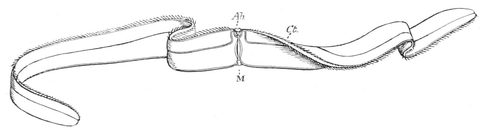
F�� 182 —Cestus pectenalis Ab, aboral sense-organ; Ct, the sagittal ribs; M, mouth. (After Bigelow.)
C. pectenalis was found in abundance off one of the Maldive Islands [431] and differs from C. veneris in having a large and prominent orange patch at each end of the body. It is said to be extremely graceful in the water, moving with slow, ribbon-like undulations, and shining in the sunlight with a violet iridescence. Vexillum, from the Mediterranean Sea and Canary Islands, is rather more pointed at the extremities than Cestus, and differs from it in some important anatomical characters.
Order IV. Platyctenea.
This order has been constituted for two remarkable genera, in which the oro-apical axis is so much reduced that distinct dorsal and ventral surfaces can be distinguished.
There is a single pair of long milky-white tentacles capable of complete retraction into tentacular sheaths.
Fam. 1. Ctenoplanidae. Ctenoplana was discovered by Korotneff in 1886 floating with the Plankton off the coast of Sumatra. In 1896 Willey [432] discovered four specimens on a cuttle-bone floating off the coast of New Guinea. To these authors we are indebted for the only accounts of this animal that have been published.
When the Ctenoplana is creeping on the bottom of a dish or with its dorsal side downwards on the surface film of the water, it has the form of a flattened disc with a notch on each side. On the upper or dorsal surface eight short rows of ctenophoral plates may be seen, and in a position corresponding with the two notches in the margin of the body are situated the two sheaths from which the long pinnate tentacles protrude. In the exact centre of the dorsal surface is situated the statolith, supported by stiff processes from adjacent cells; and forming a circlet round the statolith there is a row of short ciliated tentacles. These tentacles, however, when examined carefully in the living animal, are found to be arranged in two sets of
about nine in each, separated by narrow gaps on each side, the gaps corresponding in position with the axis through the tentacles.
When the animal is swimming it assumes a helmet-shape by depressing the sides of the body like a pair of flaps on the tentacular axis, and then the ctenophoral plates come into play and produce the progressive movements of the animals. The pinnate tentacles are opaque white in colour, and have peculiar serpentine movements. Very little is known at present concerning many details of the internal anatomy, but there is one point of considerable theoretical interest—namely, the presence of definite male genital ducts.
Three of Dr. Willey's specimens were mottled with a green pigment, whereas his fourth specimen and Korotneff's only specimen were mottled with a red pigment. It has yet to be determined whether the differences which have been observed in the individual specimens are of specific value.
Fam. 2. Coeloplanidae.—Coeloplana was originally discovered by Kowalevsky in the Red Sea, but has recently been found by Abbott [433] on the coast of Japan.
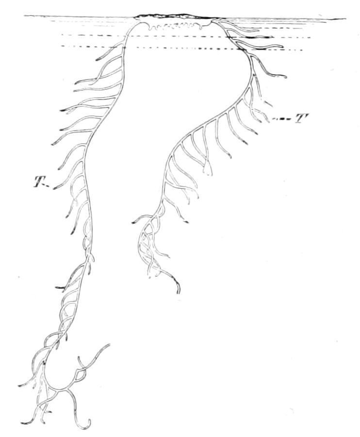
F�� 183 Coeloplana mitsukurii, floating at the surface of the sea with the dorsal side downwards T, T, the tentacles expanded (After Abbott )
The Japanese species are found principally on encrusting Algae, Zostera, Melobesia, etc., which they resemble very closely in colour. The Red Sea species is, according to Kowalevsky, ciliated all over,
but the Japanese species are ciliated only on the ventral surface. As in Ctenoplana, the body of Coeloplana is a flattened disc with a notch at each end of the tentacular axis, when creeping; but Coeloplana does not swim, nor at any time does it assume a helmetshape. The tentacles are very long and of a chalky-white colour. They can be retracted into tentacle-sheaths. When the animal is excited it throws out the whole tentacle in a cloud of white filaments, "and to watch it at such a time, shooting out and retracting the tentacles, moving along the side of the aquarium like a battleship in action is truly a remarkable spectacle."[434] On the dorsal side of the body there is a series of processes which are called the dorsal tentacles. The statolith is very small, and is not surrounded by sensory processes as it is in Ctenoplana. There are no ctenophoral plates. The colours of the Japanese species are scarlet or carmine red and dirty brown or brownish yellow. They are from 1 to 2 centimetres in diameter.
CLASS II. NUDA
Ctenophora without tentacles.
Fam. Beroidae. Beroe, the only genus of this family and class, differs from other Ctenophora in several important particulars. There are no tentacles, and the stomodaeum is so large that the body-form assumes that of a thimble with moderately thick walls. The infundibulum is small. The paragastric and longitudinal canals give rise to numerous ramifications which form a network distributed throughout the surface of the body. The statolith is unprotected by a dome, and the polar fields are bordered by a number of small branching papillae. The eight ribs extend for nearly the whole length of the body Beroe is almost cosmopolitan, and is frequently found at the surface of the sea in great numbers. B. ovata is found off the Shetlands, Hebrides, and west coast of Ireland, but is rare on the east coast of the British Islands and in the English Channel. At Valencia it is common in August and September, and sometimes
reaches the great size of 90 mm. in length by 50 mm. in breadth. It is usually of a pale pink colour.
A������� �� C���������
Hydroctena salenskii has recently been discovered by Dawydoff[435] floating with the Plankton off the island Saparua in the Malay Archipelago. It is claimed to be a connecting link between the Ctenophora and the Medusae of the Hydrozoa.
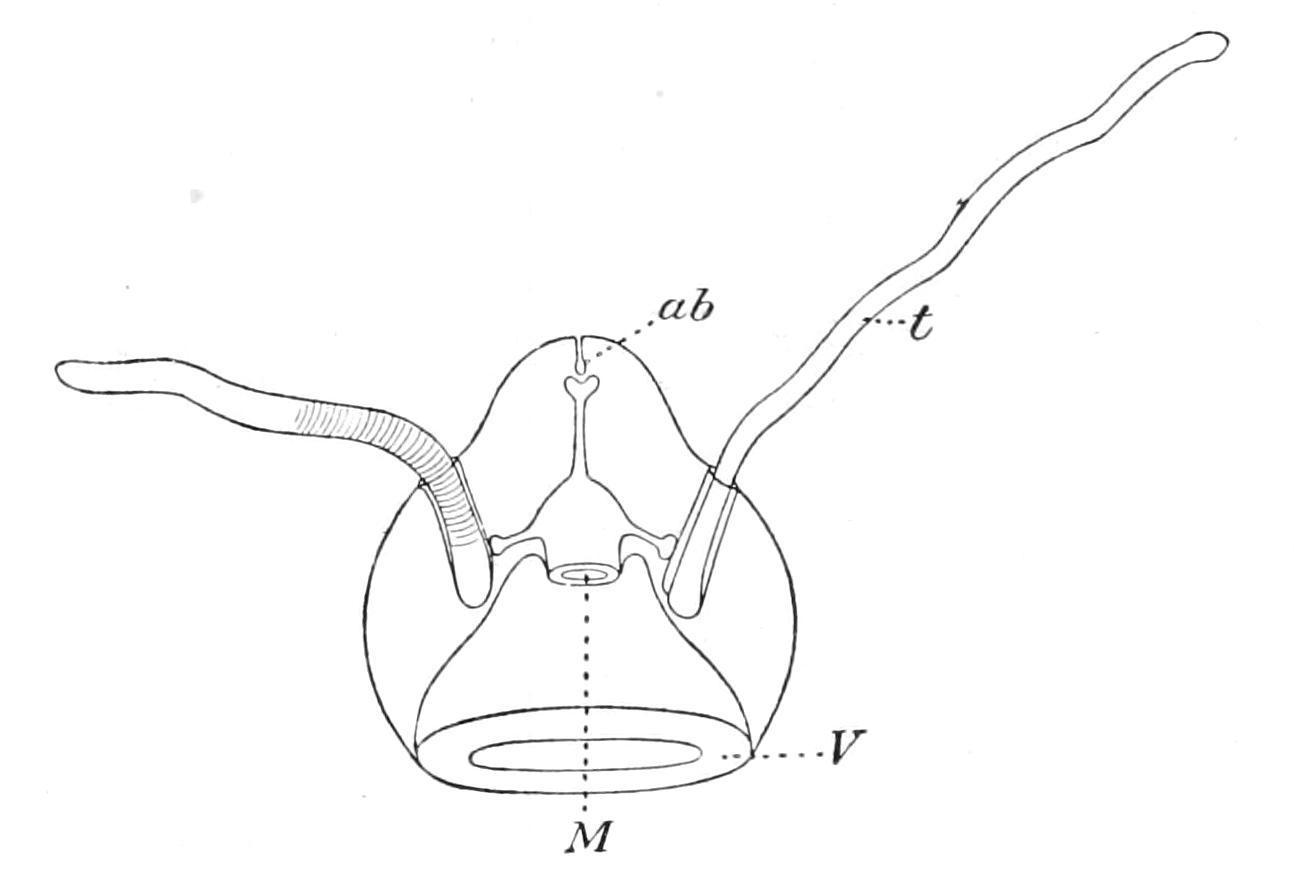
In external features it is like one of the Narcomedusae, having a transparent jelly-like bell with a wide bell-mouth guarded by a velum (Fig. 184, V). There are only two simple but solid tentacles (t), provided with tentacle-sheaths, but inserted on opposite sides of the bell—not on the margin, but, as in the Ctenophore, at a level not far removed from the aboral pole. At the aboral pole there is a minute pore surrounded by a high ciliated epithelium bearing an orange pigment. This leads into a short blind canal, which terminates in an ampulla bearing two statoliths supported by elastic processes from the ampullar epithelium.
The sub-umbrellar cavity extends for a distance of about one-half the height of the bell. The mouth (M), which opens into this cavity, leads into a wide cavity that gives off a short blind canal to the side of each tentacular sheath, and a straight tube that leads straight to the statocyst, where it also ends blindly. There are no radial canals and no ring canal at the margin of the umbrella. There are also no ctenophoral plates. In the absence of any information concerning the position of the genital glands, the character of the epithelium of the tentacles and the development, we are not justified in regarding Hydroctena either as a Ctenophore or as a connecting link between the Ctenophora and the Hydromedusae. It may be regarded simply as a Craspedote Medusa, probably related to the Narcomedusae, with a remarkable aberrant aboral sense-organ.
Formerly Fellow of St. John's College Professor of Zoology in McGill University, Montreal.
CHAPTER XVI
ECHINODERMATA INTRODUCTION CLASSIFICATION ANATOMY OF A STARFISH SYSTEMATIC ACCOUNT OF ASTEROIDEA
The name Echinodermata[436] means literally "spiny-skinned," and thus brings into prominence one very conspicuous feature of most of the animals belonging to this phylum. All, it is true, do not possess spines; but with one or two doubtful exceptions, all have calcareous plates embedded in the skin, and these plates, in many cases, push out projections which raise the skin into corresponding elevations, which are called the spines. The spines are, like the other plates, inside the skin, and to speak of an Echinoderm living in its shell, as we speak of a Snail, is a serious error. The shell of a Mollusc is fundamentally a secretion poured forth from the skin, and is thus entirely external to the real living parts; but the plates and spines of an Echinoderm may be compared to our own bones, which are embedded deeply in the flesh. Hence the name ossicle (little bone) is used to designate these organs.
Besides the possession of these spines, Echinoderms are characterised by having their organisation pervaded by a fundamental radial symmetry The principal organs of the body are repeated and are arranged like the spokes of a wheel round a
central axis instead of being, as, for example, in Chaetopoda, arranged behind one another in longitudinal series.
In addition to these striking peculiarities, Echinoderms possess a most interesting internal organisation, being in this respect almost exactly intermediate between the Coelenterata and the higher Invertebrata. Like so many of the latter, the Echinodermata have an anus, that is, a second opening to the alimentary canal through which indigestible material is rejected; like them also, they have a body-cavity or coelom surrounding the alimentary canal—from the lining of which the genital cells are developed. On the other hand, there is no definite circulatory system, nor any specialised excretory organ, and the nervous system exhibits no concentration which could be called a brain, and is, moreover, in close connexion with the skin. In all these points the Echinodermata resemble the Coelenterata.
One of the most characteristic features of the internal anatomy of Echinodermata is the presence of a peculiar series of organs, known collectively as the water-vascular system or hydrocoel This is really a special division of the coelom or body-cavity which takes on the form of a ring-shaped canal embracing the mouth, from which are given off long radial canals, usually five in number, running to the more peripheral parts of the body.[437] Each radial canal carries a double series of lateral branches, which push out the skin so as to appear as appendages of the body. These appendages are known as tentacles or tube-feet; they are both sensory and respiratory in function, and often in addition, as the name tube-foot indicates, assist in locomotion. As a general term for these appendages, to be applied in all cases without reference to their function, the name podium has been suggested and will be employed here. A system of canals, in many ways resembling the water-vascular system, is found in Brachiopoda, Gephyrea and Polyzoa, but the peculiarity of Echinodermata is the way in which it is kept filled with fluid. From the ring-canal in the interval (or interradius) between two radial canals, a vertical canal, termed the stone-canal, is given off, which
communicates with the exterior by means of a sieve-like plate, the madreporite, pierced by fine canals. These canals and the stonecanal itself are lined with powerful cilia, which produce a strong inward current, and keep the water-vascular system tensely filled with sea water.
The phylum includes the familiar Starfish and Sea-urchins, which in sheltered spots are found between tide-marks; the Brittle Stars and Sea-cucumbers, which can be dredged up from below low-water mark, and lastly the beautiful Feather-stars, of which there are comparatively few species still living, although huge beds of limestone are composed of the remains of fossil Feather-stars.
One species of Sea-cucumber (Synapta similis)[438] is said to enter brackish water in the mangrove swamps of the tropics; but, with this exception, the whole phylum is marine. A few species can endure partial exposure to the air when left bare by the receding tide, but the overwhelming majority are only found beneath low-water mark, and a considerable number live in the deepest recesses of the ocean.
Their distribution is, no doubt, partly determined by food, a number of species being strictly confined to the neighbourhood of the shore. On the other hand, since a very large number of species live on the layer of mud impregnated with animal remains which forms the superficial layer of the deposit covering the sea-floor, it is not surprising to learn that many have an exceedingly wide range, since this deposit is very widely distributed. Another equally important factor in determining distribution is wave-disturbance, and it is surprising to learn to what a depth this extends. Off the west coast of Ireland a large wave literally breaks on a submerged rock 15 fathoms beneath the surface. Speaking generally, it is useless to look for Echinoderms on an exposed coast, and the same species, which in the sheltered waters of the Clyde are exposed at low water, must be dredged up from 20 to 30 fathoms outside Plymouth Sound.
The ordinary collector is attracted to the group chiefly by the regularity and beauty of the patterns produced by the radial symmetry, but to the scientific zoologist they are interesting from many other points of view. Differing widely nevertheless from the higher Invertebrata in their symmetry when adult, they have as larvae a marked bilateral symmetry, and the secondary development of the radial symmetry constitutes one of the most remarkable lifehistories known in the animal kingdom.
Then again, owing to the possession of ossicles, the Echinodermata are one of the few groups of Invertebrata of which abundant remains occur fossilised. In attempting, therefore, to decipher the past history of life from the fossil record, it is necessary to have an exact and detailed knowledge of Echinoderm skeletons and their relation to the soft parts. Lastly, the internal organisation of Echinoderms throws valuable light on the origin of the complicated systems of organs found in the higher animals.
Echinodermata are divided into two great sub-phyla, which must have very early diverged from one another. These are:—
(1) Eleutherozoa, (2) Pelmatozoa.[439]
The sub-phylum Pelmatozoa, to which the living Feather-stars (Crinoidea) and the majority of the known fossil species belong, is characterised by the possession of a fixing organ placed in the centre of the surface opposite the mouth—the aboral surface as it is called. Ordinarily this organ takes on the form of a jointed stalk, but in most modern species it is a little knob with a tuft of rooting processes, termed cirri. In the other sub-phylum, the Eleutherozoa, no such organ is found, and the animals wander about freely during their adult life, though for a brief period of their larval existence they may be fixed by a stalk-like protuberance arising from the oral surface.
SUB-PHYLUM I. ELEUTHEROZOA
The Eleutherozoa are divided into four main classes, between which no intermediate forms are found amongst the living species, though intermediate types have been found fossil.
The four classes into which the Eleutherozoa are divided are defined as follows:—
(1) Asteroidea (Starfish).—"Star"-shaped or pentagonal Eleutherozoa with five or more triangular arms, not sharply marked off from the central disc. The mouth is in the centre of one surface, called from this circumstance the "oral"; the anus is in the centre of the opposite surface, termed the "aboral." From the mouth a groove runs out on the under surface of each arm towards its tip, termed the "ambulacral" groove. Projecting from the ambulacral groove are found the podia or tube-feet, the organs of movement and sensation of the animal.
(2) Ophiuroidea (Brittle Stars).—Eleutherozoa, in which the body consists of a round disc with long worm-like arms inserted in grooves on its under surface. No anus is present, and the ambulacral grooves are represented by closed canals. The podia are merely sensory and respiratory, locomotion being effected by muscular jerks of the arms.
(3) Echinoidea (Sea-urchins).—Globular or disc-shaped Eleutherozoa, in which the skeleton forms a compact cuirass except for a short distance round the mouth (peristome) and round the anus (periproct). The ambulacral grooves are represented by canals which, like meridians of longitude on a school-globe, run from the neighbourhood of the mouth to near the aboral pole of the body. The spines are large and movably articulated with the plates. The animals move by means of podia and spines, or by means of the latter only. The anus is usually situated at the aboral pole, but is
sometimes displaced towards the side, or even on to the ventral surface.
(4) Holothuroidea (Sea-cucumbers).—Sausage-shaped Eleutherozoa, in which the skeleton is represented only by isolated nodules of calcium carbonate, and in which the body-wall is highly muscular. The mouth and anus are situated at opposite ends of the body, and the ambulacral grooves (represented by closed canals) run from near the mouth to the proximity of the anus. Movement is accomplished by means of the podia, aided by worm-like contractions of the body.
CLASS I. ASTEROIDEA[440] (S�������)
The Starfish derive their name from their resemblance in shape to the conventional image of a star. The body consists of broad triangular arms (generally five in number) which coalesce in the centre to form a disc. The skin is soft and semi-transparent, permitting the skeleton to be easily detected; this consists of a meshwork of rods or plates, leaving between them intervals of soft skin. In a living Starfish it can be seen that many of these soft places are raised up into finger-like outgrowths, which are termed "papulae" or "dermal gills," through the thin walls of which an active interchange of gases with the surrounding water takes place, and the animal obtains in this way the oxygen necessary for its respiration.
Very few and feeble muscle-fibres exist in the body-wall, and the movements of the arms, as a whole, are very slow and limited in range. There is a membranous lip surrounding the mouth, from which five broad grooves run outwards, one on the underside of each arm. These are termed the "ambulacral grooves." Each groove is Λ-shaped, and its sides are stiffened by a series of rod-like ossicles called the "ambulacral ossicles."
The animal progresses by the aid of a large number of translucent tentacles, termed "tube-feet" or "podia," which are attached to the walls of the ambulacral grooves.
Anatomy of a Starfish.—As an introduction to the study of the anatomy not only of Starfish but of Echinodermata as a whole, we select Asterias rubens, the common Starfish of the British coasts, which in many places may be found on the beach near low-water mark.
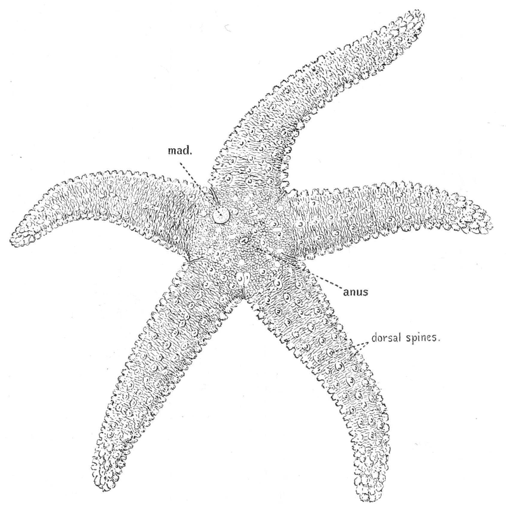
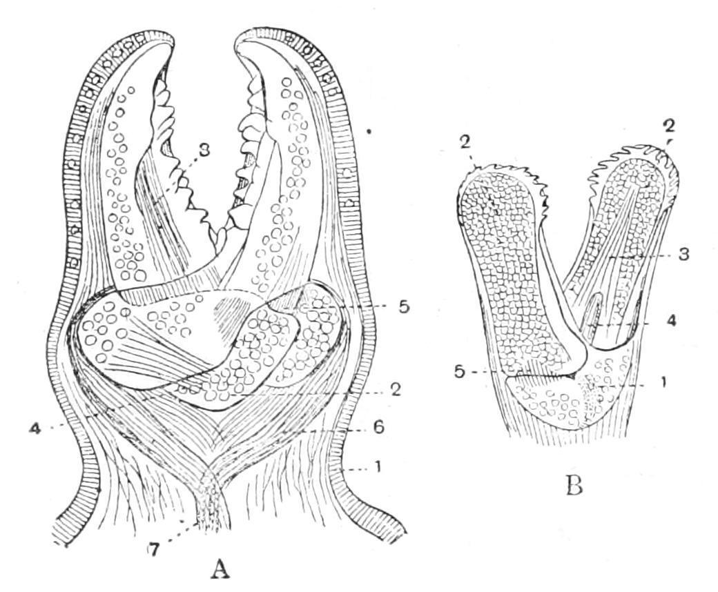
External Features.—In this species (Fig. 185) the skeleton is a network of rod-like plates, leaving wide meshes between them, through which protrude a perfect forest of transparent papulae. From the points of junction of the rods arise short blunt spines surrounded by thick cushions of skin. The surfaces of these cushions are covered with a multitude of whitish specks, which, on closer inspection, are seen to have the form of minute pincers, each consisting of two movable blades crossing each other below and articulated to a basal piece. These peculiar organs are termed "pedicellariae" (Fig. 186), and their function is to keep the animal clean by seizing hold of any minute organisms which would attempt to settle on the soft and delicate skin. When irritated the blades open and then snap together violently, and remain closed for a long time.[441] These actions are brought about by appropriate muscles attaching the blades to the basal piece.
F��. 185. Asterias rubens, seen from the aboral surface, × 1. mad, Madreporite.
The last-named ossicle increases the certainty of the grip by fixing the lower parts of each blade in the same vertical plane, and preventing lateral slipping, so that it serves the same purpose as the pivot in a pair of scissors. Each blade, in fact, fits into a groove on the side of this piece. The muscles which close the blades arise from the lower ends (handles) of the blades, and are united below to form a common muscular string which attaches the whole organ to one of the plates of the skeleton. An attempt of the victim to tear the pedicellaria out is resisted by the contraction of this string, which thus brings about a closer grip of the blades. In order that the blades may open they must first be lifted out of the grooves on the basal piece—this is effected by special lifting muscles. The opening is brought about by muscles extending from the "handle" of one blade to the upper part of the other.
Scattered about amongst the papulae between the cushions are other pedicellariae of a larger size in which the blades do not cross one another (Fig. 186, B).
In the space or "interradius" between two arms, on the aboral surface, there is found a button-shaped ossicle. This is covered with
fine grooves, and from a fancied resemblance between it and some forms of coral it has received the name "madreporite" (Fig. 185, mad). The bottoms of the grooves are perforated by capillary canals lined by flagella, through the action of which water is constantly being introduced into the water-vascular system.
The anus is situated near the centre of the upper surface of the disc, but it is so minute as to require careful inspection in order to discover its position (Fig. 185).
F�� 186 View of pedicellariae of A glacialis A, Crossed form, × 100 1, Ectoderm covering the whole organ; 2, basal piece; 3, auxiliary muscle closing the blades; 4, muscle lifting right blade out of the groove; 5, handle of left blade; 6, muscles closing the blades, and uniting to form 7, the muscular string attaching the pedicellaria to the skeleton B, straight form, × 10 1, Basal piece; 2, blades; 3 and 4, muscles closing the blades; 5, muscle opening the blades. (From Cuénot.)
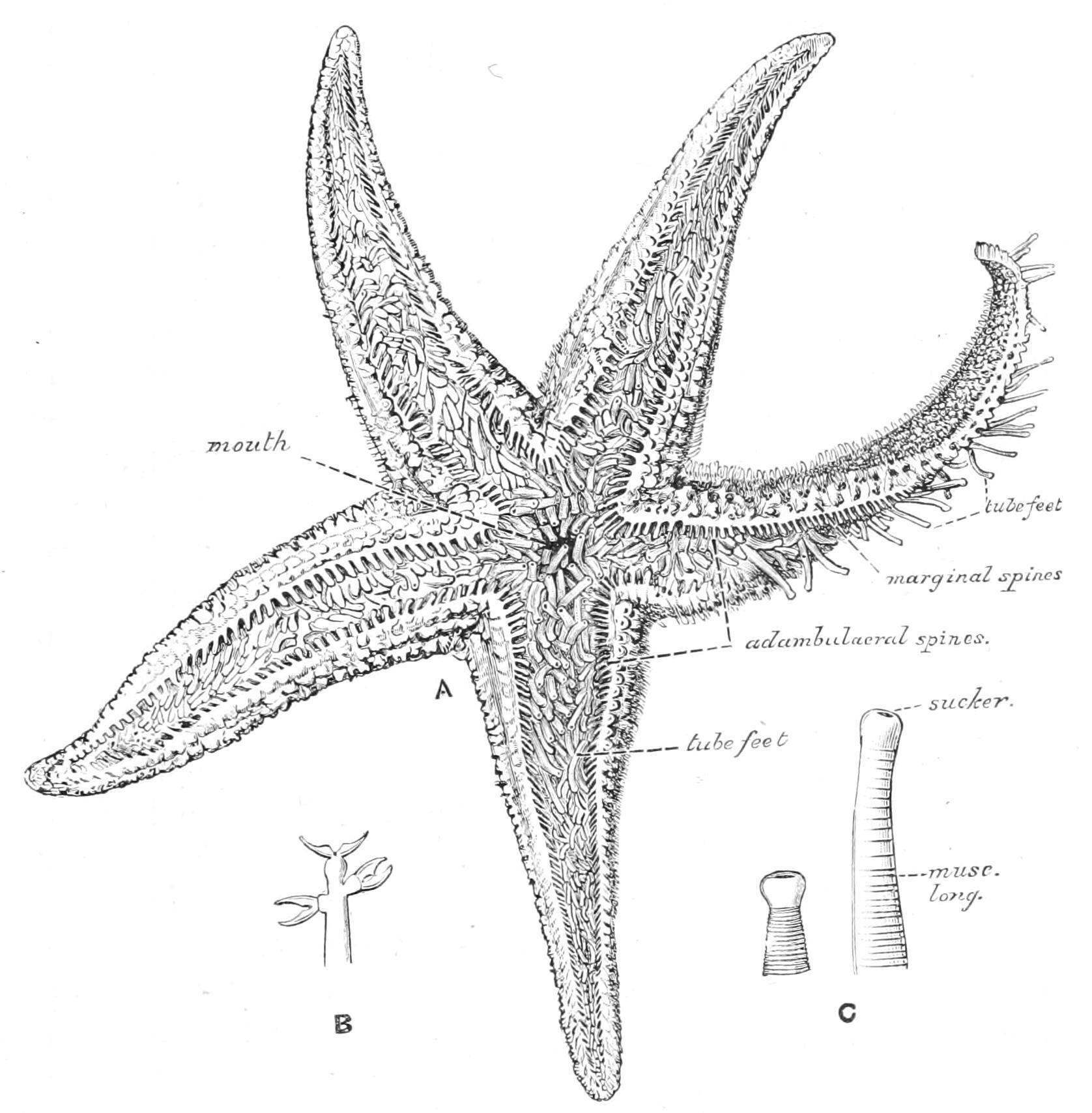
On the under side of the animal the most conspicuous features are the five ambulacral grooves which radiate out from the "peristome," a thin membranous area surrounding the central mouth. The grooves are filled with the tube-feet, which are closely crowded together and apparently arranged in four rows.
Skeleton.—The sides of the ambulacral grooves are stiffened by the rod-like "ambulacral ossicles." To the outer ends of these are articulated a set of shorter rods termed the "adambulacral ossicles" which carry each two or three rod-like spines, the "adambulacral spines," the skin covering which bears numerous pedicellariae (Fig. 187, B). When the animal is irritated the edges of the groove are
brought together, and these spines then form a trellis-work covering and protecting the delicate tube-feet; the numerous pedicellariae are then in a position to make it unpleasant for any intruder. The closure of the groove is effected by means of powerful muscles connecting each ambulacral ossicle with its fellow. There are also feebler muscles connecting these plates with their successors and predecessors, which enable the arm to be bent downwards in a vertical plane. It is raised by a muscular band running along the dorsal wall of the coelom to the point of the arm.
F�� 187 A, Asterias rubens, seen from the oral surface, drawn from a living specimen, × 1 B, an adambulacral spine, showing three straight pedicellariae; C, a tube-foot expanded and contracted
When the series of ambulacral and adambulacral ossicles is followed inwards towards the mouth it is seen that the first ambulacral ossicle is closely fixed to the second, but is widely separated from its fellow, remaining, however, connected with the latter by a powerful adductor muscle. In consequence of the separation of this pair of ossicles each is brought into closer contact with the corresponding ossicle in the adjacent radius, to which it is connected by a muscle called the abductor. The first adambulacrals in adjacent radii are also brought into closer contact and carry long spines which, when the ambulacral grooves are contracted, project like a grating over the mouth. In the
Welcome to our website – the ideal destination for book lovers and knowledge seekers. With a mission to inspire endlessly, we offer a vast collection of books, ranging from classic literary works to specialized publications, self-development books, and children's literature. Each book is a new journey of discovery, expanding knowledge and enriching the soul of the reade
Our website is not just a platform for buying books, but a bridge connecting readers to the timeless values of culture and wisdom. With an elegant, user-friendly interface and an intelligent search system, we are committed to providing a quick and convenient shopping experience. Additionally, our special promotions and home delivery services ensure that you save time and fully enjoy the joy of reading.
Let us accompany you on the journey of exploring knowledge and personal growth!