Instant digital products (PDF, ePub, MOBI) ready for you
Download now and discover formats that fit your needs...
Electrocardiography of Arrhythmias-A Comprehensive Review-A Companion to Cardiac Electrophysiology, 2e (Mar 8, 2021)_(032368050X)_(Elsevier).pdf Mithilesh Kumar Das
No part of this publication may be reproduced or transmitted in any form or by any means, electronic or mechanical, including photocopying, recording, or any information storage and retrieval system, without permission in writing from the publisher. Details on how to seek permission, further information about the Publisher’s permissions policies and our arrangements with organizations such as the Copyright Clearance Center and the Copyright Licensing Agency, can be found at our website: www.elsevier.com/permissions
This book and the individual contributions contained in it are protected under copyright by the Publisher (other than as may be noted herein).
Notice
Practitioners and researchers must always rely on their own experience and knowledge in evaluating and using any information, methods, compounds or experiments described herein. Because of rapid advances in the medical sciences, in particular, independent verification of diagnoses and drug dosages should be made. To the fullest extent of the law, no responsibility is assumed by Elsevier, authors, editors or contributors for any injury and/or damage to persons or property as a matter of products liability, negligence or otherwise, or from any use or operation of any methods, products, instructions, or ideas contained in the material herein.
Previous editions copyrighted 2006, 2011, and 2015.
Library of Congress Control Number: 2018964891
Content Strategist: Robin Carter
Content Development Specialist: Angie Breckon
Publishing Services Manager: Shereen Jameel
Senior Project Manager: Anitha Rajarathnam
Design Direction: Patrick Ferguson
To every member of my lovely family, especially to my dear wife, Su-Mei, and our wonderful children, Priscilla, Melvin, and Jessica, for all of their love, support, and patience throughout my career, and during the editing and writing of this book; to my beloved grandchildren, Ethan, Titus, Miles, and Philip, who always fill my life with joy and happiness; to my unforgettable mentors, and benefactors, Drs. Pablo Denes, Robert Hauser, Gordon Ewy, and Joseph Alpert, who have guided me like a steady lighthouse in the right direction.
Shoei K. Stephen Huang, MD
To immediate family members including my dear children, Rebekah, Jordan, and Jacob, and especially my precious wife Jeanne, who gave encouragement and sacrificed our time together for the high goal of dissemination of knowledge; as well as those who steered me carefully through my career (particularly Drs. Mark E. Josephson and Douglas P. Zipes), I dedicate my small portion of this work.
John M. Miller, MD
LIST OF CONTRIBUTORS
Amin Al-Ahmad, MD
Texas Cardiac Arrhythmia Institute
St. David’s Medical Center
Austin, Texas
Jason G. Andrade, MD
Department of Medicine
Montreal Heart Institute
University of Montreal
Montreal, Canada
Department of Medicine
University of British Columbia Vancouver, Canada
Elad Anter, MD
Associate Professor of Medicine
Harvard Medical School
Electrophysiology
Beth Israel Deaconess Medical Center Boston, Massachusetts
Rishi Arora, MD, FHRS
Professor of Medicine
Director, Experimental Cardiac Electrophysiology
Northwestern University—Feinberg School of Medicine
Chicago, Illinois
Samuel J. Asirvatham, MD
Professor of Medicine and Pediatrics
Internal Medicine
Division of Cardiovascular Diseases
Mayo Clinic
Rochester, Minnesota
Javier E. Banchs, MD, FACC, FHRS
Director of Electrophysiology and Pacing
Division of Cardiology
Baylor Scott & White Health
Temple, Texas
Mohamed Bassiouny, MD
Electrophysiologist
Texas Cardiac Arrhythmia Institute
St. David’s Medical Center
Austin, Texas
Tina Baykaner, MD, MPH
Clinical Instructor
Cardiovascular Medicine
Stanford University
Palo Alto, California
Francis Bessière, MD, MSc
Fellow
Electrophysiology
Montreal Heart Institute
Montreal, Canada
Deepak Bhakta, MD, FACP, FACC, FAHA, FHRS, CCDS
Associate Professor of Clinical Medicine
Krannert Institute of Cardiology
Indiana University School of Medicine
Indianapolis, Indiana
Frank Bogun, MD
Associate Professor of Medicine
Division of Cardiology
Cardiovascular Medicine
University of Michigan
Ann Arbor, Michigan
Chad Brodt, MD
Cardiovascular Division
CV Institute
Stanford University
Palo Alto, California
Eric Buch, MD
Associate Professor of Medicine
Cardiac Arrhythmia Center
Division of Cardiology
University of California
UCLA Cardiac Arrhythmia Center
David Geffen School of Medicine and UCLA Health System
Los Angeles, California
J. David Burkhardt, MD
Texas Cardiac Arrhythmia Institute
St. David’s Medical Center
Austin, Texas
David J. Callans, MD
Professor of Medicine
University of Pennsylvania
Associate Director of Electrophysiology
Hospital of the University of Pennsylvania Philadelphia, Pennsylvania
Jien-Jiun Chen, MD
Attending Physician
Division of Cardiology
Department of Internal Medicine
National Taiwan University Hospital, YunLin Branch
Hospital of the University of Pennsylvania Philadelphia, Pennsylvania
Jiunn-Lee Lin, MD, PhD
Chair Professor
Cardiovascular center
Taipei Medical University Shuan-Ho Hospital
New Taipei City, Taiwan
Lian-Yu Lin, MD, PhD
Professor of Medicine
Division of Cardiology
Department of Internal Medicine
National Taiwan University College of Medicine and Hospital
Taipei, Taiwan
Ting-Tse Lin, MD
Lecturer
Department of Internal Medicine
National Taiwan University Hospital, Hsin-Chu Branch
Hsin-Chu, Taiwan
Deborah Lockwood, BM, BCh, MA
Associate Professor of Medicine
Heart Rhythm Institute
University of Oklahoma Health Sciences Center
Oklahoma City, Oklahoma
Steven M. Markowitz, MD
Professor of Medicine
Division of Cardiology
Department of Medicine
Weill Cornell Medical College
New York-Presbyterian Hospital
New York, New York
Gregory F. Michaud, MD
Professor of Medicine
Chief, Arrhythmia Section
Vanderbilt University Medical Center
Nashville, Tennessee
John M. Miller, MD
Professor of Medicine
Indiana University School of Medicine
Director, Cardiac Electrophysiology Services
Indiana University Health
Indianapolis, Indiana
Marc A. Miller, MD
The Leona M. and Harry B. Helmsley Charitable Trust Professor of Medicine
Cardiac Electrophysiology; Director, Cardiac Arrhythmia Service
The Leona M. and Harry B. Helmsley Charitable Trust Center for Cardiac Electrophysiology
Icahn School of Medicine at Mount Sinai
New York, New York
Jay A. Montgomery, MD
Assistant Professor of Medicine Arrhythmia Section
Department of Medicine
Vanderbilt University Medical Center
Nashville, Tennessee
Talal Moukabary, MD
Director, Cardiac Electrophysiology
Carondelet Heart and Vascular Institute
Tucson, Arizona
J. Paul Mounsey, BSc, BM, BCh, PhD
Chief of Electrophysiology
Professor of Medicine
Cardiovascular Sciences
East Carolina Heart Institute
Greenville, North Carolina
Koonlawee Nademanee, MD
Professor of Medicine
Department of Medicine
Chulalongkorn University
Bangkok, Thailand
Hiroshi Nakagawa, MD, PhD
Professor of Medicine
Heart Rhythm Institute
University of Oklahoma Health Sciences Center
Oklahoma City, Oklahoma
Niyada Naksuk, MD
Department of Cardiovascular Diseases
Mayo Clinic
Rochester, Minnesota
Sanjiv M. Narayan, MD, PhD
Professor of Medicine
Director of AF Program and of Electrophysiology Research
Stanford University
Palo Alto, California
Andrea Natale, MD
Texas Cardiac Arrhythmia Institute
St. David’s Medical Center
Austin, Texas
Akihiko Nogami, MD, PhD
Professor
Cardiovascular Division
University of Tsukuba
Tsukuba, Ibaraki, Japan
Suk-Kyu Oh, MD
Clinical Assistant Professor
Division of Cardiology
Korea University Anam Hospital
Seoul, South Korea
Hakan Oral, MD
Professor of Internal Medicine
Cardiovascular Medicine
University of Michigan
Ann Arbor, Michigan
Santosh K. Padala, MD
Assistant Professor of Medicine
Cardiac Electrophysiology
Virginia Commonwealth University
Richmond, Virginia
Deepak Padmanabhan, MD, DM Department of Cardiovascular Diseases
Mayo Clinic Rochester, Minnesota
Hee-Soon Park, MD
Clinical Assistant Professor Division of Cardiology, Internal Medicine
Korea University Anam Hospital Seoul, South Korea
Bhupesh Pathik, MBBS, PhD Department of Cardiology
Royal Melbourne Hospital Department of Medicine University of Melbourne Melbourne, Australia
Thomas Paul, MD, FACC, FHRS Professor Department of Pediatric Cardiology and Pediatric Intensive Care Medicine
Georg-August-University Medical Center Göttingen, Germany
Basilios Petrellis, MBBS
Cardiac Electrophysiology Department of Cardiology
Sydney Adventist and Mater Hospitals New South Wales, Australia
Vivek Y. Reddy, MD
The Leona M. and Harry B. Helmsley Charitable Trust Professor of Medicine
Cardiac Electrophysiology; Director, Cardiac Arrhythmia Service
The Leona M. and Harry B. Helmsley Charitable Trust Center for Cardiac Electrophysiology
Icahn School of Medicine at Mount Sinai New York, New York
Sukit Ringwala, MD, MPH Director of Cardiac Electrophysiology Edward Hines, Jr. Veterans Administration Hospital Hines, Illinois
Assistant Professor Department of Cardiology Loyola University Medical Center Maywood, Illinois
Jaime Rivera, MD
Director of Cardiac Electrophysiology
Instituto Nacional de Ciencias Médicas y Nutrición
Hospital Médica Sur Mexico City, Mexico
Jason Roberts, MD
Director of Inherited Arrhythmia Service Heart Rhythm Program
Assistant Professor of Medicine
Western University London, Ontario, Canada
Miguel Rodrigo, PhD
Postdoctoral researcher
ITACA Institute
Universitat Politècnica de València
Valencia, Spain
Postdoctoral researcher
Cardiology Department
Stanford University
Palo Alto, California
Yuichiro Sakamoto, MD
Cardiovascular Medicine
Toyohashi Heart Center
Toyohashi, Aichi, Japan
Javier E. Sanchez, MD
Texas Cardiac Arrhythmia Institute
St. David’s Medical Center
Austin, Texas
Pasquale Santangeli, MD
Assistant Professor of Medicine
Cardiac Electrophysiology
University of Pennsylvania; Fellow
Cardiovascular Division
Hospital of the University of Pennsylvania Philadelphia, Pennsylvania
William H. Sauer, MD Professor of Medicine
Cardiac Electrophysiology Section
University of Colorado Hospital Aurora, Colorado
J. Philip Saul, MD, FHRS, FAHA, FACC Professor of Pediatrics
West Virginia University School of Medicine
Morgantown, West Virginia
Richard K. Shepard, AB, MD
Associate Professor of Medicine
Internal Medicine
Virginia Commonwealth University Richmond, Virginia
Jaemin Shim, MD, PhD
Associate Professor
Division of Cardiology
Korea University Medical Center
Seoul, South Korea
Kalyanam Shivkumar, MD, PhD, FHRS
UCLA Cardiac Arrhythmia Center
Medicine and Radiology
UCLA Health System
David Geffen School of Medicine and UCLA Health System
Los Angeles, California
Konstantinos Siontis, MD
Cardiovascular Medicine
University of Michigan
Ann Arbor, Michigan
Allan C. Skanes, MD Director, Heart Rhythm Program Professor of Medicine
Arrhythmia Service
Western University Ontario, Canada
Wilber W. Su, MD
Associate Professor and Director of Electrophysiology
Cardiology
Heart Institute, Banner-University Medical Center and University of Arizona Phoenix, Arizona
Associate Professor of Medicine
Stanford University Palo Alto, California
Edward Sze, MD
Clinical Cardiac Electrophysiology
Duke University Medical Center Durham, North Carolina
Hiroshi Tada, MD, PhD
Professor
Department of Cardiovascular Medicine, Faculty of Medical Sciences University of Fukui
Yoshida-gun, Fukui, Japan
Taresh Taneja, MD
Staff Electrophysiologist
Cardiac Electrophysiology
Kaiser Santa Clara Medical Center
Santa Clara, California
Patrick J. Tchou, MD
Robert and Suzanne Tomsich Department of Cardiovascular Medicine
Cleveland Clinic
Cleveland, Ohio
John K. Triedman, MD Professor of Pediatrics
Harvard Medical School
Boston, Massachusetts
Roderick Tung, MD
Associate Professor
Department of Medicine
Cardiology
Director of Clinical Cardiac Electrophysiology
Pritzker School of Medicine
University of Chicago Chicago, Illinois
Mohit K. Turagam, MD
Cardiac Electrophysiology
Mount Sinai
New York, New York
Wendy S. Tzou, MD
Associate Professor of Medicine
Cardiac Electrophysiology Section
University of Colorado
Aurora, Colorado
Mohan N. Viswanathan, MD
Clinical Associate Professor of Medicine
Cardiac Electrophysiology
Stanford University
Palo Alto, California
Tomos E. Walters, MBBS, PhD
Assistant Professor
Department of Electrophysiology
University of California San Francisco San Francisco, California
Paul J. Wang, MD
Professor of Medicine
Director, Cardiac Arrhythmia Service
Stanford University
Palo Alto, California
William Whang, MD, MS
Associate Professor
Department of Medicine
Icahn School of Medicine at Mount Sinai
New York, New York
Takumi Yamada, MD, PhD
Associate Professor of Medicine
Division of Cardiovascular Disease
University of Alabama at Birmingham Birmingham, Alabama
Raymond Yee, MD
Chair, Division of Cardiology
Professor of Medicine
Western University Ontario, Canada
Junaid A.B. Zaman, MA, BMBCh, MRCP
Fullbright British Heart Foundation Scholar
Cardiovascular Medicine
Stanford University
Palo Alto, California
Honorary Clinical Research Fellow
National Heart and Lung Institute
Imperial College
London, United Kingdom
Clinical Cardiac Electrophysiology Fellow
Heart Institute
Cedars Sinai Medical Center
Los Angeles, California
We are pleased to launch this 4th edition after increasing levels of demand from readers and practitioners all over the world. This newest edition tracks the rapid and exciting development of new mapping/ imaging/catheter technologies and ablation techniques in our rapidly changing field. We could not have prepared this text without the valuable input from countless readers, and we continue to welcome your comments, criticisms, and feedback to improve future editions of this book.
Major changes in this edition include the addition of several innovative chapters and new authors, particularly in the areas of atrial fibrillation and ventricular tachycardias. We also removed obsolete content and updated every chapter to match the latest in field knowledge and application methods, such as fundamental concepts of biophysics and parameters of radiofrequency lesion formation and cryothermal ablation, ablation-related anatomy, pathophysiology and diagnoses of arrhythmias, as well as 3-dimensional mapping/image-guided ablation techniques of common and uncommon arrhythmias. We also provided troubleshooting tips for helpful guidance in difficult cases. In addition to hundreds of detailed figures and tables, the number of online videos has been substantially expanded to facilitate access to and ease of learning.
We retained some features from prior editions including the same chapter format for consistency in organizational structure and content presentation. We continued our work with experienced, skilled, and acclaimed subject matter experts to write and review each chapter. Finally, we remained committed to casting a wide readership net and aimed to make this book readily accessible to all levels of cardiac electrophysiologists, trainees, and allied health professionals.
We envision this book to be a powerful and authoritative reference tool for the office; a practical guide for patient care, teaching, and preparation for the board examination in clinical cardiac electrophysiology; a comprehensive and trustworthy manual for an ablation procedure; and a valuable and accessible resource in every electrophysiology laboratory. We also hope that this book will help the next generation of practitioners be better equipped to overcome challenges and succeed in the expanding field of cardiac electrophysiology.
Shoei K. Stephen Huang, MD Temple, Texas, USA
John M. Miller, MD Indianapolis, Indiana, USA
John M. Miller, MD
Shoei K. Stephen Huang, MD
We deeply appreciate all the contributors for their extraordinary dedication to this book. We particularly thank those new authors for their incredible efforts in making this new edition fresh and relevant. Finally, special gratitude goes to Elsevier’s staff, especially Angie Breckon, Anitha Rajarathnam and Robin Carter, all of whom
exhibited the highest level of professionalism in shepherding this book to successful publication.
Shoei K. Stephen Huang, MD John M. Miller, MD
Biophysics and Pathophysiology of Radiofrequency Lesion Formation
David E. Haines
KEY POINTS
• Radiofrequency (RF) energy induces thermal lesion formation through resistive heating of myocardial tissue. Tissue temperatures of 50°C or higher are necessary for irreversible injury.
• Under controlled conditions, RF lesion size increases with increasing delivered power, electrode–tissue interface temperature, electrode diameter, and contact force.
• Power density declines with the square of distance from the source, and tissue temperature declines inversely with distance from the heat source.
• The ultimate RF lesion size is determined by the zone of acute necrosis as well as by the region of microvascular injury.
When Huang and colleagues first introduced radiofrequency (RF) catheter ablation in 19851 as a potentially useful modality for the management of cardiac arrhythmias,2 few would have predicted its meteoric rise. In the past 2 decades, it has become one of the most useful and widely used therapies in the field of cardiac electrophysiology. RF catheter ablation has enjoyed a high efficacy and safety profile, and indications for its use continue to expand. Improvements in catheter design have continued to enhance the operator’s ability to target the arrhythmogenic substrate, and modifications in RF energy delivery and electrode design have resulted in more effective energy coupling to the tissue. It is likely that most operators view RF catheter ablation as a black box in that once the target is acquired, they need only push the button on the RF generator. However, gaining insight into the biophysics of RF energy delivery and the mechanisms of tissue injury in response to this intervention will help the clinician to optimize catheter ablation, which may ultimately enhance its efficacy and safety.
BIOPHYSICS OF TISSUE HEATING
Resistive Heating
The RF energy is a form of alternating electrical current that generates a lesion in the heart by electrical heating of the myocardium. A common form of RF ablation found in the medical environment is the electrocautery, which is used for tissue cutting and coagulation during surgical procedures. The goal of catheter ablation with RF energy is to transform electromagnetic energy into thermal energy in the tissue effectively and to destroy the arrhythmogenic tissues by heating them to a lethal temperature. The mode of tissue heating by RF energy is resistive (electrical) heating. As electrical current passes through a resistive medium, the voltage drops and heat is produced (similar to the heat that is created in an incandescent light bulb). The RF electrical current is typically delivered in a unipolar fashion with completion of the circuit through an indifferent electrode placed on the skin. Typically, an oscillation frequency of 500 to 750 kHz is selected. Lower
• Electrode cooling reduces the efficiency of tissue heating. For a fixed energy delivery, blood flow over the electrode–tissue interface reduces lesion width on the surface by convective tissue cooling. Cooled ablation increases lesion size by allowing the operator to increase the power that can be delivered before limiting electrode–tissue interface temperatures are achieved.
• Avoiding collateral injury while maintaining lesion transmurality when ablating thin-walled structures is challenging because present technology does not allow the operator to monitor lesion formation in real time.
frequencies are more likely to stimulate cardiac muscle and nerves, resulting in arrhythmias and pain sensation. Higher frequencies will result in tissue heating; however, in the megahertz range, the mode of energy transfer changes from electrical (resistive) heating to dielectric heating (as observed with microwave energy). With very high frequencies, conventional electrode catheters become less effective at transferring the electromagnetic energy to the tissue, and therefore complex and expensive catheter antenna designs must be used.3
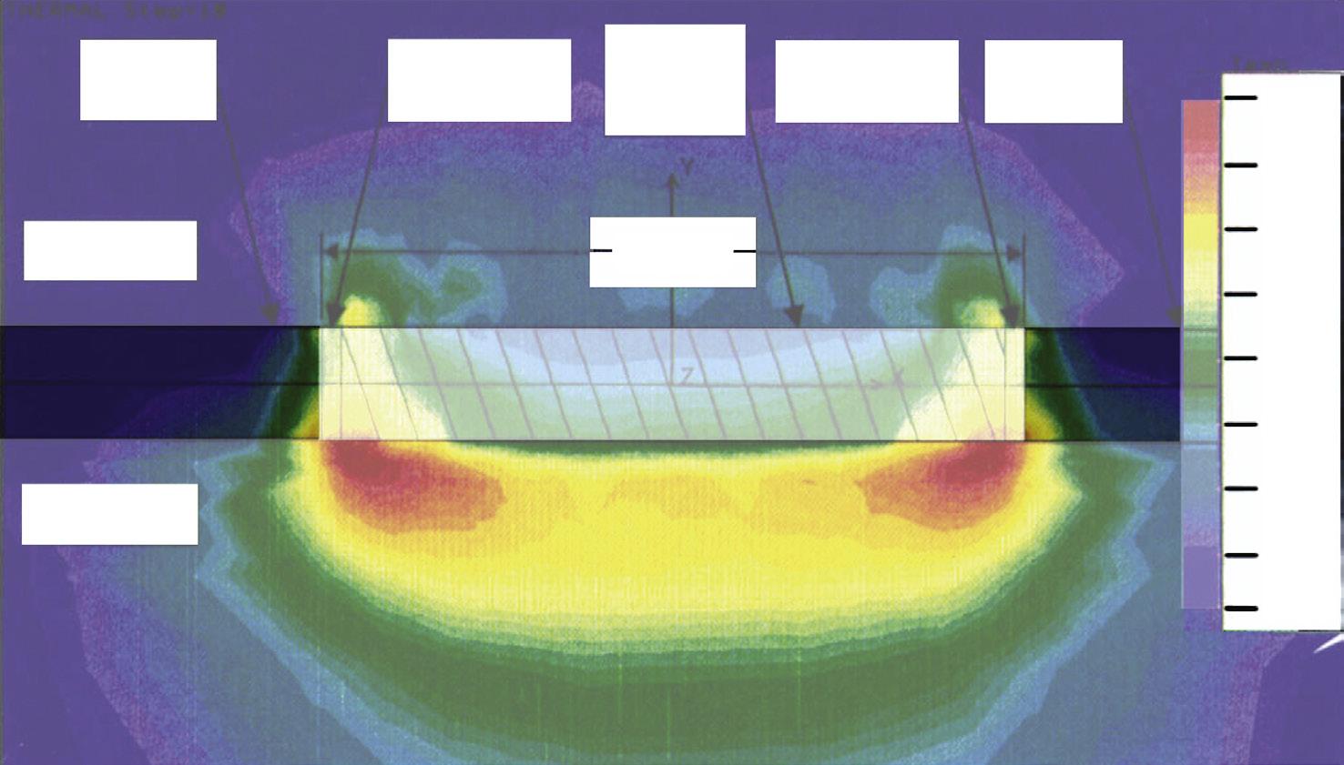
Resistive heat production within the tissue is proportional to the RF power density, and that, in turn, is proportional to the square of the current density (Table 1.1). When RF energy is delivered in a unipolar fashion, the current distributes radially from the source. The current density decreases in proportion to the square of the distance from the RF electrode source. Thus direct resistive heating of the tissue decreases proportionally with the distance from the electrode to the fourth power (Fig. 1.1). As a result, only the narrow rim of tissue in close contact with the catheter electrode (2–3 mm) is heated directly. All heating of deeper tissue layers occurs passively through heat conduction.4 If higher power levels are used, both the depth of direct resistive heating and the volume and radius of the virtual heat source will increase.
Thermal Conduction
Most of the tissue heating resulting in the formation of lesion during RF catheter ablation occurs as a result of thermal conduction from the direct resistive heat source. Transfer of heat through tissue follows basic thermodynamic principles and is represented by the bioheat transfer equation.5 Change in tissue temperature with increasing distance from the heat source is called the radial temperature gradient At the onset of RF energy delivery, the temperature is very high at the source of heating and falls off rapidly over a short distance (see Fig. 1.1 and Videos 1.1 and 1.2 on Expert Consult). As time progresses, more thermal energy is transferred to deeper tissue layers by thermal conduction. The tissue temperature at any given distance from the heat source increases in a monoexponential fashion over time. Sites close to
TABLE 1.1 Equations Describing Biophysics of
Radiofrequency Ablation
V = I R Ohm’s Law
V – voltage
I – current
R – resistance
Power = V I (cos ά) Cos ά – phase shift between voltage (V) and current (I) in alternating current
Current Density = I/4 π r2 I – total electrode current
SAR =│E│2
σ/r = │J│2 │ σ r
r – distance from electrode center
SAR – heat production per unit of volume of tissue
σ – tissue electrical conductivity
r – tissue mass density
E – electrical field strength
J – electrical current density
J = I │ π r2 I – current, r – distance of spherical boundary from electrode center in conductive medium
SAR at boundary α I2/ r4
T (t) = Tss + (Tinitial – Tss)
e-t/τ
r/ri = (to – T) / (t – T)
Monoexponential relationship between tissue temperature (T) and duration of radiofrequency energy delivery (t).
Tinitial – starting tissue temperature
Tss – tissue temperature at steady state
τ – time constant
Relationship between tissue temperature and distance from heat source in ideal system.
r – distance from center of heat source, ri – radius of heat source, to – temperature at electrode tissue interface, T – basal tissue temperature, t – temperature at radius r.
the heat source have a rapid rise in temperature (a short half-time of temperature rise), whereas sites remote from the source heat up more slowly.6 Eventually, the entire electrode–tissue system reaches steady state, meaning that the amount of energy entering the tissue at the thermal source equals the amount of energy that is being dissipated at the tissue margins beyond the lesion border. At steady state, the radial temperature gradient becomes constant. If RF power delivery is interrupted before steady state is achieved, tissue temperature will continue to rise in deeper tissue planes as a result of thermal conduction from more superficial layers heated to higher temperatures. In one study, the duration of continued rise in temperature at the lesion border zone after a 10-second RF energy delivery was 6 seconds. The temperature increased by an additional 3.4°C and remained above the temperature recorded at the termination of energy delivery for more than 18 seconds. This phenomenon, termed thermal latency, has important clinical implications because active ablation, with beneficial or adverse effects, will continue for a certain period despite cessation of RF current flow.7 Because the mechanism of tissue injury in response to RF ablation is thermal, the final peak temperature at the border zone of the ablative lesion should be relatively constant. Experimental studies predict this temperature with hyperthermic ablation to be approximately 50°C,3 although alternative methods propose that this critical temperature may be higher.8 This is called the isotherm of irreversible tissue injury The point at which the radial temperature gradient crosses the 50°C isothermal line defines the lesion radius in that dimension. One may predict the three-dimensional temperature gradients with thermodynamic modeling and finite-element analysis and by doing so can
predict the anticipated lesion dimensions and geometry with the 50°C isotherm. In an idealized medium of uniform thermal conduction without convective heat loss, a number of relationships can be defined using boundary conditions when a steady-state radial temperature gradient is achieved. In this theoretical model, it is predicted that radial temperature gradient is inversely proportional to the distance from the heat source. The 50°C isotherm boundary (lesion radius) increases in distance from the source in direct proportion to the temperature at that source. It was predicted, then demonstrated experimentally, that in the absence of significant heat loss because of convective cooling, the lesion depth and diameter are best predicted by the electrode–tissue interface temperature.4 In the clinical setting, however, the opposing effects of convective cooling by circulating blood flow diminish the value of electrode-tip temperature monitoring to assess lesion size.
The idealized thermodynamic model of catheter ablation by tissue heating predicted, then demonstrated, that the radius of the lesion is directly proportional to the radius of the heat source (Fig. 1.2).9 When one considers the virtual heat source radius as the shell of direct resistive heating in tissue contiguous to the electrode, it is not surprising that larger electrode diameter, length, and contact area all result in a larger source radius and larger lesion size, and that this may result in enhanced procedural success. Higher power delivery not only increases the source temperature but also increases the radius of directly heated tissue (i.e., the heat source), thereby increasing lesion size in two ways. These theoretical means of increasing the efficacy of RF catheter ablation have been realized in the clinical setting with large-tip catheters and cooled-tip catheters.10–12
The relationship of ablation catheter distance from the ablation target to the power requirements for clinical effect was tested in a Langendorff-perfused canine heart preparation. Catheter ablation of the right bundle branch was attempted at varying distances, and during the delivery of RF energy, power was increased in a stepwise fashion. The RF power required to block the right bundle branch conduction increased exponentially with increasing distance from the catheter. At a distance of 4 mm, most RF energy deliveries reached the threshold of impedance rise before block was achieved. When pulsatile flow was streamed past the ablation electrode, the power requirements to cause block increased fourfold.13 Thus the efficiency of heating diminished with cooling from circulating blood, and small increases in distances from the ablation target corresponded with large increases in ablation power requirements, emphasizing the importance of optimal targeting for successful catheter ablation.
Sudden Impedance Rise
While the peak tissue temperature increases during ablation, the temperature at greater tissue depths also increases. A very high source temperature therefore should theoretically yield a very deep 50°C isotherm temperature and, in turn, very large ablative lesions. Unfortunately, this process is prevented in the biological setting because of the formation of coagulum and char at the electrode–tissue interface when temperatures exceed 100°C. At 100°C, blood literally begins to boil. This can be observed in the clinical setting with generation of showers of microbubbles if tissue heating is excessive.14 As the blood and tissue in contact with the electrode catheter desiccate, the residue of denatured proteins adheres to the electrode surface. These substances are electrically insulating and result in a smaller electrode surface area available for electrical conduction. In turn, the same magnitude of power is concentrated over a smaller surface area, and the power density increases. With higher power density, the heat production increases, and more coagulum is formed. Thus in a positive-feedback fashion, the electrode becomes completely encased in coagulum within 1 to 2 seconds. In a study testing ablation with a 2-mm-tip electrode in vitro and in vivo, a
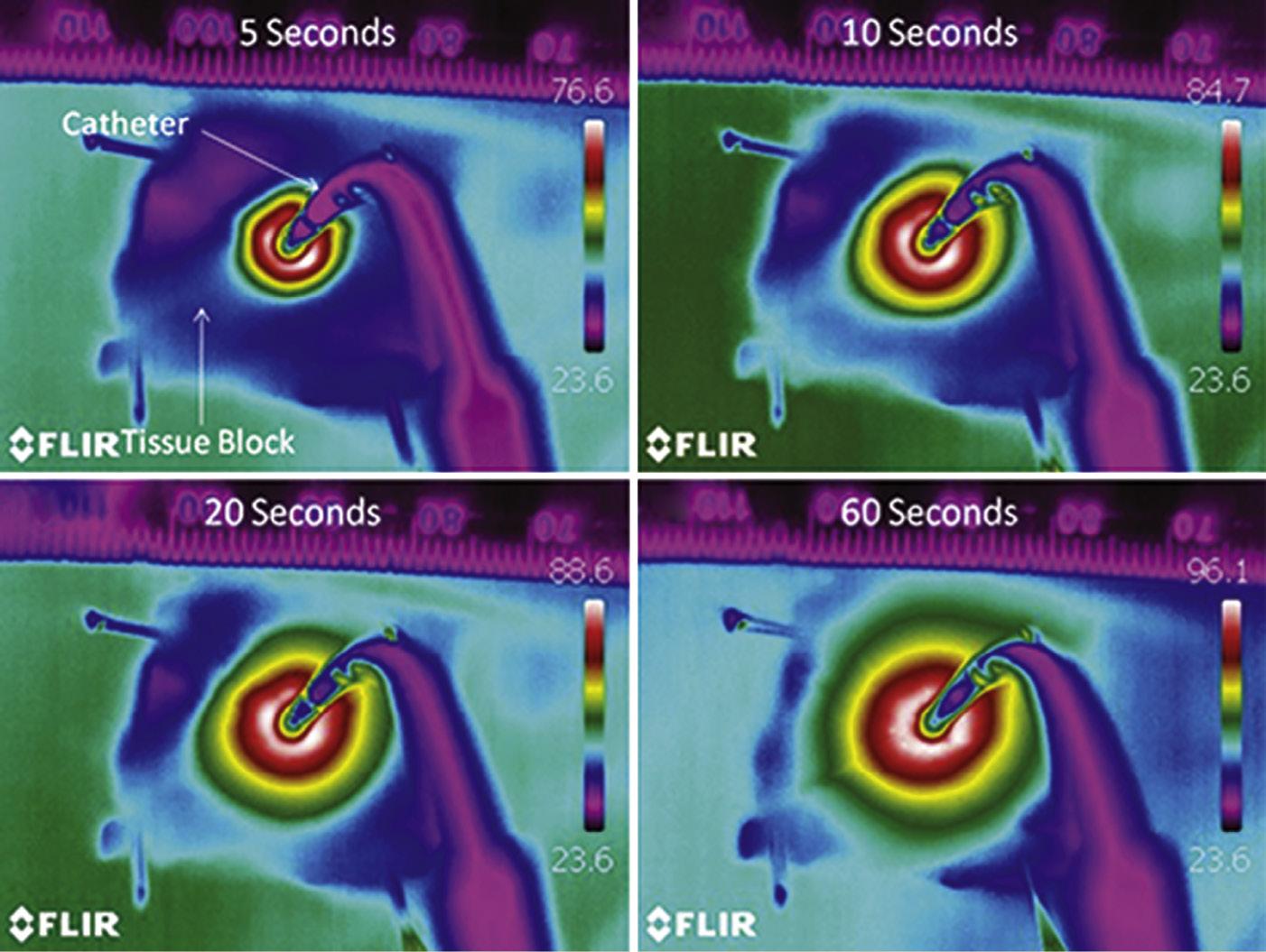
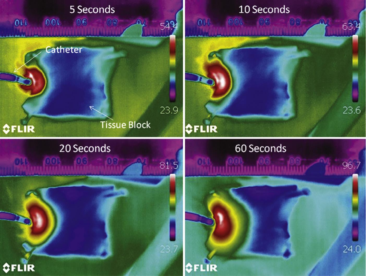
Fig. 1.1 Infrared thermal imaging of tissue heating during radiofrequency ablation with a closed irrigation catheter. Power is delivered at 30 W to blocks of porcine myocardium in a tissue bath. The surface of the tissue is just above the fluid level to permit thermal imaging of tissue and not the fluid. Temperature scale (right) and a millimeter scale (top) are shown in each panel. A, When viewed from the surface, there is radial heating of the tissue from the electrode. B, Tissue heating visualized in cross section. The electrode is partially submerged in the fluid bath and perpendicular to the upper edge of the tissue. In both cases, very high tissue temperatures (>96°C) are achieved at 60 seconds because of the absence of fluid flow over the tissue surface.
measured temperature of at least 100°C correlated closely with a sudden rise in electrical impedance (Fig. 1.3).15 All modern RF energy ablation systems have an automatic energy cutoff if a rapid rise in electrical impedance is observed. Some experimenters have described accumulation of soft thrombus when temperatures exceed 80°C.16 This is likely caused by blood protein denaturation and accumulation, but fortunately appears to be more of a laboratory phenomenon than one
observed in the clinical setting. When high temperatures and sudden rises in electrical impedance are observed, there is concern about the accumulation of char and coagulum, with the subsequent risk of char embolism. Reports of asymptomatic cerebral embolic lesions on diffusion weighted imaging–magnetic resonance imaging (MRI) images after atrial fibrillation ablation highlight the clinical significance of microembolism.17,18 Anticoagulation and antiplatelet therapies have
Fig. 1.2 A, Radial temperature gradients measured during in vitro catheter ablation with source temperatures varying from 50°C to 80°C. The tissue temperature falls in an inverse proportion to distance. The horizontal line represents the 50°C isothermal line. The point at which the radial temperature gradient crosses the 50°C isotherm determines the boundary of the lesion. A higher source temperature results in a greater lesion depth. B, Lesion depth and diameter are compared with the electrode radius in temperature-feedback power-controlled radiofrequency ablation. A larger diameter ablation electrode results in higher power delivery and a proportional increase in lesion dimension.
been proposed as preventative measures,19 but meticulous sheath management avoidance of excessive heating at the electrode–tissue interface remains the best strategy to avoid this risk.20
Convective Cooling
The major thermodynamic factor opposing the transfer of thermal energy to tissue is convective cooling. Convection is the process in which heat is rapidly distributed through a medium by active mixing of that medium. In the case of RF catheter ablation, the heat is produced by resistive heating and transferred to deeper layers by thermal conduction. Simultaneously, the heat is conducted back into the circulating blood pool and metal electrode tip. Because the blood is moving rapidly past the electrode and over the endocardial surface, and because water (the main constituent of blood) has a high heat capacity, a large amount of the heat produced at the site of ablation can be carried away by the blood. Convective cooling is such an important factor that it dominates the thermodynamics of catheter ablation.21 Efficiency of energy coupling to the tissue can be as low as 10%, depending on electrode size, catheter stability, and position relative to intracavitary blood flow.22 Unstable, sliding catheter contact results in significant tip cooling and decreased efficiency of tissue heating.23 This is most often observed with ablation along the tricuspid or mitral valve annuli, or on the left pulmonary vein ridge.
Paradoxically, the convective cooling phenomenon has been used to increase lesion size. As noted earlier, maximal power delivery during RF ablation is limited by boiling of blood and coagulum formation at the
Fig. 1.3 The association of measured electrode-tip temperature and sudden rise in electrical impedance is shown in this study of radiofrequency catheter ablation with a 2-mm-tip ablation electrode in vitro (blue circles) and in vivo (yellow squares). The peak temperature recorded at the electrode–tissue interface is shown. Almost all ablations without a sudden rise in electrical impedance had a peak temperature of 100°C or less, whereas all but one ablation manifesting a sudden rise in electrical impedance had peak temperatures of 100°C or more. (From Haines DE, Verow AF. Observations on electrode–tissue interface temperature and effect on electrical impedance during radiofrequency ablation of ventricular myocardium. Circulation. 1990;82:1034-1038. With permission.)
electrode tip. However, if the tip is cooled, a higher magnitude of power may be delivered without a sudden rise in electrical impedance. The higher magnitude of power increases the depth of direct resistive heating and, in turn, increases the radius of the effective heat source. In addition, higher temperatures are achieved 3 to 4 mm below the surface, and the entire radial temperature curve is shifted to a higher temperature over greater tissue depths. The result is a greater 50°C isotherm radius and a greater depth and diameter of the lesion. Nakagawa and coworkers demonstrated this phenomenon in a blood-superfused exposed thigh muscle preparation. In this study, intramural tissue temperatures 3.5 mm from the surface averaged 95°C with an irrigated-tip catheter despite a mean electrode–tissue interface temperature of 69°C. Lesion depths were 9.9 mm compared with 6.1 mm in a comparison group of temperature-feedback power control delivery with no electrode irrigation (Fig. 1.4). An important finding of this study was that six of 75 lesions had a sudden rise in electrical impedance associated with an audible pop. In these cases, the intramural temperature exceeded 100°C, resulting in sudden steam formation and a steam pop. The clinical concern about pop lesions is that sudden venting of steam to the endocardial or epicardial surface (or both) can potentially cause perforation and tamponade.24 Monitoring intramyocardial steam formation with near field ultrasound to terminate energy delivery before steam venting can occur has been proposed as a method of mitigating this risk.25
The observation of increasing lesion size with ablation-tip cooling holds true only when the ablation is not power limited. If the level of power used is insufficient to overcome the heat lost by convection, the resulting tissue heating may be inadequate. In this case, convective cooling will dissipate a greater proportion of energy, and less of the available RF energy will be converted into tissue heat. The resulting lesion may be smaller than it would be if there was no convective cooling. As power is increased to a higher level, more energy will be converted into tissue heat, which results in larger lesions. If power is unlimited and temperature-feedback power control delivery is used, greater magnitudes of convective cooling will allow for higher power
Fig. 1.4 Current, voltage, and temperatures measured during radiofrequency catheter ablation with a perfused-tip electrode catheter in a canine exposed thigh muscle preparation are shown. Temperatures were recorded within the electrode, at the electrode–tissue interface, and within the muscle below the ablation catheter at depths of 3.5 and 7 mm. Because the electrode–tissue interface is actively cooled, high current and high voltage levels can be used. This results in an increased depth of direct resistive heating and superheating of the tissue below the surface of ablation. The peak temperature in this example at a depth of 3.5 mm was 102°C, and at 7 mm was 67°C, indicating that the 50°C isotherm defining the lesion border was significantly deeper than 7 mm. (From Nakagawa H, Yamanashi WS, Pitha JV, et al. Comparison of in vivo tissue temperature profile and lesion geometry for radiofrequency ablation with a saline-irrigated electrode versus temperature control in a canine thigh muscle preparation. Circulation. 1995;91:22642273. With permission.)
levels and very large lesions. Thus paradoxically in this situation, lesion size may be inversely related to the electrode–tissue interface temperature if the ablation is not power limited.26 However, if power level is fixed (most commercial RF generators limit power delivery to 50 W for use with standard catheters), lesion size increases in proportion to the electrode–tissue interface temperature even in the setting of significant convective cooling (Fig. 1.5).27
The magnitude of convective cooling that is achieved with irrigated catheters is relatively small compared to the circulating blood pool, but this cooling can occur in the highly localized region at the electrode–tissue interface. The main effect of irrigation is to prevent excessive surface heating, boiling, char formation, and impedance rise despite use of high power amplitude. High versus low irrigation rates do not affect the ablation lesion depth if power is constant. However, high irrigation rates do result in smaller lesion diameter on the endocardial surface. Thus if thin-walled tissues are being ablated (e.g., posterior left atrial wall), then using high irrigation flow rate will not alter risk of injury to collateral structures, but it will decrease ablation efficacy by reducing superficial lesion size.28
Electrode-tip cooling can be achieved passively or actively. Passive tip cooling occurs when the circulating blood flow cools the mass of the ablation electrode and cools the electrode–tissue interface. This can be enhanced by using a large ablation electrode29 or by using an electrode material with high thermal conductivity, such as gold30,31 or diamond.32
Active tip cooling can be realized with a closed or open perfused-tip system. One design recirculates the saline through a return port, and the opposing design infuses the saline through weep holes in the electrode into the bloodstream. Both designs are effective and result in larger lesions and greater procedure efficacy than standard RF catheter ablation,33,34 although open irrigation is preferred because operators observe less tendency for formation of thrombus compared to closed systems. Present-day electrode geometries vary considerably, tip irrigation is used routinely, and standard
Group 1
Group 2
Group 1
Group 2
Fig. 1.5 Temperatures measured at the tip of the electrode during experimental radiofrequency ablation and power are compared with the resulting lesion volume in this study. A maximal power of 70 W was used. If lesion creation was not power limited (group 1), the lesion volume was a function of the delivered power. However, if lesion production was limited by the 70-W available power maximum (group 2), the temperature measured at the electrode tip correlated with lesion size. (From Petersen HH, Chen X, Pietersen A, et al. Lesion dimensions during temperature-controlled radiofrequency catheter ablation of left ventricular porcine myocardium: impact of ablation site, electrode size, and convective cooling. Circulation. 1999;99:319-325. With permission.)
catheters embed their thermocouples within the mass of the electrode tip, thereby providing evidence of tip cooling with irrigation, but no ability to detect tissue heating. A new generation of temperature sensing catheters offers to improve this scenario. Six miniature thermocouple sensors have been positioned immediately below the electrode surface and distributed around the tip of a force sensing catheter, significantly improving ability to predict catheter orientation and lesion depth.35
Because the peak tissue temperature is shifted from the endocardial surface to deeper intramyocardial layers, there is a risk of excessive intramural heating and pop lesions. The challenge for the clinician lies with the fact that with varying degrees of convective cooling, there is no reliable method for monitoring whether tissue heating is inadequate, optimal, or excessive. Cooling at the electrode–tissue interface limits the value of temperature monitoring to prevent excess power delivery and steam pops. New technologies such as MR thermography36 or near-field ultrasound-guided ablation37 may allow the operator to visualize lesion formation real time during energy delivery and more precisely adjust power and magnitude of convective cooling to optimize lesion formation. Catheter ablation in the pericardial space is a unique condition. Because there is no circulating blood, there is no convective cooling whatsoever. Ablations performed with conventional RF catheters yield very small lesions. Perfused-electrode catheters are usually used in this setting to provide some surface cooling and allow ablation at higher powers. In particular, linear ablation tools designed for surgical ablation of atrial fibrillation from the epicardial surface require cooling to achieve transmural atrial lesions.38,39
Catheter Contact Force and Orientation
It has long been appreciated that electrode–tissue contact force is an important factor in successful RF energy lesion formation. With increasing force, greater proportion of the electrode surface area is in contact with the tissue, and there is more efficient energy coupling. In addition, with thin-walled tissues, the endocardial surface is slightly depressed with increasing contact force and the tissue is somewhat thinner at the contact point. This increases the likelihood that the RF lesion will be transmural. In recent years, force-sensing catheters have been developed that use either fiber–optic or piezoelectric components attached to a flexible catheter tip that can accurately measure the force applied to the tip electrode of the catheter. Lesions created with higher contact force (>20 g–force) were larger and required lower powers than lesions created with lower contact force.40 Contact force catheters have also been used to determine that the average force needed for atrial perforation in a swine model was 175 g–force (range, 77–376 g–force).41 Force-sensing catheters have been tested extensively in the clinical setting. Successful catheter ablation has been associated with higher contact forces, and higher forces that have been applied over longer durations (the force-time integral).42,43 However, there is a concern that using greater contact force and longer durations during ablation of the posterior left atrial wall may have contributed to a higher risk of atrial-esophageal fistula.44
Catheter orientation will affect lesion size and geometry. Perpendicular catheter orientation results in less electrode surface area in contact with the tissue and more surface area in contact with the circulating blood pool. Parallel catheter orientation provides more electrode–tissue contact. With unrestricted power delivery, the parallel orientation should produce the larger lesion. In perfused-tip catheters, parallel orientation also results in more active tissue cooling and smaller lesion sizes than a perpendicular orientation.45 The resultant interplay among active cooling, passive cooling, and power availability or limitation determines whether the lesions will be larger or smaller in these varying conditions. If perfused-tip catheters are positioned in a parallel orientation with greater tissue cooling, the lesions are smaller in vitro because of diminished efficiency of energy delivery. The effects of catheter orientation are less important with 4- or 5-mm-tip catheters but become more dominant when 8- or 10-mm tips are used.
Electrical Current Distribution
Catheter ablation depends on the passage of RF electrical current through tissues. As tissue contact improves, the impedance of the RF electrical circuit decreases since there is lower impedance at an electrode–tissue interface than at an electrode-blood interface.46,47 A strong correlation is observed between effective lesion formation and rate of impedance fall during energy delivery because hotter tissue has a lower impedance than cooler tissue. When electrode–tissue interface temperature monitoring is unreliable because of high-magnitude convective cooling, an observed impedance drop is a useful sign that tissue heating is occurring. An initial impedance drop greater than 10 Ω is an indicator of good catheter contact as assessed by force-sensing catheters.48 With the progressive fall in impedance during ablation, the delivered current increases along with tissue heating. If no impedance drop is observed, catheter repositioning is warranted.49,50
The magnitude of tissue heating is determined by the current density; in turn, the distribution of RF field around the electrodes in unipolar, bipolar, or phased RF energy delivery will determine the distribution of tissue heating. If energy is delivered in a unipolar fashion in an isotropic medium from a spherical electrode to an indifferent electrode with infinite surface area, current density around the electrode
should be entirely uniform. As geometries and tissue properties change, heating becomes nonuniform. Standard 4- or 5-mm electrode tips are small enough so that heating around the tip is fairly evenly distributed, even with varying tip contact angle to the tissue. One study of ablation with a nonirrigated catheter equipped with multiple surface mounted thermistors showed that the single thermistor located at the tip of the 4-mm electrode accurately represented the maximum recorded temperature from all thermistors 96% of the time, and failed to predict sudden impedance rise in only one of 17 of cases where that occurred.51
It has been proposed that power distribution during RF ablation can be modulated by altering the tonicity of the irrigation solution. RF power is dissipated throughout the entire circuit, including the catheter, catheter-tissue interface, myocardium, tissue between the heart and the dispersive electrode, skin-dispersive electrode interface, wire conductors, and RF generator electronics. If more power is channeled through this tissue rather than directly into the blood pool, lesion size should be larger. This was accomplished by Nguyen et al. by replacing 0.9% saline with 0.45% saline as the irrigation liquid.52 The hypotonic solution produced a higher impedance environment in the blood pool around the electrode and resulted in a greater proportion of RF current passing directly into the tissue. This resulted in 60% larger lesion volumes in experimental testing in vivo. This may be a useful strategy for ablation of deep intramyocardial targets.52
Fat is distributed widely on the epicardium of the heart and reduces both electrical and thermal conductivity. Epicardial ablation over fat will result in minimal ablation of the underlying myocardium. Conversely, ablation of tissue insulated by fat outside of the ablation target will produce an insulating effect, with higher temperatures for longer durations after cessation of energy delivery.53
Bipolar ablation uses a second active ablation electrode rather than a dispersive electrode, and the current flows between the two electrodes with heating occurring at both electrodes. For symmetrical ablation at both active electrodes of the bipole, it is important that the electrodes are similar in size, because heating is proportional to power density, which is a function of both power and electrode surface area. With bipolar delivery, the electrical field is densest between the electrodes, and so some additional volume heating may be achieved in the intervening tissues if the interelectrode spacing is close. If energy is delivered in a bipolar mode between contiguous electrodes on a catheter positioned parallel to the tissue, there may be improved lesion formation between the electrodes, but lesion depth will be less than that achieved with multipolar ablation in the unipolar mode. It is possible to deliver both unipolar RF energy to multiple poles and bipolar energy between poles simultaneously by altering the phase of RF signal between the two electrodes, or by using a duty cycle to alternate from unipolar to bipolar. The blended unipolar–bipolar RF lesion from contiguous electrodes will be deeper than a pure bipolar ablation but more continuous than a pure unipolar ablation.54
Dispersive Electrode
For unipolar RF energy delivery, the power dissipated in the complete electrical circuit is proportional to the impedance and voltage drop for each component of the series circuit. The impedances of the ablation system generator electronics and transmission lines are low relative to the impedance of the tissue interposed between the catheter and the dispersive electrode, so most of the energy dissipation occurs within the body. The site of greatest impedance, voltage drop, and power dissipation is at the electrode–tissue interface. However, most power is consumed with electrical conduction through the body and blood pool and into the dispersive electrode. In fact, only a fraction of the total delivered power is actually deposited in the myocardial tissue (Fig. 1.6). The return path of current to the indifferent electrode will certainly affect the current density close to that indifferent electrode, but its placement
Fig. 1.6 Circuit diagrams for radiofrequency (RF) ablation. A, From the RF generator, the cables and catheter present minimal resistance. The myocardial tissue and blood pool represent resistance circuits in parallel from the distal electrode. The return path from the ablation electrode to the generator comprises the patient’s body and dispersive electrode in series. B, Hypothetical resistances for RF ablation circuit path. The resistance of the blood pool is about half than that of the myocardial tissue. In this situation, for 50 W of energy delivered to the catheter, only 5 W is deposited in the myocardial tissue because of shunting of current through the lower resistance blood pool and power loss in the return path. C, Effect of adding a second dispersive skin electrode to the circuit. Assuming that the impedance of each dispersive electrode is 45 Ω and the generator voltage is constant, the total ablation circuit impedance is decreased by 12%. This allows for greater current delivery through the circuit and a proportional increase in power delivered to the tissue.
anterior versus posterior, and high versus low on the torso, has only a small effect on the distribution of RF current field lines within millimeters of the electrode. Therefore lesion geometry should not be affected greatly by dispersive electrode placement. However, the proportion of RF energy contributing to lesion formation will be reduced if a greater proportion of that energy is dissipated in a long return pathway to the dispersive electrode. When the ablation is power limited, it is advantageous to minimize the proportion of energy that is dissipated along the current pathway at sites other than the electrode–tissue interface to achieve the greatest magnitude of tissue heating and the largest lesion. In an experiment that tested placement of the dispersive electrode directly
opposite to the ablation electrode versus at a more remote site, lesion depth increased by 26% with optimal placement.55 Vigorous skin preparation to minimize impedance at the skin interface with the dispersive electrode, closer placement of the dispersive electrode to the heart, and use of multiple dispersive electrodes to increase skin contact area will all increase tissue heating in a power-limited energy delivery. Nath and associates reported that in the setting of system impedance higher than 100 Ω, adding a second dispersive electrode increased the peak electrode-tip temperature during clinical catheter ablation (Fig. 1.7).56 The dispersive electrode has a large surface area relative to that of the ablation electrode so that the power density at the skin surface is
Cables and catheter Body and skin electrode
Single dispersive electrode Double dispersive electrode
Fig. 1.7 Impedance, voltage, current, and catheter-tip temperature readings during radiofrequency catheter ablation in a subset of patients with a baseline system impedance of more than 100 Ω. Ablations using a single dispersive electrode were compared with those using a double dispersive electrode. A lower system impedance was observed with addition of the second dispersive patch. This resulted in a greater current delivery and higher temperatures measured at the electrode–tissue interface.
(From Nath S, DiMarco JP, Gallop RG, et al. Effects of dispersive electrode position and surface area on electrical parameters and temperature during radiofrequency catheter ablation. Am J Cardiol. 1996;77:765-767. With permission.)
uniformly low. As a consequence, there is minimal skin heating during RF catheter ablation. However, if high powers are used, and/or the contact surface area of the dispersive electrode is reduced (e.g., a partially detached electrode), excess power density with resultant skin heating can occur. Case reports of serious skin burns from the dispersive electrode emphasize this point.57 Sequential activation of two ground pads to allow intermittent cooling of each pad results in lower skin temperatures during high-power delivery.58
Edge Effect
Electrical field lines are not entirely uniform around the tip of a unipolar ablation electrode. The distribution of field lines from an electrode source is affected by changes in electrode geometry. At points of geometric transition, the field lines become more concentrated. This so-called edge effect can result in significant nonuniformity of heating around electrodes. The less symmetrical the electrode design (such as the ones found with long electrodes), the greater the degree of nonuniform heating. McRury and coworkers tested ablation with 12.5-mm length electrodes and found that a centrally placed temperature sensor significantly underestimated the peak electrode–tissue interface temperature.59 Finite-element analysis demonstrated a concentration of electrical current at each edge of the electrode (Fig. 1.8). When dual thermocouples were placed on the edge of the electrode, the risk of coagulum formation and impedance rise was significantly reduced during ablation testing in vivo.
TISSUE PATHOLOGY AND PATHOPHYSIOLOGIC RESPONSE TO RADIOFREQUENCY ABLATION
Gross Pathology and Histopathology of the Ablative Lesion
The endocardial surface in contact with the ablation catheter shows pallor and sometimes a small depression caused by volume loss of the
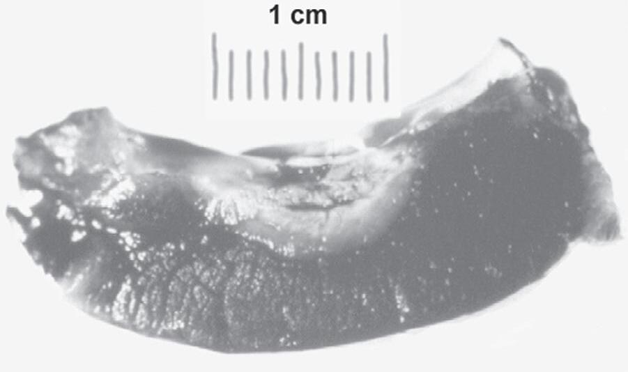
acute lesion. If excessive power has been applied, there may be visible coagulum or char adherent to the ablation site. On sectioning the acute lesion produced by RF energy, a central zone of pallor and tissue desiccation characterizes its gross appearance. There is volume loss, and the lesion frequently has a teardrop shape with a narrower lesion width immediately subendocardially and a wider width 2 to 3 mm below the endocardial surface. This is because of surface convective cooling by the endocardial blood flow. Immediately outside the pale central zone is a band of hemorrhagic tissue. Beyond that border, the tissue appears relatively normal. The acute lesion border, as assessed by vital staining, correlates with the border between the hemorrhagic and normal tissue (Fig. 1.9). The histologic appearance of the lesion is consistent with coagulation necrosis. There are contraction bands in the sarcomeres, nuclear pyknosis, and basophilic stippling consistent with intracellular calcium overload.60
The temperature at the border zone of an acute hyperthermic lesion assessed by vital staining with nitro blue tetrazolium was observed to be 52°C to 55°C in one study,3 and 60°C in another.61 However, it is likely that the actual isotherm of irreversible thermal injury occurs at a lower temperature boundary outside the lesion boundary, and that it cannot be identified acutely. Coagulation necrosis is a manifestation of thermal inactivation of the contractile and cytoskeletal proteins in the cell. Changes in the appearance of vital stains are caused by loss of enzyme activity, as is the case with nitro blue tetrazolium or triphenyl tetrazolium chloride staining and dehydrogenase activity.62 Therefore the acute assessment of the lesion border represents the border of thermal inactivation of various proteins, but the ultimate viability of the cell may depend on the integrity of more thermally sensitive organelles such as the plasma membrane (see later discussion). In the clinical setting, recorded temperature does correlate with response to ablation. In patients with manifest Wolff–Parkinson–White syndrome, the reversible accessory pathway conduction block with a nonirrigated catheter was observed at a mean electrode temperature of 50°C ± 8°C, whereas the permanent block occurred at a temperature of 62°C ± 15°C.63 In a study of electrode-tip temperature monitoring during atrioventricular junctional ablation, an accelerated junctional rhythm was observed at a mean temperature of 51°C ± 4°C. Permanent complete heart block was observed at ablation temperatures of 60°C ± 7°C.64 Because the targeted tissue was likely millimeters below the endocardial surface, the temperatures recorded by the catheter were expected to be higher than those achieved intramurally at the critical site of ablation.
The subacute pathology of the RF lesion is similar to what is observed with other types of injury. Although the appearance of typical coagulation necrosis persists, the lesion border becomes more sharply demarcated with infiltration of mononuclear inflammatory cells. A layer of fibrin adheres to the lesion surface, coating the area of endothelial injury. After 4 to 5 days, the transition zone at the lesion border is lost, and the border between the RF lesion and surrounding tissue becomes sharply demarcated. The changes in the transition zone within the first hours and days after ablation likely account for the phenomena of early arrhythmia recurrence (injury with recovery)65 or delayed cure (progressive injury caused by the secondary inflammatory response).66 The coagulation necrosis in the body of the lesion shows early evidence of fatty infiltration. By 8 weeks after the ablation procedure, the necrotic zone is replaced with fatty tissue, cartilage, and fibrosis and can be surrounded by chronic inflammation.67 The chronic RF ablative lesion evolves into a uniform scar. Like any fibrotic scar, there is significant contraction of the scar with healing. Relatively large and wide acute linear lesions have the final gross appearance of narrow lines of glistening scar when examined 6 months after the ablation procedure.68
Fig. 1.8 Steady-state temperature distribution derived from a finite-element analysis of radiofrequency ablation with a 12-mm-long coil electrode. In this analysis, the electrode temperature at the center of the electrode was maintained at 71°C. The legend of temperatures is shown at the right of the graph and ranges from the physiologic normal (violet = 37°C) to the maximal tissue temperature (red = 161°C) located below the electrode edges. There is a significant gradient of heating between the peak temperatures at the electrode edges and the center of the electrode. UV, Ultraviolet. (From McRury ID, Panescu D, Mitchell MA, Haines DE. Nonuniform heating during radiofrequency catheter ablation with long electrodes: monitoring the edge effect. Circulation. 1997;96:4057-4064. With permission.)
Fig. 1.9 Typical appearance of radiofrequency catheter ablation lesion. There is a small central depression with volume loss, surrounded by an area of pallor, then a hemorrhagic border zone. The specimen has been stained with nitro blue tetrazolium to differentiate viable from nonviable tissue.
The uniformity of the healed lesion accounts for the absence of any proarrhythmic effect of RF catheter ablation, unless multiple lesions with gaps are made. A group of patients who underwent pulmonary vein ablation and had clinical recurrence had full-thickness pulmonary vein antral biopsies at the time of a follow-up surgical maze procedure. Fifty percent of those specimens showed viable myocardium with or without scar on histopathologic analysis, explaining the reestablishment of pulmonary vein conduction after the acute catheter procedure.69
Radiofrequency Lesion Ultrastructure
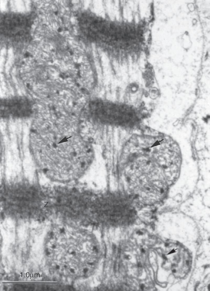
The ultrastructural appearance of the acute RF lesion offers some insight into the mechanism of tissue injury at the lesion border zone. In cases of experimental RF ablation in vivo, ventricular myocardium was examined in a band 3 mm from the edge of the acute pathologic lesion as defined by vital staining (Fig. 1.10). It showed marked disruption in cellular architecture characterized by dissolution of lipid membranes and inactivation of structural proteins. The plasma
Fig. 1.10 Electron micrograph of a myocardial sample 3 mm outside of the border zone of acute injury created by radiofrequency catheter ablation. There is severe disruption of the sarcomere with contracted Z bands, disorganized mitochondria, and basophilic stippling (arrows). Bar scale is 1.0 μm. M, mitochondria; Z, Z-bands. (From Nath S, Redick JA, Whayne JG, Haines DE. Ultrastructural observations in the myocardium beyond the region of acute coagulation necrosis following radiofrequency catheter ablation. J Cardiovasc Electrophysiol. 1994;5:838-845. With permission.)
membranes were severely disrupted or missing. There was extravasation of erythrocytes and complete absence of basement membrane. The mitochondria showed marked distortion of architecture with swollen
and discontinuous cristae membranes. The sarcomeres were extended with loss of myofilament structure or were severely contracted. The T tubules and sarcoplasmic reticulum were either absent or severely disrupted. Gap junctions were also either severely distorted or absent. Thus despite the fact that the tissue examined was outside of the border of the acute pathologic lesion, the changes were profound enough to conclude that some progression of necrosis would occur within this border zone. The band of tissue 3 to 6 mm from the edge of the pathologic lesion was examined, and it showed significant ultrastructural abnormalities, but not as severe as those described closer to the lesion core. Severe abnormalities of the plasma membrane were still present, but gap junctions and mitochondria were mainly intact. The sarcomeres were variable in appearance, with some being relatively normal and some partially contracted. Although ultrastructural disarray was observed in the 3- to 6-mm zone, the myocytes appeared to be viable and would likely recover from the injury.70
Radiofrequency Ablation and Arterial Perfusion
In addition to direct injury to the myocytes, RF-induced hyperthermia has an effect on the myocardial vasculature and the myocardial perfusion. Impairment of the microcirculation likely contributes to lesion formation by an ischemic mechanism. A previous study examined the effects of microvascular perfusion during acute RF lesion formation. In open-chest canine preparations, the left ventricle was imaged with ultrasound from the epicardial surface, and a myocardial echocardiographic contrast agent was injected into the left anterior descending artery during endocardial RF catheter ablation. After ablation, the center of the lesion showed no echo contrast, consistent with severe vascular injury and absence of blood flow to that region. In the border zone of the lesion, a halo effect of retained myocardial contrast was observed. This suggested marked slowing of contrast transit rate through these tissues. The measured contrast transit rate at the boundary of the gross pathologic lesion was 25% ± 12% of the transit rate in normal tissue. In the 3-mm band of myocardium outside of the lesion edge, the contrast transit was 48% ± 27% of normal, and in the band of myocardium 3 to 6 mm outside of the lesion edge, the transit rate was 82% ± 28% of normal (P < .05 for all comparisons). The ultrastructural appearance of the arterioles demonstrated marked disruption of the plasma membrane and basement membrane and extravasation of red blood cells in these regions of impaired myocardial perfusion. Although the relative contribution of microvascular injury and myocardial ischemia to ultimate lesion formation is unknown, it may play a role in lesion extension during the early phases after ablation.71 Clinically, this phenomenon has been demonstrated with late gadolinium enhancement MRI. Regions of nonenhancement, indicating microvascular injury, on the MRI acquired immediately after ablation correlated best with scar observed on MRI 3 months later.72
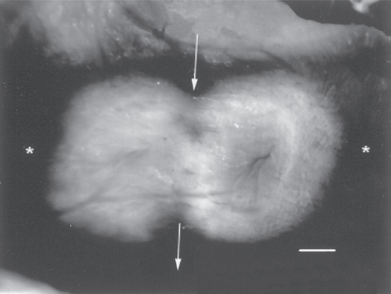
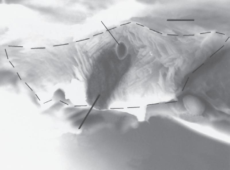
The effect of RF heating on larger arteries is a function of the size of the artery, the arterial flow rate, and the proximity to the RF source. The heat-sink effect of coronary blood flow is protective of the vascular endothelium. With higher power output of new ablation technologies, however, the convective cooling of the arterial flow may be overwhelmed, and there may be an increased risk of vascular injury. In one study, flow rate through a marginal artery (or intramural perfusion cannula) in an in vitro rabbit heart preparation was varied between 0 and 10 mL/minute. A pair of epicardial ablations was produced with epicardial RF energy applications. Even at low flow rates, there was substantial sparing of the artery and the surrounding tissue owing to the heat-sink effect of the arterial flow (Fig. 1.11). However, if 45 W of power was applied along with RF electrode-tip cooling, complete ablation of the tissue contiguous to the intramural perfusion cannula was achieved.73 Although this
60C
Marginal ar ter y 1 mm
Epicardial surface
Preser ved myocardium
Endocardial surface
60C Sequential lesions 12 mL/min Perfusion Rate
Fig. 1.11 Top, Epicardial view of two radiofrequency lesions created during perfusion of a penetrating marginal artery in a rabbit heart. The lesions show central pallor that is apparent after vital staining. The course of the artery is marked. The asterisks mark the line used for perpendicular sectioning of the lesion. Bottom, Cross-section through the middle of lesion perpendicular to marginal artery. The broken lines outline the lesion boundary. A region of myocardial sparing contiguous to the penetrating marginal artery (labeled) is apparent. Electrical conduction was present across this bridge of viable myocardium postablation. (From Fuller IA, Wood MA. Intramural coronary vasculature prevents transmural radiofrequency lesion formation: implications for linear ablation. Circulation. 2003;107:1797-1803. With permission.)
may be a desirable effect in the setting of small perfusing arteries through a region of conduction critical for arrhythmia propagation, it is not desirable if the artery is a large epicardial artery that happens to be contiguous to an ablation site, as is sometimes the case with accessory pathway or slow atrioventricular nodal pathway ablation, ablation in the tricuspid–subeustachian isthmus for atrial flutter, or epicardial ablation. Cases of arterial injury have been reported, particularly with the use of large-tip catheters or tip-cooling technologies that allow for application of high RF powers.74,75 In particular, when high-power ablation is required within the coronary sinus or great cardiac vein, it is prudent to define the course of the arterial anatomy to avoid unwanted arterial thermal injury. Because greater destructive power is possible, the operators need to be aware of using only enough power that is required to achieve complete ablation of the targeted tissue to achieve the goal of arrhythmia ablation.
Collateral Injury From Ablation
The injury to targeted myocardium is usually achieved if an effort is made to optimize electrode–tissue contact. To ensure procedural success, particularly with ablation of more complex substrates such as those found with atrial fibrillation, operators have used a number of large-lesion RF technologies such as cooled-tip, perfused-tip, or largetip catheters. However, with deep lesions sometimes comes unintended
collateral injury to contiguous structures. Therefore an understanding of the anatomic relationships and careful titration of RF energy delivery can avoid adverse consequences of ablation in most cases. A rare but dangerous complication of ablation of the posterior left atrium is esophageal injury, often leading to atrioesophageal fistula or esophageal perforation.76 The esophagus is located immediately contiguous to the atrium in most patients, with a distance from atrial endocardium to esophagus as small as 1.6 mm.77 Hyperthermic injury leads to damage to structural proteins resulting in significant reduction in tensile strength of the esophageal musculature.78 This, coupled with esophageal mucosal injury and ulcer formation, likely leads to ultimate perforation with a high case-fatality rate. Other structures that can be damaged with pulmonary vein isolation procedures are vagal and phrenic nerves.79,80 Although these nerves usually regenerate after several months, permanent palsy can occur. Avoiding injury to these structures while achieving reliable transmural ablation of the myocardium can be challenging.
The standard approach to minimizing heating of extracardiac structures has been to limit power amplitude and carefully monitor heating with multiple modalities (temperature, impedance drop, microbubbles on intracardiac echocardiogram imaging). However, longer durations of delivery that are required when lower powers are used promote heating of deeper tissue layers. Conversely, high power with short ablation duration exploits different rates of temperature rise between superficial and deep tissue layers. Whereas superficial tissues heat very rapidly, deeper tissues require heat conduction from the source of resistive heating and heating is delayed. Thus short duration RF delivery preferentially heats the superficial tissues resulting in successful ablation of the thin-walled myocardium while avoiding excess heating of deeper structures. Reduction in catheter irrigation will also increase superficial lesion width and promote continuity and contiguity of adjacent lesions.28
A complication of ablation of atrial fibrillation that was prevalent when ablation was being performed within the vein was pulmonary vein stenosis.81 If the temperature rise of the venous wall is excessive, irreversible changes in the collagen and elastin of the vein wall will occur. In vitro heating of pulmonary vein rings showed a 53% reduction in circumference and a loss of compliance with hyperthermic exposure at or above 70°C. After exposure to those temperatures, results of the histologic examination showed loss of the typical collagen structure, presumably caused by thermal denaturation of that protein.82 For this reason, most pulmonary vein isolation is now performed outside the vein in the pulmonary vein antrum.
CELLULAR MECHANISMS OF THERMAL INJURY
The therapeutic effect of RF catheter ablation is caused by electrical heating of tissue and thermal injury. The field of hyperthermia is broad, and the effects of long-duration exposures to mild and moderate hyperthermia have been well characterized in the oncology literature. Thermal injury is dependent on both time and temperature. For example, when human bone marrow cells in culture are exposed to a temperature of 42°C, cell survival is 45% at 300 minutes. However, when those cells are heated to 45.5°C, survival at 20 minutes is only 1%.83
Data regarding the effects of brief exposure of myocardium to higher temperatures, as is the case during catheter ablation, are more limited and are reviewed in this section. The central zone of the ablation lesion reaches high temperatures and is simply coagulated. Lower temperatures are reached during the ablation in the border zones of the lesion. The responses of the various cellular components to low and moderate hyperthermia determine the pathophysiologic response to ablation. The thermally sensitive elements that contribute to overall thermal
injury to the myocyte are the plasma membrane with its integrated channel proteins, the nucleus, and the cytoskeleton. Changes in these structures that occur during hyperthermic exposure all contribute to the ultimate demise of the cell.
Plasma Membrane
The plasma membrane is very thermally sensitive. A pure phospholipid bilayer will undergo phase transitions from a relatively solid form to a semiliquid form as temperature rises. Addition of integral proteins and the varying composition of the phospholipids with regard to the saturation of the hydrocarbon side chains affect the degree of membrane fluidity in eukaryotic cells. In one study, cultured mammalian cell membranes were found to have a phase transition at 8°C and a second transition between 22°C and 36°C. No phase changes were seen in the 37°C to 45°C temperature range, but studies have not been performed examining this phenomenon in sarcomeres or at temperatures above 45°C.84 Regarding the function of integral plasma membrane proteins during exposure to heating, both inhibition and accentuation of protein activity have been observed. Stevenson and colleagues reported an increase in intracellular potassium (K+) uptake in cultured Chinese hamster ovary cells upon heating up to 42°C. This was blocked by ouabain, indicating an increased activity of the sodium (Na+), K+–adenosine triphosphatase pump.85 Nath and colleagues examined action potentials in vitro in a superfused guinea pig papillary muscle preparation. In the low hyperthermic range between 38°C and 45°C, there was an increase in the maximal dV/dt (rate of phase 0 depolarization) of the action potential, indicating enhanced Na+ channel kinetics. In the moderate hyperthermia range from 45°C to 50°C, the maximal dV/dt decreased below baseline values. The mechanism of this Na+ channel inhibition was hypothesized to be either partial thermal inactivation of the Na+ channel or, more likely, voltage-dependent Na+ channel inactivation caused by thermally mediated cellular depolarization86 (see later discussion).
Cytoskeleton
The cytoskeleton is composed of structural proteins that form microtubules, microfilaments, and intermediate filaments. The microfilaments coalesce into stress filaments. These include the proteins actin, actinin, and tropomyosin and form the framework to which the contractile elements of the myocyte attach. The cytoskeletal elements may have varying degrees of thermal sensitivity depending on the cell type. For example, in human erythrocytes, the cytoskeleton is composed predominantly of the protein spectrin. Spectrin is thermally inactivated at 50°C. When erythrocytes are exposed to temperatures above 50°C, the erythrocytes rapidly lose their biconcave shape.87 There is no scientific literature reporting the inactivation temperature of the cytoskeletal proteins in myocytes. However, electron micrographs of the border zone of RF lesions show significant disruption in the cellular architecture with loss of the myofilament structure.70 In the central portion of the RF lesion, thermal inactivation of the cytoskeleton contributes to the typical appearance of coagulation necrosis.
Nucleus
The eukaryote nucleus shows evidence of thermal sensitivity in both structure and function. Nuclear membrane vesiculation, condensation of cytoplasmic elements in the perinuclear region, and a decrease in heterochromatin content have been described.88,89 The nucleolus appears to be the most heat-sensitive component of the nucleus. Whether or not hyperthermia induces DNA strand breaks is controversial. One reproducible finding after hyperthermic exposure is the elaboration of nuclear proteins called heat shock proteins (HSPs). Although the function of HSPs has not been entirely elucidated, they
appear to exert a protective effect on the cell. It is hypothesized that 70-kDa HSP facilitates the effective production and folding of proteins and assists their transit among organelles.90
Cellular Electrophysiology
Hyperthermia leads to dramatic changes on the electrophysiology of myocardium. The thermal sensitivity of myocytes has been tested in a variety of experimental systems, and the mechanisms of the electrophysiologic responses to catheter ablation have been elucidated. In one series of in vitro experiments, isolated superfused guinea pig papillary muscles were subjected to 60 seconds of exposure to hyperthermic superfusate at temperatures varying from 38°C to 55°C. Action potentials were recorded continuously during and after the hyperthermic pulse. If resting membrane potential was not restored after return to normothermia, the muscle was discarded, and testing proceeded with a new tissue sample. The resting membrane potential was assessed in unpaced preparations, and the action potential amplitude, duration, dV/dt, and excitability were tested during pacing. The preparations maintained a normal resting membrane potential in the low hyperthermic range (<45°C). In the intermediate hyperthermic range (45°C–50°C), the myocytes showed a temperature-dependent depolarization that was reversible on return to normothermic superfusion. Finally, experiments in the high hyperthermic range (>50°C) typically resulted in irreversible depolarization, contracture, and death (Fig. 1.12). There was a temperature-dependent decrease in action potential amplitude between 37°C and 50°C as well as an inverse linear relationship between temperature and action potential duration. With increasing temperatures, the dV/dt increased, but above 46°C, this measurement began to decrease in preparations that had a greater magnitude of resting membrane potential depolarization.
Spontaneous automaticity was observed in both paced and unpaced preparations at a median temperature of 50°C, compared with a temperature of 44°C in preparations without automaticity. The occurrence of automaticity in unpaced preparations in the setting of hyperthermia-induced depolarization suggested abnormal automaticity as the mechanism. Beginning at temperatures higher than 42°C, loss of excitability to external-field stimulation was seen in some paced preparations and was dependent on the resting membrane potential. Mean resting membrane potential observed with loss of excitability was −44 mV, compared with −82 mV for normal excitability. The superfusate temperature measured during reversible loss of excitability was 43°C to 51°C, but irreversible loss of excitability (cell death) occurred only at temperatures of 50°C or higher.86 Thus it appeared from these experiments that there was increased cationic entry into the hyperthermic cell and that the resultant depolarization led to loss of excitability and cell death.
Calcium Overload and Cellular Injury
In a preparation similar to that described previously, Everett and colleagues further elucidated the specific mechanisms for cellular depolarization and death in response to hyperthermia.91 Isolated superfused guinea pig papillary muscles were attached to a force transducer to assess the pattern of contractility with varying hyperthermic exposure. Consistent with the observations of resting membrane potential changes during heating, there was a reversible increase in tonic resting muscle tension at temperatures between 45°C and 50°C. Above 50°C, the preparations showed evidence of irreversible contracture. This suggested that hyperthermia was causing calcium entry into the cell and ultimately calcium overload. This hypothesis was confirmed with calcium-sensitive Fluo-3-acetoxymethyl (AM)
Fig. 1.12 The magnitude of depolarization of guinea pig papillary muscle cells exposed to 1-minute pulses of hyperthermic perfusate versus perfusate temperature. At temperatures below 45°C, little depolarization is seen. The cells have progressive depolarization between 45°C and 50°C. Above 50°C, few recordings are made because most cells have irreversible contracture and death. (From Nath S, Lynch C III, Whayne JG, Haines DE. Cellular electrophysiological effects of hyperthermia on isolated guinea pig papillary muscle: implications for catheter ablation. Circulation. 1993;88:1826-1831. With permission.)
dye. Hyperthermic increases in papillary muscle tension correlated well with Fluo-3-AM luminescence. To elucidate the mechanism of calcium entry into the cell and its role in cellular injury, preparations were pretreated with either a calcium-channel blocker (cadmium or verapamil) or an inhibitor of the sarcoplasmic reticulum calcium pump (thapsigargin). Preparations heated to 42°C to 44°C showed no significant changes in tension at baseline or with drug treatment. Upon exposure to 48°C, treatment with calcium-channel blockers did not reduce the increase in resting tension or Fluo-3-AM fluorescence, suggesting that the increase in cytosolic calcium was not the consequence of channel-specific calcium entry into the cell. By contrast, thapsigargin treatment led to irreversible papillary muscle contracture at lower temperatures (45°C–50°C) than that observed without this agent. For preparations heated to 48°C, there was a greater increase in muscle tension and Fluo-3-AM fluorescence in the thapsigargin group compared with controls (Fig. 1.13). The authors concluded that hyperthermia results in significant increases in intracellular calcium, probably as a result of nonspecific transmembrane transit through thermally induced sarcolemmal pores. With increased intracellular calcium entry, the sarcoplasmic reticulum acts as a protective buffer against calcium overload, unless this function is blocked with an agent like thapsigargin. In this case, cell contracture and death occur at lower temperatures than expected.91
Conduction Velocity
Simmers and coworkers have examined the effects of hyperthermia on impulse conduction in vitro in a preparation of superfused canine myocardium.92 The average conduction velocity at baseline temperatures of 37°C was 0.35 m/second. When the superfusate temperature was raised, conduction velocity increased to supernormal values, reaching a maximum of 114% of baseline at 42.5°C. At temperatures above 45.4°C, conduction velocity slowed down. Transient conduction block was observed between 49.5°C and 51.5°C, and above 51.7°C permanent block was observed (Fig. 1.14).92 These findings are exactly concordant with the temperature-related changes in cellular electrophysiology described previously. In a related experiment, the
Fig. 1.13 The effects of hyperthermic exposure on calcium entry into cells were tested in isolated superfused guinea pig papillary muscles. A change in resting tension was used as a surrogate measure for cytosolic calcium concentration (A, C), and a change in Fluo-3-acetoxymethyl (Fluo-3-AM) fluorescence was used as a direct measure of free cytosolic calcium (B, D). With exposure to mild hyperthermia (42°C–44°C), little change in calcium levels was observed. With moderate hyperthermia (48°C), however, muscle tension and Fluo3-AM fluorescence increased significantly. This increase was not channel specific because calcium-channel blockade with cadmium or verapamil did not alter this response (A, B). The response was accentuated by thapsigargin (C, D), an agent that blocks calcium reuptake by the sarcoplasmic reticulum. (From Everett TH, Nath S, Lynch C III, et al. Role of calcium in acute hyperthermic myocardial injury. J Cardiovasc Electrophysiol 2001;12:563-569. With permission.)
authors assessed myocardial conduction across a surgically created isthmus during heating with RF energy. The temperatures recorded during transient conduction block (50.7°C ± 3.0°C) and permanent conduction block (58.0°C ± 3.4°C) were nearly identical to those temperature ranges recorded in the experiments performed with hyperthermic perfusate. The authors concluded that the sole effects of RF ablation on the electrophysiologic properties of the myocardium were hyperthermic, and that there was no additional pathophysiologic response that could be attributed to direct effects of passage of electrical current through the tissue.93 It is unknown whether these changes in conduction velocity are solely caused by changes in intracellular ionic concentrations or whether thermal injury to gap junctions may also be implicated.
Fig. 1.14 Conduction velocity of myocardium in superfused canine myocardium in vitro versus the temperature of the superfusate. A mild augmentation of conduction velocity caused by an increase in dV/dt is observed at temperatures up to 45°C. Between 45°C and 50°C, conduction velocity falls, and above 50°C, conduction is blocked. (From Simmers TA, de Bakker JM, Wittkampf FH, Hauer RN. Effects of heating on impulse propagation in superfused canine myocardium. J Am Coll Cardiol. 1995;25:1457-1464. With permission.)
DETERMINANTS OF EFFECTIVE LESION FORMATION
Targeting
The success of catheter ablation is dependent on several factors. The first and foremost factor is optimizing the targeting of the arrhythmogenic substrate. It is intuitive that increasing the size and depth of an
ablative lesion will not improve the ablation success if the site selected for ablation is poor. To optimize site selection, it is necessary to understand the physiology and the anatomy of the arrhythmia in its entirety. The proximity of the electrode to the target will be the most important factor for ablation success.
Tissue Composition
Lesion sizes are decreased in areas of dense scar. In addition, an insulating layer of fat as thin as 2 mm overlying myocardial tissue (as in epicardial ablation) will prevent formation of a lesion with RF energy delivery.94
Power
Lesion size is proportional to power. Any method that will allow for greater power deposition into the tissue will result in more tissue heating and greater depth of thermal injury. In addition to power amplitude, efficiency of power coupling to the tissue (i.e., how much power is converted into tissue heat and how much is “wasted” with convective cooling) will affect ultimate lesion size.
Electrode Temperature
The electrode is passively heated by conduction of heat from the tissue during ablation. Lesion size increases directly with electrode temperature up until the point of coagulum formation and impedance rise. The relationship between lesion size and electrode temperature is confounded by the effects of convective cooling and catheter motion in vivo.
Peak Tissue Temperature
Because of convective cooling, electrode temperature underestimates peak tissue temperature—the real determinant of lesion size. Future sensors such as infrared, microwave, or ultrasound detectors may allow the operator to monitor actual lesion growth. Real-time MRI may guide ablation and provide real-time thermography or imaging of lesion formation.
Electrode Contact Force
Greater electrode–tissue contact force increases lesion size by improving electrical coupling with the tissue, increasing the electrode surface area in contact with the tissue, and reducing the shunting of current to the blood pool. In addition, greater contact force may prevent the electrode from sliding with cardiac motion. The optimal electrode contact force is believed to be 20 to 40 g–force.6,40,42,43 Excessive contact that buries the electrode in the tissue, however, may prevent convective cooling of the electrode and reduce current delivery.
Convective Cooling
Ultimately, lesion size is a function of tissue heating, and tissue heating is a function of the magnitude of RF power that is converted into heat in the tissues. The greater the magnitude of power delivered to the tissue, the greater the lesion size. Convective cooling at the electrode–tissue interface, either active or passive, will allow the operator to increase safely the power amplitude before impedance rises. However, if the ablation is power limited (i.e., the power amplitude is fixed throughout the ablation), greater degrees of convective cooling will draw heat from the tissue surface to create a smaller endocardial lesion diameter, especially at the endocardial surface, and smaller lesion volume. The two factors that affect passive cooling at the electrode–tissue interface are the magnitude of regional blood flow and the stability of the electrode catheter on the tissue surface. Catheter motion over the tissue greatly
increases the loss of heat to the blood pool. Intramyocardial blood flow draws heat from the tissue and not from the electrode, and therefore decreases lesion size.
Electrode Size
When the goal is to maximize lesion size, larger electrodes will always be better than smaller electrodes. Larger electrodes increase the surface area and allow the operator to deliver higher total power without excessive current density at the electrode–tissue interface. Thus coagulum formation with a sudden rise in electrical impedance can be avoided despite high total power delivery. The higher power delivery to the tissue increases the depth of direct volume heating and in turn increases the size of the virtual heat source. This translates directly into a larger lesion. As is the case with cooled electrodes, a large electrode will result in larger lesion formation only if it is accompanied by higher power delivery. If a large electrode is used with lower power, a larger endocardial surface area may be ablated, but the lesion will not be as deep. The RF energy delivery to multiple electrodes may simultaneously produce a large lesion as well, but other issues such as catheter and target geometry may limit energy coupling to the tissue if the electrode–tissue contact is poor.
Duration of Energy Delivery
Tissue temperature follows a monoexponential rise during RF delivery (Table 1.2) until steady state is achieved. The half-time for lesion formation is 5 to 10 seconds for conventional ablation. Therefore lesion formation is assumed to be nearly complete after 45 to 60 seconds (five half-lives). Large lesion technologies such as irrigated-tip catheters have a longer half-time of lesion growth, and in these cases continued energy application for 2 minutes or more may result in some incremental gain in lesion depth. High power, short duration energy delivery will maximize heating in the first 2 to 3 mm and lessen heating in deeper layers.28
Ablation Circuit Impedance
For RF ablation, reducing resistance within the cables and dispersive electrode current path will increase current delivery to the tissue. Using 0.45% saline as an irrigation solution may result in greater current density and heating within the tissue. Placing dispersive electrodes on the trunk rather than on the thigh and using two dispersive electrodes particularly when baseline impedance is high will maximize tissue heating.
Electrode Orientation
For nonirrigated electrodes with unrestricted power, an orientation parallel to the tissue generally results in larger lesions because of a larger electrode area in contact with the tissue and less current shunting to the blood pool. For irrigated electrodes delivering high-power outputs, the parallel electrode orientation may result in smaller lesion sizes because of a greater magnitude of tissue cooling.45
Electrode Geometry
Very long electrodes will provide greater surface area, allow higher power delivery, and usually yield larger lesions. If the electrode is too long, however, efficiency of electrode coupling to the tissue is lost, and lesion size is not increased.27 In addition, power is concentrated at points of geometric transitions (the edge effect), resulting in the possibility of excess heating at the electrode edges and less heating in the middle of the electrode.59
Targeting Close proximity to the target improves likelihood of success even with a small lesion size
Tissue composition Smaller lesion sizes in scar and fat
Power Directly proportional to lesion size
Ablation electrode temperature Underestimates peak tissue temperature because of convective cooling effects
Peak tissue temperature Directly proportional to lesion size
Electrode–tissue contact force Directly proportional to lesion size
Convective cooling
–
Electrode–tissue interface
Active – perfused tip catheter
Passive – large tip, sliding contact
– Intramyocardial arterial flow
With fixed energy delivery, reduces lesion size; with unlimited energy, increases lesion size – Reduces lesion size
Electrode size Directly proportional to lesion size provided unrestricted power
Duration energy delivery Monoexponential relation to lesion size with half-time lesion formation of 5–10 seconds
Ablation circuit impedance Lower body and dispersive (skin) patch resistance increases current delivery. Shunting current through blood decreases impedance but can reduced lesion size.
Electrode orientation For nonirrigated electrode, parallel orientation increases lesion size. For irrigated electrode, perpendicular orientation increases lesion size.
Electrode geometry Effects lesion size and shape by concentrating current density at electrode edges and asymmetries
Electrode material Higher heat conductive materials increase lesion size by electrode cooling
RF characteristics
Pulsed
Phased
Frequency
May increase lesion size by allowing electrode cooling
Increases continuity of linear lesions formed with multi-electrode arrays
Reduced heating efficiency at higher (MHz) frequencies
Electrode Material
Electrode materials with high heat-transfer characteristics (such as gold and diamond) are more effectively cooled by passive blood flow and may allow for greater energy deliveries.30,31
Fig. 1.15 A, Unmodulated and modulated patterns of radiofrequency (RF) power. The modulated waveform is pulsed with periods of oscillating voltage separated by periods of quiescence. B, Unipolar and phased RF deliveries from a multielectrode array. With the unipolar delivery, the oscillating voltages among the electrodes are all in phase, and therefore there is no electrical potential for current flow between electrodes. With phased RF, the oscillations in voltage among contiguous electrodes are out of phase, creating an electrical potential for current to flow between electrodes as well as to the dispersive skin electrode.
Characteristics of Radiofrequency Energy
Very high frequencies of alternating current lead to less efficient tissue heating, and lower frequencies may result in tissue stimulation causing pain and arrhythmias. Pulsed RF current may allow for more electrode cooling than unmodulated RF, and therefore increase power delivery. With multielectrode ablation arrays, phased RF among the electrodes allows for more continuous linear lesions (Fig. 1.15).
CONCLUSION
The RF catheter ablation remains the dominant modality for ablative therapy of arrhythmias. This technology is simple, has a high success rate, and has a low complication rate. Despite the fact that new ablation technologies such as ultrasound, laser, microwave, and cryothermy are being tested and promoted as being easier, safer, or more efficacious, they are unlikely to supplant RF energy as the first choice for ablation of many arrhythmias. An appreciation of the biophysics and pathophysiology of RF energy heating of myocardium during catheter ablation will help the operator to make the proper adjustments to optimize ablation safety and success. A tissue temperature of 50°C needs to be reached to achieve irreversible tissue injury. This likely occurs as a result of sarcolemmal membrane injury
and intracellular calcium overload. The 50°C isotherm determines the boundary of the lesion. Greater lesion size is achieved with higher power delivery and higher intramural tissue temperatures. Energy coupling to tissue is optimized by assuring optimal catheter contact force into tissue. Monitoring surface temperature in noncooled catheters is useful in preventing boiling of blood with coagulum formation and a sudden increase in electrical impedance. Irrigated-tip catheters are effective at allowing the operator to increase power resulting in increased lesion depth. A full understanding of the biophysics of catheter ablation, and the anatomy and physiology of the arrhythmogenic substrate, will allow the operator to select the optimal ablation approach.