CONTRIBUTORS
Alexander W. Aleem
Resident
Department of Orthopaedic Surgery
Washington University in St. Louis St. Louis, Missouri
Laith M. Al-Shihabi, MD
Resident, Department of Orthopaedic Surgery
Rush University Medical Center Chicago, Illinois
Howard S. An, MD
Professor, Department of Orthopaedic Surgery
Rush University Medical Center Chicago, Illinois
Bernard R. Bach, Jr., MD
The Claude Lambert-Susam Thomsen Professor of Orthopaedic Surgery
Director of the Division of Sports Medicine Director Sports Medicine Fellowship
Orthopaedic Surgery
Rush University Medical Center Chicago, Illinois
John P. Begly, MD
Resident, Department of Orthopaedics
NYU Hospital for Joint Diseases New York, New York
Sanjeev Bhatia, MD Fellow
The Steadman Clinic and The Steadman Philippon Research Institute Vail, Colorado
Randip Bindra, MCh, Orth, FRCS
Professor of Orthopaedic Surgery
Griffith University and Gold Coast University Hospital Gold Coast, Australia
Nicholas M. Brown, MD
Resident, Department of Orthopaedic Surgery
Rush University Medical Center Chicago, Illinois
Lisa K. Cannada, MD
Associate Professor
Orthopaedic Surgery
Saint Louis University St. Louis, Missouri
Emily E. Carmody, MD
Assistant Professor Orthopaedic Oncology and Metabolic Bone Disease
Department of Orthopaedics and Rehabilitation University of Rochester Medical Center and Wilmot Cancer Center Rochester, New York
Peter N. Chalmers, MD
Orthopaedic Resident
Orthopaedic Surgery
Rush University Medical Center Chicago, Illinois
Peter Chimenti, MD
Orthopaedic Surgery Resident Department of Orthopaedics and Rehabilitation University of Rochester Medical Center Rochester, New York
Cara A. Cipriano, MD
Assistant Professor
Department of Orthopaedic Surgery Division of Musculoskeletal Oncology
Washington University in St. Louis St. Louis, Missouri
Mark S. Cohen, MD
Professor, Department of Orthopaedic Surgery
Rush University Medical Center Chicago, Illinois
Brian J. Cole, MD, MBA
Professor
Department of Orthopaedics
Department of Anatomy and Cell Biology
Section Head, Cartilage Restoration Center at Rush Rush University Medical Center Chicago, Illinois
Michael Collins, MD
Research Fellow
Midwest Orthopaedics at RUSH
RUSH University Medical Center
Chicago, Illinois
Gregory L. Cvetanovich, MD
Orthopaedic Surgery Resident
Rush University
Department of Orthopaedic Surgery Chicago, Illinois
Miguel S. Daccarett, MD
Assistant Professor of Orthopaedic Trauma and Sports Medicine
Department of Orthopaedic Surgery
University of Nebraska Omaha, Nebraska
Matthew B. Dobbs, MD
Professor and Director of Strategic Planning
Department of Orthopaedic Surgery
Washington University School of Medicine
St. Louis, Missouri
Scott M. Doroshow, DO
Orthopaedic Surgery Resident
Philadelphia College of Osteopathic Medicine
Philadelphia, Pennsylvania
Kenneth A. Egol, MD
Vice Chair and Professor
Division of Orthopaedic Trauma, Department of Orthopaedic Trauma
NYU Hospital for Joint Diseases
New York, New York
Brandon J. Erickson, MD
Orthopaedic Surgery Resident
Rush University
Department of Orthopaedic Surgery Chicago, Illinois
Yale A. Fillingham, MD
Orthopaedic Surgery Resident
Rush University Medical Center
Department of Orthopaedic Surgery
Chicago, Ilinois
Brian Forsythe, MD
Assistant Professor, Division of Sports Medicine
Midwest Orthopaedics at Rush, Rush University Medical Center Chicago, Illinois
Rachel M. Frank, MD
Department of Orthopaedic Surgery
Rush University Medical Center Chicago, Illinois
Nicole A. Friel, MD, MS
Resident, Department of Orthopaedic Surgery
University of Pittsburgh Medical Center Pittsburgh, Pennsylvania
Todd Gaddie, MD
Resident, Orthopaedic Surgery
University of Nebraska Medical Center Omaha, Nebraska
Leesa M. Galatz, MD
Professor, Orthopaedic Surgery
Department of Orthopaedic Surgery
Washington University in St. Louis St. Louis, Missouri
Tad Gerlinger, MD
Associate Professor, Department of Orthopaedic Surgery
Rush University Medical Center Chicago, Illinois
Hilton Phillip Gottschalk, MD
Vice-Chief
Pediatric Orthopaedic Surgery
Central Texas Pediatric Orthopaedics
Hand and Upper Extremity Program
Pediatric Orthopaedics
Dell Children’s Medical Center Austin, Texas
Joshua A. Greenspoon, BSc
Research Assistant
Steadman Philippon Research Institute Vail, Colorado
Christopher E. Gross, MD
Assistant Professor
Department of Orthopaedic Surgery
Medical University of South Carolina Charleston, South Carolina
Steven L. Haddad, MD
Senior Attending Physician
Department of Orthopaedic Surgery
Illinois Bone and Joint Institute, LLC Glenview, Illinois
Matthew P. Sullivan, MD
Resident
Orthopaedic Surgery
University of Pennsylvania Philadelphia, Pennsylvania
Stephanie J. Swensen, MD
Resident Physician
Department of Orthopaedic Surgery
NYU Hospital for Joint Diseases New York, New York
Ivan S. Tarkin, MD
Chief of Orthopaedic Trauma
Department of Orthopaedic Surgery
University of Pittsburgh Medical Center Pittsburgh, Pennsylvania
Brandon M. Tauberg, MD
Resident, Department of Orthopaedic Surgery
Albert Einstein College of Medicine/Montefiore Medical Center
Bronx, New York
Nikhil N. Verma, MD
Orthopaedics Sports Medicine Physician
Midwest Orthopaedics at Rush Professor, Department of Orthopaedic Surgery, Rush University Medical Center Chicago, Illinois
Arvind von Keudell, MD
Resident
Harvard Combined Orthopaedic Surgery Residency Program
Harvard Medical School Boston, Massachusetts
David Walton, MD
Foot and Ankle Fellow, Department of Orthopaedics
Duke University Medical Center Durham, North Carolina
Jonathan P. Watling, MD
Resident Physician
New York Presbyterian/Columbia University Medical Center
Department of Orthopaedic Surgery
New York, New York
Michael C. Willey, MD
Clinical Associate Professor
Department of Orthopaedics and Rehabilitation University of Iowa Hospitals and Clinics Iowa City, Iowa
Jennifer Moriatis Wolf, MD
Professor
Department of Orthopaedic Surgery University of Connecticut Health Center
New England Musculoskeletal Institute Farmington, Connecticut
Paul Hyunsoo Yi, MD
Resident Physician
Department of Orthopaedic Surgery University of California, San Francisco San Francisco, California
Marc A. Zussman, MD
Assistant Professor, Department of Orthopaedic Surgery, Rush University Medical Center
Clinical Assistant Professor, Department of Surgery, University of Illinois OrthoIllinois Rockford, Illinois
VIDEO CONTENTS
DIAGNOSTIC KNEE ARTHROSCOPY
Chapter 1, Video 1—Rachel M. Frank, Bernard R. Bach, Jr.
DIAGNOSTIC SHOULDER ARTHROSCOPY
Chapter 2, Video 1—Rachel M. Frank, Brian J. Cole
ACL RECONSTRUCTION
Chapter 3, Video 1—Michael Collins, Brian Forsythe
FEMORAL NECK FRACTURE ORIF (WATSON JONES APPROACH)
Chapter 6, Video 1—Daniel J. Stinner
CEPHALOMEDULLARY HIP NAIL
Chapter 7, Video 1—Ivan S. Tarkin, Greg Meloy
CARPAL TUNNEL RELEASE
Chapter 9, Video 1—Randip Bindra
LUMBAR SPINE L4-5 DECOMPRESSION
Chapter 10, Video 1—Howard S. An
CLOSED REDUCTION OF DISTAL RADIUS FRACTURES
Chapter 14, Video 1—Sophia A. Strike, Dawn M. LaPorte
PEDIATRIC SUPRACONDYLAR HUMERUS FRACTURE CRPP
Chapter 18, Video 1—Scott M. Doroshow, Martin J. Herman, and Brandon M. Tauberg
PONSETI CLUBFOOT CASTING METHOD
Chapter 19, Video 1—Matthew B. Dobbs, Daniel K. Moon
EPIPHYSIODESIS FOR LIMB LENGTH
DISCREPANCY AND ANGULAR DEFORMITY
Chapter 21, Video 1—Yale A. Fillingham, Monica Kogan
BIOPSY TECHNIQUES
Chapter 22, Video 1—Peter Chimenti, Emily E. Carmody
PROXIMAL TIBIAL TRACTION PIN INSERTION
Chapter 25, Video 1—Amar Arun Patel, Stephen M. Quinnan
FEMORAL TRACTION PIN INSERTION
Chapter 25, Video 2—Amar Arun Patel, Stephen M. Quinnan
CALCANEAL TRACTION PIN INSERTION
Chapter 25, Video 3—Amar Arun Patel, Stephen M. Quinnan
KNEE INJECTION OR ASPIRATION VIA A SUPEROLATERAL APPROACH
Chapter 26, Video 1—Brandon J. Erickson, Nikhil N. Verma
KNEE INJECTION OR ASPIRATION VIA AN SUPEROMEDIAL APPROACH
Chapter 26, Video 2—Brandon J. Erickson, Nikhil N. Verma
KNEE INJECTION OR ASPIRATION VIA AN INFEROLATERAL APPROACH
Chapter 26, Video 3—Brandon J. Erickson, Nikhil N. Verma
KNEE INJECTION OR ASPIRATION VIA AN INFEROMEDIAL APPROACH
Chapter 26, Video 4—Brandon J. Erickson, Nikhil N. Verma
ANKLE INJECTION OR ASPIRATION VIA AN ANTEROMEDIAL APPROACH
Chapter 26, Video 5—Brandon J. Erickson, Nikhil N. Verma
ANKLE INJECTION OR ASPIRATION VIA AN ANTEROLATERAL APPROACH
Chapter 26, Video 6—Brandon J. Erickson, Nikhil N. Verma
HIP INJECTION OR ASPIRATION VIA AN ANTEROLATERAL APPROACH
Chapter 26, Video 7—Brandon J. Erickson, Nikhil N. Verma
HIP INJECTION OR ASPIRATION VIA A STRAIGHT ANTERIOR APPROACH
Chapter 26, Video 8—Brandon J. Erickson, Nikhil N. Verma
SHOULDER INJECTION OR ASPIRATION VIA A POSTERIOR APPROACH
Chapter 26, Video 9—Brandon J. Erickson, Nikhil N. Verma
SHOULDER INJECTION OR ASPIRATION VIA AN ANTERIOR APPROACH
Chapter 26, Video 10—Brandon J. Erickson, Nikhil N. Verma
ELBOW INJECTION OR ASPIRATION VIA A LATERAL APPROACH
Chapter 26, Video 11—Brandon J. Erickson, Nikhil N. Verma
• CPT Code: 29871—Arthroscopy, knee, surgical; for infection, lavage and drainage
• CPT Code: 29873—Arthroscopy, knee, surgical; with lateral release
• CPT Code: 29874—Arthroscopy, knee, surgical; for removal of loose body or foreign body (e.g., osteochondritis dissecans fragmentation, chondral fragmentation)
• CPT Code: 29875—Arthroscopy, knee, surgical; synovectomy, limited (e.g., plica or shelf resection; separate procedure)
• CPT Code: 29876—Arthroscopy, knee, surgical; synovectomy, major, two or more compartments (e.g., medial or lateral)
• CPT Code: 29877—Arthroscopy, knee, surgical; débridement/shaving of articular cartilage (chondroplasty)
• CPT Code: 29879—Arthroscopy, knee, surgical; abrasion arthroplasty (includes chondroplasty where necessary) or multiple drilling or microfracture
• CPT Code: 29880—Arthroscopy, knee, surgical; with meniscectomy (medial and lateral, including any meniscal shaving)
• CPT Code: 29881—Arthroscopy, knee, surgical; with meniscectomy (medial or lateral, including any meniscal shaving)
• CPT Code: 29882—Arthroscopy, knee, surgical; with meniscus repair (medial or lateral)
• CPT Code: 29883—Arthroscopy, knee, surgical; with meniscus repair (medial and lateral)
• CPT Code: 29884—Arthroscopy, knee, surgical; with lysis of adhesions, with or without manipulation (separate procedure)
• CPT Code: 29885—Arthroscopy, knee, surgical; drilling for osteochondritis dissecans with bone grafting, with or without internal fixation (including débridement of base of lesion)
• CPT Code: 29886—Arthroscopy, knee, surgical; drilling for intact osteochondritis dissecans lesion
• CPT Code: 29887—Arthroscopy, knee, surgical; drilling for intact osteochondritis dissecans lesion with internal fixation
• CPT Code: 29888—Arthroscopically aided anterior cruciate ligament repair/augmentation or reconstruction
• CPT Code: 29889—Arthroscopically aided posterior cruciate ligament repair/augmentation or reconstruction
■ A lateral leg post is placed on the outside of the operating table at the level of the midthigh and is positioned so a valgus stress can be applied to allow improved access to the medial compartment. The post should allow the surgeon to stand between the bed and the patient’s ankle (as the thigh is pressed against the leg post); often surgeons may need to use their hip against the patient’s leg if no assistance is available.
• Alternatively, a circumferential leg holder can be used, with placement in the same position along the thigh as the leg post. This leg holder is typically placed at the level of the tourniquet.
■ An examination of the knee with anesthesia should be performed after appropriate patient positioning, and various motions, including varus/valgus stress, should be performed to confirm that the position is adequate to permit a thorough examination of the knee.
■ A time-out should be performed to ensure patient safety and to confirm the procedure to be performed.
Prepping and Draping
■ Skin preparation is performed per surgeon/institution preference; the authors typically use alcohol followed by a chlorhexidine preparation solution while the assistant holds the foot in sterile fashion.
■ The extremity is then draped in layers, as follows:
• Down sheet under the operative leg, over the contralateral leg
• Sticky-u drape with tails aimed proximally around the thigh, just distal to the plastic drape applied before prepping
• Impervious stockinette applied over the foot to the midcalf, followed by Coban wrapping (3M, Minneapolis, MN) around the stockinette
• Arthroscopy extremity drape over the leg to the level of the midthigh, creating the final sterile field; this drape typically has a hole in the center that creates a seal
• The arthroscopy extremity drape is used by anesthesia to create a barrier to the surgical field.
• Before draping, a mayo stand can be placed near the head of the bed over the patient’s torso; after draping, this can be used to hold some of the arthroscopic equipment that is needed during the case.
Landmarks and Portal Placement
■ Helpful landmarks are the patella, patellar tendon, and femoral condyles.
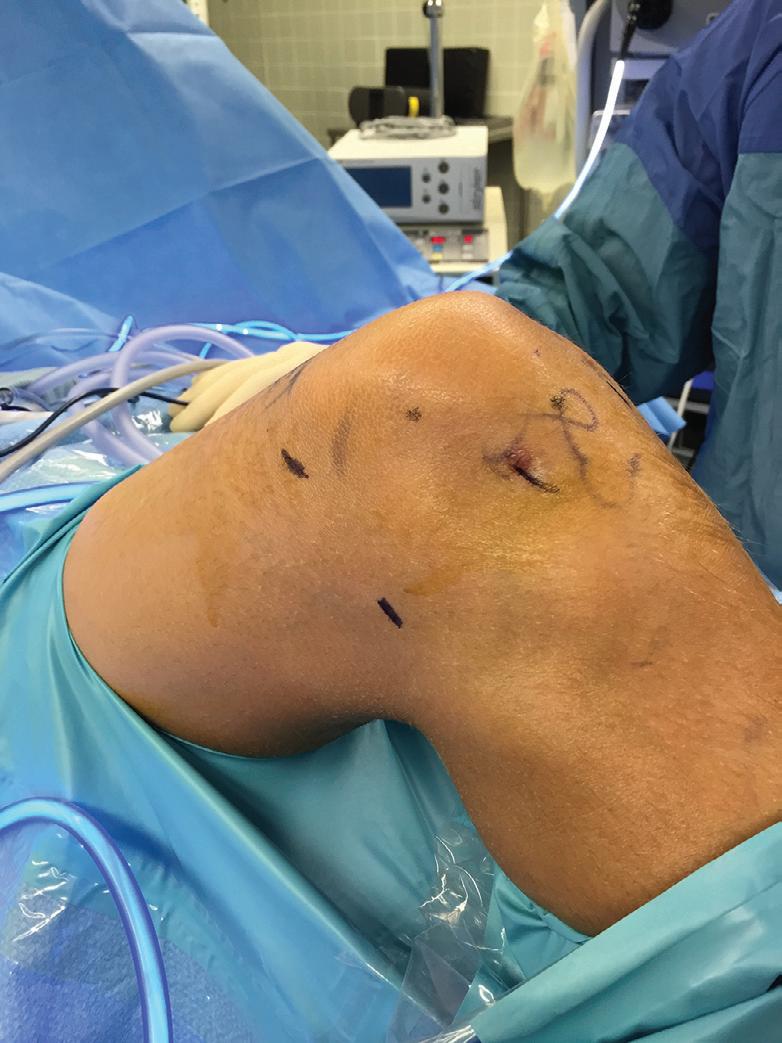
■ Standard portals used for diagnostic arthroscopy include the anterolateral (AL), anteromedial (AM), superomedial (SM), and superolateral (SL) portals (Fig. 1-1).
• With the knee flexed to 90 degrees, the landmarks become more visible.
■ The AL and AM portals are primarily used for diagnostic knee arthroscopy; the SM and SL portals are often but not always used.
■ The AL and AM portals are located in the “soft spot” on either side of the inferior pole of the patella.
• AL portal: Between the lateral femoral condyle and lateral proximal tibia (AL); primary viewing portal
• AM portal: Between the medial femoral condyle and medial proximal tibia (AM); primary working portal
■ SM and SL portals are made approximately 4 cm proximal to the medial and lateral poles of the patella, respectively.
• The SM and SL portals are often used for water flow; although these portals are not always created, they can be helpful in cases that involve extensive synovectomies and procedures within the patellofemoral joint.
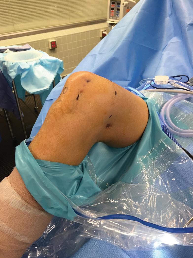
■ Additional portals: The posteromedial (PM) and posterolateral (PL) portals also occasionally are used in diagnostic knee arthroscopy, although these portals tend
superomedially.
■ From the medial gutter, the arthroscope is slightly withdrawn and moved laterally as the knee is placed into flexion with approximately 10 degrees of external rotation.
■ A valgus force is applied to the leg, and the camera is directed posterior to visualize the medial compartment from within the notch.
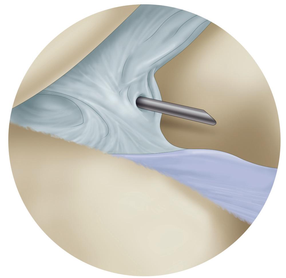
■ At this point, an 18-gauge spinal needle is placed into the portal site for the AM portal and is visualized arthroscopically (Fig. 1-6).
Figure 1-2 Right knee indicates locations for (A) posterolateral and (B) posteromedial portals before a diagnostic knee arthroscopy.
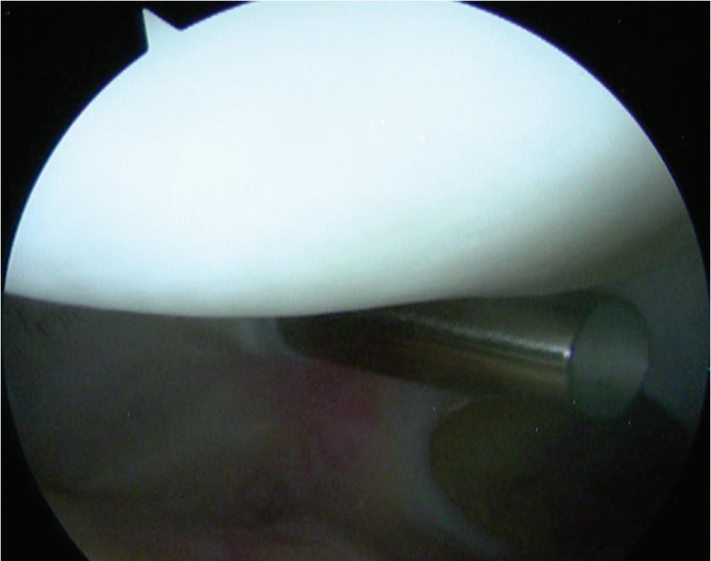
Figure 1-3 Arthroscopic photograph of the left knee shows the patellofemoral joint with the outflow cannula placed
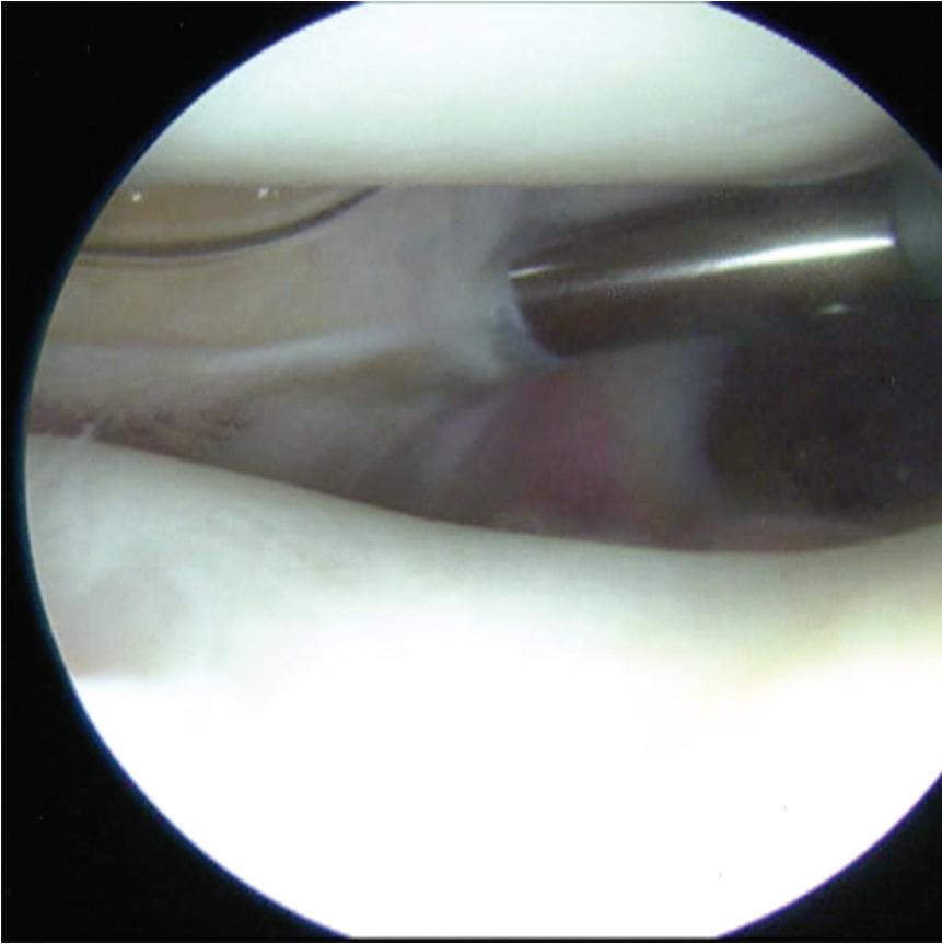
Figure 1-4 Arthroscopic photograph of the left knee shows the patellofemoral joint in extension.
■ The knee is brought into the figure-four position with flexion and application of a varus load with internal rotation.
■ The foot of the operative leg is rested on the anterior tibia of the contralateral leg.
■ As the leg is brought up into the figure-four position, the hand holding the arthroscope should supinate to rotate approximately 90 degrees while aiming posterior with the camera.
■ Thus, the correct visual orientation of the lateral compartment is maintained. The entire lateral meniscus should be probed and inspected, especially the posterior horn where tears are often missed (Figs. 1-10 and 1-11).
■ The hand holding the arthroscope should be raised toward the ceiling and pushed posterior to facilitate adequate visualization. Gentle increases in varus stress help open up this area.
■ The popliteus hiatus should be well visualized.
■ The posterior horn of the lateral meniscus is naturally more lax than the posterior horn of the medial meniscus.
■ The camera should be gently moved laterally, with the eyes oriented laterally to view the midbody of the meniscus followed by the anterior horn.
■ To truly visualize the anterior horn, the camera is slowly and gently withdrawn.
■ The anterior horn is sometimes better visualized with the arthroscope through the AM portal.
■ The tibial plateau and lateral femoral condyle articular surfaces are subsequently assessed.
Posterior Diagnostic Knee Arthroscopy (Video 1-1)
Posterior Compartments (Fig. 1-12)
■ Although many authors agree that posterior knee arthroscopy should be performed as part of most, if not all, diagnostic arthroscopic procedures, visualization of the posterior compartments of the knee is especially helpful in evaluation for loose bodies and in repair of meniscus root tears.
■ For access to the posteromedial and posterolateral compartments of the knee, the modified Gillquist maneuver is typically performed.
• This maneuver is referred to as a contralateral drive-through maneuver.
■ For visualization of the posteromedial compartment, the knee is flexed to 90 degrees and a blunt trocar is placed through the AL portal toward the anterolateral wall of the medial femoral condyle.
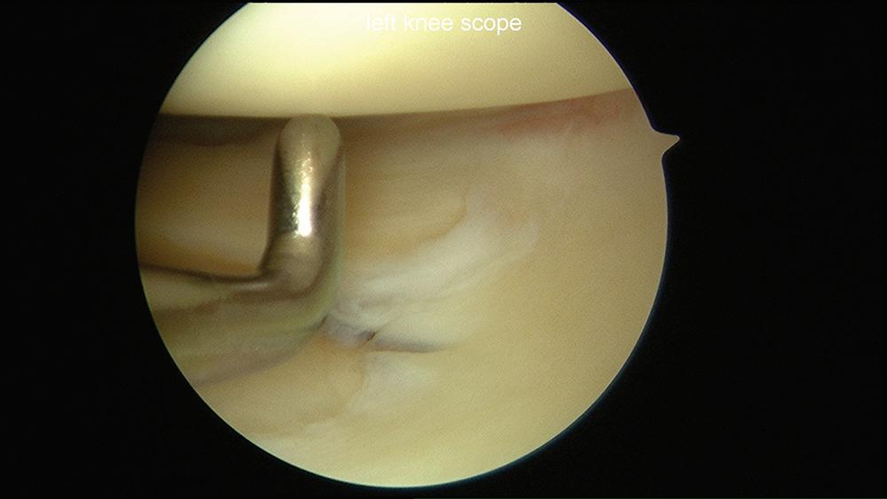
Figure 1-10 Arthroscopic photograph of the left knee shows assessment of the lateral compartment.
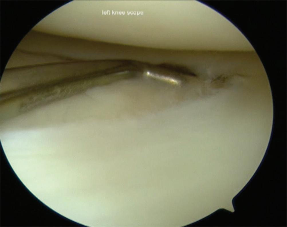
Figure 1-11 Arthroscopic photograph of the left knee shows assessment of the lateral meniscus.
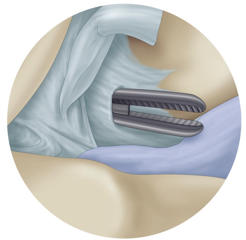
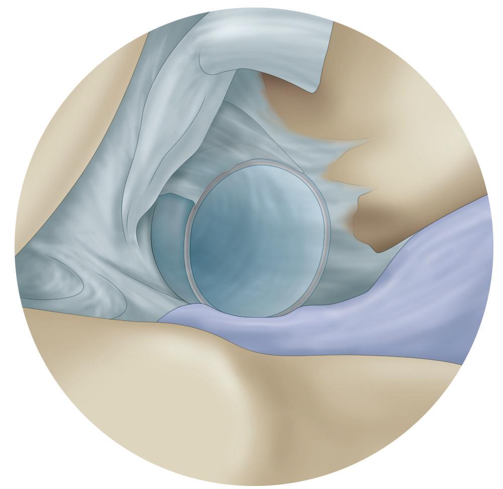
Figure 1-12 Schematics show establishment of the posteromedial portal, including (A) needle localization of the portal site with a spinal needle with direct visualization, (B) blunt dissection of the portal through the capsule (in this case with a hemostat), and (C) insertion of a plastic cannula to complete portal establishment.
■ The obturator is slowly advanced posteriorly while the knee is slowly extended until it “pops” through the interval between the medial femoral condyle and the PCL; a valgus stress is applied to help facilitate access.
■ Care should be taken to avoid injury to either the PCL or the medial wall of the intercondylar notch.
■ The arthroscope is advanced over the trocar. The same technique is used to access the posterolateral compartment, with the trocar inserted into the AM portal and gently pushed and advanced through the interval between the medial aspect of the lateral femoral condyle and the ACL; a varus stress is applied to help facilitate access. Pending surgeon comfort, the arthroscope can be used directly instead of the blunt trocar. Often, the use of a 70-degree arthroscope is helpful for visualization of the posterior compartments of the knee.
■ The authors have also found that an ipsilateral drive-through maneuver can be helpful for accessing the posterior compartments.
■ When this maneuver is performed, the arthroscope is placed from the AL portal and slid in the interval between the ACL origin and the lateral wall of the intercondylar notch; conversely the AL portal may be used to slide into the posteromedial portal between the PCL and the medial wall of the intercondylar notch.
■ Dependent on the visualization and relative joint tightness, varying degrees of knee flexion from 70 degrees (i.e., figure 2-4 position) to 30 degrees may facilitate this maneuver.
■ Although the contralateral drive-through maneuver is generally easier to perform, on occasion a larger loose body blocks visualization in either the medial or lateral compartment, which makes visualization for creation of an accessory PM or PL portal difficult.
■ In general, transitioning into the posterior compartments may be necessary to access for meniscocapsular tears, meniscal root tear repairs, loose bodies, visualization of the posterior cruciate tibial footprint during PCL reconstruction, synovectomy, and in unusual situations, posterior capsular releases or Baker’s cyst decompression.
BRIEF SUMMARY OF SURGICAL STEPS
• Suprapatellar pouch
• Patellofemoral joint
• Trochlear groove
• Medial gutter
• Lateral gutter
• Medial compartment
• Intercondylar notch
• Cruciate ligaments
• Lateral compartment
• Posterior compartments
TECHNICAL PEARLS
• Suprapatellar pouch → eyes at 12 o’clock to identify the proximal patellar pole when retracting
• Patellofemoral joint → eyes laterally with scope astride 30 degrees
• Trochlear groove → eyes at 6 o’clock
• Lateral gutter → eyes aimed medially, raising scope up when synovial folds visualized
• Medial gutter → eyes at 6 o’clock or aiming medially
• Medial compartment → eyes aimed laterally at the notch; in a tight knee, eyes may need to look up as well
• Placement of the scope on the anterior horn of the meniscus medially may provide a second way to visualize the posterior horn of the meniscus
• Intercondylar notch → the anterior cruciate ligament femoral insertion is best visualized with the eyes placed at 10 or 2 o’clock
• Lateral compartment → eyes at 12 o’clock to visualize the posterior horn of the meniscus, rotating laterally to inspect the midbody and anterior horn
REQUIRED EQUIPMENT
Tourniquet
30-Degree arthroscope
Arthroscopy tower, fluid system, pump, tubing
Arthroscopic graspers, scissors
Arthroscopic probe, Wissinger rod, switching sticks, knot tier
Cannulas
Spinal needle
COMMON PITFALLS
(When to call for the attending physician)
• Portals placed too inferiorly risk damage to the meniscus and prohibition of adequate visualization of the medial joint
• Aggressive débridement of fat pad may cause bleeding and an increase in postoperative pain
• Significant valgus stress to visualize the medial compartment may risk injury to the medial collateral ligament
• Aggressive insertion of trocar, scope, or probe may cause iatrogenic injury to articular cartilage
• A stiff knee may make entering the gutters difficult; starting in the patellofemoral joint and entering the compartments via the intercondylar notch is helpful in these cases
• Be careful with radiofrequency near the gutters; a blister can be caused by being too close to the skin
Patient Positioning
Beach Chair
■ A leg pad is placed securely against the patient’s buttocks to ensure that the buttocks and back are firmly against the beach chair; this placement prevents sciatic, lower back, and pelvic injuries related to positioning.
■ Place a facemask over the patient, with care taken to not obstruct the airway; protect the eyes, ears, and nose at all times.
• If a facemask is not used, tape the patient’s head into place in a neutral position with a towel over the forehead.
■ A team effort then moves the patient from the supine to the beach chair position on the operating table (head of bed is elevated approximately 60 degrees). Confirm that the airway and facemask remain in a secured position, and confirm that the leg pad is firmly against the patient’s buttocks and that the patient’s back is firmly against the operating table.
■ The upper portion of the operating table can then be adjusted to improve exposure of the posterior aspect of the shoulder, typically with sliding the back of the table toward the contralateral shoulder while shifting the patient’s torso toward the operative side. Take care to confirm that the patient’s head and neck remain in a neutral position.
■ Folded towels can be placed behind the medial border of the ipsilateral scapula to stabilize it on the operating table.
■ Take the shoulder through the range of motion (ROM) that is necessary for the intended operative procedure.
■ Ensure that the patient’s knees and elbows are appropriately padded.
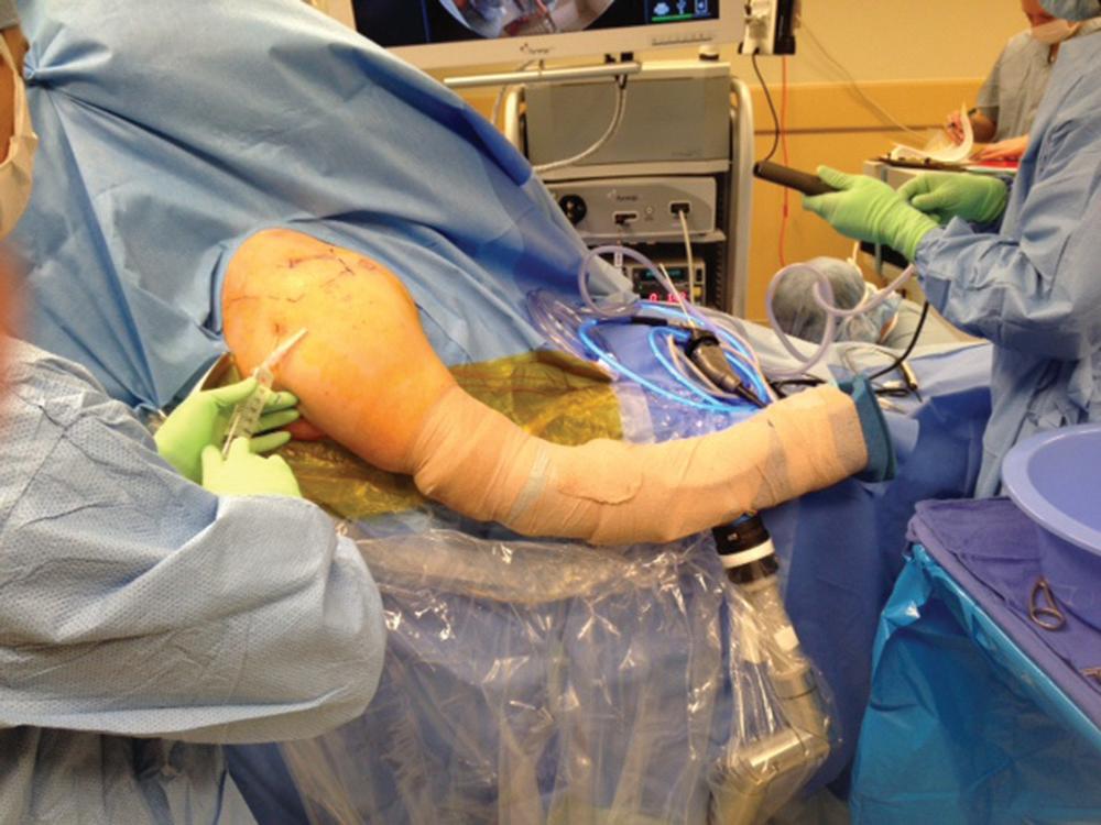
■ Turn the table 45 to 90 degrees to improve access to the patient for both the anesthesia team and the surgical team (Fig. 2-1).
Lateral Decubitus
■ Ensure that the bean bag is on the operating table before attempting the transfer. A sheet should be under and on top of the bean bag.
■ Transfer the patient to the operating table.
■ A team effort is used to roll the patient into the lateral decubitus position on the bean bag, with the operative extremity up.
■ Place the axillary roll under the patient, approximately 2 to 3 fingerbreadths distal to the axilla against the rib cage. This placement minimzes the pressure on the brachial plexus during the case.
■ Position the bean bag as desired to ensure optimal exposure and access to the shoulder and all necessary portals.
COMMONLY USED ICD9 CODES
• 715.91—Degenerative joint disease (DJD)/osteoarthritis (OA) shoulder
• 716.91—Cuff tear arthropathy
• 715.11—Acromioclavicular (AC) arthritis
• 840.4—Rotator cuff tear, traumatic rotator cuff (capsule) sprain
• 840.7—Superior labral tear from anterior to posterior (SLAP) superior glenoid labrum lesion
• 840.0—AC (joint) (ligament) sprain
• 840.9—Shoulder strain, sprain of unspecified site of shoulder and upper arm
• 718.31—Recurrent dislocation of shoulder
• 726.0—Frozen shoulder, adhesive capsulitis of shoulder
• 718.41—Contracture of shoulder, contracture of joint of shoulder region
• 718.11—Loose body in shoulder, loose body in joint of shoulder region
• 718.01—Shoulder cartilage issue, articular cartilage disorder involving shoulder region
COMMONLY USED ICD10 CODES
• M19.01—Primary osteoarthritis, shoulder
• M19.11—Posttraumatic osteoarthritis, shoulder
• M75.1—Rotator cuff tear or rupture, not specified as traumatic
• S43.43—Superior glenoid labrum lesion
• S43.5—Sprain of acromioclavicular joint
• S43.0—Subluxation and dislocation of shoulder joint
• M75.0—Adhesive capsulitis of shoulder
• M24.51—Contracture, shoulder
• M24.01—Loose body in shoulder
• M24.11—Other articular cartilage disorders, shoulder
Figure 2-1 Intraoperative photograph shows final set-up for shoulder arthroscopy in the beach chair position.