https://ebookmass.com/product/campbells-operative-
Instant digital products (PDF, ePub, MOBI) ready for you
Download now and discover formats that fit your needs...
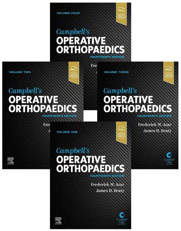
Campbell's Operative Orthopaedics 4-Volume Set 14th Edition Frederick M. Azar
https://ebookmass.com/product/campbells-operativeorthopaedics-4-volume-set-14th-edition-frederick-m-azar/
ebookmass.com
campbell’s operative orthopaedics 14th edition Frederick M.Azar
https://ebookmass.com/product/campbells-operative-orthopaedics-14thedition-frederick-m-azar/
ebookmass.com
Green's Operative Hand Surgery: 2-Volume Set 8th Edition Scott W. Wolfe Md
https://ebookmass.com/product/greens-operative-hand-surgery-2-volumeset-8th-edition-scott-w-wolfe-md/
ebookmass.com
Complete Vanish Valley Witch Mysteries: Books 1-6 Webb
https://ebookmass.com/product/complete-vanish-valley-witch-mysteriesbooks-1-6-webb/
ebookmass.com
ASVAB: 1001 Practice Questions For Dummies 2nd Edition
Angie Papple Johnston
https://ebookmass.com/product/asvab-1001-practice-questions-fordummies-2nd-edition-angie-papple-johnston/
ebookmass.com
A Cowboy of Legend (Lone Star Legends #1) 1st Edition
Linda Broday
https://ebookmass.com/product/a-cowboy-of-legend-lone-starlegends-1-1st-edition-linda-broday/
ebookmass.com
Report on the State of the European Union: Volume 5: The Euro at 20 and the Futures of Europe 1st ed. Edition
Jérôme Creel
https://ebookmass.com/product/report-on-the-state-of-the-europeanunion-volume-5-the-euro-at-20-and-the-futures-of-europe-1st-ededition-jerome-creel/ ebookmass.com
We Are Your Children Too: Black Students, White Supremacists, and the Battle for America's Schools in Prince Edward County, Virginia P. O'Connell Pearson
https://ebookmass.com/product/we-are-your-children-too-black-studentswhite-supremacists-and-the-battle-for-americas-schools-in-princeedward-county-virginia-p-oconnell-pearson/
ebookmass.com
Materials for Biomedical Engineering: Thermoset and Thermoplastic Polymers Valentina Grumezescu
https://ebookmass.com/product/materials-for-biomedical-engineeringthermoset-and-thermoplastic-polymers-valentina-grumezescu/
ebookmass.com
A Study Guide for Danez Smith's "Tonight, in Oakland" Gale
https://ebookmass.com/product/a-study-guide-for-danez-smiths-tonightin-oakland-gale/
ebookmass.com
Campbell’s Operative Orthopaedics, 14th ed. List of Techniques VOLUME I Surgical Techniques
1.1 Fixation of Tendon to Bone, 14
1.2 Tendon Fixation Into the Intramedullary Canal, 15
1.3 Tendon to Bone Fixation Using Locking Loop Suture, 16
1.4 Tendon to Bone Fixation Using Wire Suture, 16
1.5 Fixation of Osseous Attachment of Tendon to Bone, 17
1.6 Removal of a Tibial Graft, 22
1.7 Removal of Fibular Grafts, 23
1.8 Removal of an Iliac Bone Graft, 26
1.9 Approach to the Interphalangeal Joints, 28
1.10 Medial Approach to the Great Toe Metatarsophalangeal Joint, 28
1.11 Dorsomedial Approach to Great Toe Metatarsophalangeal Joint, 29
1.12 Approach to the Lesser Toe Metatarsophalangeal Joints, 29
1.13 Medial Approach to the Calcaneus, 29
1.14 Lateral Approach to the Calcaneus, 29
1.15 Extended Lateral Approach to the Calcaneus, 29 1.16 Sinus Tarsi Approach, 31
1.17 U–Shaped Approach to the Calcaneus, 31
1.18 Kocher Approach (Curved L) to the Calcaneus, 32
1.19 Anterolateral Approach to Chopart Joint, 33
1.20 Anterior Approach to Expose the Ankle Joint and Both Malleoli, 33
1.21 Kocher Lateral Approach to the Tarsus and Ankle, 34
1.22 Ollier Approach to the Tarsus, 34
1.23 Single-Incision Posterolateral Approach to the Lateral and Posterior Malleoli, 35
1.24 Posterolateral Approach to the Ankle (Gatellier and Chastang), 35
1.25 Anterolateral Approach to the Lateral Dome of the Talus (Tochigi, Amendola, Muir, and Saltzman), 35
1.26 Posterior Approach to the Ankle, 36
1.27 Medial Approach to the Tarsus (Knupp et al.), 37
1.28 Medial Approach to the Ankle (Koenig and Schaefer), 37
1.29 Medial Approach to the Posterior Lip of the Tibia (Colonna and Ralston), 38
1.30 Anterolateral Approach to the Tibia, 39
1.31 Medial Approach to the Tibia (Phemister), 39
1.32 Posterolateral Approach to the Tibial Shaft (Harmon, Modified), 39
1.33 Anterolateral Approach to the Lateral Tibial Plateau (Kandemir and MacLean), 39
1.34 Medial Approach to the Medial Tibial Plateau, 41
1.35 Posteromedial Approach to the Medial Tibial Plateau (Supine), 41
1.36 Posteromedial Approach (Prone) to the Superomedial Tibia (Banks and Laufman), 42
1.37 Posterolateral Approach to the Tibial Plateau (Solomon et al.), 43
1.38 Posterolateral Approach to the Tibial Plateau Without Fibular Osteotomy (Frosch et al.), 44
1.39 Tscherne-Johnson Extensile Approach to the Lateral Tibial Plateau (Johnson et al.), 44
1.40 Anterolateral Approach for Access to Posterolateral Corner (Sun et al.), 45
1.41 Posterolateral Approach to the Fibula (Henry), 46
1.42 Anteromedial Parapatellar Approach (von Langenbeck), 47
1.43 Subvastus (Southern) Anteromedial Approach to the Knee (Erkes, as Described by Hofmann, Plaster, and Murdock), 47
1.44 Anterolateral Approach to the Knee (Kocher), 48
1.45 Posterolateral Approach to the Knee (Henderson), 49
1.46 Posteromedial Approach to the Knee (Henderson), 51
1.47 Medial Approach to the Knee (Cave), 52
1.48 Medial Approach to the Knee (Hoppenfeld and deBoer), 53
1.49 Transverse Approach to the Meniscus, 53
1.50 Lateral Approach to the Knee (Bruser), 55
1.51 Lateral Approach to the Knee (Brown et al.), 56
1.52 Lateral Approach to the Knee (Hoppenfeld and deBoer), 57
1.53 Extensile Approach to the Knee (Fernandez), 58
1.54 Direct Posterior Approach to the Knee (Brackett and Osgood; Putti; Abbott and Carpenter), 58
1.55 Direct Posteromedial Approach to the Knee for Tibial Plateau Fracture (Galla and Lobenhoffer as Described by Fakler et al.), 61
1.56 Direct Posterolateral Approach to the Knee (Minkoff, Jaffe, and Menendez), 62
1.57 Anterolateral Approach to the Femur (Thompson), 62
1.58 Lateral Approach to the Femoral Shaft, 63
1.59 Posterolateral Approach to the Femoral Shaft, 64
1.60 Posterior Approach to the Femur (Bosworth), 64
1.61 Medial Approach to the Posterior Surface of the Femur in the Popliteal Space (Henry), 67
1.62 Lateral Approach to the Posterior Surface of the Femur in the Popliteal Space (Henry), 67
1.63 Lateral Approach to the Proximal Shaft and the Trochanteric Region, 68
1.64 Anterior Iliofemoral Approach to the Hip (Smith-Petersen), 70
1.65 Anterior Approach to the Hip Using a Transverse Incision (Somerville), 71
1.66 Modified Anterolateral Iliofemoral Approach to the Hip (Smith–Petersen), 71
1.67 Lateral Approach to the Hip (Watson-Jones), 73
1.68 Lateral Approach for Extensive Exposure of the Hip (Harris), 73
1.69 Lateral Approach to the Hip Preserving the Gluteus Medius (McFarland and Osborne), 75
1.70 Lateral Transgluteal Approach to the Hip (Hardinge), 77
1.71 Lateral Transgluteal Approach to the Hip (Hay as Described by McLauchlan), 77
1.72 Posterolateral Approach (Gibson), 78
1.73 Posterior Approach to the Hip (Osborne), 80
1.74 Posterior Approach to the Hip (Moore), 82
1.75 Medial Approach to the Hip (Ferguson; Hoppenfeld and DeBoer), 84
1.76 Stoppa Approach (AO Foundation), 85
1.77 Ilioinguinal Approach to the Acetabulum (Letournel and Judet, as Described by Matta), 87
1.78 Iliofemoral Approach to the Acetabulum (Letournel and Judet), 90
1.79 Kocher-Langenbeck Approach (Kocher-Langenbeck; Letournel and Judet), 91
1.80 Modified Gibson Approach (Modified Gibson Approach, Moed), 93 1.81 Extensile Iliofemoral Approach (Letournel and Judet), 94
1.82 Extensile Iliofemoral Approach (Reinert et al.), 94
1.83 Triradiate Extensile Approach to the Acetabulum (Mears and Rubash), 97
1.84 Extensile Approach to the Acetabulum (Carnesale), 99
1.85 Approach to the Ilium, 99
1.86 Approach to the Symphysis Pubis (Pfannenstiel), 100
1.87 Posterior Approach to the Sacroiliac Joint, 102
1.88 Anterior Approach to the Sacroiliac Joint (Avila), 102
1.89 Approach to Both Sacroiliac Joints or Sacrum (Modified from Mears and Rubash), 103
1.90 Approach to the Sternoclavicular Joint, 104
1.91 Approach to the Acromioclavicular Joint and Coracoid Process (Roberts), 104
1.92 Anteromedial Approach to the Shoulder (Thompson; Henry), 105
1.93 Anteromedial/Posteromedial Approach to the Shoulder (Cubbins, Callahan, and Scuderi), 106
1.94 Anterior Axillary Approach to the Shoulder (Leslie and Ryan), 106
1.95 Anterolateral Limited Deltoid-Splitting Approach to the Shoulder, 106
1.96 Extensile Anterolateral Approach to the Shoulder (Gardner et al.), 109
1.97 Transacromial Approach to the Shoulder (Darrach; McLaughlin), 109
1.98 Posterior Deltoid-Splitting Approach to the Shoulder (Wirth et al.), 110
1.99 Posterior Approach to the Shoulder (Modified Judet), 111
1.100 Simplified Posterior Approach to the Shoulder (King, as Described by Brodsky et al.), 111
1.101 Posterior Inverted-U Approach to the Shoulder (Abbott and Lucas), 113
1.102 Anterolateral Approach to the Shaft of the Humerus (Thompson; Henr y), 114
1.103 Subbrachial Approach to the Humerus (Boschi et al.), 116
1.104 Posterior Approach to the Proximal Humerus (Berger and Buckwalter), 117
1.105 Posterolateral Approach to the Distal Humeral Shaft (Moran), 118
1.106 Posterolateral Extensile (Cold) Approach to the Distal Humerus (Lewicky, Sheppard, and Ruth), 120
1.107 Posterolateral Approach to the Elbow (Campbell), 121
1.108 Extensile Posterolateral Approach to the Elbow (Wadsworth), 121
1.109 Posterior Approach to the Elbow by Olecranon Osteotomy (MacAusland and Müller), 123
1.110 Extensile Posterior Approach to the Elbow (Bryan and Morrey), 123
1.111 Lateral Approach to the Elbow, 124
1.112 Lateral J–Shaped Approach to the Elbow (Kocher), 126
1.113 Medial Approach with Osteotomy of the Medial Epicondyle (Molesworth; Campbell), 127
1.114 Medial and Lateral Approach to the Elbow, 127
1.115 Global Approach to the Elbow (Patterson, Bain, and Mehta), 127
1.116 Posterolateral Approach to the Radial Head and Neck, 130
1.117 Approach to the Proximal and Middle Thirds of the Posterior Surface of the Radius (Thompson), 131
1.118 Anterolateral Approach to the Proximal Shaft and Elbow Joint (Henr y), 132
1.119 Anterior Approach to the Distal Half of the Radius (Henry), 132
1.120 Anterior Approach to the Coronoid Process of the Proximal Ulna (Yang et al.), 134
1.121 Approach to the Proximal Third of the Ulna and the Proximal Fourth of the Radius (Boyd), 135
1.122 Dorsal Approach to the Wrist, 137
1.123 Dorsal Approach to the Wrist, 137
1.124 Volar Approach to the Wrist, 137
1.125 Lateral Approach to the Wrist, 138
1.126 Medial Approach to the Wrist, 138
Arthroplasty of the Hip
3.1 Preoperative Templating for Total Hip Arthroplasty (Capello), 203
3.2 Posterolateral Approach with Posterior Dislocation of the Hip, 207
3.3 Implantation of Cementless Acetabular Component, 210
3.4 Implantation of Cemented Acetabular Component, 212
3.5 Implantation of Cementless Femoral Component, 214
3.6 Implantation of Cemented Femoral Component, 218
3.7 Direct Anterior Approach with Anterior Dislocation of the Hip, 222
3.8 Gluteus Maximus and Tensor Fascia Lata Transfer for Primary Deficiency of the Abductors of the Hip, 269
3.9 Revision After Adverse Local Tissue Reaction, 285
3.10 Transtrochanteric Approach for Revision Total Hip Arthroplasty, 289
3.11 Removal of Cemented Femoral Component, 289
3.12 Removal of Cementless Femoral Component, 290
3.13 Removal of Implants with Extensive Distal Bone Ingrowth (Glassman and Engh), 291
3.14 Extended Trochanteric Osteotomy (Younger et al.), 292
3.15 Removal of a Broken Stem—Proximal Window (Moreland, Marder, and Anspach), 294
3.16 Removal of a Broken Stem—Distal Window, 295
3.17 Removal of Femoral Cement, 295
3.18 Removal of Distal Cement with a High-Speed Burr (Turner et al.), 296
3.19 Removal of Distal Cement with a High-Speed Burr and Cortical Window (Mallory), 298
3.20 Removal of a Loose All-Polyethylene Cup, 299
3.21 Removal of a Metal-Backed, Cemented Acetabular Component, 299
3.22 Cementless Acetabular Component (Mitchell), 301
3.23 Management of Acetabular Cavitary Deficits, 302
3.24 Management of Segmental Acetabular Deficit with Femoral Head Allograft, 307
3.25 Management of Segmental Acetabular Deficit with Metal Augment (Jenkins et al., Modified), 307
3.26 Management of Combined Deficits with Structural Grafting (Sporer et al.), 308
3.27 Acetabular Distraction for Management of Pelvic Discontinuity (Sheth et al.), 310
3.28 Cup-Cage Technique for Management of Pelvic Discontinuity (Abdel et al., Modified), 311
3.29 Management of Pelvic Discontinuity with Allografting and Custom Component (DeBoer et al.), 311
3.30 Management of Femoral Deficit with Modular Femoral Component (Cameron), 316
3.31 Revision with Extensively Porous-Coated Femoral Stem (Mallory and Head), 316
3.32 Management of Proximal Femoral Bone Loss with Modular Tapered Fluted Stem (Kwong et al.), 317
3.33 Management of Proximal Femoral Deficiencies with Impaction Bone Grafting and Cemented Revision Stem (Gie, Modified), 317
3.34 Management of Massive Deficits with Proximal Femoral AllograftProsthesis Composite, 319
3.35 Management of Massive Deficits with Modular Megaprosthesis (Klein et al.), 321
Surface Replacement Hip Arthroplasty
4.1 Hip Resurfacing Technique—Birmingham Hip Replacement, 336 Arthrodesis of the Hip
5.1 Arthrodesis with Cancellous Screw Fixation (Benaroch et al.), 349
5.2 Arthrodesis with Anterior Fixation (Matta et al.), 349
5.3 Arthrodesis with Double-Plate Fixation (Müller et al.), 350
5.4 Arthrodesis with Cobra Plate Fixation (Murrell and Fitch), 351
5.5 Arthrodesis with Hip Compression Screw Fixation (Pagnano and Cabanela), 353
5.6 Arthrodesis in the Absence of the Femoral Head (Abbott, Fischer, and Lucas), 353
Hip Pain in the Young Adult and Hip Preservation Surgery
6.1 Surgical Dislocation of the Hip (Ganz et al.), 367
6.2 Combined Hip Arthroscopy and Limited Open Osteochondroplasty (Clohisy and McClure), 371
6.3 Mini-Open Direct Anterior Approach (Ribas et al.), 373
6.4 Bernese Periacetabular Osteotomy (Matheney et al.), 381
6.5 Rectus-Sparing Modification of Bernese Osteotomy (Novais et al.), 385
6.6 Step-Cut Lengthening of the Iliotibial Band (White et al.), 388
6.7 Core Decompression (Hungerford), 393
6.8 Core Decompression—Percutaneous Technique (Mont et al.), 394 Arthroplasty of the Knee
7.1 Surgical Approach for Primary Total Knee Arthroplasty, 436
7.2 Bone Preparation for Primary Total Knee Arthroplasty, 439
7.3 Pie-Crusting, 444
7.4 Posterior Stabilized Total Knee Arthroplasty In A Varus Knee, 445
7.5 Posterior Cruciate–Retaining Total Knee Arthroplasty of a Varus Knee, 445
7.6 Valgus Deformity Correction, 446
7.7 Flexion Contracture Correction, 447
7.8 Recurvatum Correction, 448
7.9 Posterior Cruciate Ligament Balancing, 448
7.10 Bone Grafting of Peripheral Tibial Defects (Windsor, Insall, and Sculco), 450
7.11 Component Implantation, 453
7.12 Unicondylar Knee Arthroplasty, 455
7.13 Patellofemoral Arthroplasty, 456
7.14 Arthrodesis with an Intramedullary Nail for an Infected Total Knee Arthroplasty, 463
Arthrodesis of the Knee
8.1 Compression Arthrodesis Using External Fixation, 486
8.2 Arthrodesis Using Intramedullary Nail Fixation, 487
8.3 Knee Arthrodesis with Locked Intramedullary Nail After Failed Total Knee Arthroplasty, 489
8.4 Arthrodesis Using Plate Fixation, 490
Soft-Tissue Procedures and Osteotomies About the Knee
9.1 Proximal Release of Quadriceps (Sengupta), 494
9.2 Quadricepsplasty for Posttraumatic Contracture of the Knee (Modified Thompson, Described by Hahn et al.), 494
9.3 Drainage of Bursa, 499
9.4 Excision of Bursa, 499
9.5 Popliteal Cyst Excision (Hughston, Baker, and Mello), 501
9.6 Medial Gastrocnemius Bursa Excision (Meyerding and Van Demark), 502
9.7 Semimembranosus Bursa Excision, 503
9.8 Semitendinosus Tendon Transfer (Ray, Clancy, and Lemon), 503
9.9 Lateral Closing Wedge Osteotomy (Modified Coventry; Hofmann, Wyatt, and Beck), 513
9.10 Opening Wedge Hemicallotasis (Turi et al.), 518
9.11 Varus Distal Femoral Osteotomy (Coventry), 522
Total Ankle Arthroplasty
10.1 Total Ankle Arthroplasty, 533
10.2 Dome Osteotomy for Correction of Varus Deformity Above the Ankle Deformity (Tan and Myerson), 536
10.3 Medial Tibial Plafondplasty for Varus Deformity at the Ankle Joint (Tan and Myerson), 537
10.4 Reconstruction of Lateral Ankle Ligaments for Chronic Instability as an Adjunct to Total Ankle Arthroplasty (Coetzee), 538
10.5 Tibiotalar Arthrodesis Conversion to Total Ankle Arthroplasty (Pellegrini et al.), 540
10.6 Revision Total Ankle Arthroplasty (Meeker et al.), 555 Ankle Arthrodesis
11.1 Opening Wedge Osteotomy of the Tibia For Varus Deformity and Medial Joint Arthrosis, 564
11.2 Intraarticular Opening Medial Wedge Osteotomy (Plafondplasty) of the Tibia for Intraarticular Varus Arthritis and Instability (Mann, Filippi, and Myerson), 566
11.3 Distraction Arthroplasty of the Ankle, 568
11.4 Mini-Incision Technique, 575
11.5 Transfibular (Transmalleolar) Arthrodesis with Fibular Strut Graft, 576
11.6 Anterior Approach with Plate Fixation, 580
11.7 Lateral Approach with Fibular Sparing (Smith, Chiodo, Singh, Wilson), 580
11.8 Tibiotalocalcaneal Arthrodesis, 581
11.9 Posterior Approach for Arthrodesis of Ankle and Subtalar Joints (Campbell), 584
11.10 Arthrodesis with a Thin-Wire External Fixation, 584
11.11 Tibiotalar Arthrodesis with a Sliding Bone Graft (Blair; Morris et al.), 590
11.12 Tibiotalar or Tibiotalocalcaneal Fusion with Structural Allograft and Internal Fixation for Salvage of Failed Total Ankle Arthroplasty (Berkowitz et al.), 591
11.13 Bone Graft Harvest from the Proximal Tibia (Whitehouse et al.), 593
Shoulder and Elbow Arthroplasty
12.1 Hemiarthroplasty, 608
12.2 Total Shoulder Arthroplasty, 612
12.3 Reverse Total Shoulder Arthroplasty, 617
12.4 Debridement Arthroplasty (Wada et al.), 633
12.5 Interposition Arthroplasty, 637
12.6 Radial Head Arthroplasty, 639
12.7 Coonrad-Morrey Prosthesis, 642
12.8 Elbow Resection Arthroplasty (Campbell), 648
Salvage Operations for the Shoulder and Elbow
13.1 External Fixation (Charnley and Houston), 660
13.2 Plate Fixation (AO Group), 660
13.3 Pelvic Reconstruction Plate (Modification of Richards et al.), 661
13.4 Shoulder Arthrodesis After Failed Prosthetic Shoulder Arthroplasty (Scalise and Iannotti), 662
13.5 Arthroscopic Shoulder Arthrodesis for Brachial Plexus Injury (lenoir), 664
13.6 Elbow Arthrodesis (Staples), 665
13.7 Elbow Arthrodesis (Müller et al.), 666
13.8 Elbow Arthrodesis (Spier), 666
13.9 Latissimus Dorsi Transfer, Open Technique (Gerber et al.), 668
13.10 Latissimus Dorsi Transfer, Arthroscopically Assisted Technique (Castricini et al.), 669
13.11 Lower Trapezius Transfer, Open Technique (Elhassan et al.), 671
13.12 Lower Trapezius Transfer, Arthroscopically Assisted Technique (Elhassan et al.), 672
13.13 Pectoralis Major Transfer (Modification of Resch et al.,), 673
13.14 Latissimus Dorsi Tendon Transfer (Mun et al.), 675
Amputations of the Foot
15.1 Terminal Syme Amputation, 700
15.2 Amputation at the Base of the Proximal Phalanx, 700
15.3 Metatarsophalangeal Joint Disarticulation, 703
15.4 Metatarsophalangeal Joint Disarticulation, 703
15.5 First or Fifth Ray Amputation (Border Ray Amputation), 703
15.6 Central Ray Amputation, 704
15.7 Transmetatarsal Amputation, 707
15.8 Chopart Amputation, 711
15.9 Syme Amputation, 713
15.10 Two-Stage Syme Amputation (Wyss et al.; Malone et al.; Wagner), 717
15.11 Boyd Amputation, 717
Amputations of the Lower Extremity
16.1 Transtibial Amputation, 722
16.2 Transtibial Amputation (Modified Ertl; Taylor and Poka), 723
16.3 Transtibial Amputation Using Long Posterior Skin Flap (Burgess), 725
16.4 Knee Disarticulation (Batch, Spittler, and McFaddin), 726
16.5 Knee Disarticulation (Mazet and Hennessy), 728
16.6 Knee Disarticulation (Kjøble), 728
16.7 Transfemoral (Above-Knee) Amputation of Nonischemic Limbs, 730
16.8 Transfemoral (Above-Knee) Amputation of Nonischemic Limbs (Gottschalk), 731
Amputations of the Hip and Pelvis
17.1 Anatomic Hip Disarticulation (Boyd), 733
17.2 Posterior Flap (Slocum), 735
17.3 Standard Hemipelvectomy, 736
17.4 Anterior Flap Hemipelvectomy, 736
17.5 Conservative Hemipelvectomy, 739
Major Amputations of the Upper Extremity
18.1 Amputation at the Wrist, 743
18.2 Disarticulation of the Wrist, 743
18.3 Distal Forearm (Distal Transradial) Amputation, 744
18.4 Proximal Third of Forearm (Proximal Transradial) Amputation, 745
18.5 Disarticulation of the Elbow, 745
18.6 Supracondylar Area, 746
18.7 Amputation Proximal to the Supracondylar Area, 748
18.8 Amputation Through the Surgical Neck of the Humerus, 748
18.9 Disarticulation of the Shoulder, 750
18.10 Anterior Approach (Berger), 752
18.11 Posterior Approach (Littlewood), 753
18.12 Targeted Muscle Reinnervation After Transhumeral Amputation (O’Shaughnessy et al.), 756
Amputations of the Hand
19.1 Kutler V-Y or Atasoy Triangular Advancement Flaps (Kutler; Fisher), 764
19.2 Atasoy Triangular Advancement Flaps (Atasoy et al.), 766
19.3 Bipedicle Dorsal Flaps, 767
19.4 Adipofascial Turnover Flap, 768
19.5 Thenar Flap, 768
19.6 Local Neurovascular Island Flap, 769
19.7 Island Pedicle Flap, 769
19.8 Retrograde Island Pedicle Flap, 771
19.9 Ulnar Hypothenar Flap, 771
19.10 Index Ray Amputation, 771
19.11 Transposing the Index Ray (Peacock), 774
19.12 Advancement Pedicle Flap for Thumb Injuries, 776
19.13 Phalangization of Fifth Metacarpal, 778
19.14 Krukenberg Reconstruction (Krukenberg; Swanson), 779
19.15 Lengthening of the Metacarpal and Transfer of Local Flap (Gillies and Millard, Modified), 781
19.16 Osteoplastic Reconstruction and Transfer of Neurovascular Island Graft (Verdan), 782
19.17 Riordan Pollicization (Riordan), 784
19.18 Buck-Gramcko Pollicization (Buck-Gramcko), 785
19.19 Foucher Pollicization, 787
Osteomyelitis
21.1 Drainage of Acute Hematogenous Osteomyelitis, 821
21.2 Sequestrectomy and Curettage for Chronic Osteomyelitis, 827
21.3 Open Bone Grafting (Papineau et al.; Archdeacon and Messerschmitt), 828
21.4 Antibiotic Bead Pouch (Henry, Ostermann, and Seligson), 829
21.5 Intramedullary Antibiotic Cement Nail, 829
21.6 Split-Heel Incision (Gaenslen), 834
21.7 Distal Third of the Femur, 835
21.8 Drainage, 835
21.9 Resection of the Metatarsals, 836
21.10 Partial Calcanectomy, 837
21.11 Resection of the Fibula, 837
21.12 Resection of the Iliac Wing (Badgley), 838
Infectious Arthritis
22.1 Surgical Drainage of the Tarsal Joint, 846
22.2 Anterolateral Drainage of the Ankle, 847
22.3 Posterolateral Drainage of the Ankle, 847
22.4 Anteromedial Drainage of the Ankle, 847
22.5 Posteromedial Drainage of the Ankle, 847
22.6 Arthroscopic Drainage of the Knee, 848
22.7 Anterior Drainage of the Knee, 849
22.8 Posterolateral and Posteromedial Drainage of the Knee (Henderson), 849
22.9 Posteromedial Drainage of the Knee (Klein), 850
22.10 Posteromedial and Posterolateral Drainage of the Knee (Kelikian), 850
22.11 Lateral Aspiration of the Hip, 851
22.12 Anterior Aspiration of the Hip, 851
22.13 Medial Aspiration of the Hip, 851
22.14 Posterior Drainage of the Hip (Ober), 852
22.15 Anterior Drainage of the Hip, 852
22.16 Lateral Drainage of the Hip, 852
22.17 Medial Drainage of the Hip (Ludloff), 853
22.18 Arthroscopic Debridement and Partial Synovectomy of the Hip in an Adult, 853
22.19 Resection of the Hip (Girdlestone), 854
22.20 Anterior Drainage of the Shoulder, 856
22.21 Posterior Drainage of the Shoulder, 856
22.22 Medial Drainage of the Elbow, 857
22.23 Lateral Drainage of the Elbow, 857
22.24 Posterior Drainage of the Elbow, 858
22.25 Lateral Drainage of the Wrist, 858
22.26 Medial Drainage of the Wrist, 859
22.27 Dorsal Drainage of the Wrist, 859
22.28 Osteotomy of the Ankle, 859
22.29 Transverse Supracondylar Osteotomy of the Femur, 859
22.30 V-Osteotomy of the Femur (Thompson), 860
22.31 Supracondylar Cuneiform Osteotomy of the Femur, 860
22.32 Supracondylar Controlled Rotational Osteotomy of the Femur, 861
22.33 Intraarticular Osteotomy, 861
22.34 Reconstruction After Hip Sepsis (Harmon), 864
22.35 Transverse Opening Wedge Osteotomy of the Hip, 864
22.36 Transverse Closing Wedge Osteotomy of the Hip, 865
22.37 Brackett Osteotomy of the Hip (Brackett), 865
Tuberculosis and Other Unusual Infections
23.1 Curettage for Tuberculous Lesions in the Foot, 874
23.2 Excision of Metatarsal, 874
23.3 Excision of Cuneiform Bones, 874
23.4 Excision of Navicular, 875
23.5 Excision of Cuboid, 875
23.6 Excision of Calcaneus, 875
23.7 Excision of Talus, 876
23.8 Partial Synovectomy and Curettage (Wilkinson), 877
23.9 Lesions above Acetabulum, 878
23.10 Lesions of the Femoral Neck, 878
23.11 Lesions of the Trochanteric Area (Ahern), 878
23.12 Excision of the Hip Joint, 879
23.13 Excision of Elbow Joint, 880
23.14 Excision of Wrist Joint, 880
General Principles of Tumors
24.1 Resection of the Shoulder Girdle (Marcove, Lewis, and Huvos), 909
24.2 Resection of the Scapula (Das Gupta), 913
24.3 Resection of the Proximal Humerus, 916
24.4 Intercalary Resection of the Humeral Shaft (Lewis), 920
24.5 Resection of the Distal Humerus, 920
24.6 Resection of the Proximal Radius, 920
24.7 Resection of the Proximal Ulna, 921
24.8 Resection of the Distal Radius, 921
24.9 Resection of the Pubis and Ischium (Radley, Liebig, and Brown), 928
24.10 Resection of the Acetabulum, 932
24.11 Resection of the Innominate Bone (Internal Hemipelvectomy) (Karakousis and Vezeridis), 934
24.12 Resection of the Sacroiliac Joint, 934
24.13 Resection of the Sacrum (Stener and Gunterberg), 936
24.14 Resection of the Sacrum (Localio, Francis, and Rossano), 937
24.15 Resection of the Sacrum Through Posterior Approach (MacCarty et al.), 937
24.16 Resection of the Proximal Femur (Lewis and Chekofsky), 938
24.17 Resection of Entire Femur (Lewis), 938
24.18 Intraarticular Resection of the Distal Femur with Endoprosthetic Reconstruction, 943
24.19 Resection of the Proximal Tibia (Malawer), 944
24.20 Resection of the Proximal Fibula (Malawer), 945
24.21 Resection of the Distal Third of the Fibula, 945
24.22 Rotationplasty for a Lesion in the Distal Femur (Kotz and Salzer), 950
24.23 Rotationplasty for a Lesion of the Proximal Femur Without Involvement of the Hip Joint (Winkelmann), 950
24.24 Rotationplasty for a Lesion of the Proximal Femur Involving the Hip Joint (Winkelmann), 952
Operative Orthopaedics, 14th ed.
List of Techniques
VOLUME II Congenital Anomalies of the Lower Extremity
29.1 Amputation of an Extra Toe (Simple Postaxial Polydactyly), 1081
29.2 Tsuge Ray Reduction (Tsuge), 1082
29.3 Ray Reduction, 1083
29.4 Ray Amputation, 1083
29.5 Simplified Cleft Closure (Wood, Peppers, and Shook), 1087
29.6 Correction of Angulated Toe, 1088
29.7 Arthroplasty of the Fifth Metatarsophalangeal Joint (Butler), 1088
29.8 Creation of Syndactyly of the Great Toe and Second Toe for Hallux Varus (Farmer), 1090
29.9 Dome-Shaped Osteotomies of Metatarsal Bases (Berman and Gartland), 1092
29.10 Cuneiform and Cuboid Osteotomies (McHale and Lenhart), 1095
29.11 Anterior Tibial Tendon Transfer, 1100
29.12 Transverse Circumferential (Cincinnati) Incision (Crawford, Marxen, and Osterfeld), 1102
29.13 Extensile Posteromedial and Posterolateral Release (McKay, Modified), 1103
29.14 Achilles Tendon Lengthening and Posterior Capsulotomy (Selective Approach), 1106
29.15 First Metatarsal Osteotomy and Tendon Transfer for Dorsal Bunion, 1108
29.16 Osteotomy of the Calcaneus for Persistent Varus Deformity of the Heel (Dwyer, Modified), 1109
29.17 Medial Release with Osteotomy of the Distal Calcaneus (Lichtblau), 1109
29.18 Selective Joint-Sparing Osteotomies for Residual Cavovarus Deformity (Mubrak and Van Valin), 1110
29.19 Triple Arthrodesis, 1112
29.20 Talectomy (Trumble et al.), 1112
29.21 Open Reduction and Realignment of Talonavicular and Subtalar Joints (Kumar, Cowell, and Ramsey), 1115
29.22 Open Reduction and Extraarticular Subtalar Fusion (Grice- Green), 1116
29.23 Tibiofibular Synostosis (Langenskiöld), 1120
29.24 Insertion of Williams Intramedullary Rod and Bone Grafting (Anderson et al.), 1123
29.25 One-Stage or Two-Stage Release of Circumferential Constricting Band (Greene), 1126
29.26 Capsular Release and Quadriceps Lengthening for Correction of Congenital Knee Dislocation (Curtis and Fisher), 1127
29.27 Lateral Release and Medial Plication (Beaty; Modified from Gao et al. and Langenskiöld), 1129
29.28 Distal Fibulotalar Arthrodesis, 1136
29.29 Proximal Tibiofibular Synostosis, 1137
29.30 Varus Supramalleolar Osteotomy of the Ankle (Wiltse), 1139
29.31 Knee Fusion for Proximal Femoral Focal Deficiency (King), 1145
29.32 Rotationplasty (Van Nes), 1148
29.33 Syme Amputation, 1150
29.34 Boyd Amputation, 1152
29.35 Physeal Exposure Around the Knee (Abbott and Gill, Modified), 1160
29.36 Percutaneous Epiphysiodesis (Canale et al.), 1161
29.37 Percutaneous Transepiphyseal Screw Epiphysiodesis (Métaizeau et al.), 1162
29.38 Tension Plate Epiphysiodesis, 1164
29.39 Proximal Femoral Metaphyseal Shortening (Wagner), 1165
29.40 Distal Femoral Metaphyseal Shortening (Wagner), 1165
29.41 Proximal Tibial Metaphyseal Shortening (Wagner), 1166
29.42 Tibial Diaphyseal Shortening (Broughton, Olney, and Menelaus), 1166
29.43 Closed Femoral Diaphyseal Shortening (Winquist, Hansen, and Pearson), 1166
29.44 Transiliac Lengthening (Millis and Hall), 1168
29.45 Tibial Lengthening (DeBastiani et al.), 1170
29.46 Tibial Lengthening (Ilizarov, Modified), 1171
29.47 Tibial Lengthening Over Intramedullary Nail (PRECICE Intramedullary Lengthening System, Ellipse Technologies, Irvine, CA); (Herzenberg, Standard, Green), 1174
29.48 Femoral Lengthening (DeBastiani et al.), 1175
29.49 Femoral Lengthening (Ilizarov, Modified), 1175
29.50 Femoral Lengthening Over Intramedullary Nail (PRECICE); (Standard, Herzenberg, and Green), 1179
Congenital and Developmental Abnormalities of the Hip and Pelvis
30.1 Arthrography of the Hip in DDH, 1193
30.2 Application of a Hip Spica Cast (Kumar), 1195
30.3 Anterior Approach (Beaty; After Somerville), 1197
30.4 Medial Approach (Ludloff), 1199
30.5 Trochanteric Advancement (Lloyd-Roberts and Swann), 1202
30.6 Varus Derotational Osteotomy of the Femur In Hip Dysplasia, with Pediatric Hip Screw Fixation, 1203
30.7 Primary Femoral Shortening, 1206
30.8 Innominate Osteotomy Including Open Reduction (Salter), 1209
30.9 Pericapsular Osteotomy of the Ilium (Pemberton), 1211
30.10 Triple Innominate Osteotomy (Steel), 1214
30.11 Transiliac (Dega) Osteotomy (Grudziak and Ward), 1216
30.12 Slotted Acetabular Augmentation (Staheli), 1219
30.13 Chiari Osteotomy, 1222
30.14 Valgus Osteotomy for Developmental Coxa Vara, 1225
30.15 Bilateral Anterior Iliac Osteotomies (Sponseller, Gearhart, and Jeffs), 1227
Congenital Anomalies of the Trunk and Upper Extremity
31.1 Woodward Operation, 1232
31.2 Morcellation of the Clavicle, 1233
31.3 Unipolar Release, 1236
31.4 Bipolar Release (Ferkel et al.), 1237
31.5 Open Reduction and Iliac Bone Grafting for Congenital Pseudarthrosis of the Clavicle, 1239
31.6 Radial and Ulnar Osteotomies for Correction of Congenital Radioulnar Synostosis (Two-Stage) (Lin et al.), 1243
Osteochondrosis or Epiphysitis and Other Miscellaneous Affections
32.1 Innominate Osteotomy for Legg-Calvé-Perthes Disease (Canale et al.), 1250
32.2 Lateral Shelf Procedure (Labral Support) for Legg-Calvé-Perthes Disease (Willett et al.), 1252
32.3 Varus Derotational Osteotomy of the Proximal Femur for Legg-Calvé-Perthes Disease (Stricker), 1253
32.4 Reversed or Closing Wedge Technique for Legg-Calvé-Perthes Disease, 1256
32.5 Arthrodiastasis for Legg-Calvé-Perthes Disease (Segev et al.), 1257
32.6 Osteochondroplasty Surgical Dislocation of the Hip (Ganz), 1258
32.7 Trochanteric Advancement for Trochanteric Overgrowth (Wagner), 1261
32.8 Trochanteric Advancement for Trochanteric Overgrowth (MacNicol and Makris), 1262
32.9 Greater Trochanteric Epiphysiodesis for Trochanteric Overgrowth, 1263
32.10 Tibial Tuberosity and Ossicle Excision (Pihlajamäki et al.), 1267
32.11 Excision of Ununited Tibial Tuberosity for Osgood-Schlatter Disease (Ferciot and Thomson), 1268
32.12 Arthroscopic Ossicle and Tibial Tuberosity Debridement for Osgood-Schlatter Disease, 1269
32.13 Extraarticular Drilling for Stable Osteochondritis Dissecans of the Knee (Donaldson and Wojtys), 1270
32.14 Reconstruction of the Articular Surface with Osteochondral Plug Grafts for Osteochondrosis of the Capitellum (Takahara et al.), 1276
32.15 Metaphyseal Osteotomy for Tibia Vara (Rab), 1281
32.16 Chevron Osteotomy for Tibia Vara (Greene), 1282
32.17 Epiphyseal and Metaphyseal Osteotomy for Tibia Vara (Ingram, Canale, Beaty), 1283
32.18 Intraepiphyseal Osteotomy for Tibia Vara (Siffert, Støren, Johnson et al.), 1285
32.19 Hemielevation of the Epiphysis Osteotomy with Leg Lengthening Using an Ilizarov Frame for Tibia Vara (Jones et al., Hefny et al.), 1285
32.20 Synovectomy of the Knee In Hemophilia, 1293
32.21 Synoviorthesis for Treatment of Hemophilic Arthropathy, 1293
32.22 Open Ankle Synovectomy in Hemophilia (Greene), 1293
32.23 Fassier-Duval Telescoping Rod, Femur (Open Osteotomy), 1297
32.24 Tibial Lengthening Over an Intramedullary Nail with External Fixation in Dwarfism (Park et al.), 1305
32.25 Bony Bridge Resection for Physeal Arrest (Langenskiöld), 1306
32.26 Bony Bridge Resection and Angulation Osteotomy for Physeal Arrest (Ingram), 1306
32.27 Peripheral and Linear Physeal Bar Resection for Physeal Arrest (Birch et al.), 1308
32.28 Central Physeal Bar Resection for Physeal Arrest (Peterson), 1308
Cerebral Palsy
33.1 Adductor Tenotomy and Release, 1328
33.2 Iliopsoas Recession, 1329
33.3 Iliopsoas Release at the Lesser Trochanter, 1329
33.4 Combined One-Stage Correction of Spastic Dislocated Hip, 1333
33.5 Proximal Femoral Resection, 1336
33.6 Redirectional Osteotomy (McHale Procedure for Neglected Hip Dislocation), (McHale et al.), 1337
33.7 Hip Arthrodesis, 1337
33.8 Fractional Lengthening of Hamstring Tendons, 1339
33.9 Distal Femoral Extension Osteotomy and Patellar Tendon Advancement (Stout et al.), 1341
33.10 Rectus Femoris Transfer (Gage et al.), 1343
33.11 Z-Plasty Lengthening of the Achilles Tendon, 1346
33.12 Percutaneous Lengthening of the Achilles Tendon, 1347
33.13 Gastrocnemius-Soleus Lengthening, 1348
33.14 Musculotendinous Recession of the Posterior Tibial Tendon, 1349
33.15 Split Posterior Tibial Tendon Transfer, 1350
33.16 Split Anterior Tibial Tendon Transfer (Hoffer et al.), 1351
33.17 Lateral Closing-Wedge Calcaneal Osteotomy (Dwyer), 1353
33.18 Medial Displacement Calcaneal Osteotomy, 1354
33.19 Hindfoot Arthrodesis, 1355
33.20 Release of Elbow Flexion Contracture, 1359
33.21 Correction of Talipes Equinovarus, 1362
33.22 Release of Internal Rotation Contracture of the Shoulder, 1363
33.23 Fractional Lengthening of Pectoralis Major, Latissimus Dorsi, Teres Major, 1364
Paralytic Disorders
34.1 Posterior Transfer of Anterior Tibial Tendon (Drennan), 1374
34.2 Subtalar Arthrodesis (Grice and Green), 1376
34.3 Subtalar Arthrodesis (Dennyson and Fulford), 1377
34.4 Triple Arthrodesis, 1378
34.5 Correction of Cavus Deformity, 1380
34.6 Lambrinudi Arthrodesis (Lambrinudi), 1380
34.7 Anterior Transfer of Posterior Tibial Tendon (Barr), 1382
34.8 Anterior Transfer of Posterior Tibial Tendon (Ober), 1382
34.9 Split Transfer of Anterior Tibial Tendon, 1383
34.10 Peroneal Tendon Transfer, 1384
34.11 Peroneus Longus, Flexor Digitorum Longus, or Flexor or Extensor Hallucis Longus Tendon Transfer (Fried and Hendel), 1385
34.12 Tenodesis of the Achilles Tendon (Westin), 1386
34.13 Posterior Transfer of Peroneus Longus, Peroneus Brevis, and Posterior Tibial Tendons, 1387
34.14 Posterior Transfer of Posterior Tibial, Peroneus Longus, and Flexor Hallucis Longus Tendons (Green and Grice), 1388
34.15 Transfer of Biceps Femoris and Semitendinosus Tendons, 1389
34.16 Osteotomy of the Tibia for Genu Recurvatum (Irwin), 1391
34.17 Triple Tenodesis for Genu Recurvatum (Perry, O’Brien, and Hodgson), 1392
34.18 Complete Release of Hip Flexion, Abduction, and External Rotation Contracture (Ober; Yount), 1394
34.19 Complete Release of Muscles from Iliac Wing and Transfer of Crest of Ilium (Campbell), 1395
34.20 Posterior Transfer of the Iliopsoas for Paralysis of the Gluteus Medius and Maximus Muscles (Sharrard), 1396
34.21 Trapezius Transfer for Paralysis of Deltoid (Bateman), 1401
34.22 Trapezius Transfer for Paralysis of Deltoid (Saha), 1402
34.23 Transfer of Deltoid Origin for Partial Paralysis (Harmon), 1402
34.24 Transfer of Latissimus Dorsi or Teres Major or Both for Paralysis of Subscapularis or Infraspinatus (Saha), 1403
34.25 Flexorplasty (Bunnell), 1404
34.26 Anterior Transfer of the Triceps (Bunnell), 1405
34.27 Transfer of the Pectoralis Major Tendon (Brooks and Seddon), 1405
34.28 Transfer of the Latissimus Dorsi Muscle (Hovnanian), 1406
34.29 Rerouting of Biceps Tendon for Supination Deformities of Forearm (Zancolli), 1408
34.30 V-O Procedure, 1416
34.31 Anterolateral Release, 1418
34.32 Transfer of the Anterior Tibial Tendon to the Calcaneus, 1418
34.33 Screw Epiphysiodesis, 1422
34.34 Supramalleolar Varus Derotation Osteotomy, 1422
34.35 Radical Flexor Release, 1424
34.36 Anterior Hip Release, 1426
34.37 Fascial Release, 1427
34.38 Adductor Release, 1427
34.39 Transfer of Adductors, External Oblique, and Tensor Fasciae Latae (Phillips and Lindseth), 1428
34.40 Proximal Femoral Resection and Interposition Arthroplasty (Baxter and D’Astous), 1429
34.41 Pelvic Osteotomy (Lindseth), 1430
34.42 Correction of Knee Flexion Contracture with Circular-Frame External Fixation (Van Bosse et al.), 1436
34.43 Correction of Knee Flexion Contracture with Anterior Stapling (Palocaren et al.), 1438
34.44 Reorientational Proximal Femoral Osteotomy for Hip Contractures in Arthrogryposis (Van Bosse), 1439
34.45 Posterior Elbow Capsulotomy with Triceps Lengthening for Elbow Extension Contracture (Van Heest et al.), 1442
34.46 Posterior Release of Elbow Extension Contracture and Triceps Tendon Transfer (Tachdjian), 1442
34.47 Dorsal Closing Wedge Osteotomy of the Wrist (Van Heest and Rodriguez, Ezaki, and Carter), 1443
34.48 Anterior Shoulder Release (Fairbank, Sever), 1449
34.49 Rotational Osteotomy of the Humerus (Rogers), 1449
34.50 Derotational Osteotomy with Plate and Screw Fixation (Abzug et al.), 1450
34.51 Glenoid Anteversion Osteotomy and Tendon Transfer (Dodwell et al.), 1450
34.52 Release of the Internal Rotation Contracture and Transfer of the Latissimus Dorsi and Teres Major (Sever-L’Episcopo, Green), 1451
34.53 Arthroscopic Release and Transfer of the Latissimus Dorsi (Pearl et al.), 1455
Neuromuscular Disorders
35.1 Open Muscle Biopsy, 1463
35.2 Percutaneous Muscle Biopsy (Mubarak, Chambers, and Wenger), 1463
35.3 Percutaneous Release of Hip Flexion and Abduction Contractures and Achilles Tendon Contracture (Green), 1467
35.4 Transfer of the Posterior Tibial Tendon to the Dorsum of the Foot (Greene), 1467
35.5 Transfer of the Posterior Tibial Tendon to the Dorsum of the Base of the Second Metatarsal (Mubarak), 1469
35.6 Scapulothoracic Fusion (Diab et al.), 1472
35.7 Plantar Fasciotomy, Osteotomies, and Arthrodesis for CharcotMarie-Tooth Disease (Faldini et al.), 1477
35.8 Radical Plantar-Medial Release and Dorsal Closing Wedge Osteotomy (Coleman), 1481
35.9 Transfer of the Extensor Hallucis Longus Tendon for Claw Toe Deformity (Jones), 1481
35.10 Transfer of the Extensor Tendons to the Middle Cuneiform (Hibbs), 1482
35.11 Stepwise Joint-Sparing Foot Osteotomies (Mubarak and Van Valin), 1482
Fractures and Dislocations in Children
36.1 Closed Reduction and Percutaneous Pinning (or Screw Fixation) of Proximal Humerus, 1501
36.2 Closed Reduction and Intramedullary Nailing of Proximal Humerus, 1501
36.3 Closed/Open Reduction and Intramedullary Nailing of Humeral Shaft, 1502
36.4 Closed Reduction and Percutaneous Pinning of Supracondylar Fractures (Two Lateral Pins), 1504
36.5 Anterior Approach, 1507
36.6 Lateral Closing Wedge Osteotomy for Cubitus Varus, 1509
36.7 Open Reduction and Internal Fixation of Lateral Condylar Fracture, 1512
36.8 Osteotomy for Established Cubitus Valgus Secondary to Nonunion or Growth Arrest, 1513
36.9 Open Reduction and Internal Fixation of Medial Condylar Fracture, 1515
36.10 Open Reduction and Internal Fixation for Displaced or Entrapped Medial Epicondyle, 1518
36.11 Closed and Open Reduction of Radial Neck Fractures, 1526
36.12 Percutaneous Reduction and Pinning, 1527
36.13 Closed Intramedullary Nailing, 1527
36.14 Overcorrection Osteotomy and Ligamentous Repair or Reconstruction (Shah and Waters), 1535
36.15 Intramedullary Forearm Nailing, 1538
36.16 Closed Reduction and Percutaneous Pinning of Fractures of the Distal Radius, 1540
36.17 Open Reduction and Internal Fixation of Physeal Fractures of Phalanges and Metacarpals, 1543
36.18 Closed Reduction and Internal Fixation, 1561
36.19 Open Reduction and Internal Fixation (Weber et al.; Boitzy), 1561
36.20 Valgus Subtrochanteric Osteotomy for Acquired Coxa Vara or Nonunion, 1561
36.21 Modified Pauwels Intertrochanteric Osteotomy for Acquired Coxa Vara or Nonunion (Magu et al.), 1564
36.22 Determining the Entry Point for Cannulated Screw Fixation of a Slipped Epiphysis (Canale et al.), 1569
36.23 Determining the Entry Point for Cannulated Screw Fixation of a Slipped Epiphysis (Morrissy), 1570
36.24 Positional Reduction and Fixation for SCFE (Chen, Schoenecker, Dobbs, et al.), 1572
36.25 Subcapital Realignment of the Epiphysis (Modified Dunn) for SCFE (Leunig, Slongo, and Ganz), 1573
36.26 Compensatory Basilar Osteotomy of the Femoral Neck (Kramer et al.), 1575
36.27 Extracapsular Base-of-Neck Osteotomy (Abraham et al.), 1576
36.28 Intertrochanteric Osteotomy (Imhäuser), 1578
36.29 Spica Cast Application, 1586
36.30 Flexible Intramedullary Nail Fixation, 1589
36.31 Closed or Open Reduction, 1595
36.32 Reconstruction of the Patellofemoral and Patellotibial Ligaments with a Semitendinosus Tendon Graft (Nietosvaara et al.), 1598
36.33 3-In-1 Procedure for Recurrent Dislocation of the Patella: Lateral Release, Vastus Medialis Obliquus Muscle Advancement, and Transfer of the Medial Third of the Patellar Tendon to the Medial Collateral Ligament (Oliva et al.), 1599
36.34 Open Reduction and Internal Fixation of Sleeve Fracture (Houghton and Ackroyd), 1600
36.35 Open Reduction and Internal Fixation of Tibial Eminence Fracture, 1602
36.36 Arthroscopic Reduction of Tibial Eminence Fracture and Internal Fixation with Bioabsorbable Nails (Liljeros et al.), 1603
36.37 Open Reduction and Internal Fixation, 1606
36.38 Open Reduction and Removal of Interposed Tissue (Weber et al.), 1611
36.39 Elastic Stable Intramedullary Nailing of Tibial Fracture (O’Brien et al.), 1614
36.40 Open Reduction and Internal Fixation, 1617
36.41 Open Reduction and Internal Fixation, 1618
36.42 Excision of Osteochondral Fragment of the Talus, 1625
36.43 Open Reduction and Internal Fixation of Cuboid Compression (Nutcracker) Fracture (Ceroni et al.), 1629
Anatomic Approaches to the Spine
37.1 Anterior Transoral Approach (Spetzler), 1648
37.2 Anterior Retropharyngeal Approach (McAfee et al.), 1649
37.3 Subtotal Maxillectomy (Cocke et al.), 1651
37.4 Extended Maxillotomy, 1652
37.5 Anterior Approach, C3 to C7 (Southwick and Robinson), 1653
37.6 Anterolateral Approach, C2 to C7 (Bruneau et al., Chibbaro et al.), 1655
37.7 Low Anterior Cervical Approach, 1657
37.8 High Transthoracic Approach, 1657
37.9 Transsternal Approach, 1657
37.10 Modified Anterior Approach to Cervicothoracic Junction (Darling et al.), 1658
37.11 Anterior Approach to the Cervicothoracic Junction Without Sternotomy (Pointillart et al.), 1659
37.12 Anterior Approach to the Thoracic Spine, 1661
37.13 Video-Assisted Thoracic Surgery (Mack et al.), 1661
37.14 Anterior Approach to the Thoracolumbar Junction, 1663
37.15 Minimally Invasive Approach to the Thoracolumbar Junction, 1663
37.16 Anterior Retroperitoneal Approach, L1 to L5, 1664
37.17 Percutaneous Lateral Approach, L1 to L4-5 (Ozgur et al.), 1667
37.18 Anterior Transperitoneal Approach, L5 to S1, 1669
37.19 Oblique Approach for Lumbar Interbody Fusion, L1-L5 and L5-S1 (Mehren et al.), 1670
37.20 Video-Assisted Lumbar Surgery (Onimus et al.), 1673
37.21 Posterior Approach to the Cervical Spine, Occiput to C2, 1673
37.22 Posterior Approach to the Cervical Spine, C3 to C7, 1674
37.23 Posterior Approach to the Thoracic Spine, T1 to T12, 1675
37.24 Costotransversectomy, 1676
37.25 Posterior Approach to the Lumbar Spine, L1 to L5, 1677
37.26 Paraspinal Approach to Lumbar Spine (Wiltse and Spencer), 1677
37.27 Posterior Approach to the Lumbosacral Spine, L1 to Sacrum (Wagoner), 1677
37.28 Posterior Approach to the Sacrum and Sacroiliac Joint (Ebraheim et al.), 1679
Degenerative Disorders of the Cervical Spine
38.1 Interlaminar Cervical Epidural Injection, 1688
38.2 Cer vical Medial Branch Block Injection, 1689
38.3 Cer vical Discography (Falco), 1690
38.4 Removal of Posterolateral Herniations by Posterior Approach (Posterior Cervical Foraminotomy), 1695
38.5 Minimally Invasive Posterior Cervical Foraminotomy with Tubular Distractors (Gala, O’Toole, Voyadzis, and Fessler), 1697
38.6 Full-Endoscopic Posterior Cervical Foraminotomy (Ruetten et al.), 1697
38.7 Tissue-Sparing Posterior Cervical Fusion (Mccormack and Dhawan), 1699
38.8 Smith-Robinson Anterior Cervical Fusion (Smith-Robinson et al.), 1703
38.9 Anterior Occipitocervical Arthrodesis by Extrapharyngeal Exposure (De Andrade and MacNab), 1705
38.10 Fibular Strut Graft in Cervical Spine Arthrodesis with Corpectomy (Whitecloud and Larocca), 1705
Degenerative Disorders of the Thoracic and Lumbar Spine
39.1 Myelography, 1724
39.2 Interlaminar Thoracic Epidural Injection, 1728
39.3 Interlaminar Lumbar Epidural Injection, 1729
39.4 Transforaminal Lumbar and Sacral Epidural Injection, 1730
39.5 Caudal Sacral Epidural Injection, 1730
39.6 Lumbar Intraarticular Injection, 1732
39.7 Lumbar Medial Branch Block Injection, 1732
39.8 Sacroiliac Joint Injection, 1734
39.9 Lumbar Discography (Falco), 1735
39.10 Thoracic Discography (Falco), 1736
39.11 Thoracic Costotransversectomy, 1738
39.12 Thoracic Discectomy—Anterior Approach, 1738
39.13 Thorascopic Thoracic Discectomy (Rosenthal et al.), 1740
39.14 Minimally Invasive Thoracic Discectomy, 1740
39.15 Transforaminal Endoscopic Thoracic Discectomy, 1741
39.16 Microscopic Lumbar Discectomy, 1747
39.17 Transforaminal Endoscopic Lumbar Discectomy, 1750
39.18 Interlaminar Endoscopic Lumbar Discectomy, 1750
39.19 Dural Repair Augmented with Fibrin Glue, 1754
39.20 Repeat Lumbar Disc Excision, 1755
39.21 Transthoracic Approach to the Thoracic Spine, 1756
39.22 Anterior Interbody Fusion of the Lumbar Spine (Goldner et al.), 1757
39.23 Percutaneous Anterior Lumbar Arthrodesis—Lateral Approach to L1 to L4-5, 1758
39.24 Hibbs Fusion (Hibbs, as Described by Howarth), 1759
39.25 Posterolateral Lumbar Fusion (Watkins), 1760
39.26 Intertransverse Lumbar Fusion (Adkins), 1761
39.27 Minimally Invasive Transforaminal Lumbar Interbody Fusion (Gardock), 1762
39.28 Pseudarthrosis Repair (Ralston and Thompson), 1764
39.29 Midline Decompression (Neural Arch Resection), 1780
39.30 Spinous Process Osteotomy (Decompression) (Weiner et al.), 1781
39.31 Microdecompression (McCulloch), 1782
39.32 Pedicle Subtraction Osteotomy (Bridwell et al.), 1792
39.33 Coccygeal Injection, 1795
Spondylolisthesis
40.1 Repair of Pars Interarticularis Defect with V-Rod Technique (Gillet and Petit), 1810
40.2 In Situ Posterolateral Instrumented Fusion: Wiltse and Spencer Approach, 1815
40.3 Posterior Instrumented Fusion with Interbody Fusion (PLIF and TLIF), 1815
40.4 L5-S1 Anterior Lumbar Interbody Fusion, 1818
40.5 Lumbar Decompression, 1823
40.6 Lumbar Decompression and Posterolateral Fusion with or without Instrumentation, 1824
40.7 Lumbar Decompression and Combined Posterolateral and Interbody Fusion (TLIF or PLIF), 1825
Fractures, Dislocations, and Fracture-Dislocations of the Spine
41.1 Stretch Test, 1838
41.2 Application of Gardner-Wells Tongs, 1843
41.3 Closed Reduction of the Cervical Spine, 1843
41.4 Halo Vest Application, 1848
41.5 Occipitocervical Fusion Using Modular Plate and Rod Construct, Segmental Fixation with Occipital Plating, C1 Lateral Mass Screw, C2 Isthmic (Pars) Screws, and Lateral Mass Fixation, 1851
41.6 Occipitocervical Fusion Using Wires and Bone Graft (Wertheim and Bohlman), 1853
41.7 Posterior Primary Osteosynthesis of C1 (Shatsky et al.), 1856
41.8 Anterior Odontoid Screw Fixation (Etter), 1858
41.9 Posterior C1-C2 Fusion Using Rod and Screw Construct with C1 Lateral Mass Screws (Harms), 1859
41.10 Posterior C1-C2 Fusion with C2 Translaminar Screws (Wright), 1862
41.11 Posterior C1-C2 Transarticular Screws (Magerl and Seemann), 1863
41.12 Posterior C1-C2 Fusion Using the Modified Gallie Posterior Wiring Technique (Gallie, Modified), 1863
41.13 Posterior C1-C2 Wiring (Brooks and Jenkins), 1864
41.14 Anterior Cervical Discectomy and Fusion with Plating, 1873
41.15 Cer vical Corpectomy and Reconstruction with Plating, 1875
41.16 Lateral Mass Screw and Rod Fixation (Magerl), 1877
41.17 Thoracic and Lumbar Segmental Fixation with Pedicle Screws, 1888
41.18 Anterior Plating, 1891
41.19 Lumbopelvic Fixation (Triangular Osteosynthesis) (Shildhauer), 1895
Infections and Tumors of the Spine
42.1 Drainage of Retropharyngeal Abscess Through Posterior Triangle of the Neck, 1934
42.2 Anterior Cervical Approach to Drainage of Retropharyngeal Abscess, 1934
42.3 Costotransversectomy for Drainage of Dorsal Spine Abscess, 1935
42.4 Drainage of Paravertebral Abscess, 1935
42.5 Drainage Through the Petit Triangle, 1936
42.6 Drainage by Lateral Incision, 1936
42.7 Drainage by Anterior Incision, 1937
42.8 Coccygectomy for Drainage of a Pelvic Abscess (Lougheed and White), 1937
42.9 Radical Debridement and Arthrodesis (Roaf et al.), 1937
42.10 Anterior Excision of Spinal Tumor, 1949
42.11 Costotransversectomy for Intralesional Excision of Spinal Tumor, 1950
42.12 Transpedicular Intralesional Excision for Tumor of the Spine, 1950
Pediatric Cervical Spine
43.1 Posterior Atlantoaxial Fusion (Gallie), 1961
43.2 Posterior Atlantoaxial Fusion Using Laminar Wiring (Brooks and Jenkins), 1963
43.3 Translaminar Screw Fixation of C2, 1963
43.4 Occipitocervical Fusion, 1964
43.5 Occipitocervical Fusion Passing Wires Through Table of Skull (Wertheim and Bohlman), 1966
43.6 Occipitocervical Fusion Without Internal Fixation (Koop et al.), 1966
43.7 Occipitocervical Fusion Using Crossed Wiring (Dormans et al.), 1967
43.8 Occipitocervical Fusion Using Contoured Rod and Segmental Rod Fixation, 1969
43.9 Occipitocervical Fusion Using a Contoured Occipital Plate, Screw, and Rod Fixation, 1970
43.10 Transoral Approach (Fang and Ong), 1970
43.11 Transoral Mandible-Splitting and Tongue-Splitting Approach (Hall, Denis, and Murray), 1971
43.12 Lateral Retropharyngeal Approach (Whitesides and Kelly), 1972
43.13 Anterior Retropharyngeal Approach (McAfee et al.), 1974
43.14 Application of Halo Device (Mubarak et al.), 1976
43.15 Posterior Fusion of C3-7, 1985
43.16 Posterior Fusion of C3 to C7 Using 16-Gauge Wire and Threaded Kirschner Wires (Hall), 1985
43.17 Posterior Fusion with Lateral Mass Screw Fixation (Roy- Camille), 1986
43.18 Posterior Fusion with Lateral Mass Screw and Rod Fixation, 1986
43.19 Rib Resection (Bonola), 1987
43.20 Posterior Spinal Fusion for Cervical Kyphosis Through a Lateral Approach (Sakaura et al.), 1992
43.21 Sternal-Splitting Approach to the Cervicothoracic Junction (Mulpuri et al.), 1994
Scoliosis and Kyphosis
44.1 Casting for Idiopathic Scoliosis, 2002
44.2 Dual Growing Rod Instrumentation Without Fusion, 2007
44.3 Shilla Guided Growth System (McCarthy et al.), 2008
44.4 Growing Rod Attachment Using Rib Anchors (Sankar and Skaggs), 2010
44.5 Anterior Vertebral Tethering, 2012
44.6 Posterior Surgeries for Idiopathic Scoliosis, 2025
44.7 Facet Fusion (Moe), 2027
44.8 Facet Fusion (Hall), 2027
44.9 Autogenous Iliac Crest Bone Graft, 2028
44.10 Thoracic Pedicle Screw Insertion Techniques, 2035
44.11 Pedicle Hook Implantation, 2039
44.12 Transverse Process Hook Implantation, 2040
44.13 Laminar Hook Implantation, 2040
44.14 Sublaminar Wires, 2040
44.15 Instrumentation Sequence in Typical Lenke 1A Curve, 2043
44.16 Deformity Correction by Direct Vertebral Rotation, 2044
44.17 Halo-Gravity Traction (Sponseller and Takenaga), 2046
44.18 Temporary Distraction Rod (Buchowski et al.), 2048
44.19 Anterior Release (Letko et al.), 2050
44.20 Osteotomy in Complex Spinal Deformity (Ponte Osteotomy), 2050
44.21 Posterior Thoracic Vertebral Column Resection (Powers et al.), 2051
44.22 Osteotomy of the Ribs (Mann et al.), 2058
44.23 Thoracoabdominal Approach, 2059
44.24 Lumbar Extraperitoneal Approach, 2059
44.25 Disc Excision, 2060
44.26 Anterior Instrumentation of a Thoracolumbar Curve, 2060
44.27 Video-Assisted Thoracoscopic Discectomy (Crawford), 2065
44.28 Thoracoscopic Vertebral Body Instrumentation for Vertebral Body Tether (Picetti), 2067
44.29 Luque Rod Instrumentation and Sublaminar Wires Without Pelvic Fixation, 2074
44.30 Sacropelvic Fixation (McCarthy), 2075
44.31 Galveston Sacropelvic Fixation (Allen and Ferguson), 2076
44.32 Unit Rod Instrumentation with Pelvic Fixation, 2078
44.33 Iliac Fixation with Iliac Screws, 2079
44.34 Iliac and Lumbosacral Fixation with Sacral-Alar-Iliac Screws, 2081
44.35 Transpedicular Convex Anterior Hemiepiphysiodesis and Posterior Arthrodesis (King), 2098
44.36 Convex Anterior and Posterior Hemiepiphysiodeses and Fusion (Winter), 2099
44.37 Hemivertebra Excision: Anteroposterior Approach (Hedequist and Emans), 2102
44.38 Hemivertebra Excision: Lateral-Posterior Approach (Li et al.), 2105
44.39 Hemivertebra Excision: Posterior Approach (Hedequist, Emans, Proctor), 2105
44.40 Transpedicular Eggshell Osteotomies with Frameless Stereotactic Guidance (Mikles et al.), 2107
44.41 Expansion Thoracoplasty (Campbell), 2110
44.42 Anterior Release and Fusion, 2120
44.43 Posterior Multiple Hook and Screw Segmental Instrumentation (Crandall), 2120
44.44 Posterior Column Shortening Procedure for Scheuermann Kyphosis (Ponte et al.), 2122
44.45 Anterior Osteotomy and Fusion (Winter et al.), 2129
44.46 Anterior Cord Decompression and Fusion (Winter and Lonstein), 2129
44.47 Anterior Vascular Rib Bone Grafting (Bradford), 2130
44.48 Circumferential Decompression and Cantilever Bending (Chang et al.), 2132
44.49 Posterior Hemivertebra Resection with Transpedicular Instrumentation (Ruf and Harms), 2133
44.50 Spondylolysis Repair (Kakiuchi), 2142
44.51 Modified Scott Repair Technique (Van Dam), 2143
44.52 Intralaminar Screw Fixation of Pars Defect (Buck Screw Technique), 2144
44.53 Spondylolysis Repair with U-Rod or V-Rod (Sumita et al.), 2144
44.54 Posterolateral Fusion and Pedicle Screw Fixation (Lenke and Bridwell), 2147
44.55 Instrumented Reduction (Crandall), 2147
44.56 Reduction and Interbody Fusion (Smith et al.), 2150
44.57 One-Stage Decompression and Posterolateral Interbody Fusion (Bohlman and Cook), 2153
44.58 Uninstrumented Circumferential In Situ Fusion (Helenius et al.), 2154
44.59 L5 Vertebrectomy (Gaines), 2156
44.60 Posterior Instrumentation and Fusion, 2160
44.61 Vertebral Excision and Reduction of Kyphosis (Lindseth and Selzer), 2163
44.62 Open Biopsy of Thoracic Vertebra (Michele and Krueger), 2173
Campbell’s Operative Orthopaedics, 14th ed. List of Techniques VOLUME III Knee Injuries
45.1 Open Meniscal Repair, 2222
45.2 Arthroscopic Partial Meniscectomy and Decompression of Meniscal Cyst, 2226
45.3 Excision of Meniscal Cyst, 2227
45.4 Repair of Medial Compartment Disruptions, 2244
45.5 Reconstruction of Medial Compartment (Slocum), 2252
45.6 Repair of Posteromedial Corner, 2256
45.7 Reconstruction of Posteromedial Corner (Hughston), 2257
45.8 Reconstruction of the Anterolateral Ligament, 2260
45.9 Repair of Lateral Compartment Disruptions, 2260
45.10 Reconstruction of the Posterolateral Structures for Mild-toModerate Posterolateral Instability (Hughston and Jacobson), 2267
45.11 Reconstruction of the Popliteal Tendon Using the Iliotibial Band for Posterolateral Instability (Müller), 2270
45.12 Rerouting of the Biceps Tendon to the Femoral Epicondyle for Posterolateral Instability (Clancy), 2273
45.13 Anatomic Posterolateral Knee Reconstruction for Grade III Posterolateral Injury (LaPrade et al.), 2275
45.14 Posterolateral Corner Reconstruction with a Single Allograft Fibular Sling (Yang et al.), 2276
45.15 Allograft Reconstruction of the Lateral Collateral Ligament (Noyes), 2277
45.16 Reconstruction of Posterolateral Structures with Semitendinosus Tendon (Larson), 2279
45.17 Valgus Tibial Osteotomy and Posterolateral Reconstruction, 2280
45.18 Repair of Bony Tibial Avulsions of Anterior Cruciate Ligament, 2287
45.19 Extraarticular Procedures (Iliotibial Band Tenodesis) (MacIntosh), 2289
45.20 Extraarticular Procedures (Iliotibial Band Tenodesis) (MacIntosh, Modified by Losee), 2289
45.21 Extraarticular Procedures (Iliotibial Band Tenodesis) (Andrews), 2290
45.22 Anterior Cruciate Ligament Reconstruction with Bone–Patellar Tendon-Bone Graft (Clancy, Modified), 2296
45.23 Anterior Cruciate Ligament Reconstruction with Hamstrings (with Proximal Release of Hamstrings), 2301
45.24 Repair of Bony Avulsion, 2316
45.25 Reconstruction of Posterior Cruciate Ligament with Patellar Tendon Graft (Clancy), 2319
45.26 Reconstruction of Posterior Cruciate Ligament with Patellar Tendon Graft (Sallay and McCarroll), 2321
45.27 Reconstruction of Posterior Cruciate Ligament with Bone-Patellar Tendon-Bone or Achilles Tendon-Bone Grafts (Berg), 2326
45.28 Reconstruction of Posterior Cruciate Ligament with Bone-Patellar Tendon-Bone or Achilles Tendon-Bone Grafts (Burks and Schaffer), 2327
45.29 Subperiosteal Release of the Lateral Quadriceps Mechanism (Ogata), 2348
45.30 Advancement of the Tibial Tuberosity (Maquet), 2351
45.31 Patellectomy (Soto-Hall), 2352
45.32 Thompson Quadricepsplasty (Thompson), 2353
45.33 Mini-Invasive Quadricepsplasty (Wang, Zhao, He), 2353
45.34 Posterior Capsulotomy (Putti, Modified), 2356
45.35 Posterior Capsulotomy (Yount), 2356
Shoulder and Elbow Injuries
46.1 Open Anterior Acromioplasty, 2388
46.2 Open Repair of Rotator Cuff Tears, 2392
46.3 Latissimus Dorsi Transfer (Gerber et al.), 2397
46.4 Decompression and Debridement of Massive Rotator Cuff Tears (Rockwood et al.), 2398
46.5 Closed Manipulation, 2402
46.6 Posterior Surgical Approach for Quadrilateral Space Syndrome (Cahill and Palmer), 2406
46.7 Posterior Surgical Approach for Suprascapular Nerve Entrapment (Post and Mayer), 2407
46.8 Suprascapular Notch Decompression, 2408
46.9 Spinoglenoid Notch Decompression, 2408
46.10 Removal of a Ganglion from the Inferior Branch of the Suprascapular Nerve (Thompson et al.), 2409
46.11 Correction of Tennis Elbow (Nirschl, Modified), 2412
46.12 Correction of Medial Epicondylitis (Nirschl), 2413
46.13 Anterior and Posterior Release of Elbow Contracture (Morrey), 2416
46.14 Excision of Heterotopic Ossification (Morrey and Harter), 2418
Recurrent Dislocations
47.1 Medial Quadriceps Tendon-Femoral Ligament Reconstruction (Phillips), 2432
47.2 Distal Realignment, 2434
47.3 Fulkerson Osteotomy, 2435
47.4 Trochleoplasty, 2435
47.5 Modified Bankart Repair (Montgomery and Jobe), 2448
47.6 Anterior Stabilization with Associated Glenoid Deficiency (Laterjet Procedure) (Walch and Boileau), 2450
47.7 Reconstruction of Anterior Glenoid Using Iliac Crest Bone Autograft (Warner et al.), 2453
47.8 Capsular Shift (Neer and Foster), 2454
47.9 Neer Inferior Capsular Shift Procedure Through a Posterior Approach (Neer and Foster), 2457
47.10 Tibone and Bradley Technique (Tibone and Bradley), 2459
47.11 Capsular Shift Reconstruction with Posterior Glenoid Osteotomy (Rockwood), 2460
47.12 McLaughlin Procedure (McLaughlin), 2461
47.13 Ulnar Collateral Ligament Reconstruction—Modified Jobe Technique, 2467
47.14 Ulnar Collateral Ligament Reconstruction—Andrews et al Technique, 2468
47.15 Ulnar Collateral Ligament Reconstruction (Altchek et al.), 2470
47.16 Ulnar Collateral Ligament Repair With an Internal Brace (Dugas et al.), 2472
47.17 Lateral Ulnar Collateral Ligament Reconstruction for Posterolateral Rotatory Instability (Nestor, Morrey, and O’Driscoll), 2473
Traumatic Disorders
48.1 Fasciotomy for Acute Compartment Syndrome of the Thigh (Tarlow et al.), 2482
48.2 Single-Incision Fasciotomy for Lower Leg Compartment Syndrome (Davey et al.), 2483
48.3 Double-Incision Fasciotomy for Lower Leg Compartment Syndrome (Mubarak and Hargens), 2484
48.4 Double Mini-Incision Fasciotomy for Chronic Anterior Compartment Syndrome (Mouhsine et al.), 2487
48.5 Single-Incision Fasciotomy for Chronic Anterior and Lateral Compartment Syndrome (Fronek et al.), 2487
48.6 Double-Incision Fasciotomy for Chronic Posterior Compartment Syndrome (Rorabeck), 2489
48.7 Open Repair of Acute Achilles Tendon Rupture, 2493
48.8 Open Repair of Achilles Tendon Rupture—Krackow et al., 2494
48.9 Open Repair of Achilles Tendon Rupture—Lindholm, 2494
48.10 Repair of Acute Achilles Tendon Rupture Using Plantaris Tendon (Lynn), 2495
48.11 Dynamic Loop Suture Technique for Acute Achilles Tendon Rupture (Teuffer), 2495
48.12 Minimally Invasive and Percutaneous Repair of Acute Achilles Tendon Rupture (Ma and Griffith), 2496
48.13 Percutaneous Achilles Tendon Repair (Hsu, Berlet, Anderson), 2497
48.14 Transfer of the Peroneus Brevis Tendon for Neglected Achilles Tendon Ruptures (Maffulli et al.), 2500
48.15 Direct Repair of Neglected Achilles Tendon Ruptures, 2502
48.16 Repair of Neglected Achilles Tendon Ruptures Using Peroneus Brevis and Plantaris Tendons (White and Kraynick; Teuffer, Modified), 2502
48.17 Repair of Neglected Achilles Tendon Ruptures Using Gastrocnemius-Soleus Turn-Down Graft (Bosworth), 2503
48.18 V-Y Repair of Neglected Achilles Tendon Ruptures (Abraham and Pankovich), 2503
48.19 Repair of Neglected Achilles Tendon Ruptures Using Flexor Hallucis Longus Tendon Transfer (Wapner et al.), 2504
48.20 Tenotomy and Repair for Chronic Patellar Tendinosis, 2507
48.21 Fixation of Patellar Stress Fracture, 2507
48.22 Suture Repair of Patellar Tendon Rupture, 2509
48.23 Suture Anchor Repair of Patellar Tendon Rupture (DeBerardino and Owens), 2510
48.24 Achilles Tendon Allograft for Chronic Patellar Tendon Rupture, 2511
48.25 Hamstring (Semitendinosus and Gracilis) Autograft Augmentation for Chronic Patellar Tendon Rupture (Ecker, Lotke, and Glazer), 2513
48.26 Hamstring Autograft Augmentation for Chronic Patellar Tendon Rupture (Mandelbaum et al.), 2514
48.27 Repair of Acute Rupture of the Tendon of the Quadriceps Femoris Muscle, 2514
48.28 Repair of Proximal Hamstring Avulsion (Birmingham et al.), 2518
48.29 Open Repair of Proximal Hamstring Avulsion (Bowman et al.), 2519
48.30 Endoscopic Repair of Proximal Hamstring Avulsion (Bowman et al.), 2519
48.31 Repair of Proximal Biceps Tendon Rupture, 2521
48.32 Subpectoral Biceps Tenodesis (Mazzoca et al.), 2521
48.33 Two-Incision Technique for Repair of the Distal Biceps Tendon (Boyd and Anderson), 2524
48.34 Single-Incision Technique for Repair of the Distal Biceps Tendon, 2525
48.35 Double-Row Repair of the Distal Triceps Tendon, 2526
48.36 Repair of the Superior Peroneal Retinaculum, 2529
48.37 Fibular Groove Deepening with Tissue Transfer (Periosteal Flap) for Recurrent Peroneal Tendon Dislocation (Zoellner and Clancy), 2529
48.38 Indirect (Impaction) Fibular Groove Deepening for Peroneal Tendon Dislocation (Shawen and Anderson), 2530
48.39 Achilles Tendon Augmentation of Superior Peroneal Retinaculum Repair (Jones), 2531
48.40 Treatment of Biceps Brachii Tendon Displacement, 2532
Arthroscopy of the Foot and Ankle
50.1 Arthroscopic Examination And Debridement of the Ankle Joint, 2553
50.2 Posterior Debridement For Ankle Impingement, 2558
50.3 Posterior Arthroscopic Subtalar Arthrodesis (Devos-bevernage et al.), 2562
50.4 Subtalar Arthroscopy, 2564
50.5 First Metatarsophalangeal Joint Arthroscopy, 2565
50.6 Tendoscopic Recession of the Gastrocnemius Tendon, 2568 Arthroscopy of the Lower Extremity
51.1 Resection of Bucket-Handle Tear, 2585
51.2 Removal of Posterior Horn Tear, 2586
51.3 Treatment of Partial Depth Meniscal Tears, 2587
51.4 Partial Excision of the Discoid Meniscus, 2588
51.5 Inside-To-Outside Technique, 2590
51.6 Outside-To-Inside Technique, 2592
51.7 Lateral Meniscal Suturing, 2593
51.8 Outside-In Repair of Complete Radial Tear of the Lateral Meniscus (Steiner et al.), 2594
51.9 Transtibial Pull-out Repair of Radial or Meniscal Root Tear (Phillips), 2597
51.10 Meniscal Replacement, 2599
51.11 Removal of Loose Bodies, 2601
51.12 Resection of Plica, 2603
51.13 Arthroscopic Drilling of an Intact Lesion of the Femoral Condyle, 2605
51.14 Arthroscopic Screw Fixation for Osteochondritis Dissecans Lesions In the Medial Femoral Condyle, 2605
51.15 Osteochondral Autograft Transfer, 2606
51.16 Anatomic Single-Bundle Endoscopic Anterior Cruciate Ligament Reconstruction Using Bone–Patellar Tendon–Bone Graft, 2610
51.17 Two-Incision Technique for Anterior Cruciate Ligament Reconstruction Using Bone–Patellar Tendon–Bone Graft, 2616
51.18 Endoscopic Quadruple Hamstring Graft, 2618
51.19 All-Inside Quadruple Hamstring Graft Anterior Cruciate Ligament Reconstruction, 2619
51.20 Anatomic Double-Bundle Anterior Cruciate Ligament Reconstruction (Karlsson et al.), 2620
51.21 Transepiphyseal Replacement of Anterior Cruciate Ligament Using Quadruple Hamstring Grafts (Anderson), 2623
51.22 Physeal-Sparing Reconstruction of the Anterior Cruciate Ligament (Kocher, Garg, and Micheli), 2625
51.23 Partial Transepiphyseal ACL Reconstruction In Skeletally Immature Athletes (Azar and Miller), 2626
51.24 Anterior Cruciate and Anterolateral Ligament Reconstruction (Phillips), 2627
51.25 Single-Tunnel Posterior Cruciate Ligament Reconstruction (Phillips), 2631
51.26 Double-Tunnel Posterior Cruciate Ligament Reconstruction (Laprade et al.), 2633
51.27 Lateral Retinacular Release, 2637
51.28 Synovectomy, 2638
51.29 Drainage and Debridement in Pyarthrosis, 2638
51.30 Arthroscopically Assisted Fracture Reduction and Percutaneous Fixation (Caspari et al.), 2639
51.31 Arthroscopic Lysis and Excision of Adhesions (Sprague), 2639
51.32 Supine Position Arthroscopy (Byrd), 2642
51.33 Lateral Position Arthroscopy (Glick et al.), 2645
51.34 Arthroscopic Repair of Labral Tears (Kelly et al.), 2649
51.35 Arthroscopic Treatment of Pincer Impingement (Larson), 2652
51.36 Arthroscopic Treatment of Cam Impingement (Mauro et al.), 2652
51.37 Arthroscopic Labral Reconstruction (Matsuda), 2653
51.38 Repair of the Adductor Tendon (Byrd), 2655
51.39 Treatment of External Snapping Hip (Ilizaliturri et al), 2655
51.40 Psoas Release at the Lesser Trochanter, 2656
51.41 Psoas Release at the Joint Level (Wettstein et al.), 2656
Arthroscopy of the Upper Extremity
52.1 Establishing a Posterior Portal, 2667
52.2 Antegrade Method, 2668
52.3 Retrograde Method, 2668
52.4 Establishing the Superior Portal (Neviaser), 2669
52.5 Arthroscopic Removal of Loose Body, 2671
52.6 Arthroscopic Fixation of Type Ii Slap Lesions (Modified from Burkhart, Morgan, and Kibler), 2673
52.7 Biceps Tendon Release, 2678
52.8 Arthroscopic Biceps Tenodesis: Percutaneous Intraarticular Transtendon Technique (Sekiya et al.), 2680
52.9 Arthroscopic “Loop ‘n’ Tack” Tenodesis (Duerr et al.), 2680
52.10 Biceps Tenodesis: Arthroscopic or Mini-Open Technique with Screw Fixation (Romeo et al. Modified), 2682
52.11 Arthroscopic Bankart Repair Technique, 2684
52.12 Posterior Shoulder Stabilization (Kim et al.), 2692
52.13 Capsular Shift, 2694
52.14 Arthroscopic Repair of Posterior Humeral Avulsion of the Glenohumeral Ligament, 2695
52.15 Remplissage (Purchase et al. [Wolf] Technique), 2695
52.16 Transosseous Bony Bankart Repair (Driscoll, Burns, and Snyder), 2697
52.17 Arthroscopic Subacromial Decompression and Acromioplasty, 2700
52.18 Chock-Block Method for Acromioplasty, 2702
52.19 Debridement of Partial-Thickness Rotator Cuff Tears, 2703
52.20 Repair of Delamination and Localized, Articular-Side PartialThickness Cuff Tears, 2704
52.21 Transtendinous Repair of A Partial Articular-Side Supraspinatus Tendon Avulsion Lesion, 2704
52.22 Rotator Cuff Repair, 2713
52.23 Repair of Large or Massive Contracted Tears Using the Interval Slide Technique (Tauro et al.), 2716
52.24 Superior Capsule Reconstruction, 2717
52.25 Subscapularis Tendon Repair (Burkhart and Tehrany), 2721
52.26 Arthroscopic Resection of the Distal End of the Clavicle (Mumford Procedure) (Tolin and Snyder), 2724
52.27 Superior Approach (Flatow et al.), 2726
52.28 Arthroscopically Assisted AC Joint Reconstruction, 2726
52.29 Release of Calcific Tendinitis, 2727
52.30 Capsular Release (Scarlat and Harryman), 2730
52.31 Suprascapular Nerve Release (Lafosse, Tomasi, and Corbett), 2731
52.32 Scapulothoracic Bursectomy, 2733
52.33 Arthroscopic Elbow Examination, 2739
52.34 Arthroscopic Treatment of Osteochondritis Dissecans, 2744
52.35 Osteochondral Autograft Transfer (Yamamoto et al.), 2744
52.36 Removal of Olecranon Tip and Osteophytes, 2746
52.37 Resection of Thickened Pathologic Synovial Plica, 2747
52.38 Arthroscopy for Arthrofibrosis (Phillips and Strasburger), 2747
52.39 Arthroscopic Tennis Elbow Release (Baker and Cummings), 2749
52.40 Arthroscopic Bursectomy (Baker and Cummings), 2750
General Principles of Fracture Treatment
53.1 Percutaneous Drainage of a Morel-Lavallée Lesion (Tseng and Tornetta), 2771
53.2 Irrigation and Debridement of Open Wounds, 2774
53.3 Harvest of Femoral or Tibial Bone Graft with the RIA Instrumentation, 2779
53.4 Screw Fixation, 2790
53.5 ASIF Cancellous Screw Technique, 2791
53.6 Pin Insertion, 2804
Fractures of the Lower Extremity
54.1 Fixation of the Lateral Malleolus, 2818
54.2 Fixation of the Medial Malleolus, 2819
54.3 Repair of the Deltoid Ligament and Internal Fixation of the Lateral Malleolus, 2821
54.4 Reduction and Fixation of Posterior Malleolar Fracture, 2823
54.5 Reduction and Fixation of Anterior Tibial Margin Fractures, 2824
54.6 Stabilization of Unstable Ankle Fracture-Dislocation (Childress), 2826
54.7 Staged Minimally Invasive Open Reduction and Internal Fixation, 2831
54.8 Posterolateral Approach to Pilon Fractures, 2832
54.9 Spanning External Fixation of Tibial Pilon Fracture (Bonar and Marsh), 2835
54.10 Definitive Ring External Fixation of Tibial Pilon Fractures (Watson), 2837
54.11 Intramedullary Nailing of Tibial Shaft Fractures, 2848
54.12 External Fixation for Tibial Shaft Fractures, 2854
54.13 Ilizarov External Fixation for Tibial Shaft Fractures, 2857
54.14 Open Reduction and Fixation of a Lateral Tibial Plateau Fracture, 2869
54.15 Posteromedial Exposure, 2871
54.16 Open Reduction and Internal Fixation of Bicondylar Injuries, 2872
54.17 Circular External Fixation (Watson), 2872
54.18 Common Approach and Technique for Patellar Fractures, 2876
54.19 Circumferential Wire Loop Fixation (Martin), 2876
54.20 Tension Band Wiring Fixation, 2877
54.21 Partial Patellectomy, 2879
54.22 Partial Patellectomy Using Figure-of-Eight Load-Sharing Wire or Cable (Perry et al.), 2879
54.23 Total Patellectomy, 2880
54.24 Fracture Fixation of the Medial Condyle, 2884
54.25 Fracture Fixation of the Posterior Part of the Medial Condyle, 2885
54.26 Swashbuckler Approach to the Distal Femur (Starr et al.), 2886
54.27 Submuscular Minimally Invasive Locking Condylar Plate Application, 2887
54.28 Double Plate Fixation (Chapman and Henley), 2888
54.29 Antegrade Femoral Nailing, 2895
54.30 Retrograde Femoral Nailing, 2901
54.31 Extraction of an Unbroken Antegrade Femoral Nail, 2904
54.32 Extraction of a Broken Femoral Antegrade Nail, 2904 Fractures and Dislocations of the Hip
55.1 Fixation of Femoral Neck Fracture with Cannulated Screws, 2912
55.2 Open Reduction and Internal Fixation (Modified Smith-Petersen), 2914
55.3 Fluoroscopically Guided Capsulotomy of the Hip, 2918
55.4 Screw–Side Plate Fixation of Intertrochanteric Femoral Fractures, 2925
55.5 Intramedullary Nailing of Intertrochanteric Femoral Fractures, 2929
55.6 Intramedullary Nailing of Intertrochanteric Femoral Fractures With Integrated Proximal Interlocking Screws (Intertan), 2933
55.7 Intramedullary Nailing in Reconstruction Mode, 2935
55.8 Fixation of Subtrochanteric Femoral Fracture with a Proximal Femoral Locking Plate, 2938
55.9 Fixation of Subtrochanteric Femoral Fracture with a Blade Plate, 2940
55.10 Open Reduction of Posterior Hip Dislocation Through a Posterior Approach, 2948
Fractures of the Acetabulum and Pelvis
56.1 Anterior Intra-Pelvic Approach, 2973
56.2 Fixation of Comminuted Posterior Wall Fracture with or without a Transverse Component, 2988
56.3 Anterior Approach for Total Hip Arthroplasty for Fractures Involving Primarily the Anterior Wall and Column (Beaulé et al.), 2988
56.4 Gluteal Pillar External Fixation, 3008
56.5 Supra-Acetabular External Fixation, 3008
56.6 Anterior Subcutaneous Internal Fixation (Vaidya et al.), 3010
56.7 Pelvic Clamps (Ganz et al.), 3011
56.8 Open Reduction and Internal Fixation of the Pubic Symphysis, 3015
56.9 Internal Fixation: Posterior Approach and Fixation of Sacral Fractures and Sacroiliac Dislocations (Prone) (Matta and Saucedo), 3016
56.10 Percutaneous Iliosacral Screw Fixation of Sacroiliac Disruptions and Sacral Fractures (Supine), 3017
56.11 Anterior Approach and Stabilization of the Sacroiliac Joint (Simpson et al.), 3019
Fractures of the Shoulder, Arm, and Forearm
57.1 Open Reduction and Internal Fixation of Clavicular Fractures (Collinge et al., Modified), 3034
57.2 Intramedullary Fixation with a Headed, Distally Threaded Pin (Rockwood Clavicle Pin), 3036
57.3 Distal Clavicular Fracture Repair with Coracoclavicular Ligament Reconstruction and Cortical Button Fixation (Yagnik et al.), 3039
57.4 Intramedullary Nailing of a Proximal Humeral Fracture, 3053
57.5 Open Reduction and Internal Fixation of Proximal Humeral Fractures, 3054
57.6 Anterolateral Acromial Approach for Internal Fixation of Proximal Humeral Fracture (Gardner et al.; Mackenzie), 3055
57.7 Open Reduction and Internal Fixation of the Humeral Shaft Through a Modified Posterior Approach (Triceps-Reflecting), 3062
57.8 Minimally Invasive Plate Osteosynthesis (Apivatthakakul et al.; Tetsworth et al.), 3064
57.9 Antegrade Intramedullary Nailing of Humeral Shaft Fractures, 3067
57.10 Open Reduction and Internal Fixation of the Distal Humerus with Olecranon Osteotomy, 3076
57.11 Open Reduction and Internal Fixation of Radial Head Fracture, 3081
57.12 Stabilization of “Terrible Triad” Elbow Fracture-Dislocation (McKee et al.), 3084
57.13 Internal Joint Stabilization for Elbow Instability (Orbay et al.), 3088
57.14 Open Reduction and Internal Fixation of Olecranon Fracture, 3093
57.15 Open Reduction and Internal Fixation of Both-Bone Forearm Fractures, 3097
57.16 Closed Reduction and Percutaneous Pinning of Distal Radial Fracture (Glickel et al.), 3103
57.17 External Fixation of Fracture of the Distal Radius, 3105
57.18 Volar Plate Fixation of Fracture of the Distal Radius (Chung), 3108
57.19 Distraction Plate Fixation (Burke and Singer as Modified by Ruch et al.), 3111
Malunited Fractures
58.1 Correction of Metatarsal Angulation, 3129
58.2 Correction of Tarsal Malunion, 3129
58.3 Posterior Subtalar Arthrodesis (Gallie), 3131
58.4 Distraction Arthrodesis (Carr et al.), 3132
58.5 Resection of Lateral Prominence of Calcaneus (Kashiwagi, Modified), 3133
58.6 Correction of Calcaneal Malunion Through Extensile Lateral Approach (Clare et al.), 3134
58.7 Correction of Valgus Malunion of Extraarticular Calcaneal Fracture (Aly), 3136
58.8 Osteotomy for Bimalleolar Fracture, 3138
58.9 Correction of Diastasis of the Tibia and Fibula, 3139
58.10 Supramalleolar Osteotomy, 3139
58.11 Oblique Tibial Osteotomy (Sanders et al.), 3142
58.12 Clamshell Osteotomy (Russell et al.), 3145
58.13 Subcondylar Osteotomy and Wedge Graft for Malunion of Lateral Condyle, 3149
58.14 Osteotomy and Internal Fixation of the Lateral Condyle, 3149
58.15 Open Reduction and Internal Fixation, 3150
58.16 Osteotomy for Femoral Malunion, 3152
58.17 Osteotomy for Femoral Malunion in Children, 3155
58.18 Correction of Cervicotrochanteric Malunion, 3157
58.19 Osteotomy and Reorientation of Scapular Neck (Cole et al.), 3160
58.20 Osteotomy and Plate Fixation, 3161
58.21 Osteotomy and Elastic Intramedullary Nailing of Midshaft Clavicular Fracture (Smekal et al.), 3164
58.22 Closing Wedge Valgus Osteotomy for Varus Malunion of Proximal Humerus (Benegas et al., Modified), 3168
58.23 Correction of Proximal Third Humeral Malunion, 3169
58.24 Correction of Radial Neck Malunion (Inhofe and Moneim, Modified), 3170
58.25 Osteotomy and Fixation of Monteggia Fracture Malunion, 3170
58.26 Resection of Proximal Part of Radial Shaft (Kamineni et al.), 3172
58.27 Osteotomy and Plating for Forearm Malunion (Trousdale and Linscheid, Modified), 3173
58.28 Correction of Forearm Malunion with Distal Radioulnar Joint Instability (Trousdale and Linscheid, Modified), 3174
58.29 Drill Osteoclasis (Blackburn et al.), 3174
58.30 Opening Wedge Metaphyseal Osteotomy with Bone Grafting And Internal Fixation with Plate and Screws (Fernandez), 3179
58.31 Volar Osteotomy (Shea et al.), 3180
58.32 Intramedullary Fixation, 3182
58.33 External Fixation (Melendez), 3183
58.34 Osteotomy for Intraarticular Malunion (Marx and Axelrod), 3184
58.35 Radiolunate Arthrodesis (Saffar), 3185
58.36 Ulnar Shortening Osteotomy (Milch), 3186
58.37 Resection of the Distal Ulna (Darrach), 3187
Delayed Union and Nonunion of Fractures
59.1 Decortication, 3199
59.2 Fibular Autograft (Nonvascularized), 3200
59.3 Intramedullary Fibular Strut Allograft (Humerus) (Willis et al.), 3200
59.4 Resection of the Distal Fragment of the Medial Malleolus, 3210
59.5 Sliding Graft, 3210
59.6 Bone Graft of Medial Malleolar Nonunion (Banks), 3211
59.7 Posterolateral Bone Grafting, 3212
59.8 Anterior Central Bone Grafting, 3212
59.9 Percutaneous Bone Marrow Injection (Connolly et al., Brinker et al.), 3213
59.10 Tibial Exchange Nailing, 3214
59.11 Plate Fixation and Bone Grafting of the Clavicle, 3222
Acute Dislocations
60.1 Open Reduction and Repair of Patellar Dislocation, 3230
60.2 Grafting of the Medial Patellar Retinaculum, 3231
60.3 Open Reduction and Repair of the Extensor Mechanism, 3231
60.4 Open Reduction of Hip Dislocation, 3236
60.5 Anatomic Reconstruction of the Conoid and Trapezoid Ligaments (Mazzocca et al.), 3240
60.6 Open Reduction of Radial Head Dislocation, 3242
Old Unreduced Dislocations
61.1 Ligamentous Reconstruction for Old Unreduced Dislocation of the Proximal Tibiofibular Joint, 3247
61.2 Open Reduction for Old Unreduced Dislocation of the Knee, 3247
61.3 Open Reduction for Old Unreduced Dislocation of the Patella, 3249
61.4 Intertrochanteric Osteotomy for Chronic Anterior Dislocation of the Hip (Aggarwal and Singh), 3249
61.5 Traction and Abduction for Chronic Posterior Hip Dislocation (Gupta and Shravat), 3250
61.6 Resection or Stabilization of the Medial End of the Clavicle for Old Anterior Sternoclavicular Joint Dislocation, 3251
61.7 Stabilization of Old Posterior Sternoclavicular Joint Dislocation (Wang et al.), 3252
61.8 Resection of the Lateral End of the Clavicle for Chronic Acromioclavicular Joint Dislocation (Mumford; Gurd), 3253
61.9 Reconstruction of the Superior Acromioclavicular Ligament for Chronic Acromioclavicular Joint Dislocation (Neviaser), 3254
61.10 Transfer of the Coracoacromial Ligament for Chronic Acromioclavicular Joint Dislocation (Rockwood), 3255
61.11 Arthroscopic Transfer of the Coracoacromial Ligament for Chronic Acromioclavicular Joint Dislocation (Boileau et al.), 3256
61.12 Open Reduction of Chronic Anterior Shoulder Dislocations (Rowe and Zarins), 3261
61.13 Open Reduction of Chronic Posterior Shoulder Dislocation from a Superior Approach (Rowe and Zarins), 3262
61.14 Open Reduction of Chronic Posterior Shoulder Dislocation Through an Anteromedial Approach (McLaughlin), 3263
61.15 Deltopectoral Approach for Chronic Posterior Shoulder Dislocation (Keppler et al.), 3264
61.16 Open Reduction and V-Y Lengthening of Triceps Muscles for Chronic Elbow Dislocation (Speed), 3266
Campbell’s Operative Orthopaedics, 14th ed. List of Techniques VOLUME IV
Peripheral Nerve Injuries
62.1 Epineurial Neurorrhaphy, 3292
62.2 Perineurial (Fascicular) Neurorrhaphy, 3292
62.3 Interfascicular Nerve Grafting (Millesi, Modified), 3293
62.4 Transfer of the Ulnar Nerve Fascicles to Nerve of the Biceps Muscle (Oberlin et al.), 3295
62.5 Double Fascicular Transfer from Ulnar and Median Nerves to Nerve of the Brachialis Branches (MacKinnon and Colbert), 3296
62.6 Neurotization of the Suprascapular Nerve with the Spinal Accessory Ner ve (MacKinnon and Colbert), 3297
62.7 Neurotization of the Axillary Nerve with Radial Nerve (MacKinnon and Colbert), 3298
62.8 Posterior Approach for Division of the Transverse Scapular Ligament (Swafford and Lichtman), 3300
62.9 Approach to the Axillary Nerve, 3301
62.10 Approach to the Musculocutaneous Nerve, 3302
62.11 Approach to the Radial Nerve, 3303
62.12 Approach to the Ulnar Nerve, 3306
62.13 Ner ve Transfer for Ulnar Nerve Reconstruction (MacKinnon and Novak), 3308
62.14 Approach to the Median Nerve, 3309
62.15 Approach to the Femoral Nerve, 3312
62.16 Approach to the Sciatic Nerve, 3314
62.17 Approach to the Common, Superficial, and Deep Peroneal Nerves, 3316
62.18 Approach to the Tibial Nerve Deep to the Soleus Muscle, 3318
Microsurgery
63.1 Microvascular Anastomosis (End-to-End), 3325
63.2 Microvascular End-to-Side Anastomosis, 3326
63.3 Microvascular Vein Grafting, 3327
63.4 Preparation for Replantation, 3334
63.5 Vessel Repair in Replantation, 3337
63.6 Ner ve Repair for Replantation, 3338
63.7 Reoperation, 3340
63.8 Pocket Technique for Microvascular Anastomosis (Arata et al.), 3341
63.9 Dissection for Free Groin Flap, 3347
63.10 Dissection for Anterolateral Thigh Flap (Javaid and Cormack), 3349
63.11 Dissection for Scapular and Parascapular Flap (Gilbert; Urbaniak et al.), 3350
63.12 Dissection for Lateral Arm Flap, 3351
63.13 Dissection for Latissimus Dorsi Transfer, 3354
63.14 Dissection for Serratus Anterior Flap, 3356
63.15 Dissection for Tensor Fasciae Latae Muscle Flap, 3357
63.16 Dissection for Gracilis Muscle Transfer, 3359
63.17 Dissection for Rectus Abdominis Transfer, 3360
63.18 Transfer of Functioning Muscle (Forearm Preparation), 3361
63.19 Posterior Approach for Harvesting Fibular Graft (Taylor), 3365
63.20 Lateral Approach for Harvesting Fibular Graft (Gilbert; Tamai et al.; Weiland), 3365
63.21 Distal Tibiofibular Fusion to Prevent Progressive Valgus Deformity, 3368
63.22 Free Iliac Crest Bone Graft (Taylor, Townsend, and Corlett; Daniel; Weiland et al.), 3369
63.23 Harvesting of Medial Femoral Condyle Corticoperiosteal Free Flap, 3369
63.24 Medial Femoral Condyle Corticoperiosteal Free Flap for Scaphoid Arthroplasty (Higgins and Burger), 3370
63.25 Dorsalis Pedis Free Tissue Transfer, 3374
63.26 Neurovascular Free Flap Transfer First Web Space, 3377
63.27 Great Toe Wraparound Flap Transfer (Morrison et al., Urbaniak et al., Steichen), 3378
63.28 Single-Stage Great Toe Transfer (Buncke, Modified), 3382
63.29 Trimmed-Toe Transfer (Wei et al.), 3384
63.30 Second or Third Toe Transplantation, 3386
Basic Surgical Technique and Postoperative Care
64.1 Midlateral Finger Incision, 3405
64.2 Z-Plasty, 3409
Acute Hand Injuries
65.1 Applying Split-Thickness Grafts, 3424
65.2 Applying Full-Thickness Grafts, 3425
65.3 Applying Cross Finger Flaps, 3427
65.4 Applying a Radial Forearm Graft (Foucher et al.), 3430
65.5 Applying a Posterior Interosseous Flap (Zancolli and Angrigiani; Chen et al.), 3433
65.6 Applying a Random Pattern Abdominal Pedicle Flap, 3435
65.7 Groin Pedicle Flap, 3436
65.8 Hypogastric (Superficial Epigastric) Flap, 3438
65.9 Applying a Filleted Graft, 3439
Flexor and Extensor Tendon Injuries
66.1 Modified Kessler-Tajima Suture (Strickland, 1995), 3447
66.2 Flexor Tendon Repair Using Six-Strand Repair (Adelaide Technique) (Savage), 3448
66.3 Four- or Six-Strand Repair (Chung, Modified Tsuge), 3449
66.4 Multiple Looped Suture Tendon Repair (Tang et al.), 3449
66.5 Six-Strand Double-Loop Suture Repair (Lim and Tsai), 3449
66.6 Eight-Strand Repair (Winters and Gelberman), 3450
66.7 End-to-Side Repair, 3451
66.8 Roll Stitch, 3452
66.9 Pull-Out Technique for Tendon Attachment, 3452
66.10 Repair in Zones I and Ii, 3458
66.11 Repair in Zones III, IV, and V, 3461
66.12 Profundus Advancement (Wagner), 3467
66.13 Reconstruction of Finger Flexors: Single-Stage Tendon Graft, 3468
66.14 Reconstruction of Flexor Tendon Pulleys, 3473
66.15 Stage 1: Excision of Tendon and Scar and Reconstruction of Flexor Pulley, 3475
66.16 Stage 2: Rod Removal and Tendon Graft Insertion, 3477
66.17 Flexor Tendon Graft, 3478
66.18 Two-Stage Flexor Tendon Graft for Flexor Pollicis Longus (Hunter), 3478
66.19 Transfer of Ring Finger Flexor Sublimis to Flexor Pollicis Longus, 3479
66.20 Flexor Tenolysis After Repair and Grafting, 3480
66.21 Freeing of Adherent Tendon (Howard), 3480
66.22 Tenodesis, 3481
66.23 Chronic Mallet Finger (Secondary Repair), 3484
66.24 Chronic Mallet Finger (Secondary Repair) (Fowler), 3485
66.25 Tendon Transfer for Correction of Old Mallet Finger Deformity (Milford), 3486
66.26 Tendon Graft for Correction of Old Mallet Finger Deformity, 3486
66.27 Repair of Central Slip of the Extensor Expansion Causing Boutonniere Deformity, 3487
66.28 Reconstruction of the Extensor Mechanism for Chronic Boutonniere Deformity (Littler, Modified), 3488
66.29 Repair of Traumatic Dislocation of the Extensor Tendon, 3491
Fractures, Dislocations, and Ligamentous Injuries of the Hand and Wrist
67.1 Closed Pinning (Wagner), 3503
67.2 Open Reduction (Wagner), 3504
67.3 Corrective Osteotomy, 3505
67.4 Open Reduction and Internal Fixation (Foster and Hastings), 3506
67.5 Open Reduction and Internal Fixation (Buchler et al.), 3507
67.6 Ligament Reconstruction for Recurrent Dislocation (Eaton and Littler), 3508
67.7 Open Reduction—Volar Approach, 3511
67.8 Repair by Suture, 3515
67.9 Anatomic Graft Reconstructions (Glickel), 3516
67.10 Jobe Four-Limb Reconstruction, 3516
67.11 Open Reduction (Kaplan), 3522
67.12 Open Reduction—Dorsal Approach (Becton et al.), 3522
67.13 Open Reduction and Fixation of Metacarpal Shaft Fracture, 3525
67.14 Percutaneous Pinning of Metacarpal Shaft Fracture, 3525
67.15 Percutaneous Pinning of a Metacarpal Shaft Fracture, 3525
67.16 Open Reduction and Plate Fixation, 3526
67.17 Open Reduction and Screw Fixation, 3527
67.18 Open Reduction (Pratt), 3530
67.19 Hemi-Hamate Autograft (Williams et al.), 3533
67.20 Open Reduction (Eaton and Malerich), 3536
67.21 Dynamic Distraction External Fixation (Ruland et al.), 3540
67.22 Dynamic Intradigital External Fixation, 3540
67.23 Tendon Graft to Replace Ruptured Collateral Ligament, 3542
67.24 Open Reduction and Fixation with a Kirschner Wire, 3544
67.25 Open Reduction and Fixation with a Pull-Out Wire and Transarticular Kirschner Wire (Doyle), 3546
67.26 Correction of Metacarpal Neck Malunion, 3551
67.27 Correction of Nonunion of the Metacarpals (Littler), 3553
67.28 Metacarpophalangeal Joint Capsulotomy, 3554
67.29 Proximal Interphalangeal Joint Capsulotomy (Curtis), 3555
67.30 Proximal Interphalangeal Joint Capsulotomy (Watson et al.), 3556
Nerve Injuries at the Level of the Hand and Wrist
68.1 Two-Point and Moving Two-Point Discrimination Testing, 3562
68.2 Conduit-Assisted Digital Nerve Repair (Weber et al.), 3566
68.3 Tension-Free Nerve Graft (Millesi, Modified), 3567
68.4 Suture of Digital Nerves, 3568
68.5 Transfer of the Proper Digital Nerve Dorsal Branch (Chen et al.), 3569
68.6 Repair of the Ulnar Nerve, 3570
68.7 Repair of the Deep Branch of the Ulnar Nerve (Boyes, Modified), 3570
68.8 Repair of the Median Nerve, 3573
68.9 Repair of the Superficial Radial Nerve, 3573
68.10 Neurovascular Island Graft Transfer, 3574
Wrist Disorders
69.1 Patient Positioning for Wrist Arthroscopy, 3585
69.2 Radiocarpal Examination, 3586
69.3 Midcarpal Examination, 3587
69.4 Distal Radioulnar Examination, 3588
69.5 Open Reduction and Internal Fixation of Acute Displaced Fractures of the Scaphoid—Volar Approach with Iliac Crest Bone Grafting, 3591
69.6 Open Reduction and Internal Fixation of Acute Displaced Fractures of the Scaphoid—Dorsal Approach, 3592
69.7 Open Reduction and Internal Fixation of Acute Displaced Fractures of the Scaphoid—Volar Approach with Distal Radial Autograft, 3593
69.8 Percutaneous Fixation of Scaphoid Fractures (Slade et al.), 3594
69.9 Excision of the Proximal Fragment, 3598
69.10 Proximal Row Carpectomy, 3600
69.11 Arthroscopic Proximal Row Carpectomy (Weiss et al.), 3601
69.12 Grafting Operations (Matti-Russe), 3603
69.13 Grafting Operations (Fernandez), 3604
69.14 Grafting Operations (Tomaino et al.), 3606
69.15 Grafting Operations (Stark et al.), 3606
69.16 Pronator-Based Graft (Kawai and Yamamoto), 3608
69.17 Vascularized Bone Grafts—1,2 Intercompartmental Supraretinacular Artery Graft (1,2 ICSRA) (Zaidemberg et al.), 3609
69.18 Vascularized Bone Grafts—Proximal Radiocarpal Artery Graft (PRCA Graft), 3609
69.19 Wrist Denervation, 3611
69.20 Excision or Reduction and Fixation of the Hook of the Hamate, 3614
69.21 Capitate Shortening with Capitate-Hamate Fusion, 3617
69.22 Radial Decompression for Treatment of Kienböck Disease (Illarramendi and De Carli), 3620
69.23 Radial Shortening, 3621
69.24 Arthroscopic Debridement of Triangular Fibrocartilage Tears, 3625
69.25 Arthroscopic Repair of Class 1B Triangular Fibrocartilage Complex Tears from the Ulna, 3626
69.26 Open Repair of Class 1B Injury, 3626
69.27 Open Repair of Class 1C Injury (Culp, Osterman, and Kaufmann, Modified), 3627
69.28 Arthroscopic Repair of Class 1D Injury (Sagerman and Short; Trumble et al.; Jantea et al., Modified), 3628
69.29 Open Repair of Class 1D Injuries (Cooney et al.), 3630
69.30 Anatomic Reconstruction of the Distal Radioulnar Ligaments (Adams and Berger), 3632
69.31 Reconstruction of the Dorsal Ligament of the Triangular Fibrocartilage Complex (Scheker et al.), 3634
69.32 Ulnar Shortening Osteotomy (Chun and Palmer), 3636
69.33 Limited Ulnar Head Excision: Hemiresection Interposition Arthroplasty (Bowers), 3637
69.34 “Matched” Distal Ulnar Resection (Watson et al.), 3639
69.35 “Wafer” Distal Ulnar Resection (Feldon, Terrono, and Belsky), 3639
69.36 Combined Arthroscopic “Wafer” Distal Ulnar Resection and Triangular Fibrocartilage Complex Debridement (Tomaino and Weiser), 3640
69.37 Distal Radioulnar Arthrodesis with Distal Ulnar Pseudarthrosis (Baldwin; Sauve-Kapandji; Lauenstein) (Sanders et al.; Vincent et al.; Lamey and Fernandez), 3641
69.38 Tenodesis of the Extensor Carpi Ulnaris and Transfer of the Pronator Quadratus (Kleinman and Greenberg), 3643
69.39 Combination Tenodesis of the Flexor Carpi Ulnaris and the Extensor Carpi Ulnaris (Jupiter and Breen, Modified), 3644
69.40 Interposition for Failed Ulna Resection (Sotereanos et al.), 3646
69.41 Arthrodesis of the Wrist (Haddad and Riordan), 3647
69.42 Compression Plate Technique, 3648
69.43 Arthrodesis of the Wrist (Weiss and Hastings), 3648
69.44 Ligament Repair, 3653
69.45 Ligament Reconstruction (Palmer, Dobyns, and Linscheid), 3656
69.46 Ligament Reconstruction (Almquist et al.), 3657
69.47 Ligament Reconstruction (Brunelli and Brunelli), 3658
69.48 Dorsal Capsulodesis (Blatt with Berger Modification), 3659
69.49 Scaphotrapezial-Trapezoid Fusion (Watson), 3660
69.50 Scaphocapitate Arthrodesis (Sennwald and Ufenast), 3662
69.51 Scaphocapitolunate Arthrodesis (Rotman et al.), 3662
69.52 Lunotriquetral Arthrodesis (Kirschenbaum et al.; Nelson et al.), 3662
Special Hand Disorders
70.1 Escharotomy (Sheridan et al.), 3675
70.2 Tangential Excision (Ruosso and Wexler Modified), 3676
70.3 Full-Thickness Excision, 3676
Paralytic Hand
71.1 Transfer of the Sublimis Tendon (Riordan), 3693
71.2 Transfer of the Sublimis Tendon (Brand), 3694
71.3 Transfer of the Extensor Indicis Proprius (Burkhalter et al.), 3694
71.4 Transfer of the Flexor Carpi Ulnaris Combined with the Sublimis Tendon (Groves and Goldner), 3695
71.5 Transfer of the Palmaris Longus Tendon to Enhance Opposition of the Thumb (Camitz), 3697
71.6 Muscle Transfer (Abductor Digiti Quinti) to Restore Opposition (Littler and Cooley), 3697
71.7 Transfer of the Brachioradialis or Radial Wrist Extensor to Restore Thumb Adduction (Boyes), 3698
71.8 Transfer of the Extensor Carpi Radialis Brevis Tendon to Restore Thumb Adduction (Smith), 3699
71.9 Royle-Thompson Transfer (Modified), 3699
71.10 Transfer of the Extensor Indicis Proprius Tendon, 3701
71.11 Transfer of a Slip of the Abductor Pollicis Longus Tendon (Neviaser, Wilson, and Gardner), 3701
71.12 Transfer of the Flexor Digitorum Sublimis of the Ring Finger (Bunnell, Modified), 3705
71.13 Transfer of the Extensor Carpi Radialis Longus or Brevis Tendon (Brand), 3707
71.14 Transfer of the Extensor Indicis Proprius and Extensor Digiti Quinti Proprius (Fowler), 3708
71.15 Capsulodesis (Zancolli), 3709
71.16 Tenodesis (Fowler), 3710
71.17 Transfer of Pronator Teres to Extensor Carpi Radialis Brevis, Flexor Carpi Radialis to Extensor Digitorum Communis, and Palmaris Longus to Extensor Pollicis Longus, 3711
71.18 Transfer of Pronator Teres to Extensor Carpi Radialis Longus and Extensor Carpi Radialis Brevis, Flexor Carpi Radialis to Extensor Pollicis Brevis and Abductor Pollicis Longus, Flexor Digitorum Sublimis Middle to Extensor Digitorum Communis, and Flexor Digitorum Sublimis Ring to Extensor Pollicis Longus and Extensor Indicis Proprius (Boyes), 3713
71.19 Distal Biceps-to-Triceps Transfer, 3719
71.20 Posterior Deltoid-to-Triceps Transfer (Moberg, Modified), 3721
71.21 Transfer of the Brachioradialis to the Extensor Carpi Radialis Brevis, 3722
71.22 Moberg Key Grip Tenodesis, 3723
71.23 Two-Stage Reconstruction to Restore Digital Flexion and Key Pinch Stage 1—Extensor Phase, 3725
71.24 Two-Stage Reconstruction to Restore Digital Flexion and Key Pinch Stage 2—Flexor Phase, 3725
71.25 Zancolli Reconstruction—First Step, 3726
71.26 Zancolli Reconstruction—Second Step, 3727
Cerebral Palsy of the Hand
72.1 Transfer of the Pronator Teres, 3735
72.2 Brachioradialis Rerouting (Ozkan et al.), 3736
72.3 Fractional Lengthening of the Flexor Carpi Radialis Muscle and Finger Flexors, 3739
72.4 Release of the Flexor-Pronator Origin (Inglis and Cooper), 3739
72.5 Extensive Release of the Flexor Pronator Origin (Williams and Haddad), 3740
72.6 Transfer of the Flexor Carpi Ulnaris (Green and Banks), 3742
72.7 Wrist Arthrodesis, 3743
72.8 Carpectomy (Omer and Capen), 3744
72.9 Myotomy, 3746
72.10 Release of Contractures, Augmentation of Weak Muscles, and Skeletal Stabilization (House et al.), 3746
72.11 Flexor Pollicis Longus Abductorplasty (Smith), 3748
72.12 Redirection of Extensor Pollicis Longus (Manske), 3748
72.13 Sublimis Tenodesis of the Proximal Interphalangeal Joint (Curtis), 3750
72.14 Intrinsic Lengthening (Matsuo et al.; Carlson et al.), 3751
72.15 Lateral Band Translocation (Tonkin, Hughes, and Smith), 3751 Arthritic Hand
73.1 Correction of Proximal Interphalangeal Joint Hyperextension Deformity (Beckenbaugh), 3766
73.2 Lateral Band Mobilization and Skin Release (Nalebuff and Millender), 3766
73.3 Correction of Mild Boutonniere Deformity by Extensor Tenotomy, 3768
73.4 Correction of Moderate Boutonniere Deformity, 3768
73.5 Correction of Severe Boutonniere Deformity, 3768
73.6 Proximal Interphalangeal Joint Volar Plate Interposition Arthroplasty, 3769
73.7 Proximal Interphalangeal Joint Arthroplasty Through a Dorsal Approach (Swanson), 3770
73.8 Proximal Interphalangeal Joint Arthroplasty Through an Anterior (Volar) Approach (Lin, Wyrick, and Stern; Schneider), 3770
73.9 Extensor Tendon Realignment and Intrinsic Rebalancing, 3773
73.10 Metacarpophalangeal Joint Arthroplasty (Swanson), 3774
73.11 Metacarpal Joint Surface Arthroplasty (Beckenbaugh), 3776
73.12 Synovectomy, 3779
73.13 Flexor Tendon Sheath Synovectomy, 3780
73.14 Metacarpophalangeal Joint Arthrodesis (Stern et al.; Segmuller, Modified), 3782
73.15 Proximal Interphalangeal Joint Arthrodesis, 3782
73.16 Thumb Interphalangeal Joint Synovectomy, 3785
73.17 Thumb Metacarpophalangeal Joint Synovectomy, 3785
73.18 Thumb Trapeziometacarpal Joint Synovectomy, 3785
73.19 Interphalangeal Soft-Tissue Reconstruction, 3785
73.20 Metacarpophalangeal Synovectomy with Extensor Tendon Reconstruction, 3786
73.21 Thumb Metacarpophalangeal Joint Reconstruction for Rheumatoid Arthritis (Inglis et al.), 3786
73.22 Metacarpophalangeal Arthroplasty, 3787
73.23 Trapeziometacarpal Ligament Reconstruction (Eaton and Littler), 3789
73.24 Distraction Arthroplasty, 3792
73.25 Tendon Interposition Arthroplasty with Ligament Reconstruction (Burton and Pellegrini), 3793
73.26 Tendon Interposition Arthroplasty with Ligament Reconstruction (Kleinman and Eckenrode), 3794
73.27 Arthroscopic Thumb CMC Arthroplasty (Slutsky), 3796
73.28 Interphalangeal Arthrodesis of the Thumb, 3801
73.29 Metacarpophalangeal Arthrodesis of the Thumb, 3801
73.30 Tension Band Arthrodesis of the Thumb Metacarpophalangeal Joint, 3801
73.31 Thumb Metacarpophalangeal Joint Arthrodesis with Intramedullary Screw Fixation, 3803
73.32 Trapeziometacarpal Arthrodesis (Stark et al.), 3803
73.33 Trapeziometacarpal Arthrodesis (Doyle), 3805
73.34 Thumb Carpometacarpal Arthrodesis with Kirschner Wire or Blade-Plate Fixation (Goldfarb and Stern), 3805
73.35 Dorsal Synovectomy, 3807
73.36 Volar Synovectomy, 3808
73.37 Arthrodesis of the Wrist (Millender and Nalebuff), 3812
Compartment Syndromes and Volkmann Contracture
74.1 Measuring Compartment Pressures in the Forearm and Hand Using a Handheld Monitoring Device (Lipschitz and Lifchez), 3821
74.2 Forearm Fasciotomy and Arterial Exploration, 3822
74.3 Hand Fasciotomies, 3823
74.4 Mini-Open Forearm Fasciotomy (Harrison et al.), 3824
74.5 Excision of Necrotic Muscles Combined with Neurolysis of Median and Ulnar Nerves for Severe Contracture, 3826
74.6 Two-Staged Free Gracilis Transfer (Oishi and Ezaki), 3826
74.7 Release of Established Intrinsic Muscle Contractures of the Hand (Littler), 3828
74.8 Release of Severe Intrinsic Contractures with Muscle Fibrosis (Smith), 3828
Dupuytren Contracture
75.1 Collagenase Injections (Hentz), 3837
75.2 Subcutaneous Fasciotomy (Luck), 3842
75.3 Partial (Selective) Fasciectomy, 3843
Stenosing Tenosynovitis of the Wrist and Hand
76.1 Surgical Treatment of De Quervain Disease, 3851
76.2 Surgical Release of Trigger Finger, 3853
76.3 Percutaneous Release of Trigger Finger, 3854
Compressive Neuropathies of the Hand, Forearm, and Elbow
77.1 “Mini-Palm” Open Carpal Tunnel Release, 3861
77.2 Extended Open Carpal Tunnel Release, 3862
77.3 Endoscopic Carpal Tunnel Release Through a Single Incision (Agee), 3865
77.4 Two-Portal Endoscopic Carpal Tunnel Release (Chow), 3868
77.5 In Situ Decompression of the Ulnar Nerve, 3872
77.6 Endoscopic Cubital Tunnel Release (Cobb), 3873
77.7 Medial Epicondylectomy, 3873
77.8 Transposition of the Ulnar Nerve, 3875
77.9 Approach to the Median Nerve, 3877
77.10 Approach to the Radial Nerve, 3880
Tumors and Tumorous Conditions of the Hand
78.1 Excision of a Dorsal Wrist Ganglion, 3911
78.2 Excision of a Volar Wrist Ganglion, 3912
78.3 Arthroscopic Resection of a Dorsal Wrist Ganglion (Osterman and Raphael; Luchetti et al.), 3912
Hand Infections
79.1 Incision and Drainage of Hand Infection, 3921
79.2 Eponychial Marsupialization (Bednar and Lane; Keyser and Eaton), 3924
79.3 Incision and Drainage of Felons, 3925
79.4 Incision and Drainage of Deep Fascial Space Infection, 3927
79.5 Postoperative Closed Irrigation (Neviaser, Modified), 3929
79.6 Open Drainage, 3930
79.7 Incision and Drainage of Radial and Ulnar Bursae, 3931
79.8 Open Drainage of Septic Finger Joints, 3932
Congenital Anomalies of the Hand
80.1 Metacarpal Lengthening (Kessler et al.), 3946
80.2 Centralization of the Hand Using Transverse Ulnar Incisions (Manske, McCarroll, and Swanson), 3952
80.3 Centralization of the Hand with Removal of the Distal Radial Anlage (Watson, Beebe, and Cruz), 3953
80.4 Centralization of the Hand and Tendon Transfers (Bora et al.), 3956
80.5 Centralization with Transfer of the Flexor Carpi Ulnaris (Bayne and Klug), 3957
80.6 Centralization of the Hand (Buck-Gramcko), 3957
80.7 Triceps Transfer to Restore Elbow Flexion (Menelaus), 3958
80.8 Rotational Osteotomy of the First Metacarpal (Broudy and Smith), 3960
80.9 Excision of an Ulnar Anlage (Flatt), 3961
80.10 Creation of a One-Bone Forearm (Straub), 3961
80.11 Resection of a Dyschondrosteosis Lesion (Vickers and Nielsen), 3963
80.12 Closing Wedge Osteotomy Combined with Darrach Excision of the Distal Ulnar Head (Ranawat, DeFiore, and Straub), 3964
80.13 Dome Osteotomy and Excision of Vickers Ligament (Carter and Ezaki), 3964
80.14 Metacarpal Lengthening (Tajima), 3966
80.15 Lengthening with Distraction Stage I (Cowen and Loftus), 3967
80.16 Lengthening with Distraction Stage II (Cowen and Loftus), 3967
80.17 Callotasis Metacarpal Lengthening (Kato et al.), 3968
80.18 Toe-Phalanx Transplantation, 3968
80.19 Toe-Phalanx Transplantation (Toby et al.), 3968
80.20 Simple Z-Plasty of the Thumb Web, 3970
80.21 Four-Flap Z-Plasty (Broadbent and Woolf, Modified), 3970
80.22 Web Deepening with a Sliding Flap (Brand and Milford), 3972
80.23 Opponensplasty (Manske and McCarroll), 3972
80.24 Ring Sublimis Opponensplasty (Riordan), 3973
80.25 Ring Sublimis Opponensplasty with Ulnar Collateral Ligament Reconstruction (Kozin and Ezaki), 3975
80.26 Abductor Digiti Quinti Opponensplasty (Huber; Littler and Cooley), 3975
80.27 Rerouting of the Flexor Pollicis Longus (Blair and Omer), 3977
80.28 Recession of the Index Finger (Flatt), 3978
80.29 Riordan Pollicization (Riordan), 3979
80.30 Buck-Gramcko Pollicization (Buck-Gramcko), 3980
80.31 Foucher Pollicization (Foucher et al.), 3981
80.32 Correction of Types I and II Bifid Thumbs (Bilhaut-Cloquet), 3986
80.33 Correction of Types III Through VI Bifid Thumbs (Lamb, Marks, and Bayne), 3986
80.34 Reduction Osteotomy (Peimer), 3988
80.35 Excision of the Proximal Ulna, 3990
80.36 Reconstruction of the Hand and Wrist, 3991
80.37 Excision of Extra Digit, 3992
80.38 Open Finger Syndactyly Release (Withey et al., Modified), 3996
80.39 Syndactyly Release with Dorsal Flap (Bauer et al.), 3997
80.40 Syndactyly Release with Matching Volar and Dorsal Proximally Based V-Shaped Flaps (Skoog), 3997
80.41 Tendon Release (Smith), 4001
80.42 Transfer of the Flexor Superficialis Tendon to the Extensor Apparatus (McFarlane et al.), 4001
80.43 Group 2 Clasped Thumb Deformity, 4002
80.44 Group 3 Clasped Thumb Deformity (Neviaser, Modified), 4003
80.45 Reverse Wedge Osteotomy (Carstam and Theander), 4005
80.46 Opening Wedge Osteotomy of the Terminal Phalanx (Carstam and Eiken), 4006
80.47 Cleft Closure (Barsky), 4013
80.48 Simple Closure of Type I and II Cleft Hands (Upton and Taghinia), 4013
80.49 Combined Cleft Closure and Release of Thumb Adduction Contracture (Snow and Littler), 4014
80.50 Cleft Closure and Release of Thumb Adduction Contracture (Miura and Komada), 4015
80.51 Palmar Cleft Closure (Ueba), 4016
80.52 Deepening of Web and Metacarpal Osteotomy, 4016
80.53 Tendon Transfer for Type II Deformities (Flatt), 4017
80.54 Reconstruction of the Hand in Apert Syndrome (Flatt), 4018
80.55 Multiple Z-Plasty Release of a Congenital Ring, 4021
80.56 Release of a Congenital Trigger Thumb, 4022
80.57 Release of a Trigger Finger, 4022
80.58 Debulking (Tsuge), 4025
80.59 Epiphysiodesis, 4025
80.60 Digital Shortening (Barsky), 4025
80.61 Thumb Shortening (Millesi), 4026
The Foot: Surgical Techniques
81.1 Application of a Tourniquet, 4031
81.2 Forefoot Block, 4034
81.3 Ankle Block, 4034
81.4 Popliteal Sciatic Nerve Block (Prone), 4037
81.5 Lateral Popliteal Nerve Block (Grosser), 4038
Disorders of the Hallux
82.1 Modified McBride Bunionectomy, 4049
82.2 Keller Resection Arthroplasty, 4056
82.3 Distal Chevron Metatarsal Osteotomy (Johnson; Corless), 4063
82.4 Modified Chevron Distal Metatarsal Osteotomy, 4065
82.5 Johnson Modified Chevron Osteotomy (Johnson), 4070
82.6 Increased Displacement, Distal Chevron Osteotomy (Murawski and Beskin), 4071
82.7 Chevron-Akin Double Osteotomy (Mitchell and Baxter), 4072
82.8 Chevron Bunionectomy, 4073
82.9 Minimally Invasive Chevron-Akin Osteotomy (Lee et al. and Holme et al.), 4075
82.10 Proximal Crescentic Osteotomy with a Distal Soft-Tissue Procedure (Mann and Coughlin), 4079
82.11 Scarf Osteotomy (Coetzee and Rippstein), 4084
82.12 Ludloff Osteotomy (Chiodo, Schon, and Myerson), 4088
82.13 Proximal Opening Wedge Osteotomy and Distal Chevron Osteotomy (Braito et al.), 4088
82.14 Medial Cuneiform Osteotomy (Riedl; Coughlin), 4089
82.15 Akin Procedure, 4091
82.16 Arthrodesis of the First Metatarsophalangeal Joint with Small Plate Fixation (Mankey and Mann), 4095
82.17 Arthrodesis of the First Metatarsophalangeal Joint with Low-Profile Contoured Dorsal Plate and Compression Screw Fixation (Kumar, Pradhan, and Rosenfeld), 4096
82.18 Truncated Cone Arthrodesis of the First Metatarsophalangeal Joint (Johnson and Alexander), 4098
82.19 Arthrodesis of the First Metatarsocuneiform Articulation (Lapidus Procedure) (Myerson et al.; Sangeorzan and Hansen; Mauldin et al.), 4099
82.20 Arthrodesis of the First Metatarsocuneiform Articulation (Lapidus Procedure) with Plate Fixation (Sorensen, Hyer, Berlet), 4101
82.21 Double First Metatarsal Osteotomies (Peterson and Newman), 4107
82.22 Modified Peterson Procedure (Aronson, Nguyen, and Aronson), 4108
82.23 First Cuneiform Osteotomy (Coughlin), 4110
82.24 First Web Space Dissection, Lateral Release, and Repeat Capsular Imbrication (Hallux Valgus Angle Less Than 30 Degrees and First-Second Intermetatarsal Angle Less Than 15 Degrees), 4113
82.25 Correcting Distal Metatarsal Osteotomy, 4117
82.26 Correction of Malunion of the Chevron Osteotomy, 4118
82.27 Distraction Osteogenesis for Metatarsal Shortening, 4120
82.28 Correction of Uniplanar (Static) Hallux Varus Deformity, 4124
82.29 Distal Metatarsal Osteotomy, Medial Capsular Release without Tendon Transfer, 4124
82.30 Transfer of Extensor Hallucis Longus with Arthrodesis of the Interphalangeal Joint of the Hallux (Johnson and Spiegl), 4126
82.31 Extensor Hallucis Brevis Tenodesis (Myerson and Komenda; Juliano et al.), 4128
82.32 Cheilectomy (Mann, Clanton, and Thompson), 4136
82.33 Distal Oblique Osteotomy for Hallux Rigidus (Voegeli et al.), 4137
82.34 “V” Resection of the First Metatarsophalangeal Joint (Valenti Procedure) (Modified Valenti, Saxena et al.), 4139
82.35 Extension Osteotomy of the Proximal Phalanx (Thomas and Smith), 4139
82.36 Excision of the Sesamoid, 4141
82.37 Fibular Sesamoidectomy: Plantar Approach, 4144
Disorders of Tendons and Fascia and Adolescent and Adult Pes Planus
83.1 Synovectomy with Repair of Incomplete Tears, 4161
83.2 Transfer of Flexor Digitorum Longus or Flexor Hallucis Longus to Tarsal Navicular, 4164
83.3 Repair of Spring Ligament, 4165
83.4 Reconstruction of the Spring Ligament Using the Peroneus Longus (Williams et al.), 4166
83.5 Anterior Calcaneal Osteotomy (Lateral Column Lengthening), 4168
83.6 Step-Cut Calcaneal Lengthening Osteotomy (Z Osteotomy) (Scott and Berlet), 4169
83.7 Medial Calcaneal Displacement Osteotomy, 4171
83.8 Opening Wedge Medial Cuneiform (Cotton) Osteotomy (Hirose and Johnson), 4171
83.9 Isolated Medial Column Arthrodesis (Greisberg et al.), 4175
83.10 Minimally Invasive Deltoid Ligament Reconstruction (Jeng et al.), 4176
83.11 Lateral Column Lengthening and Excision of Accessory Navicular, 4180
83.12 Accessory Navicular Fusion (Malicky et al.), 4181
83.13 Calcaneonavicular Bar Resection, 4184
83.14 Resection of a Middle Facet Tarsal Coalition, 4187
83.15 Calcaneal Osteotomy for Haglund Deformity, 4191
83.16 Debridement of the Tendon for Insertional Achilles Tendon Disease (Mcgarvey et al. Central Splitting Approach), 4191
83.17 Flexor Hallucis Longus Transfer for Chronic Noninsertional Achilles Tendinosis, 4193
83.18 Synovectomy of the Anterior Tibial Tendon, 4196
83.19 Debridement and Repair of the Distal Anterior Tibial Tendon (Grundy et al.), 4196
83.20 Repair of Complete Rupture of the Anterior Tibial Tendon, 4198
83.21 Minimally Invasive Tendon Reconstruction with Semitendinosus Autograft (Michels et al.), 4199
83.22 Synovectomy of the Peroneal Tendons, 4203
83.23 Fibular Groove Deepening and Repair of the Superior Retinaculum (Raikin), 4205
83.24 Repair of Rupture of the Peroneal Tendons, 4207
83.25 Peroneal Tendon Repair-Reconstruction (Sobel and Bohne), 4209
83.26 Debridement of the Peroneus Longus Tendon, Removal of Os Peroneum, and Tenodesis of Peroneus Longus to Peroneus Brevis Tendon, 4211
83.27 Release of the Fibroosseous Tunnel (Hamilton et al.), 4213
83.28 Repair of Flexor Hallucis Tear, 4214
83.29 Plantar Fascia and Nerve Release (Schon; Baxter), 4218
83.30 Two-Portal Endoscopic Plantar Fascia Release (Barrett et al.), 4219
83.31 Single-Portal Endoscopic Plantar Fascia Release (Saxena), 4220
Lesser Toe Abnormalities
84.1 Primary Plantar Plate Repair Through a Dorsal Approach, 4232
84.2 Primary Plantar Plate Repair Through a Dorsal Approach (Coughlin), 4232
84.3 Percutaneous Distal Lesser Toe Osteotomy for Grade 0-I Metatarsophalangeal Joint Instability (Magnan et al.), 4233
84.4 Modified Weil Osteotomy (Klinge et al.), 4235
84.5 Correction of Multiplanar Deformity of the Second Toe with Metatarsophalangeal Release and Extensor Brevis Reconstruction (Ellis et al.), 4236
84.6 Flexor-to-Extensor Transfer, 4238
84.7 Extensor Digitorum Brevis Transfer for Crossover Toe Deformity (Haddad), 4240
84.8 Closing Wedge Osteotomy of the Proximal Phalanx for Correction of Axial Deformity (Kilmartin and O’Kane), 4241
84.9 Correction of Moderate Hammer Toe or Claw Toe Deformity, 4244
84.10 Correction of Severe Deformity, 4246
84.11 Metatarsophalangeal Joint Arthroplasty, 4248
84.12 Shortening Metatarsal (Weil) Osteotomy (Weil), 4249
84.13 Resection Dermodesis, 4253
84.14 Terminal Syme Procedure, 4254
84.15 Proximal Interphalangeal Joint Arthrodesis with an Absorbable Intramedullary Pin (Konkel et al.), 4255
84.16 Hard Corn Treatment, 4257
84.17 Partial Syndactylization for Intractable Interdigital Corn, 4259
84.18 Arthroplasty of the Metatarsophalangeal Joint (Mann and Duvries), 4260
84.19 Dorsal Closing Wedge Osteotomy of the Metatarsals for Intractable Plantar Keratosis, 4261
84.20 Partial Resection of the Lateral Condyle of the Fifth Metatarsal Head, 4264
84.21 Resection of the Fifth Metatarsal Head for Bunionette Deformity, 4266
84.22 Subcapital Oblique Osteotomy for Bunionette Deformity (Cooper and Coughlin), 4267
84.23 Transverse Medial Slide Osteotomy for Bunionette Deformity, 4268
84.24 Oblique Diaphyseal Osteotomy of the Fifth Metatarsal for Severe Splay Foot or Metatarsus Quintus Valgus (Coughlin), 4268
84.25 Chevron Osteotomy of the Fifth Metatarsal for Bunionette Deformity, 4269
84.26 Minimally Invasive Kramer Osteotomy for Bunionette Deformity (Kramer, Lee), 4271
84.27 Minimally Invasive Distal Metatarsal Metaphyseal Osteotomy For Fifth Toe Bunionette Correction (Teoh et al.), 4272
84.28 Dorsal Closing Wedge Osteotomy for Freiberg Disease (Chao et al.), 4273
84.29 Joint Debridement and Metatarsal Head Remodeling for Freiberg Disease, 4274
84.30 Distraction Osteogenesis for Lengthening of the Metatarsal in Brachymetatarsia (Lee et al.), 4276
84.31 One-Stage Metatarsal Interposition Lengthening with Autologous Fibular Graft With Locking Plate Fixation (Waizy et al.), 4277
84.32 Metatarsal Lengthening By Circular External Fixation (Barbier et al.), 4277
Arthritis of the Foot
85.1 Arthrodesis of the First Metatarsophalangeal Joint with Resection of the Lesser Metatarsophalangeal Joints (Thompson and Mann), 4290
85.2 Cone Arthrodesis of the First Metatarsophalangeal Joint, 4292
85.3 Resection of the Lesser Metatarsal Heads Through a Plantar Approach with Arthrodesis of the First Metatarsophalangeal Joint, 4294
85.4 Correction of Flexion Deformities of the Proximal Interphalangeal Joints, 4296
85.5 Midfoot Arthrodesis, 4299
85.6 Subtalar Arthrodesis, 4301
85.7 Arthroscopic Subtalar Arthrodesis, 4303
85.8 Talonavicular Joint Arthrodesis, 4306
85.9 Triple Arthrodesis, 4307
85.10 Isolated Medial Incision for Triple Arthrodesis (Myerson), 4310
Diabetic Foot
86.1 Total Contact Cast Application (McBryde), 4323
86.2 Midfoot Reconstruction with Intramedullary Beaming (Bettin), 4335 Neurogenic Disorders
87.1 Tarsal Tunnel Release, 4347
87.2 Anterior Tarsal Tunnel Release (Mann), 4349
87.3 First Branch of Lateral Plantar Nerve Release and Partial Plantar Fascia Release (Baxter, Pfeffer, Watson, et al.), 4350
87.4 Interdigital Neuroma Excision (Dorsal) (Amis), 4354
87.5 Interdigital Neuroma Excision (Longitudinal Plantar Incision), 4356
87.6 Plantar Fascia Release, 4362
87.7 Tendon Suspension of the First Metatarsal and Interphalangeal Joint Arthrodesis (Jones), 4363
87.8 Extensor Tendon Transfer (Hibbs), 4364
87.9 Combined Proximal First Metatarsal Osteotomy, Plantar Fasciotomy, and Transfer of the Anterior Tibial Tendon (Ward et al.), 4364
87.10 Crescentic Calcaneal Osteotomy (Samilson), 4365
87.11 Crescentic Calcaneal Osteotomy (Dwyer), 4366
87.12 Triplanar Osteotomy and Lateral Ligament Reconstruction (Saxby and Myerson), 4367
87.13 Triplanar Osteotomy and Lateral Ligament Reconstruction (Knupp et al.), 4367
87.14 Plantar Fasciotomies and Closing Wedge Osteotomies (Gould), 4368
87.15 Anterior Tarsal Wedge Osteotomy (Cole), 4370
87.16 V-Osteotomy of the Tarsus (Japas), 4371
87.17 Tarsometatarsal Truncated-Wedge Arthrodesis (Jahss), 4372
87.18 Triple Arthrodesis (Siffert, Forster, and Nachamie), 4374
87.19 Triple Arthrodesis (Lambrinudi), 4375
87.20 Correction of Clawing of the Great and Lesser Toes, 4377
Disorders of Nails
88.1 Technique for Subungual Exostosis (Lokiec et al.), 4386
88.2 Technique for Subungual Exostosis (Multhopp-Stephens and Walling), 4386
88.3 Technique for Subungual and Periungual Fibromas, 4387
88.4 Technique for Glomus Tumor (Horst and Nunley), 4388
88.5 Total Nail Plate Removal, 4394
88.6 Partial Nail Plate Removal, 4395
88.7 Removal of the Nail Edge and Ablation of the Nail Matrix, 4396
88.8 Partial Nail Plate and Matrix Removal (Winograd), 4397
88.9 Nail-F Fold Reduction (Persichetti et al.), 4398
88.10 Partial Nail Fold and Nail Matrix Removal (Watson-Cheyne and Burghard; O’Donoghue; Mogensen), 4401
88.11 Complete Nail Plate and Germinal Matrix Removal (Quenu; Fowler; Zadik), 4402
88.12 Terminal Syme Procedure, 4404
Fractures and Dislocations of the Foot
89.1 Open Reduction of Calcaneal Fracture (Benirschke and Sangeorzan), 4411
89.2 Subtalar Arthrodesis, 4413
89.3 Open Reduction of Calcaneal Fracture: Sinus Tarsi Approach with or without Medial Approach, 4417
89.4 Axial Fixation of Calcaneal Fracture (Essex-Lopresti), 4420
89.5 Percutaneous Reduction and Fixation of Calcaneal Fracture (Rodemund and Mattiassich), 4421
89.6 Lateral Decompression of a Malunited Calcaneal Fracture (Braly, Bishop, and Tullos), 4426
89.7 Subtalar Distraction Bone Block Arthrodesis (Carr et al.), 4429
89.8 Open Reduction of the Talar Neck, 4438
89.9 Onlay Graft Technique Through a Posterior Approach (Johnson), 4443
89.10 Tibiocalcaneal Arthrodesis, 4445
89.11 Tibiotalar Arthrodesis (Blair), 4447
89.12 Open Reduction and Internal Fixation of Fractures of the Lateral Process of the Talus, 4448
89.13 Open Reduction of Subtalar Dislocation, 4453
89.14 Open Reduction and Internal Fixation of Tarsometatarsal (Lisfranc) Fractures, 4467
89.15 Internal Fixation with an Intramedullary Screw (Kavanaugh, Brower, and Mann), 4474
89.16 Open Reduction and Plating of Lesser Metatarsal Stress Fracture, 4475
89.17 Open Reduction of Dislocation of the Interphalangeal Joints of the Hallux, 4477
89.18 Open Reduction of First Metatarsophalangeal Joint Dislocation Using a Midline Medial Approach, 4480
89.19 Plantar Plate Repair (Anderson), 4481
89.20 Sesamoidectomy, 4484
89.21 Bone Grafting of Sesamoid Nonunion (Anders and McBryde), 4484
Sports Injuries of the Ankle
90.1 Repair of Acute Rupture of Ligaments of Distal Tibiofibular Joint, 4503
90.2 Repair of Acute Rupture of Lateral Ligaments (Broström, Gould), 4505
90.3 Lateral Repair of Chronic Instability (Watson-Jones, Modified), 4509
90.4 Lateral Repair of Chronic Instability (Evans), 4509
90.5 Lateral Repair of Chronic Instability (Chrisman-Snook), 4510
90.6 Bone Spur Resection and Anterior Impingement Syndrome (OgilvieHarris), 4515
90.7 Medial Malleolar Osteotomy (Cohen et al.), 4524
90.8 Posteromedial Arthrotomy Through Anteromedial Approach (Thompson and Loomer), 4524
90.9 Approach to Posteromedial Ankle Through Posterior Tibial Tendon Sheath (Bassett et al.), 4525
90.10 Osteochondral Autograft/Allograft Transplantation (Hangody et al.), 4526
Campbell’s OPERATIVE ORTHOPAEDICS Frederick M. Azar, MD Professor
Department of Orthopaedic Surgery and Biomedical Engineering
University of Tennessee–Campbell Clinic
Chief of Staff, Campbell Clinic
Memphis, Tennessee
James H. Beaty, MD Harold B. Boyd Professor and Chair
Department of Orthopaedic Surgery and Biomedical Engineering
University of Tennessee–Campbell Clinic
Memphis, Tennessee
Editorial Assistance
Kay Daugherty and Linda Jones
Elsevier
1600 John F. Kennedy Blvd. Ste. 1600 Philadelphia, PA 19103-2899
CAMPBELL’S OPER ATIVE ORTHOPAEDICS,
FOURTEENTH EDITION
Copyright © 2021 by Elsevier Inc.
Standard Edition: 978-0-323-67217-7
International Edition: 978-0-323-67218-4
All rights reserved. No part of this publication may be reproduced or transmitted in any form or by any means, electronic or mechanical, including photocopying, recording, or any information storage and retrieval system, without permission in writing from the publisher. Details on how to seek permission, further information about the Publisher’s permissions policies, and our arrangements with organizations such as the Copyright Clearance Center and the Copyright Licensing Agency can be found at our website: www.elsevier.com/permissions. This book and the individual contributions contained in it are protected under copyright by the Publisher (other than as may be noted herein).
Previous editions copyrighted © 2017, 2013, 2008, 2003, 1998, 1992, 1987, 1980, 1971, 1963, 1956, 1949, 1939
Notices Knowledge and best practice in this field are constantly changing. As new research and experience broaden our understanding, changes in research methods, professional practices, or medical treatment may become necessary. Practitioners and researchers must always rely on their own experience and knowledge in evaluating and using any information, methods, compounds, or experiments described herein. In using such information or methods they should be mindful of their own safety and the safety of others, including parties for whom they have a professional responsibility. With respect to any drug or pharmaceutical products identified, readers are advised to check the most current information provided (i) on procedures featured or (ii) by the manufacturer of each product to be administered, to verify the recommended dose or formula, the method and duration of administration, and contraindications. It is the responsibility of practitioners, relying on their own experience and knowledge of their patients, to make diagnoses, to determine dosages and the best treatment for each individual patient, and to take all appropriate safety precautions. To the fullest extent of the law, neither the Publisher nor the authors, contributors, or editors, assume any liability for any injury and/or damage to persons or property as a matter of products liability, negligence or otherwise, or from any use or operation of any methods, products, instructions, or ideas contained in the material herein.
The Publisher
Library of Congress Control Number: 2019949738
Senior Content Strategist: Belinda Kuhn
Senior Content Development Specialist: Jennifer Ehlers
Publishing Services Manager: Catherine Jackson
Senior Project Manager: John Casey
Book Designer: Amy Buxton
S. Terry Canale, MD It is with humble appreciation and admiration that we dedicate this edition of Campbell’s Operative Orthopaedics to Dr. S. Terry Canale, who served as editor or co-editor of five editions. He took great pride in this position and worked tirelessly to continue to improve “The Book.” As noted by one of his co-editors, “Terry is probably the only person in the world who has read every word of multiple editions of Campbell’s Operative Orthopaedics.” He considered Campbell’s Operative Orthopaedics an opportunity for worldwide orthopaedic education and made it a priority to ensure that each edition provided valuable and up-to-date information. His commitment to and enthusiasm for this work will continue to influence and inspire every future edition.
Kay C. Daugherty It is with equal appreciation and regard that we dedicate this edition to Kay C.Daugherty,themanagingeditorofthelastnineeditions Campbell’s Operative Orthopaedics. Over the last 40 years, she has faithfully and tirelessly edited, reshaped, and overseen all aspects of publication from manuscript preparationtoproofing.Shehasaprofoundtalenttoputideasanddisjointed words into comprehensible text, ensuring that each revision maintains the gold standard in readability. Each edition is a testament to her dedication to excellence in writing and education. A favorite quote of Mrs. Daugherty to one of our late authors was, “I’ll make a deal. I won’t operate if you won’t punctuate.” We are grateful for her many years of continual service to the Campbell Foundation and for the publications yet to come.
FREDERICK M. AZAR, MD
Professor
Director, Sports Medicine Fellowship University of Tennessee–Campbell Clinic Department of Orthopaedic Surgery and Biomedical Engineering Chief-of-Staff, Campbell Clinic Memphis, Tennessee
JAMES H. BEATY, MD
Harold B. Boyd Professor and Chair University of Tennessee–Campbell Clinic Department of Orthopaedic Surgery and Biomedical Engineering Memphis, Tennessee
MICHAEL J. BEEBE, MD
Instructor
University of Tennessee–Campbell Clinic Department of Orthopaedic Surgery and Biomedical Engineering Memphis, Tennessee
CLAYTON C. BETTIN, MD
Assistant Professor Director, Foot and Ankle Fellowship Associate Residency Program Director University of Tennessee–Campbell Clinic Department of Orthopaedic Surgery and Biomedical Engineering Memphis, Tennessee
TYLER J. BROLIN, MD
Assistant Professor University of Tennessee–Campbell Clinic Department of Orthopaedic Surgery and Biomedical Engineering Memphis, Tennessee
JAMES H. CALANDRUCCIO, MD
Associate Professor Director, Hand Fellowship University of Tennessee–Campbell Clinic Department of Orthopaedic Surgery and Biomedical Engineering Memphis, Tennessee
DAVID L. CANNON, MD
Associate Professor
University of Tennessee–Campbell Clinic Department of Orthopaedic Surgery and Biomedical Engineering Memphis, Tennessee
KEVIN B. CLEVELAND, MD
Instructor
University of Tennessee–Campbell Clinic Department of Orthopaedic Surgery and Biomedical Engineering Memphis, Tennessee
ANDREW H. CRENSHAW JR., MD
Professor Emeritus
University of Tennessee–Campbell Clinic Department of Orthopaedic Surgery and Biomedical Engineering Memphis, Tennessee
JOHN R. CROCKARELL, MD
Professor
University of Tennessee–Campbell Clinic Department of Orthopaedic Surgery and Biomedical Engineering Memphis, Tennessee
GREGORY D. DABOV, MD
Assistant Professor
University of Tennessee–Campbell Clinic Department of Orthopaedic Surgery and Biomedical Engineering Memphis, Tennessee
MARCUS C. FORD, MD
Instructor
University of Tennessee–Campbell Clinic Department of Orthopaedic Surgery and Biomedical Engineering Memphis, Tennessee
RAYMOND J. GARDOCKI, MD
Assistant Professor
University of Tennessee–Campbell Clinic Department of Orthopaedic Surgery and Biomedical Engineering Memphis, Tennessee
BENJAMIN J. GREAR, MD
Instructor
University of Tennessee–Campbell Clinic Department of Orthopaedic Surgery and Biomedical Engineering Memphis, Tennessee
JAMES L. GUYTON, MD
Associate Professor
University of Tennessee–Campbell Clinic Department of Orthopaedic Surgery and Biomedical Engineering Memphis, Tennessee
JAMES W. HARKESS, MD
Associate Professor
University of Tennessee–Campbell Clinic Department of Orthopaedic Surgery and Biomedical Engineering Memphis, Tennessee
ROBERT K. HECK JR., MD
Associate Professor
University of Tennessee–Campbell Clinic Department of Orthopaedic Surgery and Biomedical Engineering Memphis, Tennessee
MARK T. JOBE, MD
Associate Professor
University of Tennessee–Campbell Clinic Department of Orthopaedic Surgery and Biomedical Engineering Memphis, Tennessee
DEREK M. KELLY, MD
Professor
Director, Pediatric Orthopaedic Fellowship
Director, Resident Education
University of Tennessee–Campbell Clinic Department of Orthopaedic Surgery and Biomedical Engineering Memphis, Tennessee
SANTOS F. MARTINEZ, MD
Assistant Professor
University of Tennessee–Campbell Clinic Department of Orthopaedic Surgery and Biomedical Engineering Memphis, Tennessee
ANTHONY A. MASCIOLI, MD
Assistant Professor
University of Tennessee–Campbell Clinic Department of Orthopaedic Surgery and Biomedical Engineering Memphis, Tennessee
BENJAMIN M. MAUCK, MD
Assistant Professor
Director, Hand Fellowship
University of Tennessee–Campbell Clinic Department of Orthopaedic Surgery and Biomedical Engineering Memphis, Tennessee
MARC J. MIHALKO, MD
Assistant Professor
University of Tennessee–Campbell Clinic
Department of Orthopaedic Surgery and Biomedical Engineering Memphis, Tennessee
WILLIAM M. MIHALKO, MD PhD
Professor, H.R. Hyde Chair of Excellence in Rehabilitation Engineering
Director, Biomedical Engineering
University of Tennessee–Campbell Clinic Department of Orthopaedic Surgery and Biomedical Engineering
Memphis, Tennessee
ROBERT H. MILLER III, MD
Associate Professor
University of Tennessee–Campbell Clinic Department of Orthopaedic Surgery and Biomedical Engineering Memphis, Tennessee
G. ANDREW MURPHY, MD
Associate Professor
University of Tennessee–Campbell Clinic Department of Orthopaedic Surgery and Biomedical Engineering Memphis, Tennessee
ASHLEY L. PARK, MD
Clinical Assistant Professor
University of Tennessee–Campbell Clinic Department of Orthopaedic Surgery and Biomedical Engineering Memphis, Tennessee
EDWARD A. PEREZ, MD
Associate Professor
University of Tennessee–Campbell Clinic Department of Orthopaedic Surgery and Biomedical Engineering Memphis, Tennessee
BARRY B. PHILLIPS, MD
Professor
University of Tennessee–Campbell Clinic Department of Orthopaedic Surgery and Biomedical Engineering Memphis, Tennessee
DAVID R. RICHARDSON, MD
Associate Professor
University of Tennessee–Campbell Clinic Department of Orthopaedic Surgery and Biomedical Engineering Memphis, Tennessee
MATTHEW I. RUDLOFF, MD
Assistant Professor
Co-Director, Trauma Fellowship University of Tennessee–Campbell Clinic Department of Orthopaedic Surgery and Biomedical Engineering Memphis, Tennessee
JEFFREY R. SAWYER, MD
Professor
Co-Director, Pediatric Orthopaedic Fellowship
University of Tennessee–Campbell Clinic Department of Orthopaedic Surgery and Biomedical Engineering Memphis, Tennessee
BENJAMIN W. SHEFFER, MD
Assistant Professor
University of Tennessee–Campbell Clinic Department of Orthopaedic Surgery and Biomedical Engineering Memphis, Tennessee
DAVID D. SPENCE, MD
Assistant Professor
University of Tennessee–Campbell Clinic Department of Orthopaedic Surgery and Biomedical Engineering Memphis, Tennessee
NORFLEET B. THOMPSON, MD
Instructor
University of Tennessee–Campbell Clinic Department of Orthopaedic Surgery and Biomedical Engineering Memphis, Tennessee
THOMAS W. THROCKMORTON, MD
Professor
Co-Director, Sports Medicine Fellowship University of Tennessee–Campbell Clinic Department of Orthopaedic Surgery and Biomedical Engineering Memphis, Tennessee
PATRICK C. TOY, MD
Associate Professor
University of Tennessee–Campbell Clinic Department of Orthopaedic Surgery and Biomedical Engineering Memphis, Tennessee
WILLIAM C. WARNER JR., MD
Professor
University of Tennessee–Campbell Clinic Department of Orthopaedic Surgery and Biomedical Engineering Memphis, Tennessee
JOHN C. WEINLEIN, MD
Assistant Professor Director, Trauma Fellowship University of Tennessee–Campbell Clinic Department of Orthopaedic Surgery and Biomedical Engineering Memphis, Tennessee
WILLIAM J. WELLER, MD
Instructor
University of Tennessee–Campbell Clinic Department of Orthopaedic Surgery and Biomedical Engineering Memphis, Tennessee
A. PAIGE WHITTLE, MD
Associate Professor
University of Tennessee–Campbell Clinic Department of Orthopaedic Surgery and Biomedical Engineering Memphis, Tennessee
KEITH D. WILLIAMS, MD
Associate Professor
University of Tennessee–Campbell Clinic Department of Orthopaedic Surgery and Biomedical Engineering Memphis, Tennessee
DEXTER H. WITTE III, MD
Clinical Assistant Professor in Radiology
University of Tennessee–Campbell Clinic Department of Orthopaedic Surgery and Biomedical Engineering Memphis, Tennessee
When Dr. Willis Campbell published the first edition of Campbell’s Operative Orthopaedics in 1939, he could not have envisioned that over 80 years later it would have evolved into a four-volume text and earned the accolade of the “bible of orthopaedics” as a mainstay in orthopaedic practices and educational institutions all over the world. This expansion from some 400 pages in the first edition to over 4,500 pages in this 14th edition has not changed Dr. Campbell’s original intent: “to present to the student, the general practitioner, and the surgeon the subject of orthopaedic surgery in a simple and comprehensive manner.” In each edition since the first, authors and editors have worked diligently to fulfill these objectives. This would have not been possible without the hard work of our contributors who always strive to present the most up-to-date information while retaining “tried and true” techniques and tips. The scope of this text continues to expand in the hope that the information will be relevant to physicians no matter their location or resources.
As always, this edition also is the result of the collaboration of a group of “behind the scenes” individuals who are involved in the actual production process. The Campbell Foundation staff—Kay Daugherty, Linda Jones, and Tonya Priggel—contributed their considerable talents to editing often confusing and complex author contributions, searching the literature for obscure references, and, in general, “herding
the cats.” Special thanks to Kay and Linda who have worked on multiple editions of Campbell’s Operative Orthopaedics (nine editions for Kay and six for Linda). They probably know more about orthopaedics than most of us, and they certainly know how to make it more understandable. Thanks, too, to the Elsevier personnel who provided guidance and assistance throughout the publication process: John Casey, Senior Project Manager; Jennifer Ehlers, Senior Content Development Specialist; and Belinda Kuhn, Senior Content Strategist.
We are especially appreciative of our spouses, Julie Azar and Terry Beaty, and our families for their patience and support as we worked through this project.
The preparation and publication of this 14th edition was fraught with difficulties because of the worldwide pandemic and social unrest, but our contributors and other personnel worked tirelessly, often in creative and innovative ways, to bring it to fruition. It is our hope that these efforts have provided a text that is informative and valuable to all orthopaedists as they continue to refine and improve methods that will ensure the best outcomes for their patients.
Frederick M. Azar, MD
James H. Beaty, MD

