No part of this publication may be reproduced or transmitted in any form or by any means, electronic or mechanical, including photocopying, recording, or any information storage and retrieval system, without permission in writing from the publisher. Details on how to seek permission, further information about the Publisher's permissions policies and our arrangements with organizations such as the Copyright Clearance Center and the Copyright Licensing Agency, can be found at our website: www.elsevier.com/permissions.
This book and the individual contributions contained in it are protected under copyright by the Publisher (other than as may be noted herein).
Notice
Practitioners and researchers must always rely on their own experience and knowledge in evaluating and using any information, methods, compounds or experiments described herein. Because of rapid advances in the medical sciences, in particular, independent verification of diagnoses and drug dosages should be made. To the fullest extent of the law, no responsibility is assumed by Elsevier, authors, editors or contributors for any injury and/or damage to persons or property as a matter of products liability, negligence or otherwise, or from any use or operation of any methods, products, instructions, or ideas contained in the material herein.
International Standard Book Number: 9780323756266
Content Strategist: Belinda Kuhn
Content Development Specialist: Angie Breckon
Publishing Services Manager: Shereen Jameel
Project Manager: Aparna Venkatachalam
Design Direction: Patrick Ferguson
Printed in India
Gabi Agar, MD
Shamir Medical Center
Be’erYa’akov
Israel
Ankit Bansal, MD
Mercy Health Physicians
Department of Orthopaedic Surgery
Cincinnati, Ohio
USA
Stefano A. Bini, MD
Professor of Clinical Orthopaedics
Chief Technology Officer
Department of Orthopaedic Surgery
University of California San Francisco San Francisco, California
Tilman Calliess, MD, PhD articon
Special practice for joint surgery Bern, Switzerland
Orthopaedic Surgeon and Senior Clinical Researcher
Chair in Orthopaedic Surgery
MSK Lab
Imperial College, London University
London UK
Gil Eyal, MD
Shamir Medical Center
Be’erYa’akov
Israel
Robert Greenhow, MD, FRCSC
Orthopedic Centers of Colorado
Denver, Colorado
USA
Silvan Hess, MD
Department of Orthopaedic Surgery and Traumatology
Kantonsspital Baselland
Bruderholz, Switzerland
University of Bern
Bern
Switzerland
Michael Tobias Hirschmann, MD
Chief of Orthopaedic Surgery and Traumatology
Department of Orthopaedic Surgery and Traumatology
Kantonsspital Baselland Bruderholz Switzerland
Professor of Orthopaedic Surgery and Traumatology University of Basel Basel Switzerland
Stephen M. Howell, MD
Professor of Biomedical Engineering University of California at Davis Davis, California USA
Director of Arthroplasty Service
Adventist Health/Lodi Memorial Hospital Sacramento, California USA
Maury L. Hull, PhD
Distinguished Professor Emeritus Department of Biomedical Engineering Department of Mechanical Engineering Department of Orthopedic Surgery University of California Davis Medical Center Sacramento, California USA
Dragan Jeremic, MD
Attending Physician in the Arthroplasty Center St. Vincenz Hospital Brakel Germany
Konstantin Lamykin, MD
Shamir Medical Center
Be’erYa’akov Israel
Vincent Leclercq, MSc
Symbios
Yverdon les Bain Switzerland
David Craig Loucks, MD, FRCSC
Orthopedic Centers of Colorado Denver, Colorado USA
Peter J. McEwen, MBBS, FRACS(Orth), FAOrthA, DipModLang
Chairman
Orthopaedic Research Institute of Queensland
Consultant Orthopaedic Surgeon
Mater Private Hospital
Senior Consultant Orthopaedic Surgeon
Townsville University Hospital
Senior Adjunct Lecturer
James Cook University
Townsville, Queensland
Lukas B. Moser, MD
Department of Orthopaedic Surgery and Traumatology Kantonsspital Baselland Bruderholz Switzerland
University of Basel Basel Switzerland
Yasuo Niki, MD, PhD
Department of Orthopaedic Surgery School of Medicine
Keio University
Tokyo Japan
Charles C.J. Rivière, MD, PhD
Orthopaedic Consultant Surgeon and Clinical Researcher
The Lister Hospital London UK
Consultant Orthopaedic Surgeon Clinique du Sport
Centre de l’Arthrose Bordeaux-Mérignac
France
Emma Louise Robertson, MB ChB
Department of Orthopaedic Surgery and Traumatology
Kantonsspital Baselland Bruderholz, Switzerland
Richard F. Santore, MD
Clinical Professor (Voluntary)
Orthopedic Surgery
University of California San Diego
La Jolla, California
USA
Medical Director
Hip Preservation Center
Sharp Memorial Hospital
San Diego, California
USA
G. Daxton Steele, MD, Pharm. D
Andrews Institute
Gulf Breeze, Florida
Adjunct Professor
University of South Alabama College of Medicine
Mobile, Alabama USA
Russell Presley Swann, MD
Orthopedic Centers of Colorado Denver, Colorado USA
Pascal André Vendittoli, MD, MSc, FRCS (C)
Professor of Surgery, Montreal University
Orthopaedic Surgeon Maisonneuve-Rosemont Hospital
Director of the post graduated hip and knee reconstruction program
Senior Clinical Researcher FRQS
Montreal, Canada
Henning Windhagen, MD, Prof. Dr. Med. Director of the Department of Orthopaedic Surgery
Hannover Medical School
Hannover Germany
Yaron Bar Ziv, MD
Shamir Medical Center
Be’erYa’akov Israel
“Through the looking-glass” refers to the Lewis Carroll novel (i.e., the sequel to Alice in Wonderland) where Alice crosses over into a “bizarre universe” when she enters the flipped world on the other side of a mirror. The phrase implies unpredictable and strange happenings, a poignant metaphor for the mechanical alignment (MA) total knee arthroplasty (TKA) surgeon’s initial confrontation with kinematic alignment (KA)!
Interest in calipered KA TKA is justifiably growing because most randomized trials show clinical outcomes are better, and registry and case series analyses show implant survival is comparable or better than MA, with a negligible risk of varus tibial component failure and comparable risk of patellofemoral complications at 7 to 10 years. KA is “personalized” and threedimensional; MA is “one approach fits all” in the coronal plane.
Calipered KA is defined as the setting of the femoral and tibial components to restore the patient’s prearthritic joint lines without the release of ligaments, including the posterior cruciate ligament. Resurfacing the knee closely coaligns the components’ axes with the three kinematic axes of the native (i.e., healthy) knee, thereby preserving the posterior cruciate, collateral, and retinacular ligaments’ resting lengths, which reduces the risk of kinematic conflict between the components and soft tissues.
The surgeon uses caliper measurements of bone resections to confirm that the varus-valgus and internal-external orientations and proximal-distal and anterior-posterior positions of the femoral component are coincident within ± 0.5 mm of the prearthritic joint lines. The fine-tuning of the varus-valgus and posterior slope of the tibial cut to match the patient’s prearthritic slope creates a tight rectangular space in extension and balances the knee. These steps restore the tibial compartment forces of the native knee without ligament release, a target that compartment force studies of mechanical, functional, and restricted kinematic alignment have not achieved, even after the release of healthy ligaments.
The MA surgeon learns to “see” the knee differently, operating in the “bizarre universe” of KA. No longer are components set to the MA targets of the femoral head and ankle, the transepicondylar axis and Whiteside’s line, and a fixed posterior slope. The need for releasing ligaments disappears. Contracted and stretched ligaments are strikingly rare, even in the most deformed knees. Correction of severe varus and valgus deformities becomes surprisingly straightforward, and flexion contractures readily resolve by releasing the posterior capsule.
The calipered KA surgeon incorporates the dentist’s technique of assessing surface relationships inside the mouth to three-dimensionally fit a crown on a worn tooth without referring to a preoperative image. Similarly, the surgeon opens the knee, assesses the cartilage wear on the distal femur, sets the femoral resections’ thickness compensating for cartilage loss at 0 and 90 degrees, cuts the tibia to restore a rectangular extension space, records the resection thicknesses in their operative note, and achieves a “personalized” approach. They do not consider the tibial component is set in “varus or valgus,” because restoring the patient’s prearthritic joint line is the target. The postoperative radiographic expectation is that component alignment matches the contralateral knee’s joint lines and limb alignment within 2 to 3 degrees when the paired femora and tibia are normal.
They gain confidence from those patients with a prior contralateral MA TKA that they treat with calipered KA. In studies of patients with bilateral TKA, the recovery of the KA TKA is more often less painful and faster, and the narcotic use is less. The Forgotten Joint Score is higher and comparable to a total hip replacement. Encouraging results and a surgery free of morbidity from ligament release enable the transition from an overnight hospital stay to same-day discharge. Patients realize that self-administering motion exercises and forgoing formal physical therapy result in a less painful and faster recovery.
MA surgeons always ask the range of the varus, valgus, and posterior slope angles after calipered KA cuts the femur and tibia. This question is irrelevant and distracting to the calipered KA surgeon because they set no components in varus, valgus, or to a fixed posterior slope as the patient’s prearthritic joint line is the target. Instead, imagine the MA surgeon switches things up and examines their bone resections through the KA looking-glass. They will realize that MA’s one-approachfits-all cuts 85% of distal femurs in varus and 70% of proximal tibias in valgus relative to the patient’s prearthritic joint lines and reduces the Q-angle in 70% of patients. The higher frequency of a varus cut of the femur than a valgus cut of the tibia explains why varus knee and limb alignment are more frequent after MA than KA. The low risk of patellofemoral complications with both alignments is explained by KA restoring the prearthritic Q-angle and not decreasing it like MA, which apparently offsets KA’s internal rotation of the femoral component relative to MA.
Any TKA surgery can be like a well-planned wedding, intended to come off without a hitch yet with the unintended occurring. Whether the surgeon performs KA with manual, patient-specific, navigation, or robotic instrumentation, it is best to double-check the femoral component resurfaces the femur by measuring resections with a caliper. Intraoperative recognition of an unexpected resection deviation of 1 mm or more from the patient’s prearthritic joint lines provides an opportunity to correct component position and orientation before cementation and implantation.
Surgeons adopting KA go through a learning curve and starting with patients with simple varus deformities eases concerns. It is better to use MA than calipered KA when the surgeon is uncomfortable fully restoring the prearthritic joint lines. Restricting correction can result in instability, high tibial compartment forces, and a limb alignment unnatural to the patient.
In conclusion, several implant designs are US Food and Drug Administration (FDA) and European Union (CE) approved for use with calipered KA, fostering a spirited competition between the KA and MA techniques. Surgeons considering the transition from MA to calipered KA need to appreciate the unresolvable conflict between the two alignment philosophies. As F. Scott Fitzgerald wrote, “The test of first rate intelligence is the ability to hold two opposed ideas in the mind at the same time, and still retain the ability to function.” Those that blend the techniques experience frustration that compromises the patient’s result. It is best to follow all the principles and use the specific lookingglass for calipered KA to set and assess component positions.
FOREWORD
It is with great pleasure that I write this introduction to a textbook focusing on a new approach to total knee arthroplasty (TKA). On the surface this would appear to be a technical monograph on a technique for primary TKA, but in reality it is far more significant than that. A widely quoted study by Kurtz et al.1 indicates that close to a million TKAs are expected to be performed in the United States this year and that number is projected to grow almost exponentially to 2 to 3 million over the next decade or two. The other most widely quoted statistic in the TKA literature is that approximately 20% of TKA patients are dissatisfied with their knee,2 often to the point that they are seeking revision or regret the decision to have a knee replacement. Similar results are not seen with total hip arthroplasty, and the reason for this disparity is the topic of intense research and debate. The potential contributing factors include patient selection, implant design, and surgical technique. Certainly, there is strong evidence that performing TKA for early arthritis as seen radiographically and with only mild to moderate symptoms is associated with a high rate of dissatisfaction. In a study published from our center, a group of about 50 painful TKAs presenting with normal X-rays and laboratory exams and normal range of motion were studied. When the initial preoperative radiographs were tracked down, about half had very early osteoarthritis (OA), most often Kellgren-Lawrence grade 2 or 3.3 A national multicenter study indicated that about one-third of primary TKAs were deemed to be “inappropriate” in terms of having early-grade OA and only mild-moderate symptoms.4 Early intervention seems to be a factor, therefore, in up to onethird of dissatisfied TKA patients.
The other possibility is suboptimal implant design. Numerous designs have been introduced in an attempt to improve patient satisfaction. In a national multicenter study coordinated by our center, we used an independent blinded medical interview service to survey almost 1000 TKA patients from leading total joint centers. We discovered that there was indeed a very high level of residual symptoms, with 30% to 50% of patients reporting symptoms with normal daily activity, especially stairs and kneeling, and less than half having participated in their most favored activity in the preceding month.5 Notably, none of the recent designs (mobile bearing, high flex, or gender) performed any better than an older cruciate-retaining TKA design.6 A recent study by Kahlenberg et al.7 came to the same conclusion.
The remaining potential factor to consider is implant alignment. Using standard manual instruments, surgeons are not able to consistently hit an alignment target with outliers of greater than 3 degrees from neutral mechanical alignment (MA) observed in 10% to 30% of cases and related to surgeon volume.8 The percentage of outliers can be reduced significantly with investment in advanced technologies such as computer navigation or custom cutting guides. Unfortunately, whereas
REFERENCES
1. Kurtz S, Ong K, Lau E, Mowat F, Halpern M. Projections of primary and revision hip and knee arthroplasty in the United States from 2005 to 2030. J Bone Joint Surg Am. 2007;89(4):780–785.
2. Bourne RB, Chesworth BM, Davis AM, Mahomed NN, Charron KD. Patient satisfaction after total knee arthroplasty: who is satisfied and who is not? Clin Orthop Relat Res. 2010;468(1):57–63.
both of these options have dramatically improved the ability to achieve neutral MA and minimize outliers, this has not been associated with improvement in patient satisfaction, outcome, or revision rate.9–11 This has left knee arthroplasty surgeons at a frustrating impasse, with 20 to 30 years of techniques and technologies failing to improve TKA outcomes.
Into this challenging environment, Dr. Howell and his colleagues have pioneered a novel approach that questions the traditional gold standard of a neutral MA target for TKA. Although this initial approach was successful in introducing TKA decades ago and has been successful in millions of patients, we seem to have hit a plateau and must accept a high degree of residual symptoms and dissatisfaction unless a different approach is taken. Kinematic alignment (KA) is such a different approach. It incorporates the constitutional varus concept popularized by Bellmans et al.12 that points out that many patients, particularly athletic males, frequently have anatomy that varies substantially from neutral MA. KA also expands on the landmark work of Eckhoff et al.13 who applied three-dimensional imaging and virtual reality to redefine the anatomy and kinematics of the knee. Applying these principles revealed that the bone cuts necessary to align a TKA in neutral MA substantially changed the axis of rotation and resulting soft tissue strains in most patients, which logically could result in pain, limited motion, and dissatisfaction.
Howell and his colleagues have devised a technique to recreate the joint line obliquity and axis of rotation close to the normal anatomic state. When this was achieved with custom cutting guides in years past, his group of patients achieved a higher degree of patients feeling their knee was normal than any of the dozen or so cohorts of TKA that have been surveyed with by blinded medical interviews.14 Promising results for improving outcomes and satisfaction have been reported in a randomized clinical trial,15 case cohort studies,16 and in meta-analysis of comparative studies.17 Although further studies must be done to more definitively confirm the benefits of KA, Howell and colleagues have done a great deal to advance the state of knowledge of the anatomy and kinematics of the knee before and after TKA. Finally, his approach shows great promise in making headway in one of the greatest current challenges in orthopedics: the dissatisfied total knee patient.
Robert Barrack
Charles and Joanne Knight
Distinguished Professor of Orthopedics Orthopedic Surgery
Washington University School of Medicine
St Louis, Missouri
3. Polkowski GG 2nd, Ruh EL, Barrack TN, Nunley RM, Barrack RL. Is pain and dissatisfaction after TKA related to early-grade preoperative osteoarthritis? Clin Orthop Relat Res. 2013;471(1):162–168.
4. Riddle DL, Jiranek WA, Hayes CW. Use of a validated algorithm to judge the appropriateness of total knee arthroplasty in the United States: a multicenter longitudinal cohort study. Arthritis Rheumatol. 2014;66(8):2134–2143.
5. Parvizi J, Nunley RM, Berend KR, et al. High level of residual symptoms in young patients after total knee arthroplasty. Clin Orthop Relat Res 2014;472(1):133–137.
6. Nunley RM, Nam D, Berend KR, et al. New total knee arthroplasty designs: do young patients notice? Clin Orthop Relat Res. 2015;473(1):101–108.
7. Kahlenberg CA, Lyman S, Joseph AD, Chiu YF, Padgett DE. Comparison of patient-reported outcomes based on implant brand in total knee arthroplasty: a prospective cohort study. Bone Joint J. 2019;101-B(7 Supp C):48–54.
8. Kazarian GS, Lawrie CM, Barrack TN, et al. The impact of surgeon volume and training status on implant alignment in total knee arthroplasty. J Bone Joint Surg Am. 2019;101(19):1713–1723.
9. Burnett RS, Barrack RL. Computer-assisted total knee arthroplasty iscurrently of no proven clinical benefit: a systematic review. Clin Orthop Relat Res. 2013;471(1):264–276.
10. Kim YH, Park JW, Kim JS. 2017 Chitranjan S. Ranawat Award: does computer navigation in knee arthroplasty improve functional outcomes in young patients? A randomized study. Clin Orthop Relat Res. 2018;476(1):6–15.
11. Sassoon A, Nam D, Nunley R, Barrack R. Systematic review of patient-specific instrumentation in total knee arthroplasty: new but not improved. Clin Orthop Relat Res. 2015;473(1):151–158.
12. Bellemans J, Colyn W, Vandenneucker H, Victor J. The Chitranjan Ranawat award: is neutral mechanical alignment normal for all patients? The concept of constitutional varus. Clin Orthop Relat Res. 2012;470(1):45–53.
13. Eckhoff DG, Bach JM, Spitzer VM, et al. Three-dimensional mechanics, kinematics, and morphology of the knee viewed in virtual reality. J Bone Joint Surg Am. 2005;87(Suppl 2):71–80.
14. Nam D, Nunley RM, Barrack RL. Patient dissatisfaction following total knee replacement: a growing concern? Bone Joint J. 2014;96-B(11 Supple A):96–100.
15. Dossett HG, Estrada NA, Swartz GJ, LeFevre GW, Kwasman BG. A randomised controlled trial of kinematically and mechanically aligned total knee replacements: two-year clinical results. Bone Joint J. 2014;96-B(7):907–913.
16. Matsumoto T, Takayama K, Ishida K, Hayashi S, Hashimoto S, Kuroda R. Radiological and clinical comparison of kinematically versus mechanically aligned total knee arthroplasty. Bone Joint J. 2017;99-B(5):640–646.
17. Courtney PM, Lee GC. Early outcomes of kinematic alignment in primary total knee arthroplasty: a meta-analysis of the literature. J Arthroplasty 2017;32(6):2028–2032 e2021.
ACKNOWLEDGMENTS
In 2013, I visited with Dr. Stephen Howell to watch a kinematically aligned (KA) total knee replacement. I left relatively confused. I went back two more times to get some clarity on the concept. It was so very different from what I had been taught and was actively teaching. It took some getting used to. That “getting used to” bit took 6 months. I performed my first calipered kinematically aligned (KA) total knee arthroplasty (TKA) in the opposite knee of a patient with a perfectly performed, mechanically aligned TKA that was painful and stiff. The result was a painless KA knee with excellent range of movement and a happy patient. I never turned back: aligning implants to recreate the prearthritic geometry simply made too much sense. My physical therapists noticed, my assistants noticed, and patients coming back for contralateral surgery noticed. As with published papers, there were no stiff knees, and there was less pain, quicker recovery, and no hint of midflexion instability. I started speaking about KA at various meetings and was often greeted with skepticism. The “system” is truly allergic to any challenges to the dogma on which it is built. And yet, slowly, open-minded surgeons started to come around, some companies embraced versions of KA, and the US Food and Drug Administration (FDA) began to allow it. When, 2 years ago, we decided it was time for a textbook, we chose to take advantage of the combined perspective of an international group of experts to explain KA from as many perspectives as possible.
As for all authors and editors from time immemorial, I found the process of writing a book more challenging and time-consuming than I imagined at the outset. And, as many before me, I also found the process rewarding and educational. I have learned an incredible amount along the way. I therefore want to thank my amazing coeditors who have made the work enjoyable, the contributors that have taught me so much, and our editorial team. Above all, however, I wish to thank and acknowledge Stephen Howell, MD, for having the courage, fortitude, and stamina to challenge the status quo; for optimizing, testing, and proving every assumption both in the lab and in the clinic; and for promoting the adoption and dissemination of a new thinking about knee arthroplasty: we are all grateful for your efforts.
Stefano A. Bini, MD
Despite an army of detractors, Dr. Stephen Howell dedicated his life and career to shifting the paradigm of how knee surgeons around the world approach a total knee replacement. This manuscript will cement his contribution to the advancement of the KA technique he pioneered. The international collection of world-renowned authors is a further tribute to the breadth and depth of this topic. I, like many of the other contributors, learned the KA technique directly from Dr. Howell and witnessed more universal acceptance over the years. This project will serve as a turning point to educate the knee arthroplasty community on a scale never imagined.
I am certain that this book will most certainly draw the ire of many, but I believe it can also pique the interest of those curious surgeons, as well as the younger generation. I look forward to the scrutiny, further research, and continued evolution in KA technique that Dr. Howell’s original vision, embodied by this project, will develop from this book.
G. Daxton Steele, MD
Louis Pasteur, renowned for his discoveries of the principles of vaccination so prescient in this COVID era, stated that “in the field of observation, chance favors only the prepared mind.” As I look back over my 40-year career in orthopedics, my observation that knee arthroplasty patients needed a more “kinematic” alignment was only possible because of those colleagues, authors, and graduate students whose eye-opening instruction prepared my mind.
Distinguished Professor of Mechanical and Biomedical Engineering and my friend, Maury Hull, PhD, taught me how to see the knee from a biomedical engineer’s perspective. Based on 27 years of cadaveric knee studies at the University of California, Davis, we learned from our 6-degree-of-freedom kneetesting machine that the native knee has three rotational axes the orientations of which are either parallel or perpendicular to the joint lines and are unrelated to a line drawn from the center of the femoral head to the center of the ankle. The publications by Hollister (1993), Coughlin (2003), Freeman (2005), and Eckhoff (2005) revealed the unexpected limitation that mechanical alignment changes the joint lines and misaligns the components to the kinematic axes in nearly all knees, and that accurately setting components to resurface the knee overcomes this shortcoming.
In December 2005, Charlie Chi and Ben Ilwan Park, with PhDs in robotics and automated manufacturing, shared the concept of using a patient-specific guide for TKA with Professor Hull and me. From that seminal moment, we launched the company called OtisMed, based on the concept of placing the implants to restore the native joint lines, ignoring the dogmatic mechanical alignment targets of the centers of the femoral head and ankle. Brook Byers, my friend from Kleiner Perkins Caufield and Byers, a premier venture capital firm in the Silicon Valley, provided funding for our fledgling company, and we were off and running.
The excitement around the KA concept and its ramifications for improving patient outcomes were palpable. Accordingly, Professor Hull and I quickly changed the lab’s research orientation from anterior cruciate ligament reconstruction to TKA. My medical school friend, Gene Dossett, who performed the first randomized trial in 2007 at the Veterans Administration Hospital in Phoenix, Arizona, showed, early on, the clinical outcome superiority of KA relative to mechanical alignment.
In September 2009, the KA concept was nearly killed by the US Food and Drug Administration (FDA) when they stated that KA was substantially different from mechanical alignment and prohibited using it with patient-specific guides. As the FDA slammed one door shut, Dr. Norman Scott opened another when he asked and gave me 6 weeks to write a chapter for the Insall Scott Kelly textbook in November 2009. I first introduced calipered KA with manual instruments as an open-source concept in his authoritative text on knee surgery. The writing crystallized the need for the caliper’s use to measure and adjust the bone resections within ±0.5 mm of the target, to accurately position components coincident to the patient’s prearthritic joint lines. This enduring principle of knee arthroplasty is the focus of the present textbook.
Between 2010 and 2017, Roman Gierts, the founder of VuMedi, provided the platform for disseminating the technique of calipered kinematic alignment with manual instruments! His “YouTube” of orthopedics provided free 24-hour, 365-days-
a-year worldwide surgeon access to instructional videos. The open-source concept and ever-accessible VuMedi surgical techniques enabled calipered KA to gain traction among surgeons and researchers in North America, Europe, Asia, and the Middle East.
Randomized trials and registry studies from England, Germany, Japan, Australia, Canada, and New Zealand comparing KA and mechanical alignment TKA collectively showed better clinical outcomes and soft tissue balance with fewer ligament releases and no increased risk of complications or implant failure at 7 years. The arthroplasty community and I are indebted to the clinicians and researchers who pioneered this work, culminating with the FDA approving the use of caliper KA for two implant companies in 2017. A welcome and complete turnaround from the 2009 decision!
I am grateful to the many US and worldwide surgeons who spread the word after learning calipered KA from a visit to my operating room and clinic beginning in 2006 (you know who you are—thank you!). They showed great courage by taking up the concept against the overwhelming disapproval of the mechanical alignment thought leaders in the United States.
I am incredibly grateful for my coeditors, Stefano Bini and Dax Steele, two high-volume and exceptional fellowshiptrained arthroplasty surgeons, who are actively involved in the American Association of Hip and Knee Surgeons (AAHKS) and the Personalized Arthroplasty Society (PAS). They were the catalysts for kicking me in the pants to help them write this textbook.
So why this textbook? Drs. Bini and Steele and I hope that the open-minded and curious surgeons will read the chapters and watch the videos carefully crafted by our highly experienced international group of coauthors and, like Louis Pasteur, take the time to prepare their minds to accurately perform and see for themselves the many benefits of calipered kinematic knee alignment.
We are hopeful that those who adopt the principles, gain experience, and observe their patients will add to the understanding and further the usefulness of calipered KA. Following the tenacity of Teddy Roosevelt, as I have, they may take satisfaction that “far and away the best prize that life has to offer is the chance to work hard at work worth doing.”
Stephen M. Howell, MD
This book is a culmination of many years of work and research. I wish to dedicate its publication to the following angels: my wife and kids, who had to put up with countless hours of my being glued to the computer; my father, for teaching me to question what is possible or true; my mother, for showing the value of perseverance and hard work; and my brother, for his unwavering dedication to a mission greater than himself. I also wish to dedicate this to my partners, residents, and fellows, for their thoughtful comments along the way; your questions helped clarify my thinking.
I would personally like to dedicate this publication to my fellow editors and authors, who spent countless hours bringing this project together. To my family, who have supported me through every step in this journey. To my staff and colleagues, who never receive the credit they deserve despite being indispensable in providing patient care. And, finally, to every patient who entrusted me to improve their well-being, we cannot forget the reason we started all this in the first place.
1 Mechanical Alignment Total Knee Arthroplasty: A Thoughtful Beginning With Unanticipated Limitations 1
RICHARD F. SANTORE, MD
2 Phenotypes of the Knee and Limb: Rationale for Transitioning Toward Personalized Alignment in Total Knee Arthroplasty 6
MICHAEL TOBIAS HIRSCHMANN, MD | SILVAN HESS, MD |
LUKAS B. MOSER, MD | EMMA LOUISE ROBERTSON, MB CHB |
VINCENT LECLERCQ, MSC
3 It Is Time to Consider a Philosophical Change From Mechanical to Kinematic Alignment 13
HENNING WINDHAGEN, MD, PROF DR MED.
4 Preoperative Evaluation of the Patient for Treatment With a Calipered Kinematically Aligned Total Knee Arthroplasty 19
G. DAXTON STEELE, MD, PHARM. D
5 Calipered Kinematic Alignment Total Knee Arthroplasty Performed With Specific Manual Instrumentation, Verification Checks, and a Decision Tree 22
STEPHEN M. HOWELL, MD
6 Calipered Kinematic Alignment Using PatientSpecific Instrumentation 29
ANKIT BANSAL, MD | DAVID CRAIG LOUCKS, MD, FRCSC | ROBERT GREENHOW, MD, FRCSC | RUSSELL PRESLEY SWANN, MD
7 Calipered Kinematic Alignment With Navigation Instrumentation 39
PETER J. MCEWEN, MBBS, FRACS(ORTH), FAORTHA, DIPMODLANG
8 Kinematic Alignment With Image-Based Robotic Instrumentation 50
TILMAN CALLIESS, MD, PHD | BERNHARD CHRISTEN, MD, MHA
9 Strategies for Improving Implant Design Based on Differences in Tibiofemoral Kinematics of a Low-Conforming Total Knee Arthroplasty Implanted With Calipered Kinematic Alignment and the Native Knee 60
MAURY L. HULL, PHD
10 Strategies for Improving the Prosthetic Trochlea Design Based on Differences in Trochlea Morphology Between Femoral Components Set in Kinematic and Mechanical Alignment and the Native Knee 64
MAURY L. HULL, PHD
11 Advantages of Kinematically Aligned Total Knee Arthroplasty: A Biomechanical Perspective 69
YASUO NIKI, MD, PHD
12 Calipered Kinematically Aligned Total Knee Arthroplasty Closely Restores the Tibial Compartment Forces of the Native Knee 73
MAURY L. HULL, PHD
13 Clinical Outcome, Postoperative Alignment, and Implant Survivorship After Kinematically Aligned Total Knee Arthroplasty 78
DRAGAN JEREMIC, MD
14 Managing Severe Deformities With Calipered Kinematic Alignment 87
YARON BAR ZIV, MD | GABI AGAR, MD | KONSTANTIN LAMYKIN, MD | GIL EYAL, MD
15 Kinematic Alignment Technique for Unicompartmental Knee Arthroplasty 102
CHARLES C.J. RIVIÈRE, MD, PHD | PHILIPPE CARTIER, MD |
17 Reducing the Risk and Methods of Managing Stiffness After Calipered Kinematically Aligned Total Knee Arthroplasty 111
STEPHEN M. HOWELL, MD
18 Reducing the Risk and Management of Early and Late Tibial Component Failure After Calipered Kinematically Aligned Total Knee Arthroplasty 117
STEPHEN M. HOWELL, MD
19 Reducing the Risk and Management of Patellofemoral Instability After Calipered Kinematically Aligned Total Knee Arthroplasty 122
STEPHEN M. HOWELL, MD
20 Retaining the Posterior Cruciate Ligament and Restoring the Prearthritic Tibial Joint Line Reduces the Risk of Early-Onset Tibiofemoral Instability After Calipered Kinematically Aligned Total Knee Arthroplasty 126
STEPHEN M. HOWELL, MD
21 Revision Total Knee Arthroplasty Using Kinematic Alignment Principles 131
STEFANO A. BINI, MD
Index 143
VIDEO LIST
5 Calipered Kinematic Alignment Total Knee Arthroplasty Performed With Specific Manual Instrumentation, Verification Checks, and a Decision Tree
5.1 Caliper Measurements and Verification Checks that Minimize Flexion and Set the Femoral Component Coincident to the Native Joint Lines
5.2 Caliper Measurements and Verification Checks that Set the Tibial Component Coincident to the Native Tibial Joint Line
5.3 Follow the Decision-Tree to Restore Native Tibial Compartmetn Forces, Laxities, and Kinematics, Which Balance the Calipered KA TKA
5.4 Management of the Severe Fixed Valgus Deformity and Flexion Instability Caused By Excision or Unintended Injury to the PCL
9 Strategies for Improving Implant Design Based on Differences in Tibiofemoral Kinematics of a Low-Conforming Total Knee Arthroplasty Implanted With Calipered Kinematic Alignment and the Native Knee
9.1 Methods of Measuring Passive Knee Kinematics Using Our Custom Load Application System
12 Calipered Kinematically Aligned Total Knee Arthroplasty Closely Restores the Tibial Compartment Forces of the Native Knee
12.1 Methods of Measuring Tibial Contact Forces in Vitro
12.2 Methods of Measuring Tibial Compartment Forces Itraoperatively
14 Managing Severe Deformities With Calipered Kinematic Alignment
14.1 Preliminary trial and knee balance with shims
14.2 Patient function at the 2-month post-operative visit
14.3 Patient function at the 5 months post-operative visit
15 Kinematic Alignment Technique for Unicompartmental Knee Arthroplasty
17.5 Methods for managing stiffness and timing of manipulation under anesthesia after calipered KA TKA
18 Reducing the Risk and Management of Early and Late Tibial Component Failure After Calipered Kinematically Aligned Total Knee Arthroplasty
18.1 The incidence and causes of early and late tibial component failure after calipered KA TKA with CR implants
18.2 Verification checks that restore the native posterior slope and laxities of the trapezoidal flexion space reduce the risk of early tibial component failure
18.3 Medial stabilized implant restores A-P stability and higher function than ACL and partial meniscal deficient designs.
18.4 Revision of just the tibial implant manages tibial component failure after KA TKA
19 Reducing the Risk and Management of Patellofemoral Instability After Calipered Kinematically Aligned Total Knee Arthroplasty
19.1 The incidence, time of onset, and causes of patellofemoral instability after calipered KA TKA
19.2 Use of femoral components designed for MA do not explain patellofemoral instability after KA
19.3 Use of a verification check and an anatomic patella implant reduced the incidence of patellofemoral instability to <0.1%
19.4 A femoral component redesigned for KA should Increase the lateral coverage of the distal femur
19.5 Options for managing patellofemoral instability after calipered KA TKA
20 Retaining the Posterior Cruciate Ligament and Restoring the Prearthritic Tibial Joint Line Reduces the Risk of Early-Onset Tibiofemoral Instability After Calipered Kinematically Aligned Total Knee Arthroplasty
20.1 The incidence and causes of early-onset flexion and extension space tibiofemoral instability after calipered KA TKA
20.2 Retention of the PCL and verification checks reduce the risk of early-onset tibiofemoral instability after calipered KA TKA
20.3 Two intraoperative strategies that try to compensate for flexion space laxity when the PCL is excised or injured
20.4 Options for managing early-onset flexion and extension space tibiofemoral instability after calipered KA TKA
1 Mechanical Alignment Total Knee Arthroplasty: A Thoughtful Beginning With Unanticipated Limitations
RICHARD F. SANTORE, MD
The evolution of total knee replacement (TKR) from a novel and unreliable procedure to a highly reliable one is one of the major achievements of modern surgery over the past 50 years. Improvements in alignment theory, implant and instrument design, and surgical techniques have cumulatively contributed to this. That being said, serious surgeons all over the world can never accept what we do “now” as the ultimate solution. Every aspect of total knee surgery must be continuously subject to scrutiny for both incremental and disruptive change. The pursuit of perfection demands nothing less than acceptance of constant change. Although the focus of this book is on the role of kinematic alignment principles, it is important to put this in historical perspective and to acknowledge the roles played by perioperative patient management and implant and instrument design.
How did we get to where we are today, in 2021? The early phase of the modern era of artificial knee replacement began with the hinge prostheses in Europe by Börje Walldius of the Karolinska Institute in Stockholm, Sweden, and the interposition arthroplasty in the United States. The original Walldius prosthesis in 1951 was a hinge made of acrylic inserted without cement. The material was soon changed to the cobalt chrome alloy. His report of the technique and results in 1960 included an interesting reference to instrumentation: “No special instrument is required…”1 Although the early results were encouraging, fixed hinge designs of various types, including the Guepar, fell out of favor because of high failure rates. Aware of the work by Venable and Stuck on the use of Vitallium alloy in fracture surgery in the late 1930s, Marius Smith-Petersen, a Norwegianborn surgeon working at the Massachusetts General Hospital in Boston, used this material for his famous cup arthroplasty of the hip and experimented with an analogous cobalt-chrome resurfacing of the distal femur with a stem into the distal femur. In that same time frame, MacIntosh and Duncan McKeever developed interpositional metal hemiarthroplasties.2‒4 These were inserted without cement with a keel in the surface of the tibia for fixation and direct articulation against the bone of the femur. I was the assistant surgeon for several of these cases still being done occasionally by Richard (Dick) Scott in Boston in the late 1970s.5
In the United Kingdom in 1962, Mr. John Charnley introduced the total hip with acrylic cement fixation. There were many materials tried on the acetabular side until the introduction of polyethylene after failures with other materials such as Teflon. The combination of polymethylmethacrylate acrylic cement and polyethylene as a bearing surface by Charnley for the hip was a game changer and ushered in the true modern era of total joint replacement.6 In the late 1960s, Gunston, a Canadian who had worked in Charnley’s lab, developed a bicompartmental knee
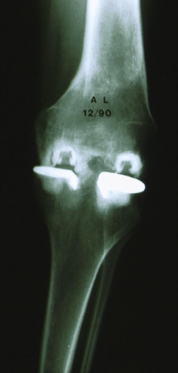
arthroplasty with cemented poly on the tibia and round metal femoral components on the condyles.7 Charnley experimented with a hemiarthroplasty that had a poly component on the femoral side and metal on the tibial side (Load Angle Inlay, Thackeray).8 The success of this was short lived. I revised one of these knees 20 years ago in a rheumatoid patient with a combined varus supracondylar osteotomy and revision arthroplasty (see Fig. 1.1).
The further development of unicompartmental arthroplasty by Marmor,9 Murray, Goodfellow and O’Connor of the Oxford group,10 Scott and the Brigham Group in Boston,11 and many others is not a focus of this book and will not be covered further in this introduction; suffice to say that a uni is an example of an anatomic versus mechanical alignment-based procedure. Intact anterior cruciate ligament (ACL) and posterior cruciate ligament (PCL) are prerequisites, and overcorrection is to be avoided. In some high-volume knee arthroplasty practices at this time (i.e., 2021), up to 60% of cases of primary knee osteoarthritis are being done with unis rather than full TKR.
In the late 1960s, also in England, Michael Freeman, surgeon, and SAV Swanson, engineer, developed a single radius of curvature, nonlinked prosthesis with a flat anterior femoral design.12 It was a “roller in trough” design—a virtual modular equivalent of the hinge prosthesis. Both cruciate ligaments were sacrificed. More important than the prosthesis, however, were Freeman’s important contributions to the history of surgical technique and instrumentation. It was Freeman who introduced parallel cuts of the two major bones that were perpendicular to the mechanical axis. Furthermore, he introduced intramedullary instrumentation to facilitate reproducibility of the cuts. Many of the knees in that era were associated with severe deformities from inflammatory arthropathies, particularly rheumatoid arthritis. He prioritized achievement of reproducible mechanical axis, rather than anatomic alignment.
A major leap forward was the development of the total condylar prosthesis by the English surgeon John Insall, working at the Hospital for Special Surgery in New York City in collaboration with the engineer Peter Walker and Drs. Ranawat and Ingless.13 Both cruciate ligaments were sacrificed. Further refinements by the engineer Burstein led to the Insall-Burstein total condylar prosthesis with an all-poly tibia with a post.14 This laid the foundation for all modern posterior cruciate–sacrificing knee replacements. Parallel with this major achievement was work in Boston by Sledge et al. on designs that preserved the posterior cruciate.15 These early generation knees had drawbacks of all-poly tibial components, which resulted in high stresses on the cement, particularly in larger sizes, and poor patellar tracking. The metal-backed modular tibia solved that problem, and posterior cruciate-retaining philosophy was established as a highly reliable alternative to the New York school,
which requires posterior cruciate sacrifice in all cases with much greater femoral bone stock loss. Patellofemoral issues were serious problems with both the cruciate-sacrificing designs of the New York school and the posterior cruciate–retaining designs of the Boston school. Implant design improvements with a central trough in the femoral component and high profile of the anterior lateral femoral flange helped greatly to improve patellofemoral mechanics.
Some confusion exists in historic terminology. The Boston surgeons at the Brigham and engineer Peter Walker designed the posterior cruciate–retaining “Kinematic Knee Prosthesis,” manufactured by Howmedica and subsequently acquired by Stryker years later. The Kinematic prosthesis was inserted using mechanical alignment instrumentation and principles. Over time, the term “kinematic alignment” has come to mean principles of insertion that respect and reproduce to varying degrees the anatomic variations of individual knees.
No two knees are identical, even in the same individual. Think of the analogy to the human face. We all have faces, yet no two faces are the same. Imagine a facial plastic surgeon dealing with posttraumatic facial deformities or elective facial reconstruction. Is the goal to make every face look the same? Is it not important to respect individual variations that make each face unique? So, too, with the knee. It is clearly understood that the human knee has a few degrees of physiologic varus in the coronal plane, yet the mechanical axis principles require a perpendicular cut of the tibia with the goal of making “every knee the same.” The groundbreaking instrumentation of Freeman was adopted and improved by others. An alternative approach was introduced by Hungerford and Krakow, a more physiologic “kinematic alignment” that respected variations in anatomy of the knee. This was paired with the first U.S. Food and Drug Administration(FDA)-approved cementless TKR in the United States in 1984, the porous-coated bead Porous Coated Anatomic (PCA).16 17 That
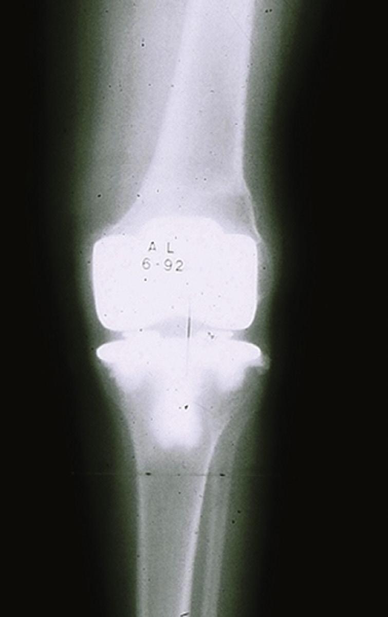
Figure 1.1 The Charnley knee arthroplasty (A) was a biocompartmental hemiarthroplasty with polyethylene femoral component and Vitallium tibial component. I revised one of these in a rheumatoid patient who also had undergone a varus supracondylar osteotomy years before the arthroplasty. The revision was done with posterior cruciate ligament sparing primary implants and a supracondylar valgus femoral osteotomy. This postop image (B) was taken after the osteotomy implant had been removed. The revision total knee replacement was done at the same time as a primary total knee replacement on the opposite knee so that the resected bone from the primary could be used as bone graft for the revision of the left side.
device went on to a much higher failure rate than expected, and porous bead technology is no longer used. This impact on alignment philosophy has lived on, however. It is important to recognize the contribution from Japan by Yamamoto, who reported on a cementless design with superior results to the cemented version in the late 1970s.18 This book explores the evolution and current status of kinematic alignment.
There is a major difference between the mindset of a surgeon and a bench scientist. A scientist postulates many ideas and seeks then to prove that the hypothesis is either wrong or right, without bias. A surgeon works with the burden and expectation of perfection in every operation. Acceptance of an error in planning or technique in any case is difficult for a surgeon to deal with. Accordingly, discarding a traditional and customary technique is difficult, even when data from a welldone study dictate that change is warranted. Consider how many years it took for the majority of surgeons to abandon the use of drains for knee replacements, even when published studies as early as 1998 showed no harm as well as benefits to doing so.19
So how does adoption of kinematic alignment fit in? Mechanical alignment has served as a straightforward and easyto-teach technique. Why change now? The real issue is that 6% to 14% of total knee patients are unhappy with their outcomes in the peer-reviewed literature.20 It is reported much higher, up to 30%, in the lay press.21 Can adoption of kinematic alignment lead to a significant improvement in patient satisfaction? The hypothesis that it will be superior is based on studies that show that the imposition of a rigid, “one size fits all” mechanical alignment approach actually reproduces the native anatomy for less than 5% of individual knees. Hungerford made an even stronger assertion in his chapter on alignment in a TKA book in 2005, when he wrote, “In single-leg stance the ankle must be brought directly under the center of gravity. This means that
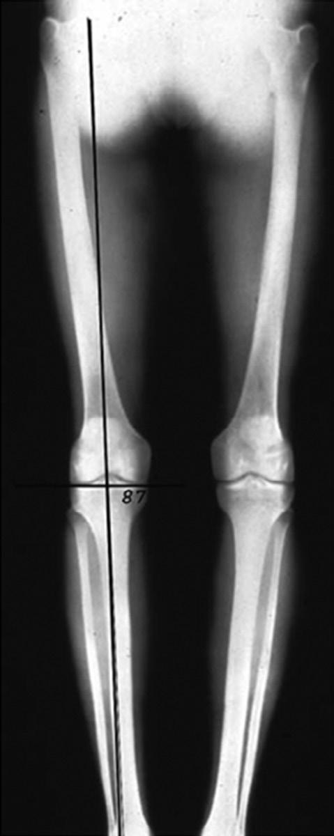
the lower leg and the mechanical axis are inclined toward the midline by 3 degrees. This can vary by as much as ±1.5 degrees depending on the breadth of the pelvis and the length of the femur…” In our experience of measuring this relationship in thousands of patients, we have seen only one patient in whom the joint line was actually perpendicular to the mechanical axis. The tibial shaft is normally parallel to the mechanical axis and is therefore 87 degrees to the joint line and not perpendicular to the joint line (see Fig. 1.2).22 Furthermore, in the peer-reviewed, Ranawat Award–winning paper by Bellemens et al. in 2012, it was reported that up to 32% of men and 17% of women have a native mechanical axis of 3 degrees or more.23 The kinematic alignment philosophy that recognizes and respects the bell curve distribution of “normal” for each individual has intuitive attractiveness. This is not a new concept, but enthusiasm for change has accelerated. However, it is important to include in the introduction that one prospective, randomized study by Young et al., also a Ranawat Award winner of the Knee Society, found no difference between kinematic technique with custom jigs and mechanical axis technique with computer-assisted technique.24 This must be taken into account in the overall equation of what really matters in the quest to make total knee patients as happy as those who get total hips. Is it possible that the way in which the kinematic alignment was performed in that study was significantly different from that proposed in this book? Regardless, the key to widespread acceptance of the principles and adoption of the kinematic technique by the majority of surgeons will hinge on successful results reported by independent surgeons
performing similar studies. One study alone cannot be accepted as definitive, but it must be acknowledged and respected for its impact on the discussion. Other studies have shown improved flexion and improved patient-reported outcomes in kinematically aligned knees, including the prospective study by Dossett et al.25 This is a dynamic and evolving area of clinical practice and research.
When I was a Harvard resident in Boston at the Robert Brigham specialty Rheumatoid and Total Joint Hospital in the late 1970s, total knee patients were admitted 2 days before surgery for “workup” and kept in the hospital for 2 weeks. After the surgery, a bulky Jones dressing was kept on for 5 days with elevation before initiation of partial weight-bearing ambulation and gentle range-of-motion exercises. As an aside, it is no wonder that the incidence of postoperative deep venous thrombosis was so high in that era. I vividly recall the excitement when Dr. Sledge and I got a patient home on day 7 after his total knee in 1979. Now, in 2020, almost all my total knee patients go home without pain on the day of their surgery and take no narcotic medications during their postop recovery period. This is commonplace in many centers in the United States. Within the large Kaiser system in California, over 80% of total knee patients go home on the day of surgery.
In Japan, patients stayed in the hospital for 6 weeks, not because of any inferior surgical techniques, but for cultural and tradition reasons. In fact, Japanese orthopedic surgeons are among the best in the world and surgical standards are very high.
The real issue is that a significant number of patients with a TKR have undesirable residual symptoms or are not satisfied with their surgical outcome. Focus on improvements in alignment are part of the quest for a marked reduction in this very disappointing situation. It is clear to any experienced surgeon who does both hip and knee replacements that the “I forget that I ever had surgery” comment, codified in the “Forgotten Joint Score-12,” is much more common after hip replacements than after knee replacements.26 Many approaches have been taken to address the outcome disparity with the knees. Psychological factors such as high preoperative scores on anxiety or anxiety/depression screening tools (K10, PHQ-9, STAI, etc.) have been studied. Reactive depression postoperatively that can rise to the level of diagnosable posttraumatic stress disorder (PTSD) has been described. Surgical technique factors such as excessively large components; tibial components that overhang the bony margins; tibial components that have reverse slope in the sagittal plane, that is, slope upward from anterior to posterior; malrotated femoral or tibial components, or both; elevation of the joint line; damage to or avulsion of the infrapatellar tendon; chronic, unrecognized, low-grade infection; arthrofibrosis; annoying “clicking and clunking” sounds; excessive laxity in extension only, or in flexion only, or both; patellar dislocation; excessively thick patella because of insufficient bone resection; or neuroma of the infrapatellar branch of the saphenous nerve are just some of the factors that can be associated with patient dissatisfaction after a “routine” TKR. To this day surgeons remain frustrated by the lack of guidance from computer-based surgical planning tools or intraoperative computer-assisted or robotic technology to guide rotation of the tibia component, which remains largely a “judgment call” at the time of surgery.
A new approach to alignment is a potentially important part of the solution to making patients much happier after their total
Figure 1.2 Long standing X-ray with normal alignment. With the ankles together, single-leg stance is stimulated. The mechanical axis is 87 degrees to the joint line, which is horizontal in the stance position. (From Hungerford DS, Hungerford MW. Alignment of the human knee: relationship to total knee replacement. In: Bellemans J, Ries M, Victor J, eds. Total Knee Arthroplasty: A Guide to
knees. The intuitive appeal of a kinematic approach to alignment is that it respects the unique differences of each knee. Just as in the example of the human face, no two knees are completely identical even in the same individual. The patient with the windswept knee appearance is the perfect example, with one knee in varus and the other in valgus. One interesting piece of history with regard to alignment in unis is that Hernigou reported on long-term follow-up of medial compartment unis of the Guepar group (Mark 1, Howmedica). The best 10-year outcomes were in the knees with mild varus alignment postoperatively.27
The mechanical axis approach had the advantage of simple instrumentation for a perpendicular cut of the tibia with matching rectangular cuts of the femur. It was easy to teach and has given satisfactory overall results. However, there are some readily identifiable theoretical shortcomings. First, the upper tibia in humans is not biologically designed perpendicular to the long axis of the leg in either the coronal or the sagittal plane. If it were, you would not be reading this book. Second, the imposition of perpendicularity in the coronal plane causes tightness medially that leads to the need for medial releases and surgical weakening of the important medical collateral ligament. It is particularly problematic in knees that have outlier amounts of native varus. Next, the knee becomes too tight in flexion on the medial side, hence the rationale of the “standard 3 degrees of external rotation” of the femoral component. Resection of more bone from the posterior aspect of the medial femoral condyle gives more room for the artificially elevated medial aspect of the tibia in flexion. This is a modification based on an arbitrary commitment to a nonphysiologic perpendicular cut of the tibia.
Every surgeon has a favorite surgical approach, a favorite implant, a preferred alignment philosophy, and a preferred approach to rehabilitation. What matters? There is a big difference between passion for a certain aspect of surgical strategy by surgeon innovators and demonstration of a significant impact on outcomes of that strategy that would compel sceptics and the broad community of knee arthroplasty surgeons to change their own practices to adopt that strategy and abandon their own prior preferred surgical strategy. One example of a strategy that compels widespread adoption is the use of tranexamic acid (TXA) to reduce operative blood loss. Because of this, blood transfusions after knee arthroplasty are now very
REFERENCES
1. Walldius B. Arthroplasty of the knee using an endoprosthesis. 8 years’ experience. Acta Orthop Scand. 1960;30:137–148.
2. Venable CS, Stuck WG. Ann Surg. 1941;114(2):309–315.
4. MacIntosh DL. Hemi-arthroplasty of the knee using a space-occupying prosthesis for painful varus and valgus deformities. Proceedings of the joint meeting of the Orthopaedic Associations of the English Speaking World. J Bone Joint Surg Am. 1958;40:1431.
5. Scott RD, Joyce MS, Ewald FC, Thomas WH. McKeever metallic hemiarthroplasty of the knee in unicompartmental degenerative arthritis. J Bone Joint Surg Am. 1985;67:203–207.
6. Charnley J. Arthroplasty of the hip. A new operation. Lancet. 1961;1(7187): 1129–1132.
7. Gunston FH. Polycentric knee arthroplasty. Prosthetic simulation of normal knee movement: interim report. Clin Orthop Relat Res. 1973;94:128–135.
8. Minns RJ, Hardinge K. Failure of one design of surface replacement knee arthroplasty due to loosening deformation and wear of the plastic femoral component. Biomaterials. 1983;4(3):147–152.
9. Marmor L. The modular knee. Clinical Orthop Relat Res. 1973;94:242–248.
uncommon. Twenty-five years ago, it was common for patients to drop hemoglobin level four or more points after surgery and require two or more units of allogenic blood transfusions. Use of TXA is now a “standard of care” for all arthroplasty surgeons. To propose a randomized prospective trial of TXA versus no TXA for TKA would now not be ethically justifiable. This is not the case with matters such as PCL-sacrificing versus PCL-preserving approach, rotating versus fixed platform tibial components, use of patient-specific versus standard instrumentation, use of computer-assisted versus conventional technique, and so on. Surgeons have their own reasons for selecting one of these options for their cases, but there is no consensus comparable to that on the use of TXA. We do not really know the causes of the high dissatisfaction rate after knee replacement surgery or how alignment contributes to the mix. It is exciting to see serious efforts to pursue the potential benefit of kinematic alignment with novel instruments designed to help achieve the intended outcome reliably. Validation of meaningful, game-changing improvement in patient satisfaction because of adoption of kinematic alignment will require confirmation and validation from independent surgeon researchers who perform well-controlled prospective studies with excellent technique for both mechanical alignment and kinematic alignment approaches. The data need to be so impressive that proponents of mechanical alignment feel convinced to switch over to kinematic alignment. Availability of reliable, easy-to-use, and easy-to-teach instruments is a necessity.
Hopefully, the chapters of this book will stimulate interest in exploring the potential of kinematic alignment to improve patient satisfaction in a way that can isolate this variable from all the other incremental changes in surgical technique, implant design, and rehabilitation that are simultaneously taking place, and validate its independent contribution. The goal of incremental and disruptive change in knee reconstruction is to fundamentally improve the patient satisfaction after TKR to more than 95%. Those who are filled with a passion for change, including Howell and Bini, will lead the way. Those who are unbiased and inquisitive will prove or disprove the hypothesis that kinematic alignment is superior to mechanical axis alignment with carefully constructed, prospective, and randomized clinical research studies.
10. Murray DW, Goodfellow JW, O’Connor JJ. The Oxford medial unicompartmental arthroplasty: a ten-year survival study. J Bone Joint Surg Br 1998;80(6):983–989.
11. Scott RD, Santore RF. Unicondylar unicompartmental replacement for osteoarthritis of the knee. J Bone Joint Surg Am. 1981;63:536–544.
12. Freeman MA, Swanson SA, Todd RC. Total replacement of the knee using the Freeman-Swanson knee prosthesis. Clin Orthop Relat Res. 1973;94:153–170.
13. Insall J, Scott WN, Ranawat CS. The total condylar knee prosthesis. A report of two hundred and twenty cases. J Bone Joint Surg. 1979;61(2):173–180.
14. Insall JN, Lachiewicz PF, Burstein AH. The posterior stabilized condylar prosthesis: a modification of the total condylar design. Two to four-year clinical experience. J Bone Joint Surg. 1982;64(9):1317–1323.
15. Sledge CB, Stern PG, Thomas WH. Two year follow-up of the duo-condylar total knee replacement. Orthop Trans. 1978;2:193.
16. Hungerford DS, Kenna RV, Krackow KA. The porous-coated anatomic total knee. Orthop Clin North Am. 1982;13(1):103–122.
17. Hungerford DS, Krackow KA. Total joint arthroplasty of the knee. Clin Orthop Relat Res. 1985;192:23–33.
18. Yamamoto S. Total knee replacement with the Kodama-Yamamoto knee prosthesis. Clin Orthop Relat Res. 1979;145:60–67.
19. Adalberth G, Bystrom S, Kolstad K, Mallmin H, Milbrink J. Postoperative drainage of knee arthroplasty is not necessary: a randomized study of 90 patients. Acta Orthop Scand. 1998;69:475–478.
20. Ali A, Sundberg M, Robertsson O, et al. Dissatisfied patients after total knee arthroplasty: a registry study involving 114 patients with 8-13 years of follow-up. Acta Orthop. 2014;85(3):229–233.
21. Szabo L. Up to a third of knee replacements pack pain and regret; Kaiser Health News (December 25, 2018). https://khn.org/news/up-to-a-third-ofknee-replacements-pack-pain-and-regret/
22. Hungerford DS, Hungerford MW. Alignment of the human knee: relationship to total knee replacement. In: Bellemans J, Ries M, Victor J, eds. Total Knee Arthroplasty: A Guide to Better Performance. Heidelberg, Germany: Springer-Verlag; 2005:25–32.
23. Bellemans J, Colyn W, Vandenneucker H, Victor J. The Chitranjan Ranawat Award: is neutral mechanical alignment normal for all patients? The concept of constitutional varus. Clin Orthop Relat Res. 2012;470(1):45–53.
24. Young SW, Walker ML, Bayan A, Briant-Evans T, Pavlou P, Farrington B. The Chitranjan S. Ranawat Award: no difference in 2-year functional outcomes using kinematic versus mechanical alignment in TKA: A randomized controlled clinical trial. Clin Orthop Relat Res. 2017;475(1):9–20.
25. Dossett HG, Estrada NA, Swartz GJ, LeFevre GW, Kwasman BG. A randomised controlled trial of kinematically and mechanically aligned total knee replacements: Two-year clinical results. Bone Joint J. 2014;96-B(7): 907–913.
26. Behend H, Giesinger K, Giesinger J, Kuster M. The “forgotten joint” as the ultimate goal in joint arthroplasty. J Arthroplasty. 2012;27(3):430–436.
27. Hernigou PH, Deschamps G. Alignment influences wear in the knee after medial unicompartmental arthroplasty. Clin Orthop Relat Res. 2004;423:161–165.
2
Phenotypes of the Knee and Limb: Rationale for Transitioning Toward Personalized Alignment in Total Knee Arthroplasty
MICHAEL TOBIAS HIRSCHMANN, MD | SILVAN HESS, MD | LUKAS B. MOSER, MD | EMMA LOUISE ROBERTSON, MB CHB | VINCENT LECLERCQ, MSC
CHAPTER OUTLINE
Overview
Variability of the Native Knee Alignment
What Do We Know So Far?
The Knee Phenotype System
Impact of Alignment Concepts on Knee Alignment
Mechanical Alignment Concept
Anatomical Alignment Concept
Kinematic Alignment Concept
Overview
For a long time, the alignment goal for total knee arthroplasty (TKA) was a neutrally aligned limb with orthogonal joint lines to the mechanical axes (mechanical alignment [MA]). This had mainly to do with the belief of a better long-term durability because of more equal load distribution in mechanically aligned TKA. Recognizing the variability in individual knee alignment and compromised functional outcomes in TKA, there is an increasing interest among knee surgeons for more personalized, more anatomical alignment (AA) methods.1 Among those newer alignment concepts, the kinematic alignment (KA) method is the most promising one.2 3 The goal of the KA concept is to restore the prearthritic alignment of a patient.4 To achieve this goal, surgeons need to have a profound knowledge of the individual native anatomy of the knee and its variability.
This chapter provides a basis for all alignment methods and a detailed overview of the current knowledge regarding the variability of lower limb alignment. First, a review of the literature will be presented. Following the discussion of the literature, the functional knee phenotype concept is introduced. Based on these phenotypes, the difference between three alignment goals of the most common alignment concepts (mechanical, anatomical, and restricted kinematic) and the native alignment will be presented and thoroughly discussed.
Variability of the Native Knee Alignment
When performing TKA, a knee surgeon should consider the native knee alignment of the patient. Only with an extended knowledge of native knee alignment in all planes (coronal, sagittal, and axial) is the surgeon able to achieve the optimum
outcome for the individual patient. The basic principle in knee surgery is to be as anatomical as possible. This principle has been neglected in TKA in terms of alignment for decades and all knees were forced into an alignment not because of better function and anatomy but because of other considerations such as implant durability and implant design, as well as easier and more reliable instrumentation. However, the only constant in anatomy is its variability, as one of the godfathers in knee surgery, Werner Müller, once stated.5
WHAT DO WE KNOW SO FAR?
Numerous studies have investigated the native coronal knee alignment.6–9 The reported overall knee alignment, usually represented as the hip-knee-ankle angle (HKA), averaged around 180 degrees. However, several studies have found that the individual coronal alignment is highly variable, and a significant number of patients appears to have either a varus or valgus alignment. Bellemans et al. investigated the incidence of constitutional varus (HKA <177 degrees) in 250 patients (male:female 125:125), aged between 20 and 27 years, on long leg radiographs (LLRs) in a Belgian population.6 They found that 32.0% of the males and 17.2% of the females had a constitutional varus. Most of the patients (66% of the males and 80% of the females) had a neutral aligned lower limb (177 degrees – HKA – 183 degrees). Only 2% of males and 2.8% of the females had a so-called “constitutional valgus.” Although the ethnicity of the participants has not been described, most of them can be regarded as Caucasians. Shetty et al. investigated the distribution of HKA based on the same criteria as Bellemans et al. in an Asian population and found more knees in varus (34% Indian, 35% Korean).9 The authors suggested that femoral bowing of the Asian population might be one reason for this shift. Interestingly, Song et al. found contradictory results investigating only Korean females. More patients had a valgus than varus (25% vs. 20%) alignment.7 Clearly, to date, the influence of ethnicity and its potential interaction with sex on HKA remains unclear.
Even though there is a great interest in the coronal alignment, most previous studies only used conventional radiographs for their measurements and few used two-dimensional magnetic resonance images (MRI) or EOS images.10 More importantly, a recent systematic review by Moser et al. identified a number of limitations of the existing literature on native coronal alignment.10 For example, the systematic review found only four studies investigating the orientation of the femoral joint line (usually measured as femoral mechanical angle [FMA]) and
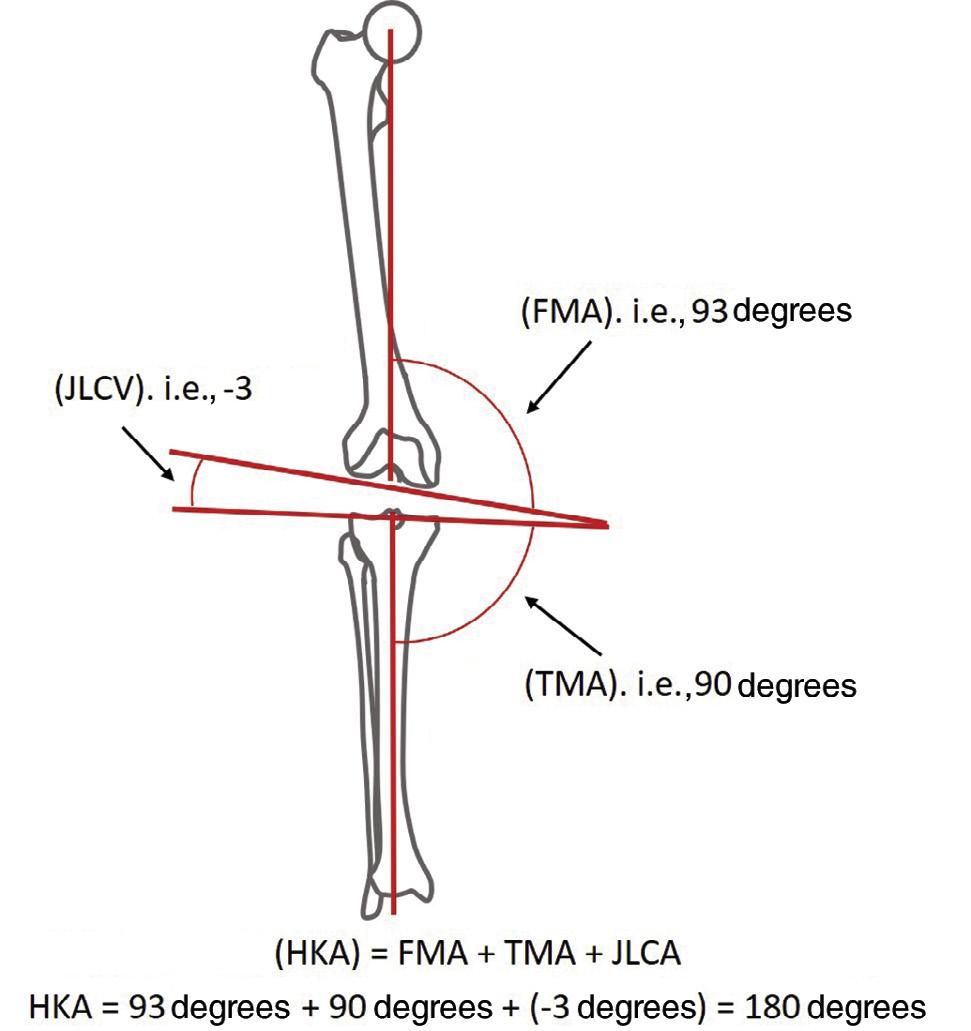
Figure 2.1 Measurement of coronal alignment and alignment equation. The hip-knee-ankle angle (HKA) is defined as the medial angle between the femoral mechanical axis and the tibial mechanical axis. The femoral mechanical angle (FMA) is defined as the medial angle between the femoral mechanical axis and a tangent to the distal femoral condyles. The tibial mechanical angle (TMA) is defined as the medial angle between the tibial mechanical axis and a tangent to the proximal tibial joint surface (tibial plateau). The joint line convergence angle (JLCA) is defined as the medial angle between the two joint lines. The JLCA thus takes negative values if the angle opens up laterally. The HKA equals the sum of FMA, TMA and JLCA (HKA = FMA + TMA + JLCA).
all studies only used LLRs. The reported mean values varied considerably (92.1 ± 1.9 degrees to 97.2 ± 2.7 degrees), and no study reported any ranges. The orientation of the tibial joint line (usually measured as tibial mechanical angle [TMA]) has been investigated by 10 studies, but sample sizes, reported mean values, and ranges varied widely (mean values ranged from 84.6 ± 2.5 degrees to 89.6 degrees). Additionally, many of these studies did not distinguish between male and female patients, although sex-related differences are well known. Therefore it seems that in the past, the research was focused on the overall alignment, whereas details about the orientation of the joint lines have been only superficially covered. As in many fields of knee surgery, for the sake of simplicity and reproducibility, more complexity was
avoided. However, in the context of TKA, the alignment of the joint lines is very important because they are not only directly accessible during surgery but also define the overall alignment. As shown by Cooke et al., the overall alignment defined by the HKA equals the sum of the FMA, TMA, and the joint line convergence angle (angle between joint surfaces [JLCA]).8 Fig. 2.1 shows a detailed description of these angles and the equation. As a consequence of this lack of knowledge, the authors of the present chapter investigated the coronal alignment parameters of a young, nonosteoarthritic population using threedimensional reconstructed computed tomography images.11–13 Their reported mean values support the findings of previous studies, but the reported ranges also demonstrate that the variability of the native anatomy had been underestimated. Mean values and ranges for HKA, FMA, and TMA are shown in Table 2.1. Based on these findings, it seems clear that a systematic MA or AA method will not fit all knees. In some knees the use of these systematic approaches will result in a major change of the alignment, which cannot be balanced by bone cuts only—extensive ligament balancing techniques need to be executed.
Looking at this huge mass of data on coronal knee alignment, one has to recognize that the conventional mindset of knee surgeons, which simply differentiates knees in varus, valgus, or neutral alignment, is too short-sighted. However, this myopic approach focusing on coronal limb alignment based on HKA is still the mainstay in TKA. Starting a more differentiated perspective by making subgroups of varus and valgus knees and including the joint lines draws a different picture, as our data show. There is a myriad of different combinations of the FMA and TMA, and the HKA is only a subordinate parameter of the FMA, TMA, and JLCA. It is therefore not sufficient to only consider HKA, and it is a prerequisite to include joint line orientation of the distal femur and proximal tibia, as well as JLCA, accordingly. An overall neutral, varus, or valgus HKA could have the theoretical combinations of the FMA and TMA shown in Table 2.2.
In fact, this might be one reason for conflicting findings in scientific articles. Some papers did not find any difference for mechanically aligned knees versus nonmechanically aligned knees, whereas others did. Clearly, the problem is that when looking at the overall coronal alignments, one fails to find any difference in outcomes with regards to alignment because patients with a distinctly different alignment are grouped together. The mass of data is in or around the mean and conceals the truth in a big foggy data cloud. We are just not able to see it yet.
Lim phenotypes (Hip-knee-ankle angle; HKA)
Femur phenotypes (Femoral mechanical angle; FMA)
Tibia phenotypes (Tibial mechanical angle; TMA)
Based on the aforementioned, it is clear that there is need for a more detailed analysis of coronal alignment. Consequently, we have developed a new classification system for knee alignment based on a native healthy population and have named it the “knee phenotype system.” This system aims first to allow a better understanding of variability in knee alignment and second, in a later stage, to give guidance for an optimal alignment in TKA for each individual knee.
THE KNEE PHENOTYPE SYSTEM
For all alignment parameters (HKA, FMA, TMA), groups with a range of 3 degrees, so-called phenotypes, were defined.11–13 The HKA groups were called “limb phenotypes,” the FMA groups “femoral phenotypes,” and the TMA “tibial phenotypes.”
The family of FMA, TMA, and HKA phenotypes are defined by the deviation from the mean value of a random sampling of a population of subjects with nonosteoarthritic knees and covers a range of ±1.5 degrees from this mean (e.g., 180 ± 1.5 degrees). To state it another way, the phenotypes represent 3-degree increments of the angle starting from the rounded overall mean angle (HKA: 180 degrees; FMA: 93 degrees; TMA: 87 degrees). Table 2.1 shows the definition of all these phenotypes.
The nomenclature of the phenotypes is organized as follows: The first part (NEU, VAR, VAL) defines the direction of alignment. The second subscripted part (HKA, FMA, and TMA) states the phenotype group. The last part (0, 3, and 6 degrees) shows the angular deviation from the mean value.
Until now, the different aspects of the coronal alignment have been investigated separately from each other. However, the coronal alignment is defined by the myriad of combinations of all these angles and, to see the bigger picture, it therefore seems important to assess the combination of the different parameters.
Thus, in the next step, the authors defined combinations of the three phenotypes, so-called “knee phenotypes” (combination of femoral and tibial phenotype) and functional knee phenotypes (combination of all three).
The alignment of the young, nonosteoarthritic population was phenotyped according to this new system. A total of 18 knee phenotypes were found out of the theoretically possible 25 knee phenotypes (by combining 5 femoral and 5 tibial phenotypes). Some 17 different knee phenotypes were found in the male population, whereas 12 different ones were found in the female population, including 11 mutual phenotypes. Table 2.3 shows the found knee phenotypes and the percentage of patients they represent.
More importantly, it was observed that there were not only 17 different knee phenotypes (e.g., combinations of femoral and tibial joint line) but that the joint line orientations varied strongly in the varus, valgus, and neutral groups. Nine different combinations of femoral and tibial phenotypes (=knee phenotypes) could be found in the varus subgroup, eight in the neutral group, and eight in the valgus group.13
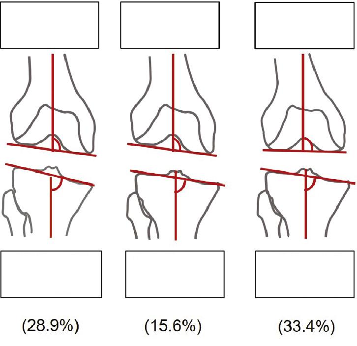
To give an example, a varus knee of 3 degrees HKA could have a distal femoral angle of 3 degrees varus and a proximal tibial angle of 6 degrees varus, whereas it could also have a distal femoral angle of 3 degrees valgus and 3 degrees varus. In TKA surgery, this is hardly the same knee. Fig. 2.2 shows the three most common knee phenotypes in the VARHKA3 degrees group and Fig. 2.3 shows two neutrally aligned patients but different joint line orientations and the clinical impact of this.
Finally, 43 different functional knee phenotypes were found in this nonosteoarthritic population. Their distribution differed significantly between males and females. Separated by sex, there were 35 male and 26 female phenotypes, and a total of 18 mutual phenotypes. The most common eight functional phenotypes
population and number and percentage of
for males and females are shown in Table 2.4 12 In conclusion, it is obvious that the description of coronal knee alignment as “varus,” “neutral,” or “valgus” is outdated and should be replaced by the knee phenotype system.
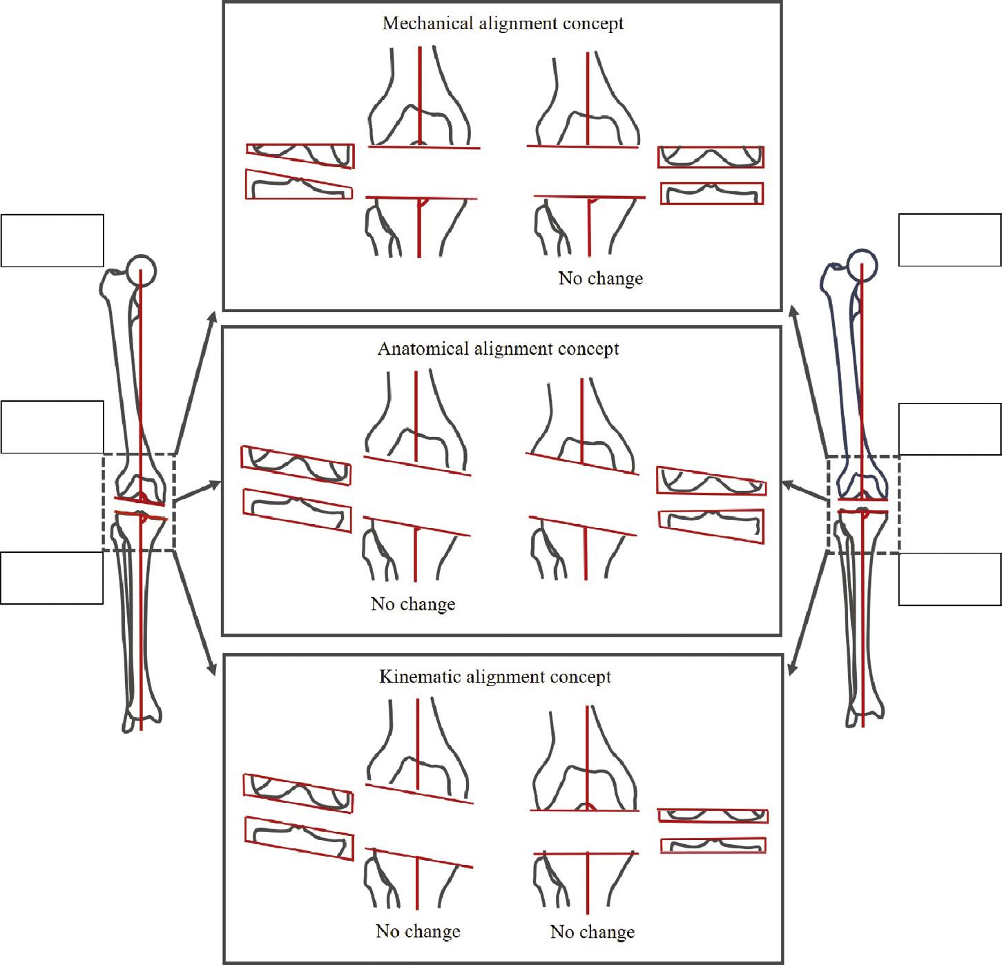
Impact of Alignment Concepts on Knee Alignment
Different alignment concepts have been promoted since the initial development of TKA, and there is ongoing debate regarding the optimal alignment method and goal.12 14 Several recent studies found better clinical outcomes after TKA, when the postoperative alignment more closely resembled the native or prearthritic alignment of the patients.3 15 However, most of the promoted alignment concepts do not aim for the native alignment of the patients.12 Surgeons need to understand both the technical
Figure 2.2 The three most common phenotypes in the VARHKA3 degrees group (patients with a slight varus overall alignment (HKA 180 ±
differences between the different alignment concepts and their impact on patients’ individual alignment. Many questions come into the mind of knee surgeons when thinking about coronal alignment in TKA. What is the rationale behind the use of each alignment system? What are the limitations? What are the pros and cons in terms of personalization? Most of these questions will be answered in this book; the latter one can be answered applying the knee phenotype system. When we aim for the least change from prearthritic native knee alignment, one can calculate the percentage of the population with an alignment (e.g., functional knee phenotype), within the target zones of the different alignment methods (mechanical, anatomical, kinematic). Clearly, the functional knee phenotype concept facilitates a more detailed discussion, as it incorporates all important coronal alignment parameters. The higher the percentage of the population covered by the chosen alignment method, the more likely it is that the native alignment of a patient is restored.
MECHANICAL ALIGNMENT CONCEPT
The alignment goal of the mechanical TKA alignment concept is a neutral limb alignment (HKA of 180 degrees) with femoral and tibial joint lines perpendicular to their mechanical axes (FMA and TMA of 90 degrees).16 Hence, the MA concept aims to purely create the following functional knee phenotype: NEUHKA0 degrees + VARFMA3 degrees + VALTMA3 degrees. This functional knee phenotype was found in 2.05% of the males and 1.77% of the females. These values are comparable to values reported by Almaawi et al., who assessed the impact of the MA and the restricted KA (rKA) on the alignment of a knee OA population.14 These very low numbers are not surprising, as the classic MA concept aims to maximize the durability of the implants rather than restore the native alignment or optimal knee function.
Indeed, TKAs implanted according to this technique showed excellent survival rates in several registries. A pooled analysis in a systematic review found an all-cause survivorship of 96.3% (95% confidence interval [CI] 95.7%–96.9%) at 15 years and
TABLE 2.3 Knee phenotypes found in the total
Figure 2.3 The schematic shows two patients with prearthritic neutral limb alignment with different femoral and tibial phenotypes and joint line orientation. Application of mechanical (top) and anatomic (middle) alignment changes the patient's prearthritic joint line orientation. Only kinematic alignment (lower) does not change the patient's prearthritic distal femoral and proximal tibial joint lines. FMA, Femoral mechanical angle; TMA, tibial mechanical angle.
94.8% (92.5%–97.1%) at 20 years.17 Furthermore, in the majority of patients a significant pain relief and improvement in function was seen.18 However, the question of why about 20% to 30% of all patients are not satisfied after TKA remains.19 20
ANATOMICAL ALIGNMENT CONCEPT
The aim of the anatomical TKA alignment concept is to neutrally align the lower limb (HKA = 180 ± 1.5 degrees) and to anatomically restore the native femoral and tibial alignment (FMA of
93 degrees and TMA of 87 degrees).21 Two biomechanical studies found better load distribution on the tibial component, better patellar kinematics, and reduced risk for lateral ligament stretching with this resulting joint line obliquity.22 23 Thus, the AA concept aims to purely restore the following functional knee phenotype: NEUHKA0 degrees+NEUFMA0 degrees+NEUTMA0 degrees. This functional knee phenotype was found in 18.97% of males and 17.70% of females. Hence, in comparison with MA, only a small adaption in the target zones for the tibial and femoral joint line orientation leads to an enormous increase in
2.4 The most common eight functional knee phenotypes for males and females demonstrate a huge variability in coronal alignment