Acknowledgements
Serendipity invariably has a role in the career pathways of scientists and academics. I was fortunate in finding a Jurassic bryozoan during my independent geological mapping project while an undergraduate at the University of Durham in the early 1970s, and equally fortunate when, unbeknownst to me at the time, my main lecturer in palaeontology, Gilbert Larwood, just happened to be a bryozoan specialist. Gilbert took me under his wing, became my first mentor, and went on to supervise my doctoral research. I then enjoyed two years as a postdoc at the University College of Swansea where I was attracted by the presence of John Ryland’s group studying living bryozoans, including Peter Hayward and John Thorpe to whom I owe a major debt for teaching me so much about bryozoan biology. Another influence in Swansea was Derek Ager. Derek was a highly original thinker about the fossil record and palaeoecology who did not receive the credit he deserved.
After Swansea I was appointed to a research post on bryozoans in the Department of Palaeontology at the then British Museum (Natural History). Pat Cook, my counterpart in the Department of Zoology, taught me never to be too dogmatic when making statements about bryozoans – there are many surprises waiting around the corner to embarrass the unwary in the study of these diverse, complex, and often enigmatic animals.
I will forever be grateful to Daphne Lee at the University of Otago who encouraged me to apply
for a William Evans Fellowship there in the late 1980s. This proved to be my personal Beagle voyage. Keith Probert at the Portobello Marine Laboratory introduced me to the wonderful diversity of animals on the Otago Shelf that interact with bryozoans, not just the hermit crabs I was there to study, while Doug Campbell and Dave MacKinnon (Canterbury University) took me in the field to see the rich Cenozoic bryozoan faunas of New Zealand (Doug’s son Hamish was later to guide me around the glorious Chatham Islands on two memorable geological fieldtrips). My collaborations with Dennis Gordon (NIWA, Wellington), the global authority on cheilostome bryozoans, describing the taxonomy of fossil and Recent bryozoans of New Zealand, began at this time. While at Portobello I was contacted by Richard Boardman (Smithsonian Institution) and F. Ken McKinney (Appalachian State University) who were partway through a study of the peculiar cyclostome bryozoan Cinctipora, one of the bioconstructional species on the Otago Shelf. Their invitation to join them in this research marked the beginning of my interest in bryozoan biomineralization, the skeleton of Cinctipora showing a striking ultrastructure (Boardman, McKinney, and Taylor 1992).
Before migrating to the world of ornithology, Mike Weedon undertook two postdocs with me at the NHM on bryozoan skeletal ultrastructures. Other scientists to whom I am indebted for various collaborative biomineralization projects
include Chiara Lombardi (ENEA, La Spezia), Piotr Kuklinski (Polish Institute of Oceanology, Sopot), Noel James (Kingston University, Ontario), and Bill Schopf (UCLA).
The late F. Ken McKinney, who has already been mentioned, deserves my special thanks. He offered so many fresh insights and was a wonderful person to work with, as well as a great friend. To Dennis Gordon and Ken McKinney, I must add a third long‐standing collaborator, Mark Wilson (College of Wooster, Ohio). It has been a delight working with Mark over the years and benefitting from his knowledge of hard substrate palaeoecology combined with a clarity of thought and expression that makes him such a superb teacher.
More recently, I have been fortunate to collaborate with two of the rising stars of bryozoology. Andrea Waeschenbach (Life Sciences, NHM) undertook a postdoc with Tim Littlewood and me on bryozoan molecular phylogeny, ferreting out the false data in GenBank and adding a lot of new data of her own to show the power of molecular sequences in understanding bryozoan evolution. Lee Hsiang Liow (University of Oslo) has been employing her analytical and modelling prowess to investigate biotic interactions through geological time, in particular competition for substrate space, a field where bryozoans have the potential to make a wider impact in macroecology and evolutionary ecology.
Supervision of doctoral students has served to broaden my perspectives. In chronological order, I thank them all: Richard Carthew, Julian Hammond, Caroline Buttler, Jon Todd, Kevin Tilbrook, Sian Evans, Phil Watts, Jo Snell, Lais Ramalho, Tanya Knowles, Scott Tompsett, Caroline Sogot, Emanuela Di Martino, and Peter Batson.
Others with whom I have been privileged to collaborate and learn from include Ehrhard Voigt, Eckart Håkansson, Andrei Grischenko,
Andrej Ernst, Roger Cuffey, Alan Cheetham, Aaron O’Dea, Eckart Håkansson, Andrew Ostrovsky, Al MacGowan, Ma Junye, Seo Ji Yun, Shun Mawatari, Kamil Zágoršek, Noel James, Jeremy Jackson, Antonietta Rosso, Joachim Scholz, Beth Okamura, Tim Palmer, Andrew Smith, Silviu Martha, Urszula Hara, Patrick Wyse Jackson, Hans Arne Nakrem, Matt Dick, Scott Lidgard, David Jablonski, Abby Smith, Seabourne Rust, Kjetil Voje, Loïc Villier, Leandro Vieira, Francoise Bigey, Helen Jenkins, Consuelo Sendino, and Mary Spencer Jones.
I am grateful to the photographers of the NHM, particularly Harry Taylor, Phil Crabb, and Phil Hurst, for their skilled macrophotography. Most of the SEM images were taken by me during countless productive hours spent in the NHM’s imaging and analysis laboratories where Alex Ball, Chris Jones, and several others were always at hand when help was needed. Piotr Kuklinski, Andrej Ernst, Caroline Buttler, and Thomas Schwaha are thanked for generously providing additional images used in this book.
The study of bryozoans has never attracted the funding it deserves – as Sir David Attenborough once remarked, you can’t care about something unless you know it exists. Alas, far too few people are aware of the existence of these perenielly ‘unfashionable’ animals, let alone have any inkling of their fascinating natural history. Nevertheless, I have benefitted over the years from grants awarded by the Natural Environment Research Council (NERC), Leverhulme Trust, European Union, Royal Society, British Council, and the Japan Society for the Promotion of Science (JSPS), all of which I gratefully acknowledge.
Finally, I would like to thank Caroline Buttler, Andrea Waeschenbach, Lee Hsiang Liow, and Emanuela Di Martino for their comments on the manuscript, and Louise Spencely for her skilled copy editing.
Introduction 1
Once known as ‘polyzoans’ or ‘ectoprocts’, bryozoans are unique in being the only animal phylum in which the great majority, if not all, species form colonies. But what exactly constitutes a colony? In the context employed here it is an aggregate of genetically identical, conjoined modules, unlike a colony of seabirds or of ants. Coloniality has evolved on numerous occasions among aquatic invertebrates. However, coloniality is not homologous between bryozoans and corals or hemichordates, although these independently evolved groups of colonial animals do exhibit several similarities. The most important feature of colonial invertebrates is their modular construction: new modules (zooids) are added during the growth of the colony and, with a few exceptions, remain physically attached to their neighbours throughout the life of the colony. The process of adding a zooid – termed budding – involves mitotic cell divisions only. Thus, all of the zooids in a bryozoan colony are genetically identical clones.
1.1 Zooids
Bryozoan zooids are small, typically measuring under a millimetre in length. However, because bryozoan colonies can contain a large number of zooids, individual colonies are typically a centimetre to a few decimetres in size. For example, seven‐year‐old colonies of Flustra foliacea, the so‐called ‘hornwrack’, less than 10 cm tall have been estimated to contain more than 100 000 zooids (Stebbing 1971). Much larger colonies occur in a few bryozoan species: a 2‐metre diameter colony of the Recent cheilostome Pentapora foliacea (the ‘Ross coral’) was recorded from British coastal waters in the early nineteenth century (Lombardi, Taylor, and Cocito 2010). In freshwater habitats, gelatinous masses formed by aggregations of colonies of the
Bryozoan Paleobiology, First Edition. Paul D. Taylor.
© 2020 Natural History Museum. Published 2020 by John Wiley & Sons Ltd.
phylactolaemate bryozoan Pectinatella magnifica can be as much as one metre in diameter (Dendy 1963; see also Cahuzac and d’Hondt 2017). And fossil trepostome bryozoan colonies up to 66 cm across have been recorded from the Late Ordovician of Kentucky (Cuffey and Fine 2005), and over a metre wide in Permian deposits of Tasmania (Reid 2003). At the other extreme are the tiny colonies of the ctenostome Monobryozoon and related genera, little more than a millimetre in length and consisting of a single feeding zooid plus stolons and detachable buds (Franzén 1960; Berge, Leinaas, and Sandøy 1985; Schwaha et al. 2019). Bryozoans are able to benefit from being simultaneously diminutive in terms of zooid size – bringing the advantages incurred by a large surface area relative to volume –but large with respect to colony size – enhancing their survival from various sources of mortality such as predation and physical disturbance.
In common with other colonial animals, bryozoan colonies are able to endure the death of one or more zooids in the colony. This phenomenon is known as partial mortality. Death of zooids can be due to their natural ageing, or external factors such as predation. In Mediterranean colonies of the palmate cheilostome Pentapora fascialis studied by Cocito, Sgorbini, and Bianchi (1998), local tissue necrosis caused by silt accumulation and algal overgrowth impacted mainly older colonies and caused collapse of their centres while younger zooids at the periphery of the colony continued to live.
As colonies grow, they accumulate an ever‐increasing number of dead zooids in their older parts, forming a necromass. For example, the basal branches of bushy and foliaceous colonies may consist entirely of dead zooids, like the dead wood of trees (e.g. McKinney and Taylor 2006, fig. 3). The skeletons of zooids in the necromass remain functional in the sense of providing structural support for the feeding zooids closer to the branch tips. Describing these old zooids as dead is not strictly accurate. In at least some species, colonies may be reactivated if, for example, the colony is broken and reparative growth ensues.
One important implication of partial mortality is that bryozoan colonies may exhibit negative
growth – with time, more zooids may be lost than new zooids budded, resulting in a net decrease in colony size. In a study of encrusting cheilostomes on settlement panels in Jamaica, Jackson and Winston (1981) found an increasing proportion of colonies showing negative growth through a period of two years. The existence of shrinking colonies means that colony age cannot always be estimated from size – colonial animals may ‘lie about their age’.
The budding of new zooids in bryozoan colonies is usually restricted to specific growth zones, for example, around the perimeters of patch‐like encrusting colonies, or at the branch tips of tree‐like erect colonies. In some species, however, new zooids may be budded more widely across the entire outer surface of the colony.
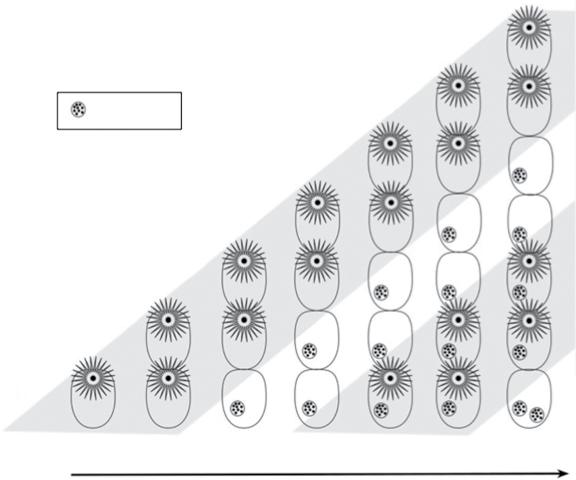
The basic bryozoan zooid (Figure 1.1) consists of a body wall (cystid) containing a fluid‐filled cavity (coelom) within which is suspended a polypide with a lophophore and gut. This arrangement has led to bryozoans occasionally being portrayed as ‘animals‐in‐a‐box’, a concept that can be misleading because the box‐like cystid, including the mineralized skeleton, if present, is actually an integral living part of the zooid rather than an inert container. Nevertheless, some degree of independence between the cystid and the polypide is evident in bryozoan zooids. This is most clearly manifested by the phenomenon of polypide cycling (Figure 1.2) in which the polypide periodically degenerates and is replaced by another within the same cystid, a process characterizing nearly all marine bryozoans (cf. the cheilostome Epistomia bursaria where the zooids have only one polypide generation: Dyrynda 1981). The lifespans of individual polypides range from a few days (Bayer and Todd 1997) to 9–10 months for some slow‐growing species from the Antarctic (Barnes 2000). When placed under stress (e.g. low pH, simulating ocean acidification), polypide cycling is diminished in favour of the budding of new zooids (Lombardi et al. 2017; Swezey et al. 2017).
The remains of the degenerated polypide form a brown body (Gordon 1973, 1977). This is either retained within the coelom of the zooid, or defecated
Figure 1.1 Basic cheilostome bryozoan labelling some of the most important anatomical features of the zooids. Growth direction is from left (proximal) to right (distal). Only the zooid on the right has its lophophore expanded for feeding; in the other three, the lophophore is retracted into the security of the box‐like skeleton (after Taylor, Lombardi, and Cocito 2015, fig. 1. © The Trustees of the Natural History Museum, London).
through the gut of the new replacement polypide. In the cheilostome Steginoporella, individual zooids can contain as many as 22 brown bodies (Palumbi and Jackson 1983), signifying an equivalent number of polypide cycles. As marine bryozoans lack a specialized excretory system, polypide cycling furnishes a means of removing waste material, crudely comparable with autumnal leaf fall in trees.
Although polypide cycling is a mechanism for prolonging the lifespans of zooids, they are not immortal and evidence for zooid senescence can be seen in the decreasing durations of successive polypides and increasing time required for a new polypide to be formed in zooids of some cheilostomes (Bayer and Todd 1997). In addition, older zooids tend to be more heavily fouled and exhibit slower regeneration of colony growth following damage (Palumbi and Jackson 1983).
Fossil evidence for the degeneration of polypides in bryozoans occurs in the form of diagenetically altered brown bodies – brown deposits – which are often seen in thin sections of Palaeozoic bryozoans
(e.g. Morrison and Anstey 1979; Ernst and Voigt 2002; Key et al. 2008; Plate 4A). Most brown deposits have a high iron content and some appear to have been pyritized. Calcified brown bodies are known in a few living cheilostomes (Cummings 1975; Gordon and Parker 1991a) but have yet to be reported in the fossil record. However, so‐called phosphatic pearls or calculi, 30–600 μm in diameter, described from within the zooidal chambers of the Silurian cystoporate bryozoan Favositella by Oakley (1934), may perhaps represent encapsulated brown bodies (Lindskog et al. 2017, p. 36).
Polypides are the food gathering and digestive organs of bryozoans. They consist of an inverted cone-, bell- or horseshoe-shaped crown of tentacles (lophophore), with a mouth at the centre where the tentacles converge (Plate 5). The mouth leads into the U‐shaped gut comprising a sucking pharynx (Nielsen 2013), stomach, pylorus, rectum and, in a few species, a gizzard (e.g. Gordon 1975). Bryozoan lophophores have between 8 and over 100 tentacles, depending on species. The largest lophophores are found in the freshwater phylactolaemates and
Figure 1.2 Schematic figure of polypide cycling in a bryozoan in which brown bodies are retained by the zooid. Starting from a single zooid (lower left), six zooids are budded in succession, making a total of seven zooids in the final growth stage depicted (vertical row on the far right). By the third growth stage, the polypide of zooid 1 has degenerated, leaving a brown body in the cystid, a fate repeated by the other, successively younger zooids as they age. However, through time, new polypides form within the old cystids, giving a second polypide generation. This in turn is followed by a second degeneration, as indicated by the presence of two brown bodies in the oldest zooid (lower right).
7 zooid 6
body first polypide first degeneration
5
second polypide
second degeneration
are horseshoe‐shaped in plan view (Wood 1983; Plate 5E). All bryozoans are active suspension feeders. Marine bryozoans mainly consume phytoplankton (Chapter 3.1), but recent research by Wood (2019) on phylactolaemates has identified ciliate protists and rotifers as major food sources, which may explain the generally larger size of their zooids. The tentacles of the lophophore bear several lines of tiny, hair‐like cilia, including a paired series of lateral cilia that beat in a metachronal wave to force water with entrained
plankton through the open end of the lophophore and towards the mouth.
A key characteristic of bryozoans is the ability of zooids to withdraw the lophophore into the sanctuary of the cystid, expanding again into the water column to resume feeding. Consequently, feeding zooids in bryozoans always have an opening in the cystid for passage of the lophophore. Such openings in the zooidal skeleton are conventionally referred to as apertures in stenolaemates, and orifices in gymnolaemates (Figure 1.3).
zooid
Figure 1.3 Comparative external skeletal morphology of the feeding zooids in a cyclostome stenolaemate (A) and a cheilostome gymnolaemate (B). Note the simple, rounded aperture at the distal end of the cyclostome but the more complex orifice with lateral processes (condyles) at the distal end of the cheilostome zooid. Both zooids in these examples have calcified exterior frontal walls, that of the cyclostome pierced by tiny pseudopores, while that of the cheilostome comprises nine overarching spines fused laterally and along the midline of the zooid, each spine containing a few large pores (pelmata). A, Reptomultisparsa incrustans (Jurassic, Bathonian, Caillasse de la Basse‐Ecarde; Ranville, Calvados, France). B, Hayamiellina aff. constans (Pleistocene, Setana Fm.; Kuromatsunai, Hokkaido, Japan). Scale bars = 200 μm.


Withdrawal of the lophophore is achieved by contraction of retractor muscles that are attached to the base of the lophophore. Protraction is a more complicated process that relies on the contraction of muscles pulling on the cystid walls or a membrane, the precise mechanism varying in different major taxonomic group. Muscular contractions squeeze the coelom and cause the tentacle sheath – a sock‐like structure enclosing the retracted lophophore – to turn inside out. This eversion of the tentacle sheath pushes the lophophore out through the orifice or aperture (Chapter 3.1). In some bryozoans, including most species belonging to the class Stenolaemata, the zooids have the shape of straight or gently curved tubes (Figure 1.4A), typically becoming progressively wider in diameter distally towards the skeletal opening for the lophophore. In other bryozoans, such as most species of the order Cheilostomata, the zooids are approximately box‐shaped (Figure 1.4B) and
have a distinct frontal surface containing a distal opening through which the lophophore extrudes. The outermost component layer of the cystid, at the interface with the external environment, is a thin organic coating called a cuticle. Like the periostraca of molluscs and brachiopods, this is secreted by an underlying epithelium. Those parts of the cystid wall that are deformed by the muscles involved in tentacle sheath eversion must be flexible and therefore cannot develop a rigid biomineralized layer beneath the cuticle. However, parts of the cystid not deformed by parietal muscles can potentially develop a biomineralized skeleton between the secretory epithelium and the outer cuticle (Chapter 2.1). Bryozoan skeletons are composed of the calcium carbonate biominerals calcite and aragonite. Both of these biominerals, calcite in particular, are relatively stable and have a high fossilization potential. The palaeontological literature often
(A) (B)
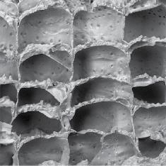
Figure 1.4 Typically tubular zooidal skeletons of a stenolaemate compared with the box‐shaped zooidal skeletons of a cheilostome: (A) fractured colony of the trepostome Stenopora crinita with long prismatic zooids intersected by prominent growth bands at the bottom and top of the image (Permian; Illawara, New South Wales, Australia); (B) broken colony of the cheilostome Schizoporella errata showing five frontally budded layers of box‐like zooids (Recent; Haifa Bay. Israel). Scale bars: A = 1 mm; B = 500 μm.
employs the terms zooecium (pl. zooecia) and zoarium (pl. zoaria), which are the skeletal parts of the zooid and colony, respectively. These terms are largely superfluous and are not used in this book.
1.2 Colonies
Based on zooids that range from tubular to box‐like in shape, bryozoans have evolved a myriad of colony‐forms (Plates 6–8, 10–12). Bryozoan zooids may be likened to building bricks that can be arranged in different ways to generate structures diverse in shape and varied in function. The inherent plasticity endowed by the modularity that characterizes bryozoans accounts for the large disparity in colony‐forms (Chapter 4.1). Some colony‐forms are common, particularly sheet‐like encrustations, whereas others are found in only a handful of taxa. Identical colony‐forms have evolved repeatedly in different bryozoan clades through the long geological history of the phylum (Chapter 9.11). Evolutionary convergence in colony‐form can be viewed in terms of fitness landscape theory: within the limits imposed by various developmental, structural,
and phylogenetic constraints, particular colony‐forms represent adaptive peaks – they are optimal morphological strategies for coping successfully with the challenges of living as a sessile benthic suspension‐feeder.
An important consequence of the rampant convergent evolution of colony‐forms in bryozoans is that the overall shape of a colony is seldom a reliable indicator of its taxonomic identity. Therefore, bryozoan taxonomy has been founded mainly on the morphological characters of the zooids. For bryozoan groups having mineralized skeletons, most of the characters used in taxonomy are skeletal, both in living and fossil bryozoans. Indeed, it is standard practise for living bryozoans to be bleached prior to identification, a process mimicking taphonomic loss of soft tissues during fossilization. This means that the same morphological characters are usually employed in the identification and classification of fossil bryozoans as those used for living taxa. Unlike some phyla, a parataxonomy for fossils has been unnecessary in bryozoans. Notwithstanding the identical taxonomic procedures and classifications employed for living and fossil bryozoans, it is becoming apparent from molecular studies that
(A)
(B)
traditional skeletal characters are not always closely correlated with phylogeny (Chapter 8). Bryozoan classifications will need to be comprehensively overhauled if they are to reflect phylogeny more clearly.
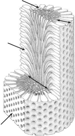
Discordance in taxonomy can also result from the different methods employed to study the skeletons of fossil bryozoans. Palaeozoic bryozoans are most often studied in thin sections, which are seldom used for post‐Palaeozoic bryozoans where surface features are employed. There are two main reasons for this contrast in study methods. The first is that Palaeozoic bryozoans more often occur in hard, strongly lithified rocks from which it is difficult to extract specimens showing reasonably well‐preserved colony surfaces. Secondly, the dominant bryozoans of the Palaeozoic belonging to the superorder Palaeostomata have fewer external characters than the cyclostome and cheilostome bryozoans of the post‐Palaeozoic. Thin sections of palaeostomate bryozoans are cut in three standard orientations – transverse, longitudinal, and tangential – to reveal internal skeletal characters and build up an understanding of the three‐dimensional morphology of the skeleton (Figure 1.5).
1.2.1 How has coloniality arisen?
Coloniality has evolved multiple times in metazoans (e.g. Ryland 1981). It is easy to envisage how a colony may have developed from a unitary ancestor by reference to the well‐known freshwater cnidarian Hydra. This animal reproduces asexually, budding clonal daughter individuals on the side of the body. These grow to a certain size and then drop off. If the buds had instead remained attached to the parent, then a colony would result. Retention of asexual buds to initiate coloniality has been termed clonoteny (Rosen 1986). In the case of bryozoans, the unitary ancestor would have resembled a modern ‘worm’ of the lophophorate phylum Phoronida. The most reliable recent molecular phylogenetic analyses (Nesnidal et al. 2013; Laumer et al. 2019) have corroborated earlier anatomical suggestions (Hyman 1959) that phoronids are the sister‐group of bryozoans. Whereas most phoronids are unitary animals, one
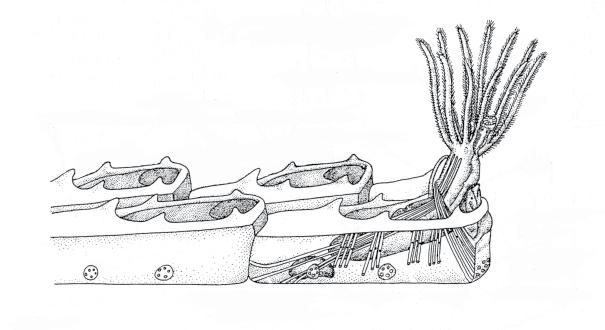
Figure 1.5 Cutaway diagram of a branch from a ramose trepostome colony (after Madsen and Håkansson 1989), showing the three standard sections employed by bryozoologists, as well as the division of the branch into an inner endozone, in which the zooids have thin walls and parallel the length of the branch, and an outer exozone in which zooids have thick walls and are oriented at right angles to branch length.
transverse section
longitudinal section
tangential section
species (Phoronis ovalis) forms true colonies by budding, while several others are able to multiply asexually. The model of Farmer, Valentine, and Cowen (1973; see also Farmer 1977) for the origin of bryozoans starts from a unitary phoronid worm in which the asexual buds are retained. This is
followed by size reduction of the zooids constituting the new colony. Because the genetic individual (genet) is the entire colony, the zooids can be small without sacrificing the advantages that large overall body size brings to competitive interactions, resistance to disturbance etc. The disproportionate relationship between body size and metabolic rate – metabolic allometry – acts as a constraint on the physiological rates of animals which can be ameliorated to some extent by having a modular colonial construction (see Hartikainen, Humphries, and Okamura 2014).
Small zooid size brings with it the physiological advantages for metabolic exchange and efficiency of having a relatively large surface area/volume ratio (Ryland and Warner 1986). Indeed, bryozoan zooids dispense with the sophisticated circulatory and excretory systems found in larger unitary animals.
1.3 Colony Propagation
Sexual reproduction in bryozoans is important not only as a means of generating genetic variation, but also because the larvae produced by sexual reproduction are the principal agents of dispersal. Most bryozoan colonies – and often their constituent zooids – are hermaphroditic and produce both testes and ovaries (see Reed 1991 and Ostrovsky 2013 for reviews of bryozoan reproduction). In contrast, a few species of crisiid cyclostomes seem to be dioecious with colonies that are either male or female (Robertson 1903; Jenkins, Bishop, and Hughes 2015). Individual zooids are typically sequential hermaphrodites, functioning first as males and then as females. In some species, however, the sexes are separate and male and female zooids are distinct polymorphs that differ morphologically and, in some instances, lack the ability to feed (Chapter 3.6).
While self‐fertilization between zooids belonging to the same colony is in theory possible, cross‐fertilization between colonies is believed to be the norm. For example, colonies of the cheilostome
Celleporella kept in isolation fail to produce larvae (Cancino, Castañeda, and Orellana 1991) and invest less in producing female zooids (Hughes, Manriquez, and Bishop 2002). In another cheilostome, Bugulina stolonifera, selfing can occur if colonies are kept in isolation but results in fewer viable larvae and these are less likely to complete metamorphosis after settling (Johnson 2010).
The production of female polymorphs (gonozooids) in cyclostomes can be considerably reduced in the absence of sperm from other colonies (Jenkins, Bishop, and Hughes 2015). Sperm are released into the water through pores in the tips of the tentacles and must be acquired by female zooids from another colony, a process which is aided by the feeding currents of the recipient zooids drawing water towards themselves. The period of viability of water‐borne sperm is limited, with an estimated half‐life of just 1.2 hours in the cheilostome Celleporella hyalina (Manríquez, Hughes, and Bishop 2001). Depending on species concerned, fertilization can occur either externally or internally in gymnolaemate bryozoans (Temkin 1996), while in stenolaemates the location is unknown but is almost certain to be internal, with the sperm being gathered by the transient polypide present during the early development of the gonozooid. After fertilization of the egg, the embryo in most bryozoan species is brooded by the parent colony before being released as a swimming larva when sufficiently developed. The diverse styles of embryonic brooding or incubation found among bryozoans (e.g. Ström 1977) are discussed in Chapter 3.
Brooded bryozoan larvae are short‐lived, incapable of feeding, and must settle quickly to establish a new colony before their provision of energy resources from the parent becomes exhausted. Therefore, settlement commonly occurs in close proximity to the parent colony (e.g. Mariani 2003), a phenomenon called philopatry. In the cheilostome Bugula neritina larvae also tend to settle close to sibling larvae (Keough 1984). Settlement is followed by metamorphosis, entailing radical changes in tissue organization and resulting in the founding individual of the new colony called
the ancestrula. The ancestrula initially adheres to the substrate using a sticky acid mucopolysaccharide secretion from the pyriform organ of the larva (Loeb and Walker 1977). The first asexually budded zooids originate directly from the ancestrula and these in turn bud further zooids to continue colony growth. Rates of budding in new colonies vary greatly: fast‐growing encrusting bryozoans such as Conopeum tenuissimum are capable of producing as many as 150 budded zooids in the first week of their life (Dudley 1973), while the bushy fouling cheilostome Bugula neritina may grow to a height of 20 mm in the first month of life (Mawatari 1951).
Although sexual reproduction is the main mechanism for colony multiplication in bryozoans, fragmentation of colonies provides an additional means of propagation in some species, especially in free‐living species (see Chapter 4.4). Barnes, Webb, and Linse (2006b) found that nearly half of the colonies of the erect palmate cheilostome Cellarinella nutti they collected from the Weddell Sea, Antarctica, had grown from fragments of pre‐existing colonies, a similar proportion to that of Neogene fossils of another palmate cheilostome, Metrarabdotos, from Venezuela (Cheetham et al. 2001).
A study of Late Cretaceous and Paleocene palmate colonies of the cheilostome family Coscinopleuridae (Håkansson and Thomsen 2001) revealed an increase in asexually propagated colonies through geological time at the expense of colonies formed by larvae. The same authors (Thomsen and Håkansson 1995) had earlier found that most of the erect species in their samples reproduced predominantly through fragmentation. In one of these – Columnotheca cribrosa – there was a strong positive correlation between the proportion of ovicellate brooding zooids in the colony and the proportion of sexually recruited colonies when samples from different habitats were compared (Figure 1.6). Colonies formed through fragmentation dominated in reef mounds, larvally recruited colonies in most other habitats.
Very few bases of attachment indicative of larvally recruited colonies are known in the
Figure 1.6 Correlation between the proportion of brooding zooids within colonies and that of sexually produced colonies in the cheilostome Columnotheca cribrosa from 10 samples collected in the Danian of Denmark (based on Thomsen and Håkansson 1995, fig. 6).
sexually recruited colonies
common Carboniferous fenestrate Archimedes (McKinney and Burdick 2001). Taken together with the abundant evidence of axial screws originating from the edges of meshworks, this led McKinney (1983) to propose that colony multiplication by fragmentation was a dominant process in Archimedes (Figure 1.7). Many of the larger colonies toppled and came to rest horizontally on the seabed. New screws grew from the edges of the prostrate colonies before eventually breaking off and forming the nuclei for a new generation of screws.
Fragments derived from a single colony are, of course, genetically identical. Borrowing from
Figure 1.7 Life history of the fenestrate bryozoan Archimedes (based on McKinney 1983, fig. 2). A colony recruited from a larva (left) is shown attached to a cylindrical substrate (as in McKinney and Burdick 2001). After toppling of the colony onto the sea‐bed, a secondary screw axis grows vertically. Subsequent detachment of this screw axis initiates an asexual cycle of propagation.

nomenclature devised for clonal plants, each fragment is called a ramet, whereas the entire complement of ramets is known as the genet. Occasionally, the ramets of bryozoans come back into contact during growth, in which case they are able to fuse. Fusion between parts of the same genet, including not only separated ramets but also separate lobes of a single integral colony, has been termed autosyndrome. Although less common than autosyndrome, fusion between two genetically distinct colonies of the same species can also occur (e.g. Craig and Wasson 2000; Hughes et al. 2004). This is known as homosyndrome. ‘Colonies’ produced by homosyndrome are chimaeras as they consist of zooids of more than one genotype. In other colonial animals (corals and tunicates), chimaeras show reduced fitness (Rinkevich and Weissman 1987), implying that homosyndrome should be selected against. Ishii and Saito (1995) described four possible outcomes when colonies of Watersipora (referred to by them as Dakaria) came into contact: (i) overgrowth of one by the other; (ii) back‐to‐back growth of the two colonies to produce a bilamellar erect structure; (iii) non‐fusion stand‐off (see Chapter 5.1);
and (iv) fusion between the colonies and subsequent growth as a continuous sheet. Fusion between colonies of another cheilostome, Membranipora membranacea, results in temporary neural integration, which enables behavioural coordination of lophophore retraction following disturbance, but there is no exchange of metabolites between the fused colonies (Shapiro 1996).
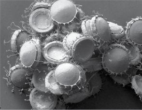
Production of asexual reproductive propagules called statoblasts (Figure 1.8) is ubiquitous in the freshwater phylactolaemate bryozoans. These resistant seed‐like bodies have a chitinous covering and are used in dispersal, sometimes by becoming attached to the plumage of waterfowl, and also in overwintering. Statoblasts are able to survive freezing and desiccation (e.g. Hengherr and Schill 2011). The fossil record of statoblasts is poorly known: most examples are from the Holocene or Pleistocene (e.g. Francis 1997; Courtney Mustaphi et al. 2016), but they have been recorded back to the Late Triassic (Kohring and Pint 2005) and possibly even the Permian (Vinogradov 1996).
Hibernacula are incipient zooids with thickened cuticles found in some ctenostomes, such as Victorella, which function as asexual overwintering
surface
Figure 1.8 Statoblasts produced by freshwater phylactolaemate bryozoans for asexual reproductive, dispersal and overwintering: (A) statoblasts of the living phylactolaemate Cristatella mucedo with hook‐like spines around the annulus (Recent; Luxembourg); (B) putative fossil statoblast showing cracked convex capsule and marginal annulus (Lower Cretaceous, Korumburra Gp; South Gippsland, Victoria, Australia). Scale bars: A = 1 mm; B = 200 μm.
propagules (e.g. Carter et al. 2010) or as resting stages at times of food shortage (e.g. Jebram 1975).
‘Overwintering’ may also be achieved among some marine cheilostome bryozoans. For example, Aetea is capable of producing a special type of zooid called a saccule that persists on the substrate after loss of the other zooids in the colony and is capable of reinitiating colony growth (Balduzzi, Barbieri, and Gristina 1991).
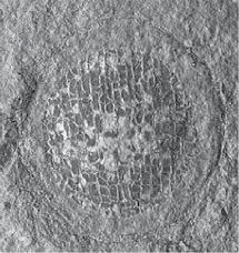
A recent study of bryozoans from the Cretaceous Chalk (Taylor, Di Martino, and Martha 2018) found that some species encrusting echinoid tests apparently underwent periods of dormancy and regeneration, comparable to the overwintering described above for the living bryozoans. In a few species of cheilostomes, sealed, non‐feeding zooids were budded around the margins of the sheet‐like colonies and growth evidently ceased, later resuming with the budding of new feeding zooids. The best example is found in ‘Micropora’ eleanorae where the sealed zooids survived on the substrate, despite the destruction of the autozooids which had more thinly calcified skeletons, to give rise to new generations of feeding zooids (Figure 1.9).
In Hislopia, specialized zooids break away from the parent colony and swim for a time before
settling to found a new clonal colony (Wood, Anurakpongsator n, and Mahujchariyawong 2006), a remarkable mode of asexual propagation known only from this single genus of freshwater ctenostomes.
1.4 Ecology
Bryozoans are widely distributed today across the globe, from polar regions to the equator. Diversities are high in some parts of the world. For example, no fewer than 556 species have been recorded living in the Mediterranean Sea (Rosso and Di Martino 2016).
Although most marine bryozoans inhabit subtidal environments on the continental shelf, they are known from the intertidal down to abyssal depths: the greatest recorded depth for a bryozoan is 8300 metres (Hayward 1981). Freshwater bryozoans are locally abundant in rivers and lakes. While most marine bryozoans can be categorized as stenohaline (i.e. intolerant of salinities differing significantly from normal marine), some species prosper in lower salinities (Winston 1977), including the brackish waters of estuaries and the inner parts of the Baltic Sea, as well as the fluctuating
(A)
(B)
Figure 1.9 Dormancy and regrowth in the Cretaceous cheilostome ‘Micropora’ eleanorae (Campanian, Chalk Gp.; Norwich, Norfolk, UK): (A) autozooids (bottom) transitioning to closed kenozooids (top) believed to be formed during periods of colony dormancy; (B) regrowth from the edge of a band of kenozooids (bottom) through the formation of a new subcolony commencing with a pseudoancestrula (arrowed). Scale bars = 500 μm.
salinities found in coastal lagoons (see Chapter 6.1). Calcification tends to be relatively weak in these bryozoans. Menon and Nair (1974) remarked that during low salinity monsoonal times the euryhaline cheilostome Einhornia crustulenta in the Cochin backwaters of India showed little calcification and the normally calcified opercula consisted only of unmineralized cuticle.
Of fundamental importance for both marine and freshwater bryozoans are hard or firm substrates onto which the larvae can settle to
establish new colonies (e.g. Eggleston 1972a). Commonly used substrates are rocks, shells, and plants, the latter including coastal algae and seagrasses in marine environments, and submerged branches of trees in freshwater environments. A few bryozoan species, most belonging to the cheilostome genus Jellyella, are specialized to grow on floating Sargassum or on the buoyant dead shells of the squid Spirula and consequently have a pelagic, pseudoplanktonic ecology (Taylor and Monks 1997). Another floating bryozoan is the Antarctic ctenostome Alcyonidium pelagosphaerum which forms hollow, ball‐like colonies up to 23 mm in diameter and lacking an obvious substrate (Peck, Hayward, and Spencer Jones 1995; Porter and Hayward 2004).
All bryozoans are suspension feeders, consuming predominantly dinoflagellates. Some other potential sources of nutrition have been suggested. Exudates from seaweeds may be used as an alternative trophic resource by epiphytic bryozoans (De Burgh and Fankboner 1978; Manríquez and Cancino 1996), dissolved organic matter has also been proposed as a source of nutrition (Best and Thorpe 1991), while uptake of amino acids directly from seawater by the cheilostome Bugula neritina has been demonstrated (Stephens and Schinske 1961). However, there is as yet no indication that these contribute significantly to the diet of bryozoans, at least not for species inhabiting shallow‐water environments.
Bacteria living within bryozoan tissues have also been hypothesized as nutritionally beneficial to the host (e.g. Karagodina et al. 2018). Perhaps of more interest in a palaeobiological context is the notion of bryozoan symbiosis with nutrition‐providing zooxanthellae, given the effects of such symbioses on host skeletons, including hypercalcification, and reef‐building capacities of other invertebrates through geological time. Zahl and McLaughlin (1957) mentioned the occurrence of zooxanthellae in the tissues of bryozoans but this has not been confirmed. Good candidates for bryozoans hosting symbiotic zooxanthellae are giant colonies of the trepostome Tabulipora sp. from the Early Permian of North Greenland with branches up to 7 cm thick. However, analysis of
(A)
(B)
the stable light isotopes in skeletons of these bryozoans failed to find the expected higher ∂13C values, leading to rejection of the zooxanthellae hypothesis (Key et al. 2005). Symbiosis was also dismissed as a cause of gigantism in some Moroccan Late Ordovician bryozoans by Jiménez‐Sánchez, Vennin, and Villas (2015) because the trepostomes concerned lived in low mesophotic to oligophotic conditions and at a high palaeolatitude.
Bryozoans themselves feature in the diets of a wide range of animals, notably pycnogonids (sea spiders) and nudibranchs (sea slugs) (see Chapter 5.2). Some bryozoan predators take single zooids at a time, while others consume the entire colony or large parts of the colony.
1.4.1 How long do bryozoan colonies live and how fast do they grow?
The lifespan of individual colonies varies according to species. Many species living in latitudes experiencing strong seasonality survive for a year or less (e.g. Eggleston 1972b). Colonies of some such species die after sexual reproduction. Their demise in the winter can be due to the deterioration or destruction of their substrates. Bryozoans such as Membranipora membranacea living as epiphytes of macroalgae may perish when the fronds they colonize become detached or decay, which occurs annually in some algae (e.g. Seed
et al. 1981; Cook, Bock, and Gordon 2018, p. 73). In polar environments winter ice‐scour can totally obliterate benthic communities annually, including any bryozoans (Conlan et al. 1988). At the other end of the spectrum are perennial colonies. Conspicuous annual growth bands are evident in the skeleton of some long‐lived cheilostomes (Plate 7A). For example, Pentapora has dense but narrow winter bands that alternate with broad summer bands (Lombardi et al. 2008). Similar bands have allowed colony ages to be estimated in some living species (Stebbing 1971; Brey et al. 1998; Barnes, Webb, and Linse 2006a, b). In the Antarctic cheilostome, Melicerita obliqua, growth bands reveal a maximum age of 45 years (Bader and Schäfer 2004). The congeneric Melicerita chathamensis from New Zealand has smaller colonies that live for about nine years, growing at a linear rate of about 5.3 mm per annum and adding approximately 110 zooids each year to the single palmate branch of the colony (Smith and Lawton 2010; Key et al. 2018).
Bryozoan growth rates, which have been quantified in several ways, vary enormously between colonies of the same species from different localities, and even more so between different species. For example, Smith (2014) compiled published data on linear growth rates which are summarized here in Figure 1.10. Most of the studied colonies
1.10
growth rates of bryozoan species using data compiled from the literature by Smith (2014, supplementary material).
Figure
Linear
grew at less than 5 mm per annum but some grew much more quickly. Possibly the highest linear growth rate recorded is from the runner‐like encrusting cheilostome Aetea in which branch tips can advance across the substrate at 72 cm per annum (Jackson and Coates 1986). Kuklinski et al. (2012) reported growth rates of sheet‐like encrusting bryozoans on settlement panels across a latitudinal gradient of 44–78°N in Europe. They found growth rates in terms of colony area to be lower at higher latitudes, both overall and for particular taxa, e.g. colonies of the cyclostome Diplosolen from the Adriatic Sea grew at a rate of about 180 mm2 per annum compared to 4 mm2 per annum in Spitsbergen.
Sokolover, Ostrovsky, and Ilan (2018) found the growth rate of Schizoporella errata to be much greater in high ambient flow velocities than in low flow, echoing findings from other taxa (e.g. Hughes and Hughes 1986). Both field‐ and laboratory‐reared colonies of the cheilostome Membranipora membranacea showed significant increases in growth rate with increasing temperature, with a roughly fivefold increase in mean growth rate between colonies in the laboratory kept at 6°C and 14°C (Saunders and Metaxas 2009). A prodigious rate of growth was recorded by Cocito et al. (2006) in colonies of the erect cheilostome Pentapora fascialis living close to submarine freshwater springs in the Adriatic Sea. Here colonies grew at almost 10 cm per year, compared to a ‘normal’ rate of growth of about 3 cm per year elsewhere in the Mediterranean. High concentrations of bicarbonate in the freshwater of the springs may be a key factor in the higher growth rate in this case.
Growth bands interpreted as annual increments have been noted in a few fossil bryozoans. Using this evidence, a large cyclostome colony from the Late Cretaceous was estimated as having lived for more than 35 years on the basis of its growth bands (Taylor and Voigt 1999), while some trepostome colonies from the Permian of Tasmania may have reached 75 years in age (Reid 2014). Cyclical variations in zooidal aperture spacing in a colony of the cryptostome Rhombopora from the Carboniferous of Ireland
(Hageman et al. 2011) could be detected at average frequencies of every 5.3, 9.4, and 23.3 zooids. On the basis of growth rates in modern bryozoans, the 23.3 zooid cycle was interpreted as annual and the 5.3 cycle as lunar/tidal, allowing the inference to be made that the 20 cm‐tall colony lived for about 20 years. There remains considerable scope for studies of geochemical signatures to estimate growth rates and colony ages in fossil bryozoans, extending research using isotopic profiles in some Recent species (e.g. Pätzold, Ristedt, and Wefer 1997).
1.5 Taxonomy
Three bryozoan classes – Phylactolaemata, Gymnolaemata, and Stenolaemata – have long been recognized on the grounds of major anatomical differences. Their status as monophyletic clades has been confirmed recently by molecular phylogenetic studies (Waeschenbach, Taylor, and Littlewood 2012). Phylactolaemates form the sister‐group of gymnolaemates + stenolaemates. Whereas all phylactolaemates living today are found in freshwater environments, the other two classes have an overwhelmingly marine distribution. Key morphological features of the three bryozoan classes are summarized in Table 1.1.
Order‐level subdivisions are employed in gymnolaemates and stenolaemates but not in the low diversity phylactolaemates in which there are fewer than 100 described species. The two gymnolaemate orders are: ctenostomes, which lack mineralized hard parts, and cheilostomes, which are characterized by biomineralized skeletons as well as zooidal orifices closed by a lid‐like operculum. Elevation of Ctenostomata and Cheilostomata to subclass level has been proposed recently by d’Hondt (2016), together with numerous other categorical rank adjustments. However, large‐scale changes to the classifications used for gymnolaemates (and other bryozoans) must await a better understanding of bryozoan phylogeny that will emerge as molecular sequence data becomes available in a greater number of taxa.
Table 1.1 Morphological characteristics of the three classes of bryozoans.
Phylactolaemata Gymnolaemata Stenolaemata
Mineralized skeletonabsent present or absent present
Zooid shape tubular tubular or box‐like tubular
Polymorphism lacking present present
Lophophore large and usually horseshoe‐shaped small to moderate in size and circular small and circular
Musculature intrinsic body wall musclesparietal muscles parietal muscles
Epistome* present absent absent
Lophophore frontal ciliapresent present absent
Habitat freshwater marine, rarely brackish or freshwater marine
* the epistome is a flap above the mouth, shared with Phoronida and believed by some to represent a separate body cavity – the protocoel – an interpretation contested by Gruhl, Grobe, and Bartolomaeus (2005) for phoronids, and later by Gruhl, Wegener, and Bartolomaeus (2009) for phylactolaemates.
1.5.1 Ctenostomata
Most modern ctenostome species are ‘weedy’ and inconspicuous, although a few develop bushy or gelatinous colonies of considerable size. For an impression of colony‐form disparity in recent ctenostomes, see d’Hondt (1983) and Hayward (1985), while the paper on Brazilian ctenostomes by Vieira, Migotto, and Winston (201a) contains excellent photographs of living colonies.
Cryptopolyzoon is unusual in agglutinating grains of sand which stick to the zooids, in some instances almost entirely covering the colony (Dendy 1889). The small pedunculate ctenostome Clavopora has a stalk of muscular heterozooids which can contract to alter the position and orientation of the head on which the feeding zooids are situated (Mawatari 1968).
Aside from their lack of a mineralized skeleton, a characteristic structure found in ctenotomes, plus a few cheilostomes (e.g. Cellaria: Perez and Banta 1996), is the setigerous or pleated collar. This is a thin membrane with a cuff‐ or collar‐like form that emerges just before the lophophore is protruded and can be reinforced by rods (M.J. McKinney and Dewel 2002). Its function may be to push aside debris and generally prevent
the lophophore from being fouled in the muddy habitats colonized by many ctenostomes. Despite their soft‐bodied nature, ctenostome bryozoans can be found in the fossil record, either as borings in calcareous substrates (Chapter 4.8), or as bioimmurations (Figure 1.11) resulting from overgrowth by other organisms with hard skeletons (e.g. Voigt 1979; Taylor 1990a, b; Todd 1994; Todd, Taylor, and Favorskaya 1997). Bioimmurations can take the form of natural moulds on the underside of the overgrowing organism (Figure 1.11A, G), or casts on the substrate surface when the spaces left by decay of the bryozoan zooids are filled by diagenetic minerals such as calcite or pyrite (Figure 1.11B–D, F). A related, non‐biogenic process – lithoimmuration – may have been responsible for the preservation of the ctenostome Pierrella larsoni encrusting the insides of the body chambers of baculite ammonites in the Late Cretaceous Western Interior Seaway of the United States (Wilson and Taylor 2012; Figure 1.11E). Early diagenetic growth of authigenic calcite associated with formation of the concretions containing the ammonites seems to have moulded the bryozoan zooids before their decomposition. In addition, outlines of encrusting ctenostome zooids


Figure 1.11 Immured fossil ctenostome bryozoans: (A) mould bioimmuration of Simplicidium brandesi on the underside of a serpulid annelid tube (Cretaceous, Berriasian; near Balki, Crimea); (B) natural cast biommuration of another colony on the outside of the same serpulid; (C) cruciate branching pattern in a natural cast bioimmuration of Simplicidium smithii (Jurassic, Kimmeridgian; South Ferriby, Lincolnshire, UK); (D) artificial cast of a bioimmured zooids of an un‐named arachnidiid (Triassic, Anisian, Upper Muschelkalk; Künzelsau‐Garnberg, Baden‐Würtemberg, Germany); (E) steinkern of the body chamber of a baculite ammonite with impressions of zooids of the ?lithoimmured Pierrella larsoni (Cretaceous, Campanian or Maastrichtian, Pierre Shale; Red Bird, Montana, USA); (F) natural cast bioimmuration of an un‐named arachnidiid, each zooid with a pair of longitudinal grooves possibly marking the edge of the frontal membrane (Jurassic, Upper Callovian or Early Oxfordian, Oxford Clay Fm.; Stanton Harcourt, Oxfordshire, UK); (G) mould bioimmuration of Buskia waiinuensis with a stolonal network to which were attached formerly erect autozooids flattened against the substrate during overgrowth by a cyclostome bryozoan from bottom right towards top left (Pleistocene, Nukumaru Limestone; Waiinu Beach, Whanganui, New Zealand). Scale bars: A, B = 2 mm; C, D, G = 500 μm; E = 10 mm; F = 200 μm.
(C) (D)
(E)
(F)
(G)