No part of this publication may be reproduced or transmitted in any form or by any means, electronic or mechanical, including photocopying, recording, or any information storage and retrieval system, without permission in writing from the publisher. Details on how to seek permission, further information about the Publisher’s permissions policies and our arrangements with organizations such as the Copyright Clearance Center and the Copyright Licensing Agency, can be found at our website: www.elsevier.com/permissions
This book and the individual contributions contained in it are protected under copyright by the Publisher (other than as may be noted herein).
Notices
Knowledge and best practice in this field are constantly changing. As new research and experience broaden our understanding, changes in research methods, professional practices, or medical treatment may become necessary.
Practitioners and researchers must always rely on their own experience and knowledge in evaluating and using any information, methods, compounds, or experiments described herein. In using such information or methods they should be mindful of their own safety and the safety of others, including parties for whom they have a professional responsibility.
With respect to any drug or pharmaceutical products identified, readers are advised to check the most current information provided (i) on procedures featured or (ii) by the manufacturer of each product to be administered, to verify the recommended dose or formula, the method and duration of administration, and contraindications. It is the responsibility of practitioners, relying on their own experience and knowledge of their patients, to make diagnoses, to determine dosages and the best treatment for each individual patient, and to take all appropriate safety precautions.
To the fullest extent of the law, neither the Publisher nor the authors, contributors, or editors, assume any liability for any injury and/or damage to persons or property as a matter of products liability, negligence or otherwise, or from any use or operation of any methods, products, instructions, or ideas contained in the material herein.
Previous edition copyrighted 2012 by Saunders, an imprint of Elsevier, Inc.
Library of Congress Cataloging-in-Publication Data
Names: Dabbs, David J., editor.
Title: Breast pathology / [edited by] David J. Dabbs.
Other titles: Breast pathology (Dabbs)
Description: Second edition. | Philadelphia, PA : Elsevier, [2017] | Includes
bibliographical references and index.
Identifiers: LCCN 2016044093 | ISBN 9780323389617 (hardcover : alk. paper)
Subjects: | MESH: Breast Neoplasms–pathology | Breast Diseases–pathology | Breast–pathology
Pathology of Neoadjuvant Therapeutic Response of Breast Carcinoma
Rare Breast Carcinomas: Adenoid Cystic Carcinoma, Neuroendocrine Carcinoma, Secretory Carcinoma, Carcinoma with Osteoclast-like Giant Cells, LipidRich Carcinoma, and Glycogen-Rich Clear Cell Carcinoma
Neoplasia of the Male Breast
Tumors of the Mammary Skin
Metastatic Tumors in the Breast
Timothy M. D’Alfonso, MD
Assistant Professor of Pathology and Laboratory
Medicine
Weill Cornell Medicine
New York, New York
Breast Tumors in Children and Adolescents
Siddhartha Deb, MBBS, BMedSci, FRCPA
Department of Pathology
Peter MacCallum Cancer Centre
Consultant Pathologist
Anapath
Melbourne, Australia
Neoplasia of the Male Breast
Ian Ellis, BMedSci, BM BS, FRCPath
Professor of Cancer Pathology
Division of Cancer and Stem Cells
University of Nottingham
Honorary Consultant Pathologist
Department of Histopathology
Nottingham University Hospitals
City Hospital Campus
Nottingham, United Kingdom
Ductal Carcinoma In Situ
Invasive Ductal Carcinoma of No Special Type and Histologic Grade
Nicole N. Esposito, MD
Acting Medical Director
Department of Pathology
St. Joseph’s Women’s Hospital
Tampa, Florida
Fibroepithelial Lesions
Papilloma and Papillary Lesions
Stephen B. Fox, Bsc(Hons), MBChB, FRCPath, FFSc, FRCPA, DPhil
Director, Pathology Department
Peter MacCallum Cancer Centre
Melbourne, Australia
Neoplasia of the Male Breast
Marie A. Ganott, MD
Associate Clinical Director of Breast Imaging
Department of Radiology
Magee-Womens Hospital of UPMC
Pittsburgh, Pennsylvania
Breast Imaging Modalities for Pathologists
Laurie M. Gay, PhD
Foundation Medicine, Inc.
Cambridge, Massachusetts
Next-Generation DNA Sequencing and the Management of Patients with Clinically Advanced Breast Cancer
Christiane M. Hakim, MD
Professor of Radiology
Chief, Department of Radiology
Magee-Womens Hospital of UPMC
Medical Director of Breast Imaging
Hillman Cancer Center
Pittsburgh, Pennsylvania
Breast Imaging Modalities for Pathologists
Erika Hissong, MD
Resident
Department of Pathology and Laboratory Medicine, Weill Cornell Medicine
New York, New York
Special Types of Invasive Breast Carcinoma: Tubular Carcinoma, Mucinous Carcinoma, Cribriform Carcinoma, Micropapillary Carcinoma, Carcinoma with Medullary Features
Syed A. Hoda, MD
Professor
Pathology and Laboratory Medicine
Weill Cornell Medical College
New York, New York
Normal Breast and Developmental Disorders
Zuzana Kos, MD, FRCPC
Assistant Professor
University of Ottawa
Department of Pathology and Laboratory Medicine
The Ottawa Hospital
Ottawa, Ontario, Canada
Molecular-Based Testing in Breast Disease for Therapeutic Decisions
Gregor Krings, MD, PhD
Assistant Professor
Department of Pathology
University of California San Francisco (UCSF)
San Francisco, California
Mesenchymal Neoplasms of the Breast
Shahla Masood, MD
Professor and Chair
Pathology and Laboratory Services
University of Florida College of Medicine–Jacksonville
Jacksonville, Florida
Patient Safety in Breast Pathology
Syed K. Mohsin, MD
Head of Breast Pathology
Riverside Methodist Hospital Columbus, Ohio
Gross Examination of Breast Specimens
Anna S. Nam, MD
Resident
Department of Pathology and Laboratory Medicine
Weill Cornell Medicine
New York, New York
Breast Tumors in Children and Adolescents
Michaela T. Nguyen, MD
Breast Pathology Fellow
Department of Pathology and Laboratory Medicine
New York Presbyterian Hospital–Weill Cornell Medicine
New York, New York
Nipple Adenoma (Florid Papillomatosis of the Nipple)
Steffi Oesterreich, PhD
Professor
Department of Pharmacology and Chemical Biology
University of Pittsburgh Cancer Institute
Director of Education
Women’s Cancer Research Center
Magee-Womens Research Institute
Pittsburgh, Pennsylvania
Lobular Neoplasia and Invasive Lobular Carcinoma
Shweta Patel, DO
Staff Pathologist
Department of Pathology and Laboratory Medicine
Allegheny General Hospital
Allegheny Health Network
Pittsburgh, Pennsylvania
Metastatic Tumors in the Breast
Joseph T. Rabban, MD, MPH
Professor
Department of Pathology
University of California San Francisco (UCSF)
San Francisco, California
Mesenchymal Neoplasms of the Breast
Emad A. Rakha, MD, PhD, FRCPath
Clinical Associate Professor
University of Nottingham
Honorary Consultant Pathologist
Nottingham University Hospitals NHS Trust
Nottingham, United Kingdom
Ductal Carcinoma In Situ
Invasive Ductal Carcinoma of No Special Type and Histologic Grade
Metaplastic Breast Carcinoma
Rathi Ramakrishnan, MD, FRCPath
Consultant
Department of Cellular Pathology
Imperial College
London, United Kingdom
Paget Disease of the Breast
Jeffrey S. Ross, MD
Albany Medical College
Albany, New York
Next-Generation DNA Sequencing and the Management of Patients with Clinically Advanced Breast Cancer
Pathology of Neoadjuvant Therapeutic Response of Breast Carcinoma
Special Types of Invasive Breast Carcinoma: Tubular Carcinoma, Mucinous Carcinoma, Cribriform Carcinoma, Micropapillary Carcinoma, Carcinoma with Medullary Features
Sandra J. Shin, MD
Professor of Pathology and Laboratory Medicine
Chief of Breast Pathology
New York Presbyterian Hospital–Weill Cornell Medicine
New York, New York
Nipple Adenoma (Florid Papillomatosis of the Nipple)
Special Types of Invasive Breast Carcinoma: Tubular Carcinoma, Mucinous Carcinoma, Cribriform Carcinoma, Micropapillary Carcinoma, Carcinoma with Medullary Features
Mesenchymal Neoplasms of the Breast
Breast Tumors in Children and Adolescents
Jan F. Silverman, MD
Professor and System Chair
Department of Pathology and Laboratory Medicine
Allegheny General Hospital
Allegheny Health Network Pittsburgh, Pennsylvania
Metastatic Tumors in the Breast
Jules H. Sumkin, DO, FACR
Professor and Chair of Radiology
UPMC Endowed Chair for Women’s Imaging Pittsburgh, Pennsylvania Breast Imaging Modalities for Pathologists
Steven H. Swerdlow, MD
Professor of Pathology
Director, Division of Hematopathology
University of Pittsburgh Medical Center Health System
University of Pittsburgh Medical Center Presbyterian Pittsburgh, Pennsylvania
Hematopoietic Tumors of the Breast
Gary M. Tse, MBBS, FRCPC, DAB, FRCPath
Senior Medical Officer
Department of Anatomical and Cellular Pathology
Prince of Wales Hospital
The Chinese University of Hong Kong
Shatin, Hong Kong
Radial Scar
Victor G. Vogel, MD, MHS
Director, Breast Medical Oncology/Research
Gesinger Health System
Danville, Pennsylvania
Epidemiology of Breast Cancer and Pathology of Heritable Breast Cancer
Amy Vogia, DO
Department of Radiology
Magee-Womens Hospital of UPMC
Pittsburgh, Pennsylvania
Breast Imaging Modalities for Pathologists
Noel Weidner, MD
Senior Consultative Pathologist
Clarient Laboratories
Aliso Viejo, California
Reactive and Inflammatory Conditions of the Breast
Infections of the Breast
Myoepithelial Lesions of the Breast
Mark R. Wick, MD
Professor of Pathology
Division of Surgical Pathology
University of Virginia Medical Center
Charlottesville, Virginia
Tumors of the Mammary Skin
INTRODUCTION
The increasing complexity and specialization of breast pathology are readily evident at the daily signout bench of clinical specimens. Today’s breast specimen reports, reflecting the complex nature of the specimens, are akin to term papers, according to one of our clinical breast pathology fellows. Only a few decades ago, the diagnostic pathology report for breast carcinoma patients was relatively straightforward and consumed perhaps not more than one piece of paper. However, because of the increasingly complex nature of pathology specimens, including the gross and microscopic reconstruction of size of lesions and semiquantitation of biomarkers for clinical care, the actual grossing and microscopic review of tissues is complex.
The pathology of breast specimens is likely no different from the increasing complexity of pathologic examination of other organ systems. The increasing complexity of cases is leading to greater specialization of pathologists to focus their attention to the clinical needs of clinicians and patients alike.
Gone are the days of simple hematoxylin and eosin–stained section examination. The progression of complexity has passed through the needs for biomarker immunohistochemistry, biomarker semiquantitation, lesional quantitation, and ascertainment of pathologic responses to therapies in the neoadjuvant setting.
As a result of this increasing complexity and specialization, along with additional demands from our colleagues in precision medicine, further extended quality assurance schemes have become relevant.
Like any other discipline in pathology, breast pathology necessitates having a relevant peer review quality assurance program to minimize diagnostic errors, many of which have played well in the media1–4 (see also Chapter 5). Peer review quality assurance of surgical pathology is migrating from postsignout to the presignout arena5 to intercept diagnostic variations.
The CLIA (Clinical Laboratory Improvement Amendments) medical director is responsible for constructing a robust peer review quality assurance program to minimize diagnostic variation and errors. Along these lines, the presignout review of breast pathology cases is clearly
1. Swapp RE, Aubry MC, Salomao DR, et al. Outside case review of surgical pathology for referred patients. Arch Pathol Lab Med 2013;137:233–240.
2. Perkins C, Balma D, Garcia R, et al. Why current breast pathology practices must be evaluated. A Susan G. Komen for the Cure white paper: June 2006. Breast J. 2007;5:443–447.
3. Staradub VL, Messenger KA, Hao N, et al. Changes in breast cancer therapy because of pathology second opinions. Ann Surg Oncol. 2002;9:982–987.
4. Landro L. What if the doctor is wrong? The Wall Street Journal. Jan 17, 2012.
5. Owens SR, Wiehagen LT, Kelly SM, Picolli AL, Lassige K, Yousem SA, Dhir R, Parwani AV. Initial experience with a novel pre-sign-out quality assurance tool for review of random surgical pathology diagnoses in a subspecialty-based university practice Am J Surg Pathol. 2010;34:1319–1323.
a step in the right direction and is clearly superior to a review done in arrears.
What do pathologists think of peer review consultations? In general, pathologists enthusiastically embrace such consultations, especially when there is a difference of interpretation among pathologists at a given institution. Pathologists will also welcome consultation when they observe that they have little experience with the lesion at question. Tissue samples of limited quantity are also a source of common consultation. Gone are the days of self-absorption and fragile egos when it comes to consultations with colleagues.
Review of outside consultation materials among external institutions has demonstrated major disagreements in up to 8% of breast pathology cases.3 These retrospective studies signal a need for comprehensive consultative peer review quality assurance programs in the CLIA laboratory setting, especially moving toward presignout peer review situations.
Even in the expert setting, targeted peer review quality assurance review in the presignout setting not only minimizes real-time diagnostic interpretative errors, but also is a focus of constant educational endeavor. In a discipline such as breast pathology, a minority of highimpact cases (breast atypias) are more likely to show variation than the diagnostically obvious cases (invasive cancers), specifically when presignout review is performed by pathologists in a collegial setting. When pathologists partake in peer review on a routine basis, pathologists will gravitate to a better understanding of the sources of interpretative variation, and this will affect their practice in a positive way. It will also affect patient management.
One of the recent studies of interobserver reproducibility in breast pathology by Elmore (see Chapter 5) is an illustration of ignorance of how pathologists practice in the real-world setting under the aegis of a CLIAmandated peer review quality assurance program. The Elmore paper garnered the media attention that was sought and unsettled women needlessly. The single positive bit of information that was demonstrated in the Elmore paper was the revelation of the variation of interpretation among the expert panel members. Experts are not immune from diagnostic variation, and, even in the expert setting, much is to be gained by intradepartmental expert peer review consultation (see Chapter 5).
Although a peer review quality assurance program is crucial in the CLIA laboratory setting for practicing pathologists, no one has addressed quality assurance of an even greater pernicious problem with the molecular testing of breast carcinoma specimens. These tests were developed with the intent of providing greater reproducibility of risk assessment for breast cancer patients—a reproducibility that, according to the vendor companies, could not be found among the grading and staging of tumors by pathologists. The reality of the results of these molecular tests, especially the laboratory developed test (LDT) variety, is that there is even greater variation of
test results among different platforms than the variation that occurs with the grading of tumors by pathologists.6 Needless to say, treatments that are based on these tests vary even more than they have before because there is little evidence for such clinical decisions. The longer a breast cancer molecular test is available, the more claims the companies can make about the prowess of the test, especially for the older LDT generation of tests. There is one thing for certain regarding all these molecular breast cancer tests regardless of vendor: those who have benefited the most are the stockholders of the companies.
These tests, introduced in 2004 as a first-generation testing platform and as a laboratory developed test, have had a significant impact on how patients are treated. However, 12 years after their introduction, there are no data that demonstrate exactly how patients benefit from the use of these tests (see Chapter 10). In our review of the role of molecular testing for breast carcinoma for prognostic and predictive interpretation, we concluded that, in fact, these tests offer very little compared with traditional pathologic data generated by a pathologist.7 These tests have proliferated into various branded vendors, and they are all based on populations of patients, sometimes heterogeneous, sometimes homogeneous, some of which are laboratory developed tests, and some of which have been cleared by the Food and Drug Administration (FDA). These in vitro diagnostic multiindex analyte assays have a high impact and are a high risk for patients and, at a minimum, command clearance by the FDA, an independent consumer-oriented agency whose intention is to maximize patient safety (see Chapter 10).
Only recently have the prospective, randomized MINDACT (Microarray in Node Negative Disease May Avoid Chemotherapy) trial data been released regarding the clinical utility of the MammaPrint (MP) test
6. Barlett JMS, Bayani J, Marshall A, et al. Comparing breast cancer multiparameter tests in the OPTIMA trial: No test is more equal than the others. J Natl Cancer Inst. 2016;108. do:10.1093/ jnci/djw050.
7. Rakha EA, Reis-Filho JF, Baehner F, et al. Breast cancer prognostic classification in the molecular era: the role of histological grade. Breast Cancer Res. 2010;12:207.
(American Association for Cancer Research, April 2016). The trial, sponsored in Europe by Agendia (Amsterdam, The Netherlands), accrued more than 6000 patients and compared their genomic classifier score of MP with Adjuvant! Online (AO) to discern if the MP test offered clinical utility beyond AO. The AO and MP tests were concordant in about two thirds of patients, whereas one third was discordant. For the discordant group, patients were randomized to chemotherapy or no therapy. The results demonstrated that patients who had a low-risk AO assessment alone in the randomized group did not benefit from chemotherapy. Overall, this resulted in a 14% reduction in chemotherapy for the high-risk MP group. These results present clinicians with a profound paradigm shift in the potential better use of the MP test with AO and should prompt clinicians to rethink the use of genomic classifiers in general.
It is predicted that this group of prognostic/predictive tests will melt away in the near future to give way to specific actionable genomic aberrations, most likely documented through massively parallel sequencing (next-generation sequencing). MATCH (Molecular Analysis and Therapy Choice) and UMBRELLA trials are currently under way by the National Cancer Institute (NCI) to determine actionable mutations for breast cancer patients.
The second edition of this multi-authored breast pathology textbook is meant to inform readers of the latest developments in diagnosis and practice in the field of breast pathology. Some topics are more amenable to updates, depending on the pace of topic information. The topics brought to your attention here “up front” reflect some of the hottest topics of recent times.
My special thanks to every contributing author, as we dedicate this volume to the patients that we serve.
David J. Dabbs, MD
Normal Breast and Developmental Disorders
Syed A. Hoda
1
Normal Breast 1
Embryology 1
Gross Anatomy 1
Structure and Histology 3
Ultrastructure 11
Arterial Supply 13
Venous Drainage 14
Lymphatic System and Regional Lymph Nodes 14
Nerve Supply 15
Hormone Regulation 15
Thelarche 16
Pregnancy, Lactation, and Milk 16
NORMAL BREAST
The breasts are the distinguishing feature of mammals and have evolved as milk-producing organs to provide appropriate nourishment to their offspring; indeed, the word mammal itself is derived from mamma, which is the Latin term for breast. There are other purported benefits of nursing. Physiologically, this act serves to help involute the uterus; and psychologically, it helps to “bond” the mother and the offspring.1 Other than the aforementioned functions of the breast, its epigamic value cannot be overemphasized.
Embryology
Breast development in utero starts in the first trimester of gestation with formation of bilateral ridges of the ectoderm on the ventral aspect of the fetus. These thickened ridges extend in a linear manner from the axilla to the groin, forming the so-called milk line (Fig. 1.1). As fetal development proceeds, all except a pair of these thickenings, one on each side of the pectoral region, regress.2–5
In its earliest stages, the aforementioned thickening is caused by condensed mesenchymal tissue around an epithelial bud. Solid epithelial cordlike columns develop from the bud. Portions of dermis increasingly envelop the epithelial columns and develop into the connective tissue of the breast. More fibrous elements of the dermis extend into the developing breast and much later form the suspensory ligaments of Cooper (after Astley Cooper, the English anatomist and surgeon, who described these structures in the 19th century). Gradually, the epithelial columns branch, canalize, and transform into ducts (and eventually into lobules). Thus each column
Menopause 18
Male Breast 18
Developmental Disorders 18
Amastia 19
Hypoplasia 19
Polymastia 19
Supernumerary Nipple 19
Aberrant Breast Tissue 19
Macromastia 20
Other Disorders of the Breast 20
Summary 20
ultimately gives rise to a lobe of the breast. A “pit” in the epidermis forms at the convergence of the major (lactiferous) ducts, and shortly thereafter, its eversion forms the protuberant nipple (Fig. 1.2).6 Rarely, the nipple may not evert, resulting in an inverted (or permanently retracted) nipple. This deformity may cause considerable difficulty in suckling.
In the third trimester, the developing mammary glands are responsive to maternal hormones and exhibit mild secretory changes. On parturition, the withdrawal of maternal hormones stimulates prolactin release, which initiates colostrum (“witch’s milk”) secretion. This occurs during the first few days after birth in approximately 90% of infants of both sexes. Colostrum is actually composed of water, fat, and debris, and its secretion dissipates within a month or so after birth. During this time, and for a period of a few weeks thereafter, the breast is palpably enlarged. Until puberty, in both sexes, the breast glandular tissue consists almost exclusively of major ducts.7
Gross Anatomy
The female breasts are rounded protuberances on either side of the anterior chest wall. The organ is present in a rudimentary form in prepubertal girls and boys, and adult males. The bulk of female breast tissue overlies the pectoralis major muscle from the second to the sixth rib in the vertical axis and from the sternal edge to the midaxilla in the horizontal axis. Breast glandular tissue usually extends beyond these arbitrary boundaries. The extension of breast tissue from the upperouter quadrant into the axilla is eponymously referred to as the tail of Spence (after James Spence, a Scottish
1.1
FIG. 1.2 Embryonic development of the breast. Schematic depiction of developing mammary bud from that in a 6-week embryo to birth: epithelial primordium (A), incipient duct formation (B), early duct formation (C), inverted nipple stage (D), and elongation of ducts and eversion of nipple (E). Area outlined in bottom arc depicts progressively growing connective tissue.
surgeon of the 19th century). This “tail” can be difficult to visualize on routine mammograms, and in earlier times, the patient was routinely placed in the socalled Cleopatra pose (a semireclining stance in which the patient turns and leans backward, thought to be a
favored pose of the famed Egyptian pharaoh), to allow such visualization.8
The breast is enveloped by fascia. Anteriorly, there is superficial pectoral fascia. Posteriorly, there is deep pectoral fascia. These two layers of fascia blend with the cervical fascia superiorly and with that overlying the abdomen inferiorly. Fibrous bands (the aforementioned Cooper ligaments), more numerous in the superior half of the breast, connect these two layers of fascia. A “space” filled with loose connective tissue lies between the deep boundary of the breast and the fascia of underlying skeletal muscle. This retromammary space allows the breast some degree of movement over the underlying pectoral fascia. The deep fascia overlying the chest wall sometimes harbors breast glandular units. These glands only rarely extend beyond this fascia into bands of underlying skeletal muscle. Such extension of breast glandular tissue into these deep structures is a normal anatomic feature that has clinical implications, most notably in modified radical mastectomies, which aim to remove as much of the breast glandular tissue as possible. Most mastectomies (short of the draconian radical mastectomies) are successful in removing no more than 90% of breast glandular tissue.
The shape and size of the breast depend not only on genetic and racial factors, but also on age, diet, parity, and menopausal status of the individual. The breast can appear hemispheric, conical, pendulous, piriform (ie, pear shaped), or thinned and flattened; however, typically, the breast is oval and hemispheric, with the long axis diagonally aligned over the chest. There is a distinct flattening of the superficial contour of the breast superior to the nipple.
The normal mature nonlactating female breast weighs approximately 200 g (±100 g).9 The typical lactating breast may weigh more than 500 g. The average adult breast typically spans 12 cm in diameter and 6 cm in thickness. In a study of breast volume in 55 women, Smith and coworkers10 found that the right breast was less voluminous: the mean volume for the right breast was 275 mL, and that for the left breast was 290 mL. This discrepancy has been correlated to handedness. There is no correlation between breast mass and risk of carcinoma because large breasts do not necessarily contain more glandular parenchyma.
The nipple, which is centrally located and typically elevated from the surrounding areola, is the most distinctive feature of the skin of the breast. The level of the nipple, vis-à-vis the thorax, varies widely but typically overlies the fourth intercostal space in younger women. Both nipple and areola are pink, light brown, or darker (depending on the general pigmentation of the body). These two structures are somewhat less pigmented in the nulliparous female and become increasingly pigmented starting in the second month of pregnancy. The tinctorial change is irreversible after pregnancy.
Between 12 and 20 minute rounded protuberances in the dermis, representing prominent sebaceous gland units usually associated with a lactiferous duct, are present on the surface of the areola.11 These protuberances are referred to as Montgomery tubercles (after Dr. William Montgomery, a 19th-century Irish obstetrician, although it is possible that Morgagni, the 18th-century
FIG.
Schematic depiction of the milk line. The milk line extends from the axilla to the inguinal region in the adult. Supernumerary nipples and/or breast tissue may persist anywhere along these lines.
Italian anatomist, detailed the same structures much earlier). Montgomery tubercles become prominent during pregnancy and lactation, reflecting the need for keeping the areola moist during feeding. The tubercles regress after menopause. Apocrine and sweat glands are also present in the immediate area. Hair follicles are present at the edge of the areola. The presence of these glands and hair follicles may be involved in the pathogenesis of persistent subareolar abscesses.
Skin incisions for breast surgery are generally based on the knowledge of the natural orientation of collagen fibers in the skin along the lines first described by Karl Langer, the 19th-century Austrian anatomist. Adherence to Langer lines of skin orientation in making surgical incisions ensures minimal scarring and better cosmetic outcome.12 These lines are based on mechanical principles rather than on any specific anatomic structures (and are founded on the somewhat macabre premise of the direction in which the human cadaver’s skin of a particular region will split if struck by a spike!).
Clinicopathologically, the breast is divided into four quadrants: upper-outer, upper-inner, lower-inner, and lower-outer; however, these quadrants do not exist in anatomic terms. In this context, the terms multifocal and multicentric merit mention. Multifocal is usually defined as disease within the same quadrant, whereas multicentric is the term generally used to describe disease in a least two quadrants (or greater than 5.0 cm apart).13 There are multiple scenarios that reveal the inadequacies of these definitions (eg, “boundary” tumors that traverse two quadrants and centrally placed tumors that span multiple quadrants). Perhaps an improved approach would be to define multicentricity as tumors that lie beyond a variable 90-degree arc, rather than a fixed quadrant based on a clock dial with two lines drawn, one between 12:00 and 6:00 o’clock, and another connecting 3:00 and 9:00 o’clock. However, even this approach “still suffers from the pie-shaped wedge that narrows to a point the closer one gets toward the center of the nipple-areola complex, culminating in those pesky subareolar and central tumors, which can touch four quadrants simultaneously even when unifocal.”14
In the current TNM (tumor-node-metastasis) staging system, breast tumors of any size with direct extension to the chest wall and/or to the overlying skin with presence of nodules or ulceration are staged as T4. The invasion of the dermis by tumor, per se, does not qualify as T4.
Structure and Histology
Several collecting ducts, each of which drains a mammary lobe, open in the nipple. The lobes are arranged around the breast in a radial (spokelike) manner (Fig. 1.3). Threedimensional depictions of the breast lobe appear as cones, with its apex at the nipple and its base in deeper breast tissue, where most lobules reside.15,16 Despite the depiction of mammary lobes in most textbooks as discrete anatomic territories within the breast, the lobes grow intricately into one another around their borders and do not constitute distinct, grossly identifiable entities. Thus the lobes cannot be visually demarcated (and dissected) during surgery. Notably, each duct system has a different
1.3 Sagittal section through the adult female breast. Three lobes are depicted in this diagram (all outlined in ellipses). The central lobe shows its basic structure from the nipple (N). Depicted herein are collecting duct (CD), lactiferous sinus (LS), segmental duct (SD), subsegmental duct (SSD), terminal duct (TD), and lobule (L).
anatomic extent: the larger ones may extend beyond a quadrant, and the smaller ones may occupy much less than a quadrant. The lobes are independent systems. It is possible that a few lobes may interconnect at some level via ducts, although the evidence for this is rather dubious. In situ (ie, noninvasive) carcinoma extends in the long axis of the lobe along the ductal system, using the latter as a scaffold. Interlobar anastomosis, if it were to exist, could potentially allow in situ carcinoma to spread beyond the primarily afflicted duct.
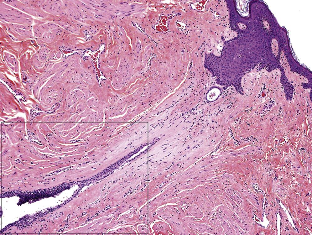
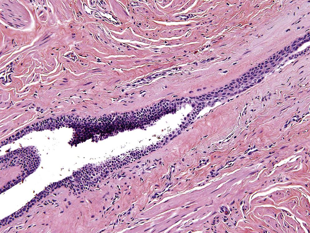
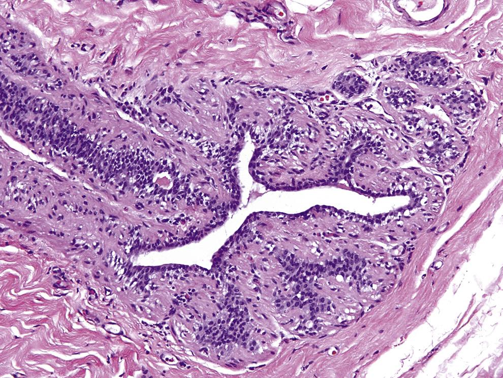
The nipple and areola are covered with stratified squamous epithelium, which is continuous with the surrounding skin over the breast. The opening of the collecting ducts at the nipple is typically plugged by keratinous debris in the nonlactating breast. The squamous epithelium of the collecting ducts undergoes gradual transition to pseudostratified columnar epithelium and, finally, to cuboidal or low-columnar epithelium (Fig. 1.4).
Approximately 20 orifices of collecting ducts, each representing a lobe of the breast, are present in the nipple. These orifices, which may be as few as 8 and as many as 24, are generally arranged as a central group and a peripheral group.17 The deeper portion of the collecting ducts has a characteristically serrated contour for a variable distance before opening into its terminal portion. The latter portion has a relatively less convoluted and smoother profile. The lactiferous ducts in the nipple are surrounded by bundles of smooth muscle. The muscle fiber arrangement is principally circular, but some fibers are also arranged vertically, interlacing among collecting and lactiferous ducts. The circular muscle fibers cause nipple erection, readying it for suckling. By cyclic contraction, the vertically arrayed muscle bundles empty the lactiferous sinuses. There is virtually no adipose tissue immediately beneath the nipple and areola.
FIG.
AB
The portion of the ductal system immediately below the collecting duct is the lactiferous sinus in which milk accumulates during lactation. This sinus communicates directly with segmental duct, which subdivides into subsegmental ducts, which in turn subdivide into terminal ducts. The latter structures drain the lobule. Each lobe contains 20 to 40 lobules. The lobule is composed of groups of small glandular structures, the acini. The latter are the terminal point of the ductal system. The serially and dichotomously branching structure of the mammary gland, from the tubular-like collecting duct to the terminal acini, leads to its classification as a compound tubuloacinar (or tubulolobular) gland (Fig. 1.5).
The lobule is inapparent to the naked eye on cut sections of breast tissue. However, with the aid of a magnifying lens, the lobules resemble minute drops of dew, and the ducts may appear as linear streaks. The size of the “normal” lobule is extremely variable, as are the number of acini in each lobule. Each lobule consists of 10 to 100 (range, 8–200) acini. The intralobular stroma consists of loose connective tissue and may also be
populated by a mixed inflammatory cell infiltrate particularly in the secretory phase of the menstrual cycle. The lobule undergoes a variety of morphologic changes under various physiologic influences (Fig. 1.6).
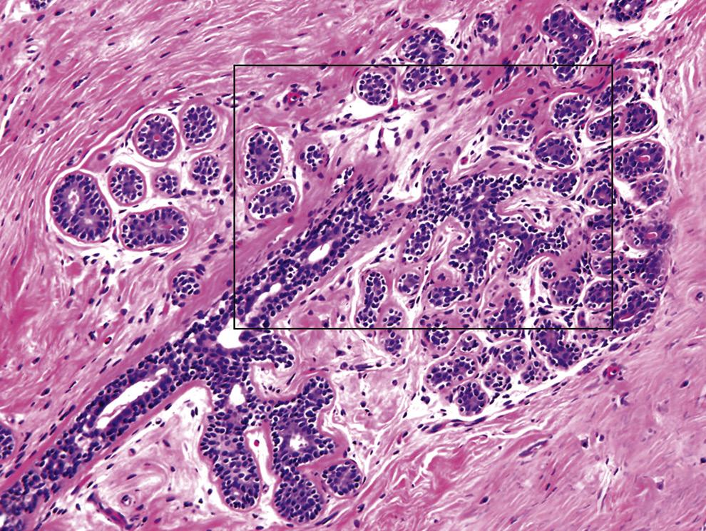
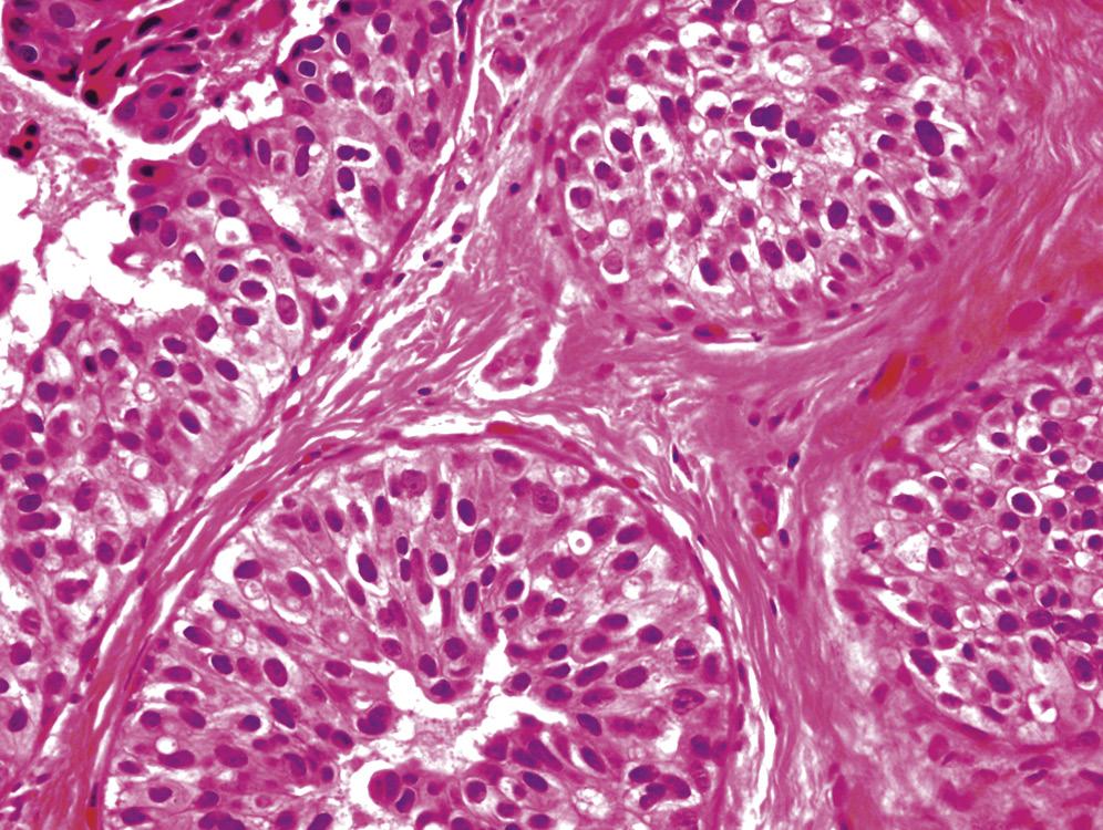
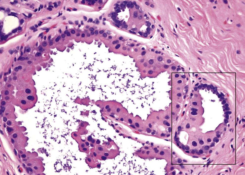
The fundamental glandular unit of the breast, and its most actively proliferating portion, is the terminal duct lobular unit (TDLU). This unit comprises the lobule and its paired terminal duct. During pregnancy and lactation, the epithelial cells of the terminal ducts and lobules undergo secretory changes, and most disease processes of the breast arise from the TDLUs (including cyst formation, which may simply represent “unfolding” of the terminal ducts and lobular units). Indeed, the only common lesion thought to be strictly of ductal origin may be the solitary intraductal papilloma (Table 1.1).
Except for the squamous epithelium–coated most distal portion of the collecting ducts, low-columnar to cuboidal epithelium lines almost the entire ductal system of the breast, including the segmental ducts, subsegmental ducts, terminal ducts, and acini. This lining epithelium is supported on its basal surface by a layer
FIG. 1.4 Vertical section through the nipple. A, A collecting duct is shown approaching the surface of the nipple (area in box is magnified in B). B, Squamous epithelium of the orifice undergoes gradual transition to the columnar epithelium of the collecting duct. A and B, Hematoxylin and eosin stain.
A B
FIG. 1.5 Terminal duct lobular unit. A, The lobule is composed of multiple acini. Acini are on the right (area in box is magnified in B). B, The terminal duct on the left is seen exiting the lobule. Note inner epithelial layer (with denser cytoplasm) and outer myoepithelial layer (with clearer cytoplasm). A and B, Hematoxylin and eosin stain.
note glandular atrophy amid largely fatty stroma. A to F, Hematoxylin and eosin stain.
of myoepithelial cells. The basement membrane (basal lamina) lies under the layer of myoepithelial cells. External to the basement membrane is connective tissue.
Myoepithelial cells facilitate milk secretion via their contractile property, which is largely under the influence of oxytocin. Receptors for the latter have been detected on the surface of myoepithelial cells,18 and this hormone
is primarily responsible for the mechanical release of milk (the milk let-down phenomenon).19
The myoepithelial cell layer is generally regarded as being spindle shaped with usually inapparent cytoplasm. Indeed, in fine-needle aspiration cytology preparations, myoepithelial cells appear to be entirely devoid of cytoplasm (ie, “naked”). The thin and compressed
FIG. 1.6 Mammary lobule at various physiological stages. A, Lobule in an adult female breast, inactive. B, Lobule in early puberty; note the incipient development of the lobule. C, Lobule in the secretory phase of the menstrual cycle; note secretions in the glands. D, Lobule after menopause, with intralobular fibrosis. E, Lobule after menopause, with intralobular adipocytes. F, Lobule in the elderly;
TABLE 1.1 Histologic Alterations in
Breast Glands and Stroma During Various Phases of the Menstrual Cyclea
PROLIFERATIVE PHASE
Epithelial cells are relatively smaller, with central nuclei and eosinophilic cytoplasm
Myoepithelial cells are relatively small
Glandular lumens are nondilated and without secretions
Stroma is relatively dense
No epithelial mitoses are present
LUTEAL PHASE
Epithelial cells are relatively larger, with minute apical snouts
Rare epithelial mitoses are present
Glandular lumens are dilated
Myoepithelial cells appear more prominent
Luminal secretions become evident
Stroma is edematous
Proliferation rate (as evidenced by Ki-67) is higher than in proliferative phase
SECRETORY/MENSTRUAL PHASE
Epithelial cells have high nuclear-to-cytoplasm ratio, with apical snouts
Epithelial mitoses are rare
Glandular lumens become smaller; luminal secretions become less evident
Myoepithelial cells are highly vacuolated
Stroma is compact
Apoptotic figures are most numerous on day 28
Lobular size almost doubles from that in early proliferative phase (from 1 mm to 2 mm)
aHistologic changes vary widely within the breast and even within lobules.
(bipolar) nuclei of the myoepithelial cells are oriented perpendicular to the layout of the epithelial cells. Myoepithelial cells extend from collecting ducts to the tip of the acini and may occasionally appear prominent either de novo (Fig. 1.7) or in certain physiologic states (eg, atrophy) and pathologic situations (eg, postradiation, adenomyoepithelioma). Myoepithelial cells appear to be inapparent in certain lesions (eg, in macrocysts, in which these cells get stretched).
The list of immunohistochemical stains that can be used to demonstrate the presence of myoepithelium around ducts is long, and newer stains are continually being introduced (Table 1.2 and Fig. 1.8), the latest one being p40.20 The lack of myoepithelial cell layer around neoplastic glands is generally considered to be diagnostic of invasive carcinoma, barring special situations such as those encountered in microglandular adenosis21 and solidpapillary carcinoma with smooth peripheral contours. Absence of myoepithelial cell layer has also been reported in some, but not all, apocrine cysts.22 The use of double (or even triple) immunolabeling with combinations of epithelial and myoepithelial immunostains is helpful in confirming early invasive carcinoma of breast (Fig. 1.9).23
The basement membrane, composed of a relatively attenuated basal lamina, lies immediately outside of the myoepithelial cell layer and divides the glands from the stroma. The basement membrane can be highlighted with appropriate immunostains (eg, laminin and collagen 4) or histochemical stains (reticulin and periodic
Sites of Origin of Common Diseases in the Breast
Paget disease, florid papillomatosis of nipple (ie, nipple adenoma)
Cysts, epithelial hyperplasia, noninvasive and invasive carcinoma
acid–Schiff). Stromal tissue lies beyond the basement membrane. The multilayered structure of the mammary gland can be highlighted with various histochemical and immunohistochemical stains (Fig. 1.10).
The mammary ducts and lobules are embedded within a variable fibrous and fatty stroma. The relative proportion of glands, fibrous tissue, and fat varies with age and body habitus; however, stromal tissues make up the bulk of the breast in adult nonlactating and nonpregnant women. Adipose tissue is typically present in the interlobar stroma and not among lobules (typically not until atrophy ensues). The fibrous tissue assists in the mechanical coherence of the gland. The fibroblastic and myofibroblastic elements in the stroma of the breast often display a deceitfully angiomatous appearance (hence, the term pseudoangiomatous stromal hyperplasia) (Fig. 1.11). The volume-fraction of collagenrich fibrous tissue is greater in younger adult women and accounts for the greater mammographic density therein.24,25 Within the United States, several states have enacted laws that require health care facilities to notify patients who are categorized as having dense breast tissue on mammograms. Such legislation is designed to help improve detection of breast carcinoma via use of additional imaging modalities.26
FIG. 1.7 Prominent myoepithelial cells in a terminal duct lobular unit. The myoepithelial cells lie external to the epithelial cells and may occasionally appear prominent (myoid hyperplasia). Hematoxylin and eosin stain.
TABLE 1.2
FROM NIPPLE
A
B
FIG. 1.8 Myoepithelial immunostain (calponin) in ductal carcinoma in situ (DCIS). A, DCIS of solid and micropapillary types. Hematoxylin and eosin stain. B, Calponin immunostain demonstrates complete myoepithelial envelope around the neoplastic cells.
AB
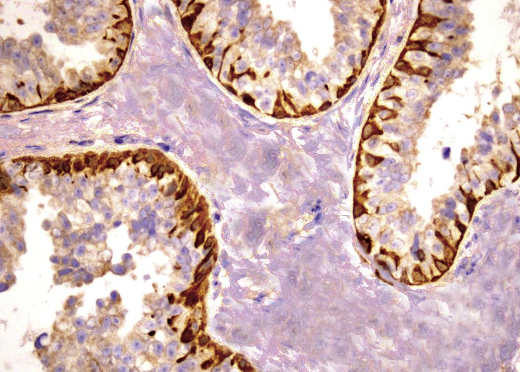
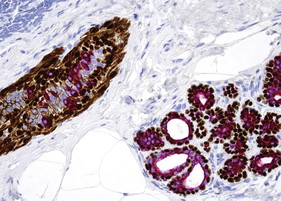
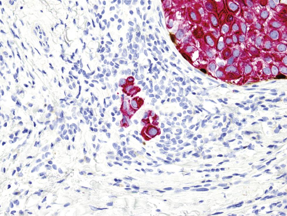
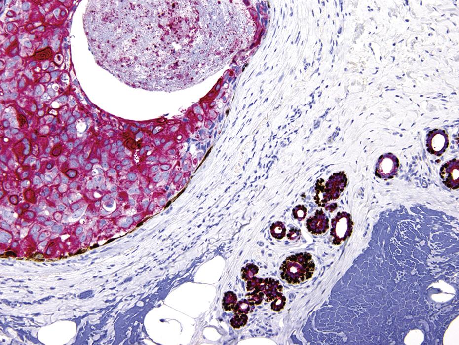
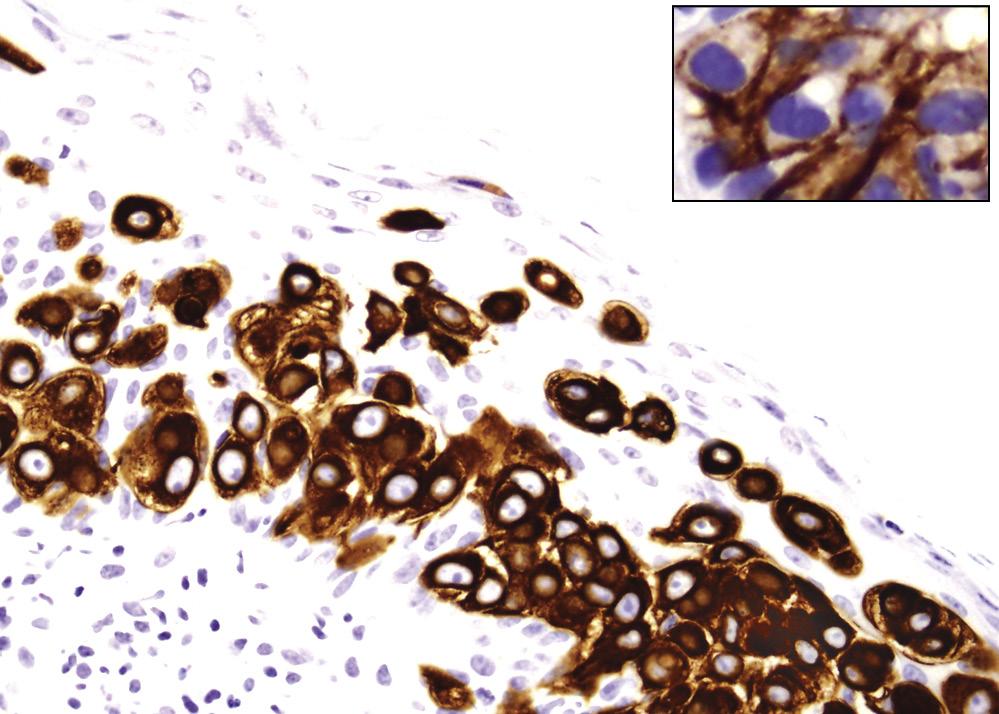
CFIG. 1.9
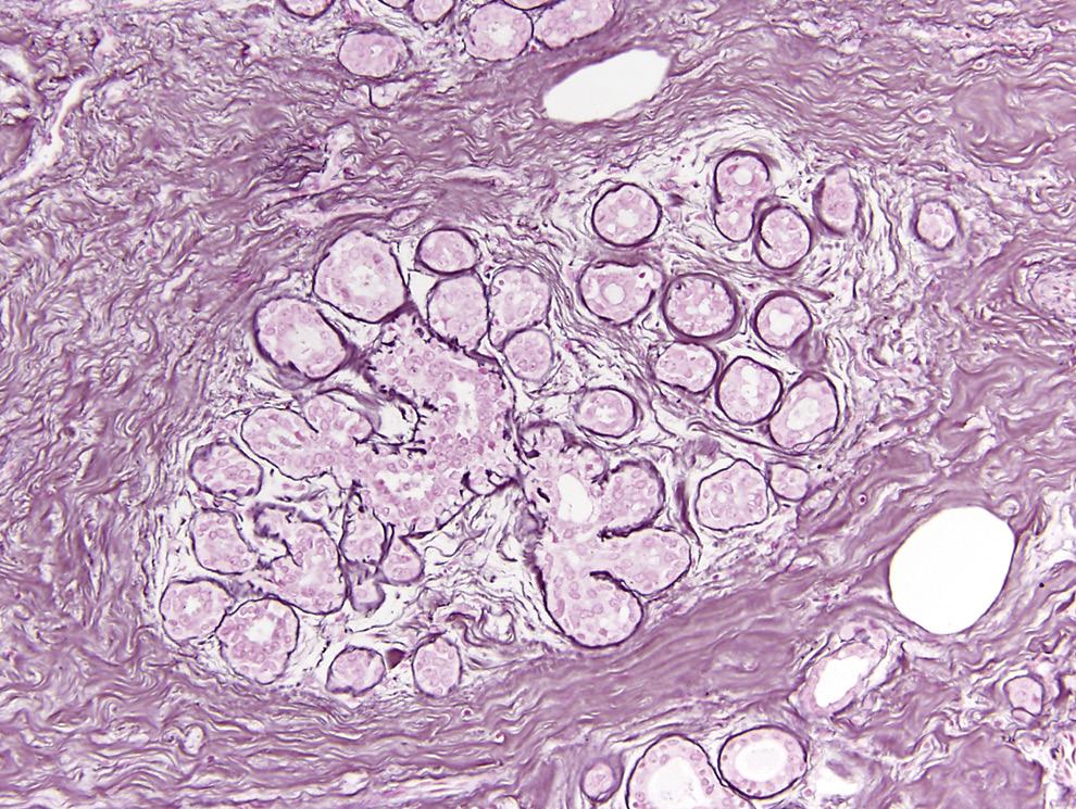
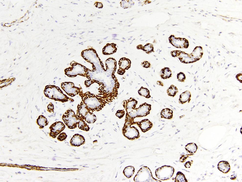
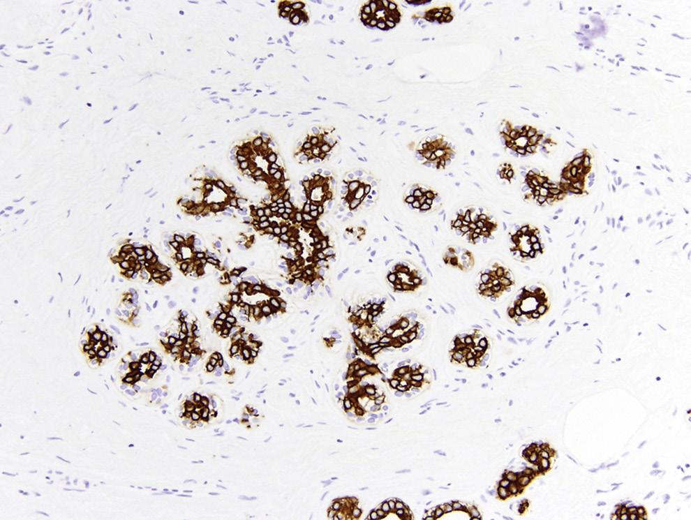
Triple stain highlights the myoepithelium and epithelium of mammary glands. The mammary ductal-lobular system is lined by a dual cell population: an inner epithelial cell layer and an outer myoepithelial cell layer. Red cytoplasmic immunostaining is seen in epithelial cells with cytokeratin. Brown cytoplasmic staining is observed in myoepithelial cells with myosin. Brown nuclear staining in myoepithelial cells is with p63. Shown here is a duct and an inactive lobule (A), ductal carcinoma in situ (B), and microinvasive carcinoma (C, center). Note absence of myoepithelium around the cells of the microinvasive carcinoma. A to C, Triple immunostain: CK AE1/3 + myosin + p63.
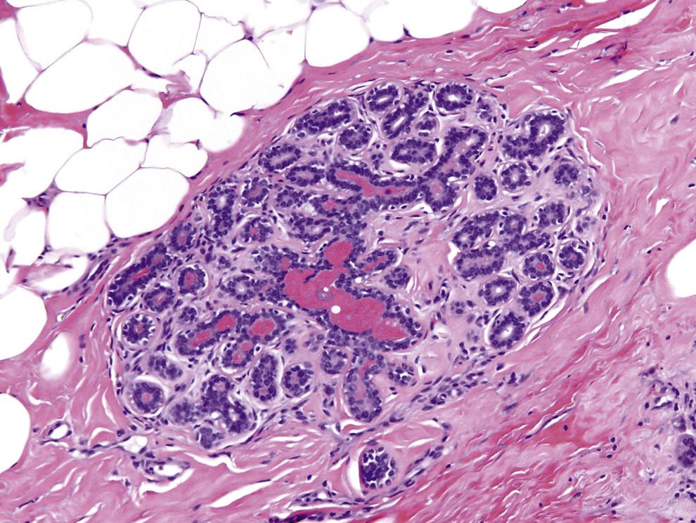
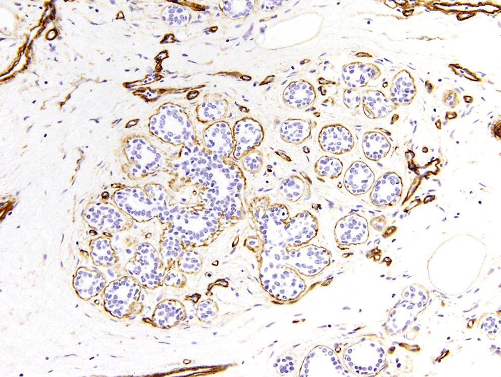
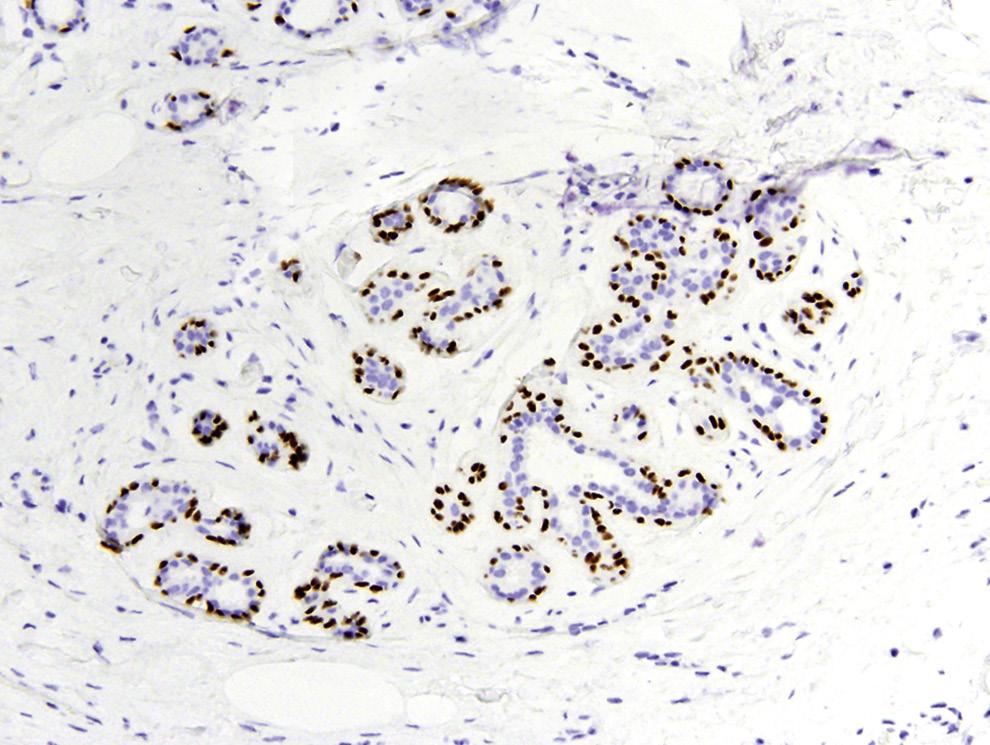
FIG. 1.10 Physiologically inactive mammary lobule: histochemical and immunohistochemical demonstration of structure. A, Normal lobule, hematoxylin and eosin stain. B, Reticulin stain decorates basement membrane. C, Collagen 4 immunostain also displays basement membrane. D, Smooth muscle myosin immunoreactivity demonstrates myoepithelial cells. E, p63 immunostain shows nuclei of myoepithelial cells. F, Cytokeratin AE1/AE3 immunostain demonstrates epithelial cells.
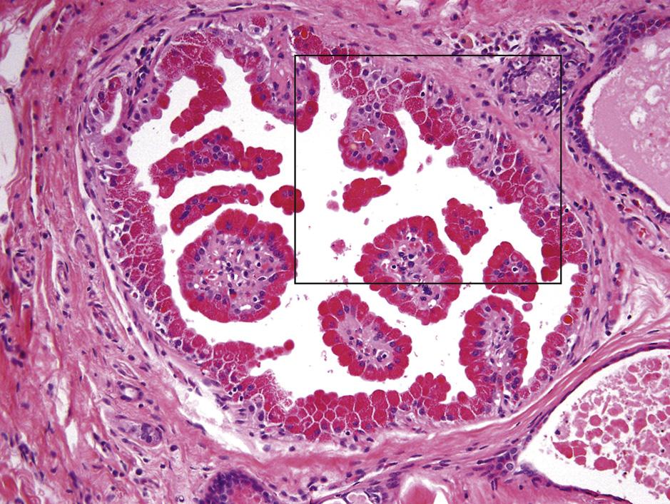
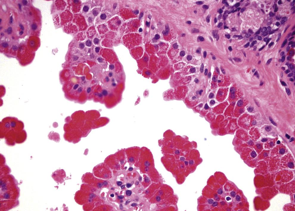
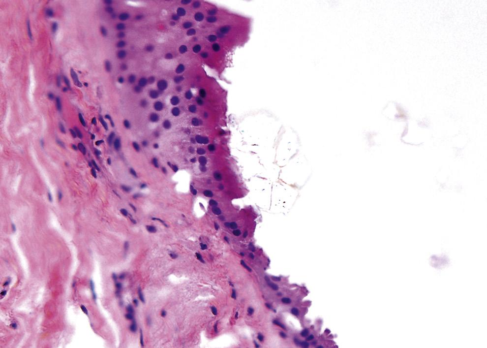
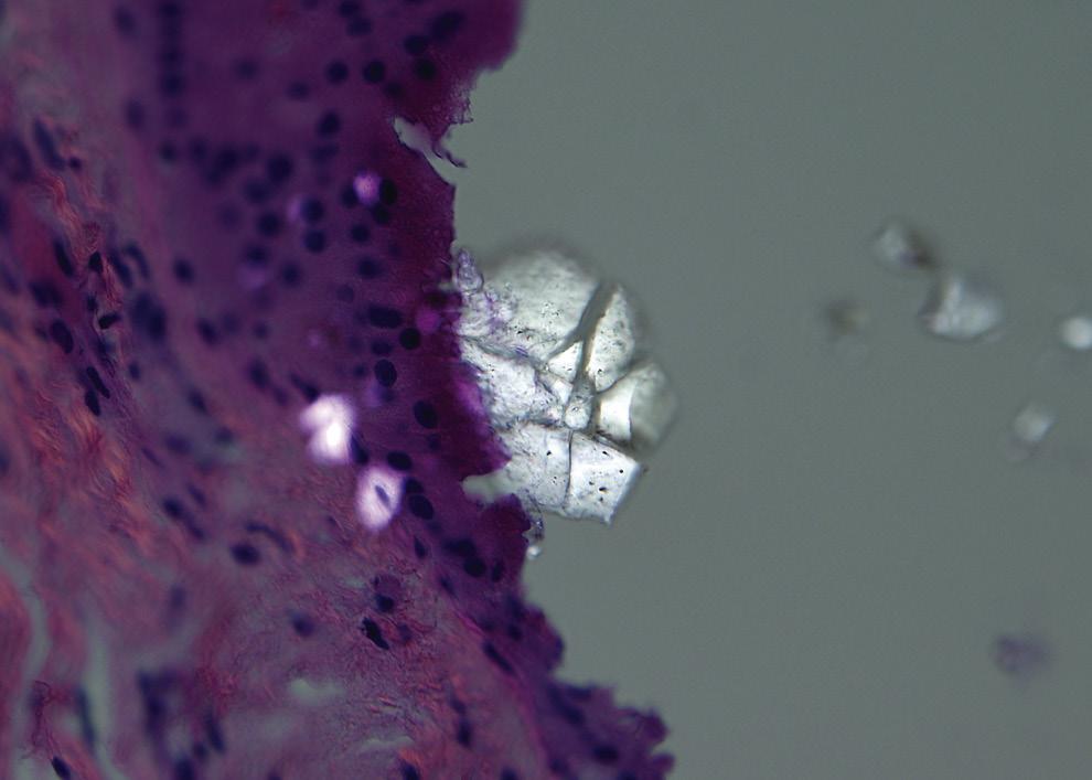
Apocrine cells are normal constituents of the glands of the breast in adult women, suggesting that this finding is a physiologic phenomenon (ie, a normal line of metaplastic differentiation) rather than a pathologic finding.27 The apocrine cells are typically pink and appear cuboidal or columnar and may exhibit a stubby apical snout (Fig. 1.12). Rarely, prominent apocrine (Lendrum) granules may become evident, particularly at the apical portions of the cells (Fig. 1.13). Inexplicably, cysts lined by apocrine epithelia are a common finding in breast lesions detected by magnetic resonance imaging 28 and typically contain calcium oxalate crystals. The latter may need polarizing microscopy to be optimally visualized (Fig. 1.14).29 Apocrine cells are almost always negative for both estrogen receptors (ERs) and progesterone receptors (PgRs) and are strongly positive for epithelial membrane antigen (EMA), gross cystic disease fluid protein-15 (GCDFP-15), and androgen receptors (ARs).
Under certain influences, as yet unknown, clear cell change can occur in epithelial cells (of both ducts and lobules) as well as in myoepithelial cells (Fig. 1.15).30–32 In epithelial cells, clear cell change can be commonly seen in association with apocrine metaplasia and following cytoplasmic accumulation of glycogen. Clear cell change can occur either spontaneously or sporadically in myoepithelial cells and may be seen in association with adenomyoepitheliosis and adenomyoepithelioma (Fig. 1.16). Such a change in either epithelium or myoepithelium has not been associated with progression to any disease process.
Foam cells are normally found within glands (typically those that are cystic) and in stroma (Fig. 1.17). Some of these foam cells are polygonal (and thus distinctly histiocytic) in appearance; others may have either an epithelioid or spindle cell appearance.33 Pigment-laden histiocytes appear in periductal connective tissue in approximately 15% of breasts (Fig. 1.18).34 These relatively large cells with low nuclearto-cytoplasmic ratio contain pale yellow to dark brown pigment. The pigment seems to have the staining qualities of lipofuscin (ie, positive for periodic acid–Schiff
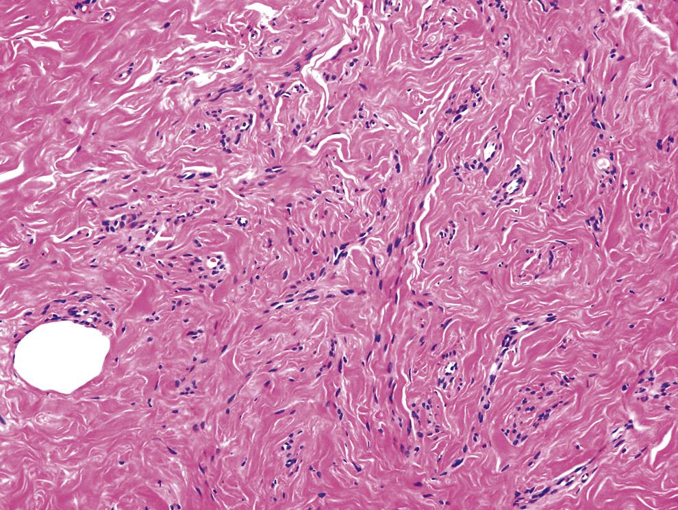
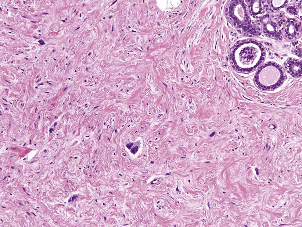
FIG. 1.11 Stromal fibrosis. Younger breasts have more stromal (mainly fibrous) component. Occasionally, the fibroblastic and myofibroblastic proliferation displays a vaguely angiomatous appearance (hence, the term pseudoangiomatous stromal hyperplasia). Hematoxylin and eosin stain.
FIG. 1.12 Apocrine metaplasia. The pink apocrine cells show bland round to ovoid nuclei. Transition of the normal cuboidal epithelium to the metaplastic apocrine epithelium is evident in the box. Hematoxylin and eosin stain.
FIG. 1.13 Cystic papillary apocrine hyperplasia with prominent apocrine granules. A, The apocrine type of metaplastic cells bear bright orange-red intracytoplasmic granules (area in box is magnified in B). A and B, Hematoxylin and eosin stain.
1.14 Cystic apocrine metaplasia with oxalate crystals. A, The apocrine cysts contain barely visible calcium oxalate crystals. B, The crystals can be better visualized under polarizing microscopy. A and B, Hematoxylin and eosin stain.
AB
1.15 Clear cell metaplasia. A and B, Acini in a lobule show cells with abundant clear cytoplasm and bland nuclei. Note unaffected glands in the vicinity. A and B, Hematoxylin and eosin stain.
FIG. 1.16 Clear cell cytoplasmic change in myoepithelial cells. Clear cell change in myoepithelia can appear pronounced. If the myoepithelial cells appear to be equal in number to the epithelial cells, the term adenomyoepitheliosis may be used. Hematoxylin and eosin stain.
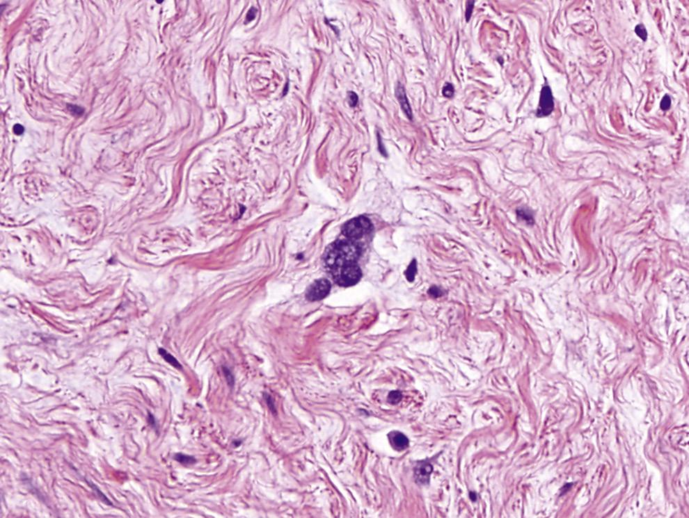
[but diastase-resistant], weakly positive for acid-fast stain, and negative for iron). Multinucleated stromal giant cells may rarely be present in the interlobular fibrous stroma, especially in the myofibroblast-dominant areas (Fig. 1.19). These giant cells have no known clinical significance.35
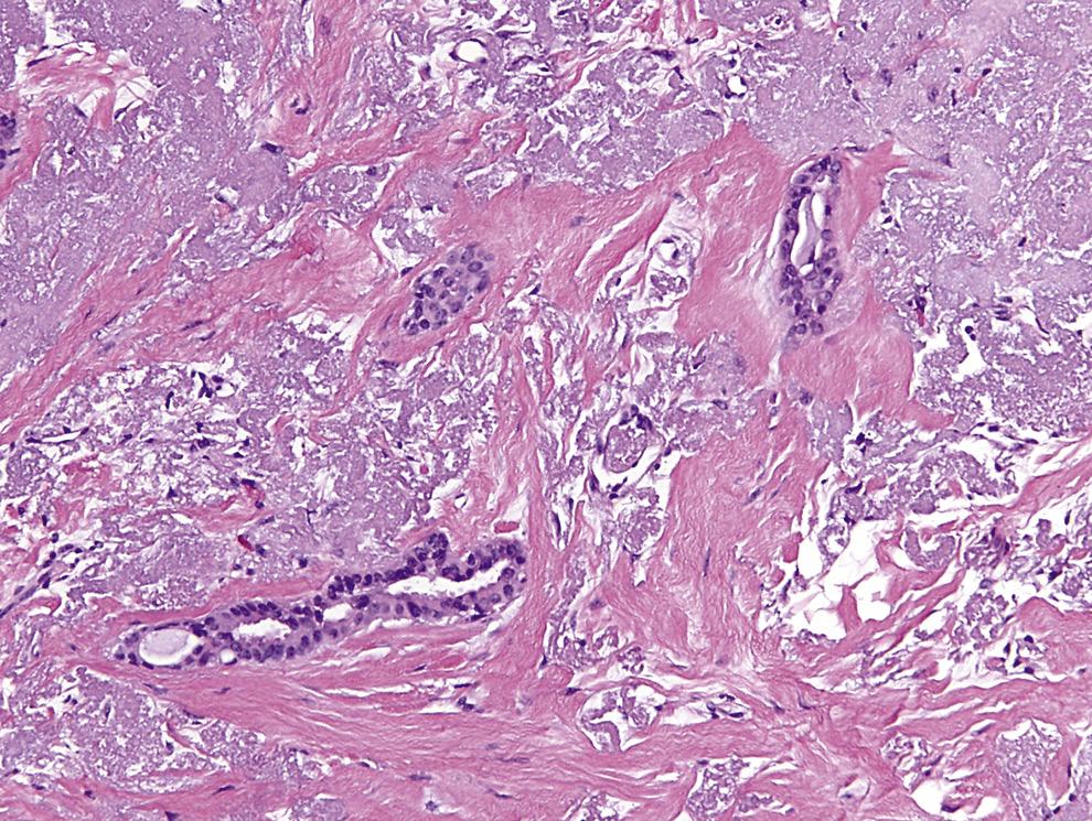
A framework of elastic tissue is present along the length of the duct system from the nipple to the subsegmental ducts. TDLUs are typically surrounded by a cuff of fibrous or myxoid connective tissues that contain virtually no elastic tissue. The larger ducts have sparse specialized connective tissue and possess relatively more elastic tissue. Bundles of elastic tissue are present in the periductal stroma of approximately 50% of women older than 50 years (Fig. 1.20). Elastosis implies an excess of elastic fibers over normal, although the baseline level of elastic tissue in the female breast remains undefined.36
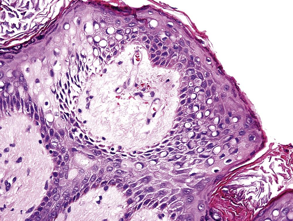
Two types of benign clear cells are present in the nipple among the stratified squamous epithelium. These are the so-called cellules claires and the Toker cells.37 The more common cellules claire (French for clear cells) type,
FIG.
FIG.
ABThese finely vacuolated histiocytic-type cells typically appear within cysts, which may (A) or may only focally (B) be lined by epithelial cells. The derivation of foam cells (epithelial or histiocytic) had been controversial in the past. A and B, Hematoxylin and eosin stain.
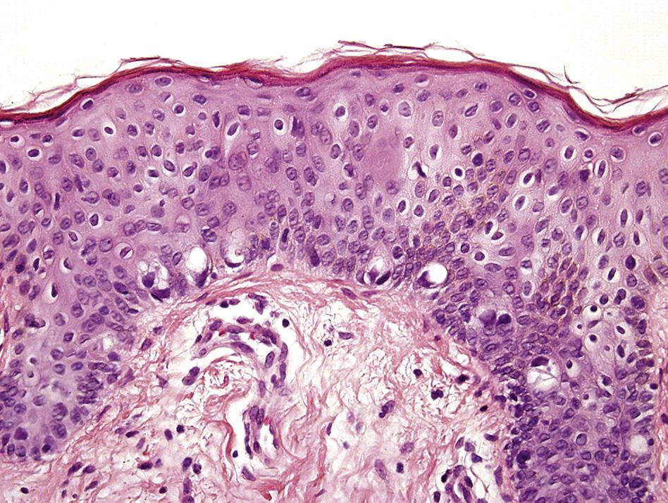
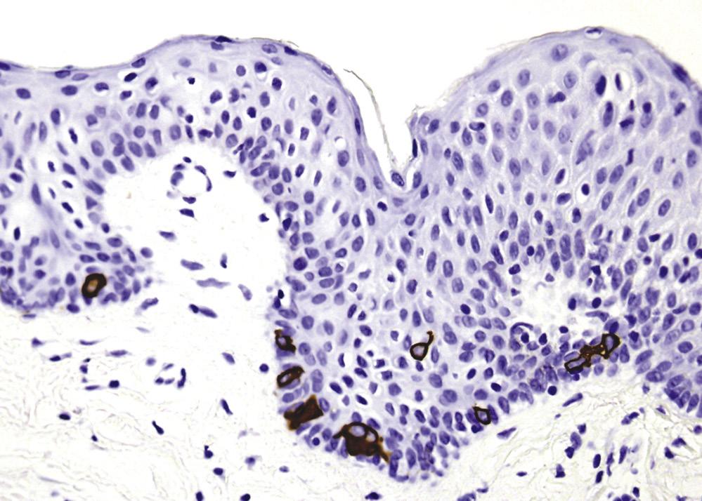
seen in about a third of the nipples, has clear cytoplasm and a semilunar nucleus that is compressed to the edge (Fig. 1.21). The clarity of the cytoplasm is likely the result of hydropic change. These clear cells are typically numerous and scattered throughout the full thickness of the epidermis. The clear portion of the cytoplasm of cellules claires is nonreactive for various cytokeratins, EMA, carcinoembryonic antigen, and papillomavirus markers. The second type of clear cells (so-called Toker cells) is more clinically significant because it can be mistaken for Paget disease of nipple. These cells, first detailed by Cyril Toker, a pathologist in New York City, are “smaller in size than typical Paget’s cells” and “larger than their squamous neighbors.”38 Toker cells are either extensions of mammary duct epithelial cells into the epidermal surface of the nipple or remnants of the embryonic nipple bud (see earlier). These cells have bland nuclei and pale cytoplasm and appear to be most numerous around the openings of lactiferous ducts.39 Toker cells occur either singly or in aggregates of a few cells. Most are commonly encountered near the basal layer but may also be found in the more superficial layers. Notably, Toker cells can
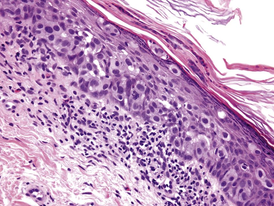
Paget disease of nipple (titled after Sir James Paget, a 19th-century British surgeon and pathologist) is the ascending extension of carcinoma cells, along the preexisting scaffold of the ductal system of the breast, to the epidermis of the nipple.40 Rarely, these Paget cells form glands. Except for HER2 (human epidermal growth factor receptor 2) (which is strongly immunoreactive in >90% of Paget cells), immunohistochemistry is generally unhelpful in the differential diagnosis of Toker and Paget cells because both cell types are reactive for various cytokeratins (including cell adhesion molecule [CAM] 5.2 and CK7) and EMA and are nonreactive for CK20 and S-100 proteins (Fig. 1.23).41–43
Nipple-sparing mastectomy has lately become a popular option for those for whom mastectomy is mandated or preferable for any reason. This procedure, which spares the nipple-areolar complex, provides a reconstructed breast with cosmetically better outcome along with the added possibility of retention of (at least some) sensation in the nipple. These advantages have to be weighed against the risks of leaving carcinoma in the nipple or the threat of carcinoma developing in residual ductal or lobular tissue in the “spared” nipple.44 In a study of 316 therapeutic nipple-sparing mastectomies, Brachtel and colleagues45 found that 71% of nipples showed no abnormality; 21% had ductal carcinoma in situ, invasive breast carcinoma, or lymphovascular channel involvement by tumor; and 8% had lobular carcinoma in situ. Lobules are present in 17% of normal nipples.46
Ultrastructure
On electron microscopy, the inactive luminal cells that line the entire length of the ductal and lobular system of the breast contain mitochondria, rough endoplasmic reticulum, and secretory granules. Surface specialization is present with microvilli projecting into the extracellular lumen. Desmosomes are present along the lateral interface with neighboring epithelial cells. Presence of the secretory granules and droplets toward the apical
FIG. 1.17 Mammary foam cells.
FIG. 1.18 Stromal histiocytes. The large, finely vacuolated cells with minute nuclei are typically seen around cystically dilated ducts. Hematoxylin and eosin stain.
AB
FIG. 1.19 Multinucleated stromal giant cells in the breast. A, Stromal giant cells (of mesenchymal phenotype) are seen here in association with stromal fibrosis. B, Detail of multinucleated stromal giant cells. A and B, Hematoxylin and eosin stain.
FIG. 1.20 Stromal elastosis. A, Periductal stromal elastosis in a 78-year-old woman. Hematoxylin and eosin stain. B, Elastic stain highlights elastic fibers in stroma.
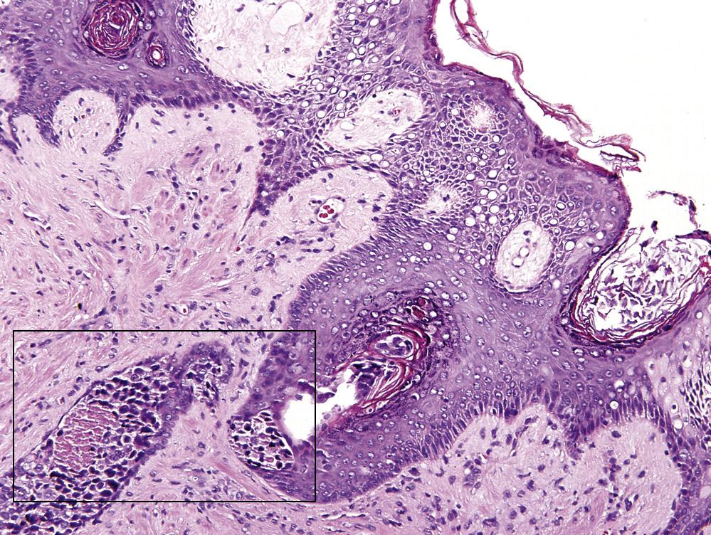
FIG. 1.21 Cellules claires (clear cells) in a nipple with Paget disease. Intraductal carcinoma in underlying collecting duct extends into the epidermis of the nipple as Paget disease (box in A) Clear cells, simulating signet-ring cells, are abundant (best seen in B). A and B, Hematoxylin and eosin stain.
AB
FIG. 1.22 Toker cells in epidermis of nipple. A, These benign seemingly vacuolated cells are scattered mainly around the basal layer and possess more abundant cytoplasm and are paler than adjacent keratinocytes. Hematoxylin and eosin stain. B, Cytokeratin 7 immunostain highlights Toker cells and imparts a dendritic appearance to these cells.
AB
FIG. 1.23 Paget disease of the nipple. A, The much larger and paler malignant cells are evident amid the native squamous epithelium of the nipple. Hematoxylin and eosin stain. B, Cytokeratin-7 immunostain highlights the presence of Paget cells. Human epidermal growth factor receptor 2 immunostain displays 3+ (on a scale of 0 to 3+) cytoplasmic membrane reactivity in Paget cells (inset).
pole of the cells depends on the physiologic state of the organ. A seemingly continuous layer of myoepithelial cells lies under the epithelial cells. This layer is oriented at right angles to the epithelial cells. Contractile actin filaments are seen in myoepithelial cells that appear more electron-dense and contain intracytoplasmic myofibrils with dense bodies and pinocytotic vesicles. The myoepithelial cells are attached to the underlying basement membrane (basal lamina) via hemidesmosomes. The epithelial cells appear to rest directly on the basal lamina wherever there is a gap between myoepithelial cells.47,48
Arterial Supply
The principal arterial supply to the breast is via the internal mammary artery, which caters to its central and medial portion. Somewhat confusing to the uninitiated is the fact that “internal mammary artery” and
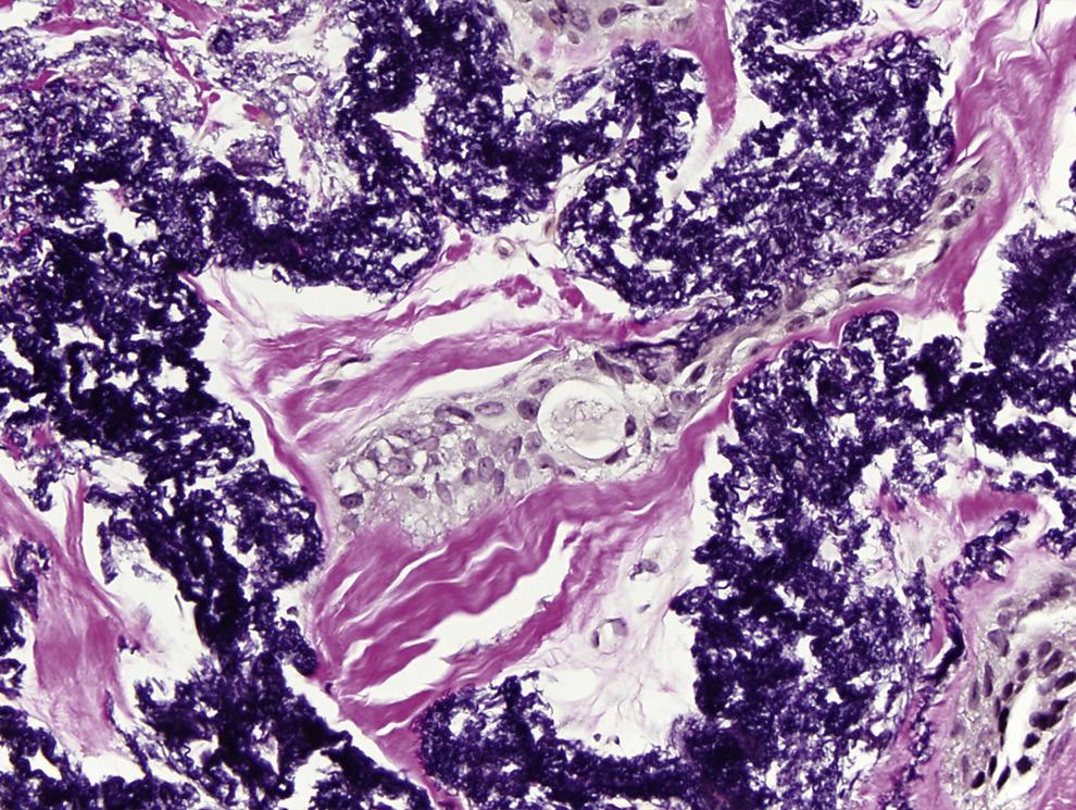
“internal thoracic artery” refer to the same arterial vessel.49 Necrosis of breast tissue after coronary artery bypass graft with segments of internal mammary artery is a rarer complication than one might expect, especially because this artery is so commonly used for this purpose.50 The lateral thoracic artery supplies the upper and outer portions of the breast. Numerous other arterial vessels, including various intercostal (mainly the second to fourth), lateral thoracic, subscapular, thoracoacromial, and thoracodorsal arteries and branches thereof, contribute to the arterial supply of the breast.51,52
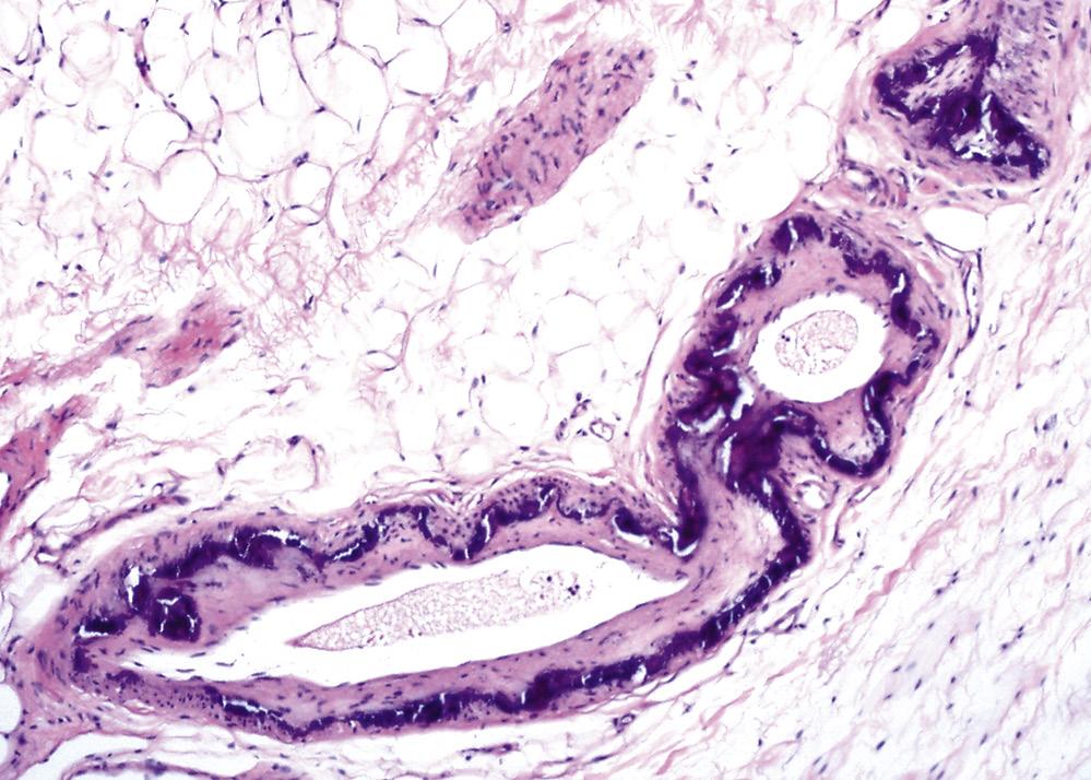
Arteries in the breast normally exhibit sclerotic changes and intramural calcifications of the type seen in so-called Monckeberg medial calcific arterial sclerosis (named after Johann Monckeberg, the German cardiovascular pathologist). Such calcified deposits are largely an aging phenomenon similar to that observed in other organs (Fig. 1.24). Up to 9% of breasts in postmenopausal women exhibit arterial calcifications detectable
on screening mammograms, and such findings are not predictive of coronary heart disease at coronary angiography.53
Given the relatively rich arterial network in the breast, it is not surprising that the vessels get traumatized by invasive procedures such as needle core biopsies. A number of cases of arterial pseudoaneurysm formation after core biopsies have been reported.54
Venous Drainage
In general, the venous drainage system of the breast follows the arterial system. However, the veins of the breast are much more variable than its arteries. The superficial venous system of the breast drains into the internal thoracic vein. The deep venous system drains into the perforating branches of the internal thoracic vein, lateral thoracic, axillary vein, and upper intercostal veins. A circular venous plexus lies around the areola.
Lymphatic System and Regional Lymph Nodes
The bulk (>75%) of the lymph drained from the breast enters the axilla.55 Most of the remainder of lymph from the organ drains into the internal mammary nodes. There are also some lesser lymphatic channels that lead to the interpectoral, internal thoracic, supraclavicular, and infraclavicular (and possibly even intramammary) lymph nodes. Lymphatic channels of the breast follow a more or less direct trail to the axillary or internal mammary nodes without involving the rich subareolar lymphatic plexus.56
The axillary lymph nodes that lie along the axillary vein and its tributaries are usually divided into three levels: level 1 nodes lie in the low-axilla, lateral to the axillary border of pectoralis minor muscle; level 2 nodes lie in the midaxilla, between the medial and the lateral borders of the pectoralis minor muscle; and level 3 nodes lie in the apex of the axilla, medial to the cranial margin of the pectoralis minor muscle and inferior to the clavicle.57 The Rotter lymph nodes (described by Josef Rotter, a
1.25 Lymphatic drainage of the breast. Schematic depiction of the breast and regional lymph nodes: axillary lymph nodes at levels I, II, and III (I, II, and III, respectively); supraclavicular lymph nodes (SC); and internal mammary lymph nodes (IM). The pectoralis minor muscle demarcates the various levels of axillary lymph nodes.
German surgeon, in the late 19th century) lie between the pectoralis major and pectoralis minor muscle, belong to the level 2 group, and may comprise up to four nodes. Level 3 lymph nodes are also known as apical or infraclavicular nodes (Fig. 1.25). Metastases to the latter group of lymph nodes portend a worse prognosis. Rotter lymph nodes are characteristically involved in breast carcinomas that arise from the upper-central and upperouter regions of the breast.58 Axillary lymph nodes usually range from 20 to 30 in number, with an average of 24; however, up to 81 lymph nodes have been dissected from this group.59 For years, conventional wisdom dictated that breast carcinoma involved the various levels of nodes in a stepwise fashion, progressing from levels 1 to 3. However, this traditional subdivision of axillary lymph nodes has been challenged by more recent studies in which the location of sentinel lymph nodes (ie, the first lymph nodes to receive lymphatic drainage from the breast) has been examined.60,61 Sentinel lymph nodes are seen at level 2 in up to 23% of patients, and metastases in level 3 lymph nodes only (skipping nodes at levels 1 and 2) are present in about 2% to 3% of cases. Of note, sentinel lymph nodes are rarely found to be in extraaxillary locations and as such are characteristically encountered in cases in which the breast has been irradiated or the axilla has been operated.62
Intramammary lymph nodes may be identified incidentally in breast biopsy samples or during mastectomies performed for another abnormality but are more often identified as ovoid, less than a 2-cm circumscribed, density on imaging studies.63 In one series, intramammary lymph nodes were identified in 28% of mastectomies performed for operable breast carcinoma.64 However, in routine practice, these nodes are encountered in less than 1% of cases. Although up to 10% of these nodes can be positive for metastatic carcinoma, they may not be part of the usual lymphatic drainage system of the breast. Before a positive intramammary lymph node is diagnosed, medullary carcinoma (with
FIG. 1.24 A mammary artery with intramural calcification. Annular intramural deposit of calcification is evident in the manner of medial calcific sclerosis of Monckeberg. Hematoxylin and eosin stain.
FIG.
its prominent lymphoid response) must be considered in the differential diagnosis. A lymph node (regardless of its location) has a capsule, subcapsular sinus, and at least one well-formed lymphoid follicle. Intramammary lymph nodes are considered as axillary lymph nodes for staging purposes.
The internal mammary lymph nodes are located in the intercostal spaces, 2 to 3 cm from the edge of the sternum in the endothoracic fascia. They are typically involved in carcinomas that are located in the upperouter quadrant. Supraclavicular lymph nodes are classified as regional nodes and lie in the supraclavicular fossa, a triangle defined by the omohyoid muscle (laterally and superiorly), the internal jugular vein (medially), and the clavicle and subclavian vein (inferiorly). Involvement of the internal mammary nodes is staged as pN3b and that of the ipsilateral supraclavicular lymph nodes as pN3c. Lymphatic drainage to the contralateral breast has not been demonstrated, although metastases to contralateral axilla have been reported.
In the current staging system, N1 implies metastatic involvement of 1 to 3 lymph nodes; N2, 4 to 9 positive lymph nodes; and N3, 10 or more positive lymph nodes. As such, a lymph node dissection with a harvest of more than 10 lymph nodes should be considered to be adequate for staging purposes.
Nerve Supply
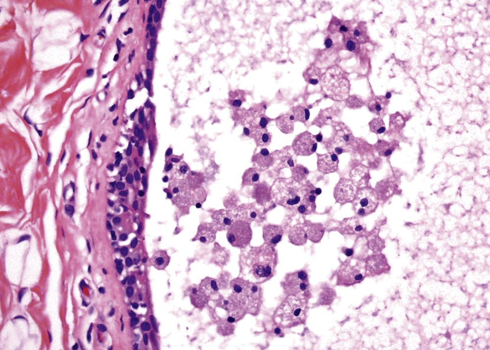
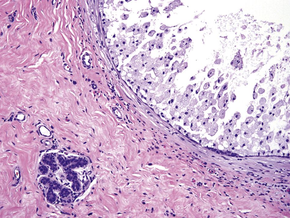
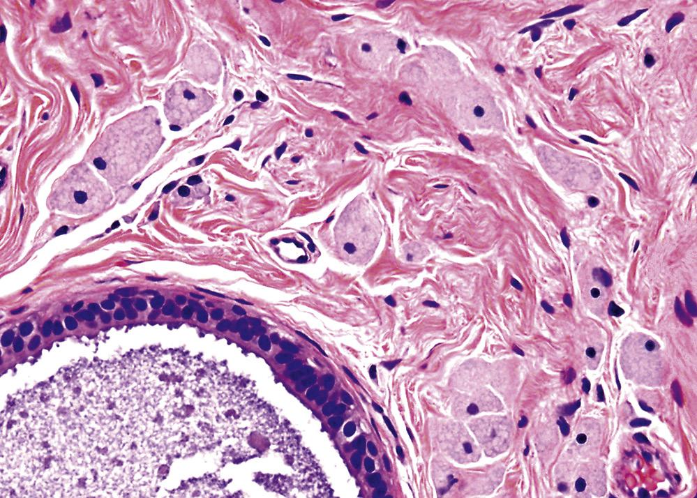
Nerve supply of the breast is derived from the anterior and lateral branches of the second to sixth intercostal (T2–6) nerves, which convey sensory and sympathetic efferent fibers. The nerve supply of the nipple is complex and is mainly from the anterior branch of the lateral cutaneous ramus of T4.65 Most sensory fibers terminate close to the epidermis as free endings, serving to signal the process of suckling to the central nervous system. Despite its well-deserved reputation as being extremely sensitive, relatively few nerves and nerve endings, including light touch receptors of Meissner (a 19thcentury German physiologist) and pressure corpuscles of Pacini (a 19th-century Italian anatomist, pronounced “pah-chee-nee”) are histologically identifiable in routinely prepared sections of the nipple. Secretory activities of the breast are mainly under hormonal control rather than regulated by efferent motor fibers. Tumoral involvement of peripheral nerves does not influence prognosis (Fig. 1.26).
Hormone Regulation
The breast is the target organ of a variety of hormones that are responsible for its physical development as well as the initiation and maintenance of lactation.66–70 Estrogen and progesterone production by the ovary at puberty influences the initial growth of the breast. Despite its predominant role, estrogen is unable to work independently of other hormones. Cyclic hormonal changes during each menstrual cycle alter the histology of mammary glands (Table 1.3). The breasts become swollen and somewhat lumpy in the latter half of the cycle. These changes are the physical manifestations
TABLE 1.3 Routine Immunohistochemical and Histochemical Stains Used to Highlight Epithelial Cells, Myoepithelial Cells, and Basement Membrane
IMMUNOSTAINS FOR EPITHELIAL CELLS
CAM 5.2, CK 7, 8, 18, 19 (lower-molecular-weight CKs)
Alpha-lactalbumin (during secretory phase)
GCDFP-15, especially in apocrine metaplastic cells
EMA reacts relatively strongly with apical region of active secretory cells
IMMUNOSTAINS FOR MYOEPITHELIAL CELLS
Smooth muscle actin
SMM-HC
Caldesmon
Calponin
CD10
p40
p63 (a p53 homologue with nuclear staining)
CK (5, 5/6, 14, 17, 34βE12 (higher-molecular-weight CK)
CK17: usually positive in ductal myoepithelial cells, rarely positive in lobular myoepithelial cells
ER, PgR, and AR: almost always negative in myoepithelial cells
IMMUNOSTAINS FOR BASEMENT MEMBRANE
Collagen IV
Laminin
Reticulin
AR, androgen receptor; CK, cytokeratin; EMA, epithelial membrane antigen; ER, estrogen receptor; GCDFP; gross cystic disease fluid protein; PgR, progesterone receptor; SMM-HC, smooth muscle myosin heavy chain.
of stromal edema and lobular proliferation. Strictly speaking, the use of the term resting breast in the premenopausal breast is inaccurate because the breast is hardly ever quiescent during these years. During pregnancy, the development of the breast is further stimulated by the continuous production of estrogen and progesterone. In this period, breast growth is further influenced by prolactin, steroids, insulin, and growth hormone.
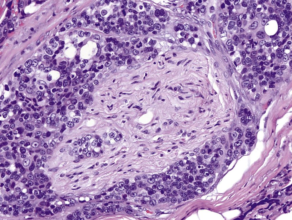
FIG. 1.26 Peripheral nerve involved by invasive carcinoma. Peripheral nerve shows circumferential involvement (perineural invasion) by invasive carcinoma. Hematoxylin and eosin stain.
At delivery, the loss of placenta and degeneration of corpus luteum causes an abrupt drop in estrogen and progesterone levels. Milk production is then brought about by prolactin and adrenocortical steroids. Suckling initiates impulses that act on the hypothalamus, resulting in the release of oxytocin from the posterior pituitary. Oxytocin stimulates the myoepithelial cells of the mammary glands, causing them to contract and discharge milk. Once the stimulation of suckling ends and feeding stops, secretion of milk ceases, and the gland gradually reverts to an inactive state.
ER is positive in epithelial cells of mammary glands in approximately 15% of cells, and both PgR and AR are sporadically positive in epithelial cells of ducts and lumen (Fig. 1.27).71 ER, PgR, and AR are almost always negative in myoepithelial cells. There is a higher frequency of ER-positivity in normal breast glandular cells during the proliferative phase of the menstrual cycle, and there is a higher frequency of PgR-positivity during the secretory phase. Oral contraceptive use decreases ER content of epithelial cells in the resting mammary epithelium.72
Two forms of ER, ER-α and ER-β, exist. ER-α can be demonstrated in nuclei of ductal and lobular epithelial cells; however, its expression varies with the phase of menstrual cycle and with the proliferative index. ER-β can be seen not only in ductal and lobular cells but also in myoepithelial and stromal cells. Relative levels of the two types of ER may have a potential, as yet unclear, role in breast carcinogenesis.73
Thelarche
The breast starts to grow at the onset of puberty, around the age of 11 years (range, 9–14 years). Thelarche refers to the onset of breast development (from the Greek thele, “nipple,” and arche, “beginning”). The process occurs under the menses-induced cyclic effect of estrogen and, to a lesser extent, of progesterone.74 Other signs of puberty follow soon thereafter. During early adulthood, stromal growth is responsible for most of the increase in breast size. At puberty, the ducts elongate and undergo repeated branching. The lobules, and acini therein, proliferate. The connective tissue becomes denser, and adipose tissue starts to accumulate. Each menstrual cycle fosters progressive gland development, the individual glands not returning to the previous cycle’s baseline. The glandular proliferation continues until the mid-30s or so and then plateaus until menopause, unless pregnancy ensues. These histologic changes, more often than not, parallel the physical alterations in the breast that occur with aging (Fig. 1.28).
Pregnancy, Lactation, and Milk
Among mammals, human beings are unique in the relatively large size of their quiescent breast; however, as in other species, the ultimate structural maturation and functional activity of the human breast occur after the completion of pregnancy and with the establishment of lactation.
The total weight gain of each breast during pregnancy is approximately 300 g. The principal changes in the
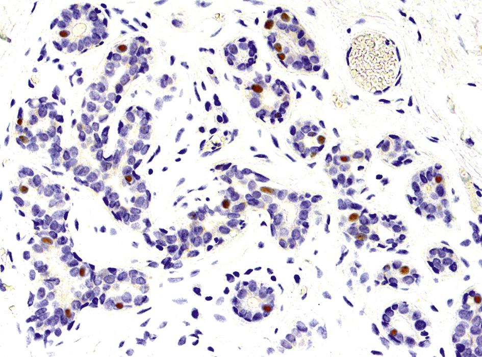
FIG. 1.27 Estrogen receptor (ER) in a normal adult lobular unit. Typically, around 15% of the glandular epithelial cells show moderate degree of ER immunoreactivity, although there is slight variation in ER reactivity in various phases of the menstrual cycle. ER 1D5 immunostain.
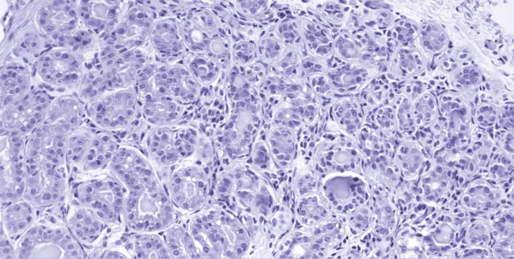
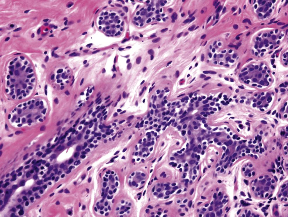
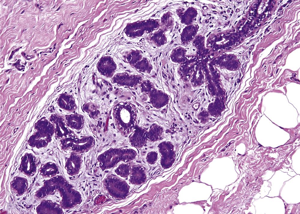
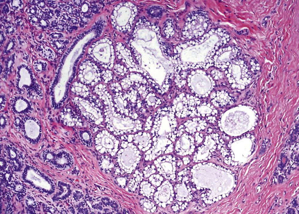
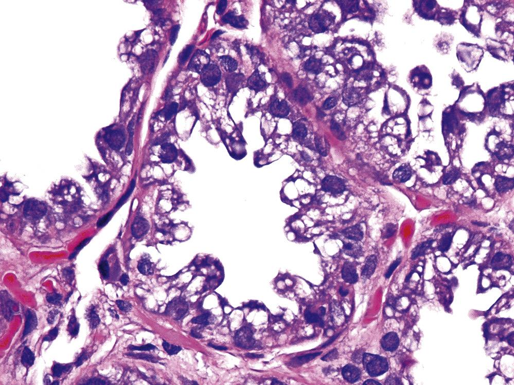
breast in pregnancy are the hyperplasia and hypertrophy of alveoli in each lobule (Fig. 1.29). The peak proliferative activity in glandular cells is observed during the first half of the pregnancy. The second half involves the maturation of the gland into a functional organ of lactation. With progression of pregnancy, the glandular cells become more vacuolated, and secretions become evident in lumens of glands. The interglandular stroma becomes relatively attenuated. By the beginning of the third trimester, the lobules form grapelike clusters.75,76
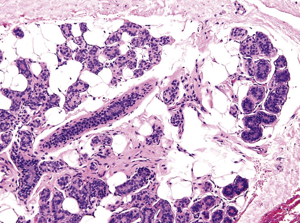
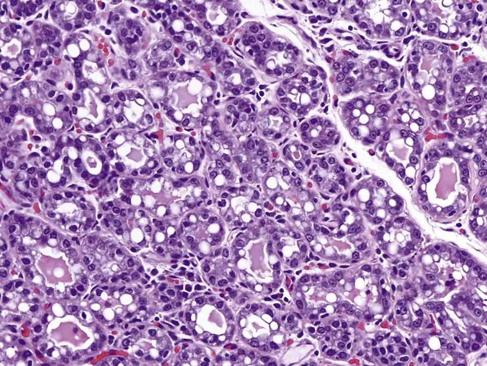
With the onset of lactation, which typically occurs within 1 to 4 days after parturition, the epithelial cells appear vacuolated with a hobnail appearance (Fig. 1.30). Luminal accumulation of secretions becomes readily evident. Milk production averages 1 to 2 mL/g of breast tissue per day. The rate of lactation is constant for the first 6 months after its onset. Lactation can continue for up to 4 years, as long as frequent suckling is maintained. On cessation of lactation, the process of involution takes a few (typically 3–4) months, although residual signs of lactation may be encountered for several months thereafter (Fig. 1.31). The process of involution affects the individual acini and lobules at different rates. After lactation ends, the breasts tend to become pendulous—a consequence of parenchymal shrinkage under the now stretched skin.
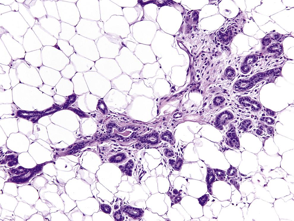
Scattered minute foci of lactational-like changes in nonlactating and nonpregnant women can be encountered in up to 3% of breast biopsy specimens.77 These changes resemble those seen in the truly lactating breasts, except for the absence of abundant secretions in acinar lumen (Fig. 1.32). Such lactational-like changes are typically uneven, and only some acini in a lobule may be affected.
Human milk is a complex fluid, composed largely of water (88%), lactose (7%), fat (4%), protein (1%, chiefly casein and lactalbumin), and various minerals including potassium, calcium, sodium, and magnesium. Vitamins and antibodies, mainly immunoglobulin A (IgA), are also present. Colostrum (milk of early lactation) is relatively richer in its antibody content.78
C
D
FIG. 1.28 Changes in contour of breast at various phases. Schematic drawing illustrates contour of the breast in a typical adult (A), fuller contour in midpregnancy (B), rounded contour in lactation (C), and droop contour in postmenopause (D).
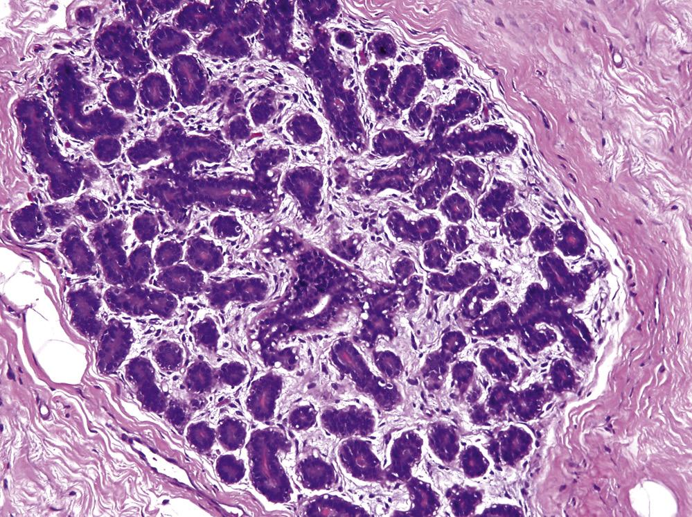
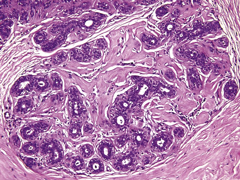
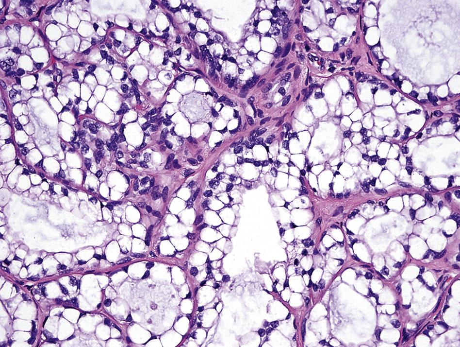
FIG. 1.29 Breast in midpregnancy. Acinar cells have a hobnail appearance with vacuolated cytoplasm. Note absence of luminal secretions. Hematoxylin and eosin stain.
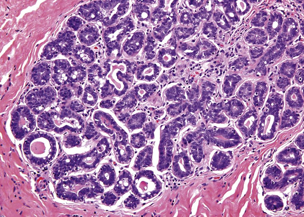
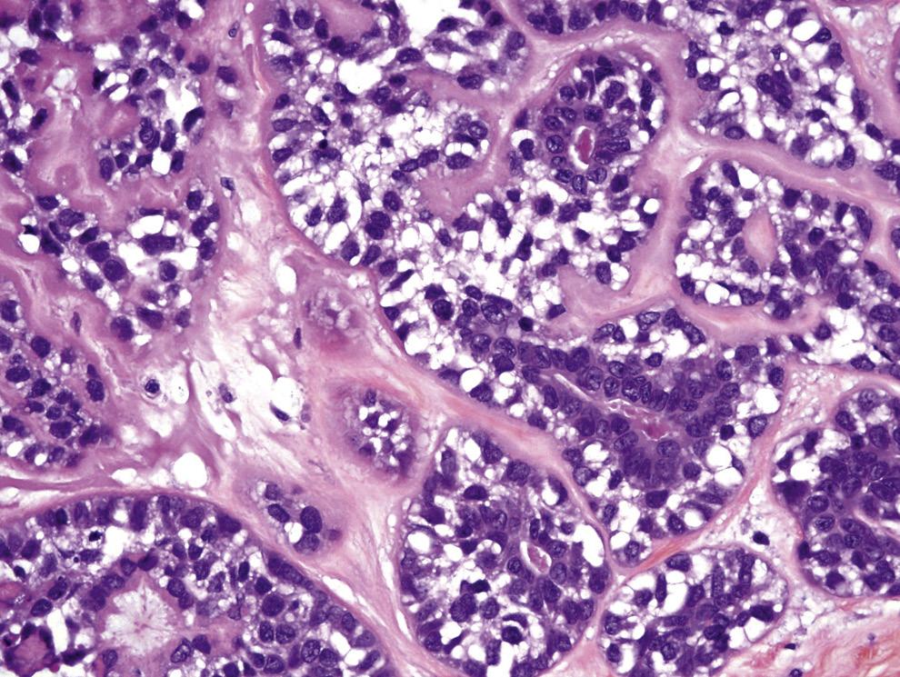
FIG. 1.30 Lactating breast. The acini are expanded with accumulation of secretions. Acinar epithelia appear finely vacuolated. Hematoxylin and eosin stain.