Activate the eBook version of this title at no additional charge.
Elsevier eBooks for Practicing Clinicians gives you the power to browse and search content, view enhanced images, highlight and take notes—both online and offline.
Unlock your eBook today.
1. Visit expertconsult.inkling.com/redeem
2. Scratch box below to reveal your code
3. Type code into “Enter Code” box
4. Click “Redeem”
5. Log in or Sign up
6. Go to “My Library” It’s that easy!
Place Peel Off Sticker Here
For technical assistance: email expertconsult.help@elsevier.com call 1-800-401-9962 (inside the US) call +1-314-447-8300 (outside the US)
THE CORE REQUISITES
BREAST IMAGING
The Core Requisites Series
Series Editor
James H. Thrall, MD
The Core Requisites Series Editor
Chairman Emeritus
Department of Radiology
Massachusetts General Hospital
Distinguished Taveras Professor of Radiology
Harvard Medical School
Boston, Massachusetts
Titles in the Series
Breast Imaging
Cardiac Imaging
Emergency Imaging
Gastrointestinal Imaging
Genitourinary Imaging
Musculoskeletal Imaging
Neuroradiology Imaging
Nuclear Medicine
Pediatric Imaging
Thoracic Imaging
Ultrasound
Vascular and Interventional Imaging
THE CORE REQUISITES
BREAST IMAGING
FOURTH EDITION
Bonnie N. Joe, MD, PhD
Professor
Department of Radiology and Biomedical Imaging
University of California San Francisco, California
Amie Y. Lee, MD
Associate Professor
Department of Radiology and Biomedical Imaging University of California San Francisco, California
Elsevier
1600 John F. Kennedy Blvd. Ste 1800 Philadelphia, PA 19103-2899
No part of this publication may be reproduced or transmitted in any form or by any means, electronic or mechanical, including photocopying, recording, or any information storage and retrieval system, without permission in writing from the publisher. Details on how to seek permission, further information about the Publisher’s permissions policies and our arrangements with organizations such as the Copyright Clearance Center and the Copyright Licensing Agency, can be found at our website: www.elsevier.com/permissions
This book and the individual contributions contained in it are protected under copyright by the Publisher (other than as may be noted herein).
Notices
Knowledge and best practice in this field are constantly changing. As new research and experience broaden our understanding, changes in research methods, professional practices, or medical treatment may become necessary.
Practitioners and researchers must always rely on their own experience and knowledge in evaluating and using any information, methods, compounds or experiments described herein. Because of rapid advances in the medical sciences, in particular, independent verification of diagnoses and drug dosages should be made. To the fullest extent of the law, no responsibility is assumed by Elsevier, authors, editors or contributors for any injury and/or damage to persons or property as a matter of products liability, negligence or otherwise, or from any use or operation of any methods, products, instructions, or ideas contained in the material herein.
Previous editions copyrighted 2017, 2011 and 2004.
Content Strategist: Melanie Tucker
Senior Content Development Manager: Somodottta Roy Choudhury
Senior Content Development Specialist: Malvika Shah
Publishing Services Manager: Shereen Jameel
Project Manager: Janish Paul / Haritha Dharmarajan
Design Direction: Patrick Ferguson
Printed in India
digit is the print number: 9 8 7 6 5 4 3 2 1
This book is dedicated to Dr. Edward A. Sickles, who has taught the science and art of breast imaging to so many of us at UCSF and around the world. He has mentored numerous breast imaging leaders and helped shape the specialty of breast imaging. In this book, we have tried to capture the fundamental lessons he has been teaching UCSF trainees for decades.
Contributors
Beatriz Adrada, MD
Radiologist
Breast Imaging
MD Anderson
Houston, Texas, United States
Shadi Aminololama-Shakeri, MD, FSBI
Professor Department of Radiology
University of California Davis Sacramento, California, United States
Dana Ataya, MD
Assistant Professor
Division of Breast Imaging
Department of Diagnostic Radiology
Moffitt Cancer Center
Tampa, Florida, United States
Assistant Professor
Department of Oncologic Sciences
University of South Florida College of Medicine Tampa, Florida, United States
Debbie L. Bennett, MD
Chief of Breast Imaging
Mallinckrodt Institute of Radiology
Washington University School of Medicine
St. Louis, Missouri, United States
Sonya Bhole, MD
Assistant professor
Northwestern University, Feinberg School of Medicine
Lynn Sage Comprehensive Breast Center Chicago, Illinois, United States
Maggie Chung, MD
Breast Imaging Fellow
Radiology & Biomedical Imaging
Breast Imaging Fellow of California, San Francisco San Francisco, California, United States
Peter R. Eby, MD, FACR, FSBI
Section Head of Breast Imaging
Department of Radiology
Virginia Mason Franciscan Health Seattle, WA, United States
Mohammad Eghtedari, MD, PhD
Associate Professor
Radiology
University of California San Diego San Diego, California, United States
Amy M. Fowler, MD, PhD
Assistant Professor
Radiology
University of Wisconsin School of Medicine and Public Health
Madison, Wisconsin, United States
Sarah Friedewald, MD, FACR, FSBI
Vice Chair of Clinical Operations and Women’s Imaging
Chief of Breast Imaging
Associate Professor, Department of Radiology
Northwestern University
Feinberg School of Medicine
Medical Director, Lynn Sage Comprehensive Breast Center
Chicago, Illinois, United States
Kimberly Funaro, MD
Assistant Member Department of Diagnostic Imaging and Interventional Radiology
H. Lee Moffitt Cancer Center
Tampa, Florida, United States
Sujata V. Ghate, MD
Associate Professor
Radiology
Duke University Medical Center
Durham, North Carolina, United States
Julie Gibbons, MD
Residency Associate Program Director Department of Radiology
Virginia Mason Franciscan Health Seattle, WA, United States
Heather Ilana Greenwood, MD
Associate Professor
Radiology & Biomedical Imaging
University of California, San Francisco
San Francisco, California, United States
Mary S. Guirguis, MD
Assistant Professor
Department of Breast Imaging
University of Texas MD Anderson Cancer Center
Houston, Texas, United States
Anne C. Hoyt, MD
Professor of Radiological Sciences
Radiological Sciences
David Geffen School of Medicine at UCLA
Los Angeles, California, United States
Debra M. Ikeda, MD, FACR, FSBI
Professor of Radiology (Breast Imaging), Emerita
Stanford University School of Medicine
Stanford, California, United States
Bonnie N. Joe, MD, PhD
Professor
Chief of Breast Imaging
Radiology & Biomedical Imaging
University of California, San Francisco San Francisco, California, United States
Andrew Nicholas Kozlov, MD
Staff Radiologist
Diagnostic Radiology
Radiology Associates of Florida Tampa, Florida, United States
Assistant Professor of Radiology
Diagnostic Radiology
University of South Florida Morsani College of Medicine
Tampa, Florida, United States
Chief of Nuclear Medicine
Department of Radiology
Tampa General Hospital Tampa, Florida, United States
Amie Y. Lee, MD
Associate Professor
Radiology & Biomedical Imaging
University of California, San Francisco San Francisco, California, United States
Christine S. Lo, MBBS, FRCR, FHKCR, FHKAM (Radiology)
Radiologist
Department of Diagnostic and Interventional Radiology
Hong Kong Sanatorium & Hospital Hong Kong, China
Jing Luo, MD
Breast Imaging Fellow
Breast Imaging
Radiology
University of Washington Seattle, Washington, United States
James Gordon Mainprize, PhD
Research Associate
Physical Sciences Sunnybrook Research Institute Toronto, Ontario, Canada
Elizabeth S. McDonald, MD, PhD, FSBI
Assistant Professor
Radiology
University of Pennsylvania Philadelphia, United States
Hannah S. Milch, MD
Assistant Clinical Professor
Radiological Sciences
David Geffen School of Medicine at UCLA Los Angeles, California, United States
Kanae Kawai Miyake, MD, PhD
Program-specific Assistant Professor
Advanced Medical Imaging and Research
Kyoto University Graduate School of Medicine
Section Chief, Nuclear Medicine
Kyoto University Hospital Kyoto, Kyoto, Japan
Linda Moy, MD
Associate Chair of Research Mentoring, Professor of Radiology
NYU Grossman School of Medicine
Center for Advanced Imaging Innovation and Research Faculty
Vilcek Institute of Graduate Biomedical Sciences
Laura and Isaac Perlmutter Cancer Center
160 East 34th Street, New York, NY 10016
New York, United States
Ramanjyot K. Muhar, MD
Associate
Radiology
Radiology Associates
San Luis Obispo, California, United States
Bethany Lynn Niell, MD, PhD
Section Chief of Breast Imaging
Department of Diagnostic Imaging and Interventional Radiology
H. Lee Moffitt Cancer Center and Research Institute
Tampa, Florida, United States
Professor
Department of Oncologic Sciences
University of South Florida Tampa, Florida, United States
Haydee Ojeda-Fournier, MD
Professor
Division Chief of Breast Imaging
UC San Diego Health
University of California San Diego La Jolla, California, United States
Dakota Orvedal, MD
Resident
Department of Radiology
Virginia Mason Franciscan Health Seattle, WA, United States
Molly Peterson, MD
Radiology Resident
Department of Radiology
University of Wisconsin Madison, Wisconsin, United States
Habib Rahbar, MD
Associate Professor
Vice Chair of Clinical Operations
Radiology
University of Washington Seattle, Washington, United States
Jocelyn Rapelyea, MD
Professor and Vice Chair of Education
Department of Diagnostic Radiology
The George Washington University Hospital Washington, District of Columbia, United States
Gaiane M. Rauch, MD, PhD
Professor Departments of Abdominal and Breast Imaging Division of Diagnostic Imaging
The University of Texas MD Anderson Cancer Center Houston Texas, United States
Kimberly M. Ray, MD
Associate Professor Radiology & Biomedical Imaging
University of California, San Francisco San Francisco, California, United States
Samantha P. Zuckerman, MD, MBE
Assistant Professor Department of Radiology Division of Breast Imaging Hospital of the University of Pennsylvania Philadelphia, Pennsylvania, United States
Congratulations to Drs. Bonnie N. Joe and Amie Y. Lee for producing The Core Requisites: Breast Imaging, 4e, now the third publication in the reimagined Core Requisites series. Drs. Joe and Lee have again successfully pivoted from a traditional narrative style to the new outline format. The new format brings out and immediately highlights the key points and concepts for each topic while minimizing the time required to sift through the text. An additional benefit of the new format is the ease of searching for information. Moreover, the books come in both print and online versions so that readers can access the information from wherever they are.
In the tradition of the Requisites in Radiology series, Breast Imaging: The Core Requisites, this book builds on the prior editions while bringing the information up to date and covering new topics that have come to the fore between editions. In that regard, Breast Imaging: The Core Requisites, 4e is a tour de force by Drs. Joe and Lee and their contributors because breast imaging has evolved rapidly, as much as or more than any subspecialty area of radiology. The evolution has progressed across multiple dimensions, all of which are reflected in this book. The diversity of imaging methods continues to grow. From a singular orientation to plain film x-ray mammography years ago, breast imaging requires knowledge of digital methods including digital tomosynthesis, multiple types of ultrasound applications, and diverse concepts in magnetic resonance imaging (MRI). Also, the need for standardized, guidelinebased approaches and accreditation has been unequivocally recognized. The challenge of harmonizing radiology interpretations with clinical information needs has been more clearly recognized and defined. Better knowledge of breast pathology has brought with it new insights into the significance of image findings and how to report them:
the radiologic–pathologic correlation. The number and complexity of interventional methods continue to grow. These important trends and others find excellent coverage in Breast Imaging: The Core Requisites, 4e.
While the format of the Core Requisites series differs substantially from the traditional Radiology Requisites series, the philosophy remains the same—a series of richly illustrated books covering the core material required across the spectrum of what radiologists need to know, from their first encounters as residents with subject material in different subspecialty areas to studying for board examinations and later for reference during clinical practice. We hope that radiologists, whether in training or practice, will find the books useful, as well as trainees and practitioners in the related fields of medical and surgical oncology. The books in the Core Requisites series are not encyclopedic; they are intended to be practical and focused on material expected on board certification examinations.
Congratulations again to Drs. Joe and Lee and their contributors for adding an excellent new resource to the Core Requisites series. I hope that this and the following books in the series will be regarded with the same fondness as earlier books in the Radiology Requisites family that have been used by radiologists at all career stages for over 30 years.
James H. Thrall, MD
The Core Requisites Series Editor Chairman Emeritus Department of Radiology Massachusetts General Hospital Distinguished Taveras Professor of Radiology Harvard Medical School Boston, Massachusetts
Preface
In keeping with the philosophy of the new The Core Requisites series, the chapters in this book are relatively short, with focused, straightforward, and high-yield content. Our goal is to allow the reader to efficiently review core information and key concepts. We hope this book serves as a useful guide to breast imaging whether you are a new resident or a seasoned radiologist looking for a concise yet comprehensive overview of the fundamentals of breast imaging.
Thank you to all of the authors who wrote and edited chapters for this book amid a global pandemic. Thank you to Dr. Deb Ikeda, editor of Breast Imaging: The Requisites, third edition, for your sponsorship and support throughout this process.
And thank you Ed (“Dr. Sickles”) for your continued mentorship and support.
Bonnie N. Joe, MD, PhD Professor Chief of Breast Imaging
Radiology and Biomedical Imaging
University of California, San Francisco San Francisco, California, United States
Amie Y. Lee Associate Professor Radiology & Biomedical Imaging
University of California, San Francisco San Francisco, California, United States
Special Populations in Breast Imaging 259 JULIE GIBBONS, DAKOTA ORVEDAL AND PETER R. EBY
Mammography Quality Standards Act (MQSA) and American College of Radiology (ACR) Accreditation Programs 280
ELIZABETH S. MCDONALD AND SAMANTHA P. ZUCKERMAN
Mammography Physics 101 285 JAMES GORDON MAINPRIZE
This page intentionally left blank
1 Introduction to Mammography: The Basics
MAGGIE CHUNG, BONNIE N. JOE AND AMIE Y. LEE
OVERVIEW This chapter is a basic introduction to the fundamentals of mammography, including standard and special views, technical adequacy, and normal anatomy.
Breast imaging is integral to the diagnosis and evaluation of breast cancer and other breast pathologies. While ultrasound and magnetic resonance radiology (MRI) also play important roles, mammography is often considered the foundation or workhorse of breast radiology. Mammography is versatile with ubiquitous roles in breast imaging, including breast cancer screening, diagnostic evaluation, and procedural guidance. It is an efficient method for evaluation of the entire breast and offers excellent visualization and characterization of breast calcifications.
Screening mammography is key to early breast cancer detection. Breast cancer is the most common non–skinrelated malignancy among women, affecting approximately one in eight women in the United States. It is also the second leading cause of cancer-related death in women and the leading cause of cancer-related death in women younger than 60 years old. Screening mammography has been consistently shown to be a cost-effective screening modality and is the only screening test that has been shown in randomized controlled trials to significantly reduce breast cancer mortality. As a result, mammography is a staple in modern medicine preventative care.
Refined by decades of technological advancements and now assisted by complementary ultrasound and MRI, mammography serves as an essential tool in the breast imager’s toolbox. This chapter will cover the basics of standard two-dimensional (2D) digital mammography. Subsequent chapters will provide more in-depth review of specific applications of mammography, including a chapter devoted to tomosynthesis.
How Is a Mammogram Performed?
COMPRESSION
When a mammogram is performed, each breast is placed on a support plate and compressed by parallel compression plates. Breast compression spreads superimposed normal parenchymal tissue for better visualization of underlying lesions (Box 1.1). Breast thickness is decreased by compression, thereby decreasing the radiation dose to the breast. Compression helps achieve more uniform thickness throughout the breast to avoid overexposure of the thinner anterior breast and underexposure of the thicker
Box 1.1 Roles of Breast Compression in Mammography
n Reduces parenchymal superimposition for better visualization
n Decreases radiation dose
n Ensures adequate x-ray penetration throughout the breast
n Reduces image blurring by decreasing patient motion and exposure time
n Reduces geometric blur and improves spatial resolution by reducing the distance between the breast tissue and the image receptor
n Improves image contrast by reducing scatter production and allowing selection of lower kV x-rays
posterior breast. It reduces geometric blur by reducing the distance between the breast tissue and the image receptor. Compression improves image contrast by reducing scatter production and allowing for selection of lower energy (kV) x-rays. Compression also immobilizes the breast and decreases exposure time, minimizing image blurring due to motion.
Patients may experience breast pain during compression. Methods to mitigate patient discomfort include performing the mammogram during day 7 to 10 of the menstrual cycle, adding foam padding between the breast and compression plates, and taking analgesics prior to receiving the mammogram.
STANDARD VIEWS
The routine mammogram consists of two complementary standard views for each breast: the craniocaudal (CC) view and the mediolateral oblique (MLO) view (Table 1.1). Based on standard mammographic terminology, each projection describes the direction of the x-ray beam. The first letter in the projection refers to the location of the x-ray source and the second refers to the location of the image receptor. For example, in the CC view the x-ray beam travels superior to inferior, from the cranial to caudal direction.
When performing the CC view, the patient faces toward the mammography unit, and the image receptor is placed inferior to the breast. The inferior breast is relatively mobile and allows the inframammary fold to be elevated when the technologist lifts and pulls the breast forward onto the
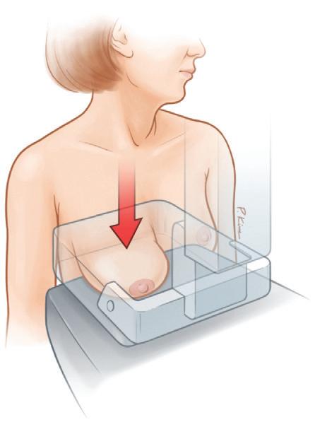
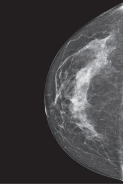
positioning platform. This maneuver allows greater visualization of the posterior and superior breast tissue. The breast is then compressed in the axial plane, perpendicular to the x-ray beam (Fig. 1.1). The CC view includes most of the breast tissue except for the far lateral and far posterior breast.
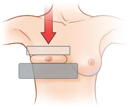
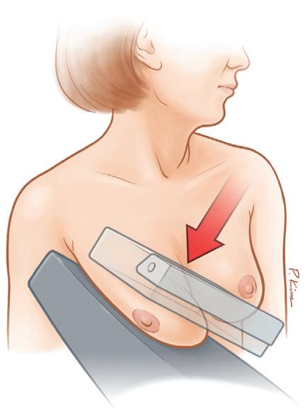
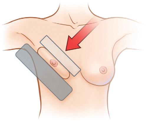
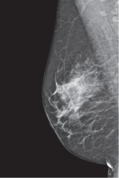
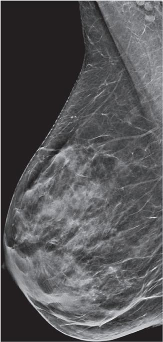
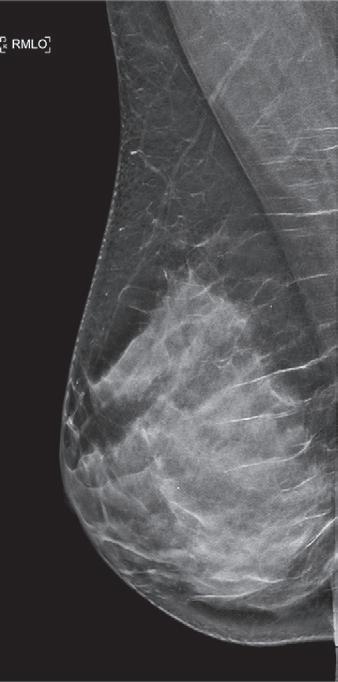
In the MLO projection, the x-ray beam travels from superomedial to inferolateral. The breast compression plane is along the angulation of underlying pectoralis major muscle, usually at 45 degrees (Fig. 1.2). However, angulation of the compression plane varies with patient anatomy and may range from 40 to 60 degrees; thinner body habitus may need a slightly steeper obliquity, and shorter or heavier body habitus may need a slightly flatter obliquity. The course of the pectoralis muscle can be approximated by drawing an imaginary line between the ipsilateral shoulder to the mid sternum. When properly positioned, the MLO view includes nearly the entire breast including the axillary tail.
These two standard views are complementary. The MLO view visualizes the most amount of breast tissue. It is the best for viewing the posterior, upper-outer quadrant, axillary tail, and lower-inner quadrant of the breast. However, there may be considerable breast tissue overlap, often with limited compression of the more anterior structures. Also, the far superior-medial breast tissue (upper-inner quadrant) may sometimes be excluded from the MLO view. The CC view allows for better visualization of the medial and posteromedial tissue, as well as better compression of the subareolar and central breast.
Standardized Labeling
Standardized image labeling is important for consistency, allowing mammograms to be accurately identified and interpreted even outside of the performing institution. According to the Mammography Quality Standards
Act (MQSA), the patient identification label is required to include the patient’s name, unique identification number and/or date of birth, facility name and location, examination date, technologist’s initials, cassette (screen) number for screen-film and computed radiography images, and mammographic unit identification (if there is more than one unit in the facility) (Box 1.2). The view and laterality label should be placed near the axilla on all mammograms. The laterality should be listed first, followed by technique (e.g., magnification, implant displacement) and projection (e.g., CC).
What Is Acceptable Quality?
Before interpreting a mammogram, it is important to first assess whether the mammogram is technically adequate (Box 1.3). Suboptimal image quality compromises mammographic evaluation and may lead to missed cancers.
IMAGE QUALITY
Each view should demonstrate adequate compression with dispersion of breast tissue and adequate x-ray penetration
n Unique identification number and/or date of birth
n Facility name and location
n Examination date
n Technologist’s initials
n Cassette/screen identification (for screen film or computed radiography; not for digital mammography)
n Mammographic unit (if there is more than one unit)
n Laterality and view placed near axilla
Fig. 1.1 The craniocaudal (CC) view. (A–B) The breast is compressed in the axial plane (at a 0-degree angle), perpendicular to the x-ray beam. (C) Example of a CC mammogram image.
Box 1.3 Evaluation of Image Quality
n Compression: Look for adequate compression with dispersion of breast tissue
n Contrast: There should be sufficient contrast to differentiate breast tissues
n Exposure: Look for adequate exposure throughout the breast
n Sharpness: Check that the breast trabeculae and skin edge are sharp and free of motion
n Noise: Noise should be minimized as it can interfere with detection of small findings such as microcalcification
n Artifacts: Images should be free of artifacts, which may obscure or mimic pathology
n Motion
n Hair
n Skin folds
n Antiperspirant
n Chin or other body parts
n Grid lines
throughout the breast. The breast trabeculae and skin edge should be sharp. Blurring may cause subtle calcifications to be missed or cause incorrect characterization of calcification morphology. Images should have appropriate contrast and minimal noise. Mammograms should be free of artifacts, including motion, hair, skin folds, and antiperspirant, which may obscure or mimic pathology (Figs. 1.3–1.5).
APPROPRIATE POSITIONING
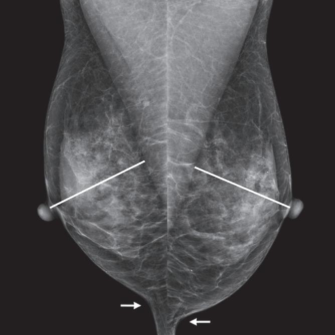
It is critical to assess if there is appropriate positioning with adequate breast tissue visualized on each view (Box 1.4) (Fig. 1.6). Appropriate positioning is important to ensure enough breast tissue is included in the mammogram to avoid potentially missing cancers. In fact, the majority of American College of Radiology (ACR) clinical image review and unit accreditation failures are due to poor positioning.
Positioning the nipple in profile helps to differentiate a nipple from a mass. As a practical approach in our practice, we will interpret a screening mammogram if the nipple is in profile on at least one view. Positioning the nipple in profile also helps identify nipple inversion or retraction that may be associated with an underlying mass. If the nipple does not naturally fall into profile, repositioning should not be performed at the expense of excluding breast tissue from the view. If the technologists cannot include both the nipple in profile and the appropriate amount of breast tissue in one view, an additional limited view can be obtained to demonstrate the nipple in profile.
Fig. 1.2 The mediolateral oblique (MLO) view. (A–B) The breast is compressed along the angulation of the underlying pectoralis muscle (usually at 45 degrees). The x-ray beam travels from superomedial to inferolateral. (C) Example of an MLO mammogram image.
Fig. 1.3 Motion artifact. Craniocaudal (CC) views from a screening mammogram. Note the marked blurring due to motion artifact in the outer right breast (A), which could limit detection of subtle cancers. Compare this to the left breast (B), where the breast trabeculae and skin edge appear sharp with no blurring.
Box 1.4 Appropriate Positioning on Standard Views
Mediolateral Oblique (MLO)
n Pectoralis muscle extends inferior to the posterior nipple line (PNL)a
n Convex anterior border of the pectoralis muscle
n Pectoralis muscle is wider superiorly and narrows inferiorly
n Open inframammary fold
n Fat is seen posterior to the fibroglandular tissue
n Nipple in profileb
Craniocaudal (CC)
n Well-visualized posterior medial breast
n Pectoralis muscle should be demonstrated when possible
n If the pectoralis muscle is not included, the length of PNLa on CC and MLO views should be within 1 cm of each other
n Fat is seen posterior to the fibroglandular tissue
n Nipple in profileb
aPNL is an imaginary line extending posteriorly and perpendicularly from the nipple to the pectoralis muscle.
bNipple should be in profile on at least one view.
view, it suggests that there is insufficient posterior breast tissue included on the CC view. It is important to clearly visualize the posterior medial breast on the CC view because it is often not well visualized on the MLO view.
The pectoralis muscle is an important landmark for evaluation of appropriate positioning. The posterior nipple line (PNL) is an imaginary line extending posteriorly and perpendicularly from the nipple to the pectoralis muscle. When the pectoralis muscle is not visible on the CC view, the PNL can be measured from the posterior nipple to the edge of the film. The length of the PNL on the CC and MLO views should be within 1 cm of each other. If the PNL length on the CC view is more than 1 cm shorter than on the MLO
On the MLO view, the visualized pectoralis muscle should extend inferior to the PNL. The pectoralis muscle should ideally demonstrate a convex anterior border and should be wider superiorly and gradually narrow inferiorly. The MLO view should attempt to show an open inframammary fold. If an open inframammary fold is not present, the breast should be pulled upward and outward as far as possible before compression is applied to reduce breast “sag” and overlapping tissue.
Fig. 1.5 Deodorant artifact. (A) Mediolateral oblique (MLO) views of the bilateral breasts shows radiopaque material over the bilateral axillae. (B) The patient was brought back for a repeat MLO views after wiping the bilateral axillae, which shows resolution of the artifact.
Fig. 1.4 Hair artifact. (A) Craniocaudal (CC) view of the left breast shows artifact from the patient’s hair obscuring the outer posterior breast. (B) The patient was brought back for a repeat CC view, which shows resolution of the hair artifact.
Special Views and Additional Views
SPOT COMPRESSION VIEW
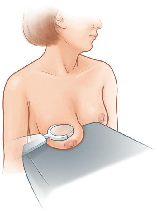
Spot compression views can be obtained for further evaluation of a region of interest. A small spot compression paddle applies pressure to a smaller area of tissue (Fig. 1.7). This increases compression on the region of interest, improving tissue separation and visualization of the area. Spot compression can help discriminate a real lesion, such as a mass or architectural distortion, from summation artifact. A real lesion will typically persist on spot compression view, retaining its shape and density, whereas a summation artifact will disperse. When a mass is adjacent to fibroglandular tissue of similar density, the margins of a mass may be at least partially obscured. By dispersing the adjacent fibroglandular tissue, spot compression can help bring out the mass margins, allowing for more complete assessment of lesion morphology. Spot compression may be performed in conjunction with magnification when using 2D imaging, as described in the subsequent section on magnification view. Spot compression may also be used with tomosynthesis images, as described further in the tomosynthesis chapter.
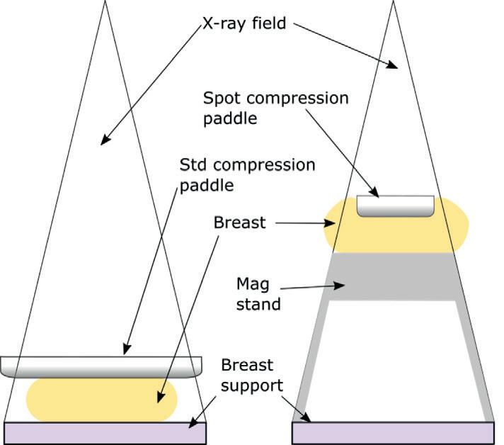
MAGNIFICATION VIEW
Magnification views, in conjunction with spot compression, can be obtained to further evaluate microcalcifications and mass margins. The magnification stand positions the breast closer to the x-ray source (decreasing the source-toobject distance) and farther from the image receptor, allowing for 1.5 to 2 times magnification of the region of interest
(Fig. 1.8). Whereas geometric magnification alone typically reduces spatial resolution, the use of a smaller focal spot and increased compression allows for higher spatial resolution. Magnification views can help enhance visualization and characterization of calcification morphology and can help reveal calcifications not seen on nonmagnified images. Magnification views can also improve visualization of mass shapes and margins not discernible on full-field nonmagnified images.
Fig. 1.6 Example of proper positioning for a bilateral screening mammogram. On the mediolateral oblique (MLO) view, note the pectoralis muscle extends inferior to the posterior nipple line (line) and the inframammary fold is open (arrow). The retroglandular fat is included and the nipple is in profile.
Fig. 1.7 Spot compression craniocaudal view.
Table 1.1 Additional Mammographic Views
Area of Interest View
Abbreviation
Retroareolar Spot compression with nipple in profile –
Outer breast Laterally exaggerated craniocaudal XCCL
Inner breast Medially exaggerated craniocaudal XCCM
Cleavage CV
Upper breast Craniocaudal (CC) from below CCFB
Upper inner breast, lower outer breast
Superior-inferior oblique SIO
ADDITIONAL VIEWS
When a portion of the breast is incompletely characterized, additional views can be obtained for further targeted evaluation (Table 1.1). These views include laterally exaggerated craniocaudal (XCCL), medially exaggerated craniocaudal (XCCM), cleavage view (CV), superior-inferior oblique (SIO), and CC from below. Use of additional views in diagnostic imaging is further discussed in Chapter 13 (Organized Approach to Diagnostic Imaging).
The Normal Mammogram
ANATOMY
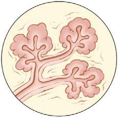
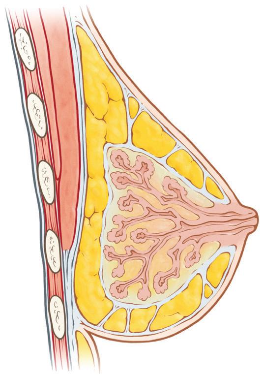
The breast is composed of overlying skin, adipose tissue, and breast parenchymal tissue, which includes glandular tissue and stroma. The breast glandular tissue is divided into 15 to 20 lobes, each drained by a lactiferous duct. The lobes contain series of branching ducts that end in terminal ductal
lobular units (TDLUs), which represent the functional units of the breast. TDLU is the origin of most breast carcinomas (e.g., ductal carcinoma in situ, invasive ductal carcinoma, and invasive lobular carcinoma). Each TDLU contains acini, ductules, and terminal ducts (Fig. 1.9). During lactation, breast milk is produced in the TDLUs and drain via the ductal system, which converge in the lactiferous sinus before opening into the nipple.
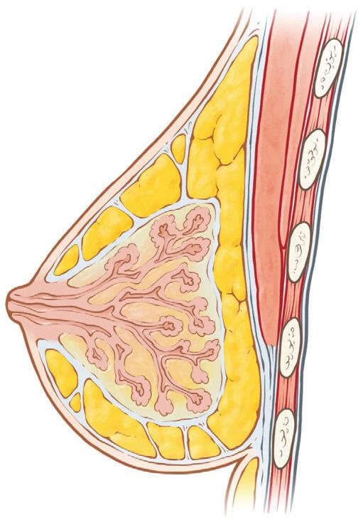
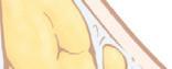
The breast is enveloped anteriorly by the superficial fascia just beneath the skin and posteriorly by the deep fascia just anterior to the pectoralis muscle. Cooper’s ligaments are thin fibrous strands that connect the two fascial layers and support the breast (Fig. 1.10). Fibroglandular tissue is found in the mammary zone. There is fat anterior and posterior to the mammary zone, called premammary (or preglandular) and retromammary (or retroglandular) fat, respectively. Most of the glandular tissue are typically found in the upper outer quadrant of the breast near the axilla. Physiologic fatty involution occurs with aging and usually spares the upper outer breast until last.
RADIOLOGIC ANATOMY OF THE BREAST
The most anterior structure on a mammogram is the overlying skin, seen as a thin, 2- to 3-mm homogenous line against the radiolucent background of the premammary fat. Posterior to the premammary fat is the mammary zone, which appears as a radiodense (white) area with scalloped borders. The retromammary fat should be predominantly radiolucent without any glandular tissue. Any isolated asymmetry or mass in this area warrants further attention (Fig. 1.11).
The pectoralis muscle is seen at the most posterior aspect of the mammogram. On the lateral and MLO views, the pectoralis muscle is shaped like an upside-down triangle along the posterior, superior border of the images. On the CC
Fig. 1.8 Standard (Std) view (left) versus spot compression magnification view (right). Note that on the magnification view, the breast is placed on a magnification stand so the imaged tissue is closer to the x-ray source and farther from the image receptor, allowing for magnification of the region of interest (Image courtesy of James Mainprize).
Alveoli Lobule
Ductule
Terminal duct
TerminalDuctal LobularUnit(TDLU)
Adiposetissue
Pectoralisminor
Pectoralismajor
Deepfascia
Superficialfascia: Deeplayer
Superficiallayer
Cooper’s ligament
Retromammary fat
Retromammary fascia
Premammar y fat
Anterior mammar yfascia
view, the pectoralis usually appears crescent-shaped when visualized.
On the CC view, only fatty tissue should be seen in the far posterior medial breast. An exception is the sternalis muscle, a normal muscular variant of the anterior thoracic wall. It appears as a flame-shaped or small triangular soft tissue density in the inner breast on the CC view. Found in approximately 8% of the population, the sternalis muscle lies superficial and perpendicular to the pectoralis major muscle and parallel to the sternum. The sternalis muscle is most often unilateral, but can be bilateral. It should not be
Nipple
Lactiferous duct
Fig. 1.9 Breast anatomy. The terminal ductal lobular unit (TDLU) is the origin of most breast carcinomas.
Rib
Fig. 1.10 Breast anatomy.
Fig. 1.11 Breast anatomy on a mammogram
Table 1.2 Image Contrast
Radiolucent (dark) Fat
mistaken for a breast mass (Fig. 1.12). Cleavage view, tomosynthesis, and/or target ultrasound could be obtained to verify as needed.
Normal axillary lymph nodes appear as circumscribed, oval, or reniform densities with radiolucent fatty hilum, typically overlying the pectoralis muscle on the MLO view. Normal lymph nodes can also be located within the breast parenchyma and are called intramammary lymph nodes. Intramammary lymph nodes are found most commonly in the lateral breast and seen in approximately 5% of all mammograms. If there is uncertainty regarding whether a lesion is a lymph node or mass, magnification views, tomosynthesis, and/or ultrasound can be obtained to better demonstrate the characteristic reniform shape and fatty hilum.
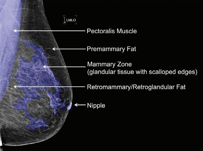
Accessory breast tissue is ectopic glandular parenchyma most commonly located in the axilla, but can be found anywhere along the milk line extending from the axilla to the abdomen. It is a normal, often bilateral variant (Fig. 1.13). Accessory breast tissue should demonstrate a similar radiographic appearance to normal glandular parenchyma. It is important to recognize the imaging appearance of accessory breast tissue to avoid mistaking the normal variant as a pathologic abnormality.
IMAGE CONTRAST
Mammography relies on the differences in x-ray attenuation between fat, fibroglandular tissue, and breast pathologies (Table 1.2). Fat is the least dense and appears radiolucent (dark) on mammography. Fibroglandular tissue, muscle, lymph nodes, neoplasms, and cysts are denser than fat and
Intermediate density (white) Fibroglandular tissue, muscle, lymph nodes, benign and malignant tumors, cysts
High density (whitest) Calcifications, metals
Box 1.5 Breast Density
According to the Breast Imaging Reporting and Data System (BI-RADS) lexicon, mammographic density is assigned to indicate the relative possibility that a lesion could be obscured by normal tissue.
A. The breasts are almost entirely fatty
B. There are scattered areas of fibroglandular density
C. The breasts are heterogeneously dense, which may obscure small masses
D. The breasts are extremely dense, which lowers the sensitivity of mammography
appear more radiopaque (whiter). Calcifications and metals are the densest (whitest) structures on mammography.
Breast pathologies are more readily detected on a background of fatty tissue due to differences in tissue density. There can be overlap in tissue density between normal and pathologic tissues, and consequently 10% to 15% of breast cancers are mammographically occult. In these cases, a cancer is present in the absence of visible mammographic findings.
Breast density refers to the relative amount of radiopaque fibroglandular tissue to radiolucent fatty elements (Box 1.5). Relative proportions of fibrous and adipose tissue in breast
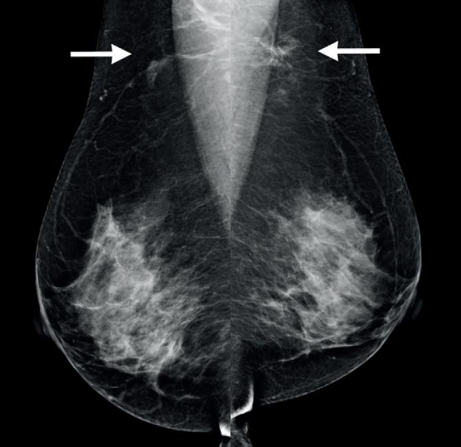
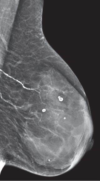
Fig. 1.12 The sternalis muscle is an anatomic variant of the chest wall musculature. The sternalis can appear as a triangular shaped asymmetry (arrow) in the inner breast at the posterior edge of the craniocaudal (CC) view.
Fig. 1.13 Mediolateral oblique (MLO) views of the bilateral breasts show accessory breast tissue in the bilateral axillae (arrows). This is a normal variant of residual breast tissue persisting from embryologic development.
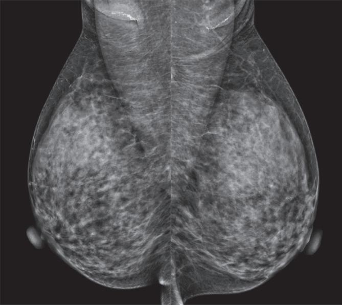
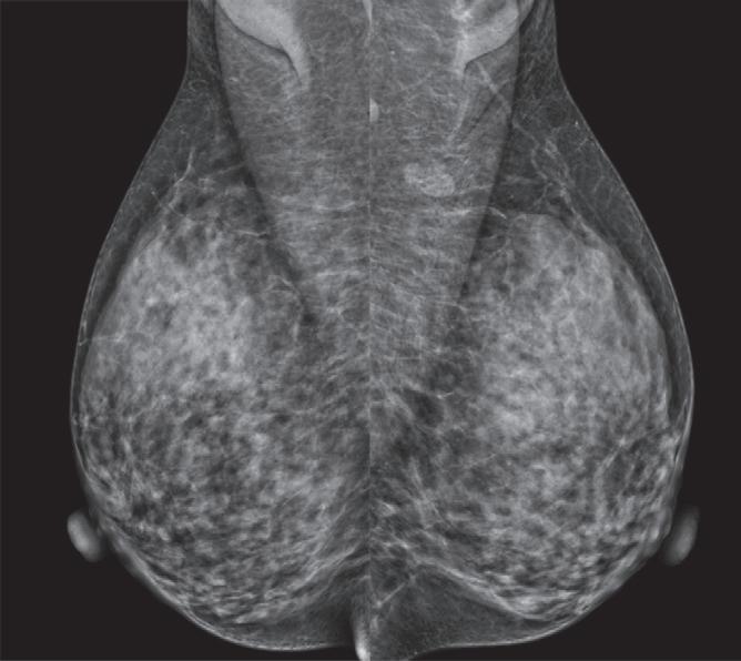
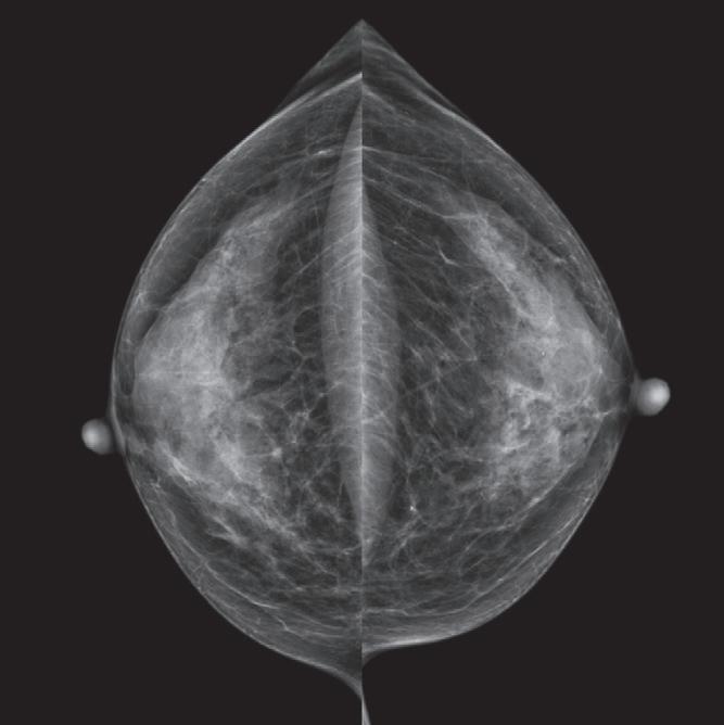
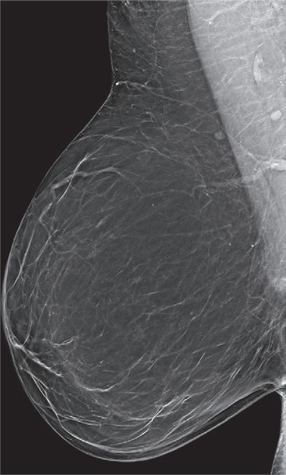
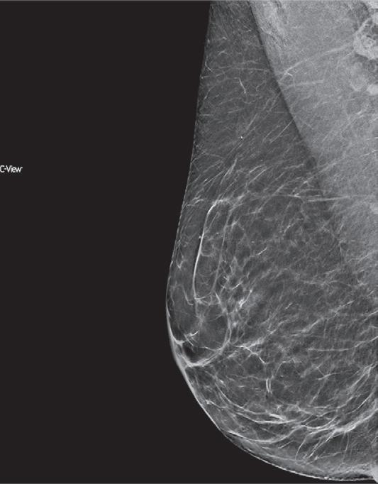
stroma vary by age and individual. Young, pregnant, and lactating women usually have a higher proportion of fibroglandular tissue. According to the Breast Imaging Reporting and Data System (BI-RADS) lexicon, mammographic density is assigned into one of four categories qualitatively indicating the relative possibility that a lesion could be obscured by normal tissue (Fig. 1.14). The categories from least to most dense are as follows: (1) The breasts are almost entirely fatty; (2) there are scattered areas of fibroglandular density; (3) the breasts are heterogeneously dense, which may obscure small masses; and (4) the breasts are extremely dense, which lowers the sensitivity of mammography. Dense fibroglandular tissue reduces the sensitivity of mammography by obscuring cancers. In addition, breast density has been shown to be an independent risk factor for developing breast cancer. Breast cancer risk and supplemental screening options are covered in detail in dedicated chapters.
Screening Versus Diagnostic Mammograms
Screening mammography is covered in detail in Chapter 12 (Organized Approach to Screening Mammography). Briefly, screening mammograms are performed in asymptomatic patients (Table 1.3). Screening mammograms are usually performed offline without a radiologist on site to guide the mammographic evaluation. After the standard CC and MLO views are obtained for each breast, patients typically leave the premises without receiving an immediate study result. The radiologist later interprets the screening mammograms in a batch and may decide to recall patients to return for additional imaging for suspicious findings requiring further workup. Patients may also be recalled for technical inadequacy (e.g., motion, positioning, artifact), referred to as a technical recall. Patients usually receive the mammogram
Table 1.3 Screening versus Diagnostic Mammograms
Screening Diagnostic
Indications
n Asymptomatic women of screening age
n Patients usually leave the premises after the mammogram is obtained
n Patients usually receive the mammogram result after leaving the premise
n Standard views: craniocaudal (CC) and mediolateral oblique (MLO) view of each breast
n Patients are recalled if additional imaging is needed
Indications
n Suspicious breast signs or symptoms
n Abnormality detected on screening mammogram
n Short interval follow-up (e.g., for a Breast Imaging Reporting and Data System [BI-RADS] 3 “probably benign” lesion)
n Radiologist is actively guiding the evaluation while patients are present
n Images are interpreted at the time of examination and results communicated to patients before they leave the premises
n Diagnostic mammography views and/or ultrasound performed at the radiologist’s discretion during the visit
results after leaving the premises, although some facilities may offer immediate interpretation.
Diagnostic mammograms are performed in patients with breast signs or symptoms and in those who need further evaluation of suspicious findings on screening mammograms. Diagnostic mammograms are also performed in patients requiring follow-up such as those who have recently received breast-conserving surgery or those with known BI-RADS 3 “probably benign” lesions. Diagnostic mammograms are performed while the radiologist is actively guiding the evaluation. Additional diagnostic imaging may include spot compression magnification views, other special views, and/or ultrasound. Diagnostic
Fig. 1.14 Examples of the four breast density categories: (A) Almost entirely fatty, (B) scattered fibroglandular densities, (C) heterogeneously dense, and (D) extremely dense.
mammograms are interpreted in real time and results are communicated to patients before patients leave the facility. Diagnostic mammography is covered in detail in Chapter 13 (Organized Approach to Diagnostic Imaging).
KEY POINTS
• Breast compression reduces parenchymal superimposition and image blurring, decreases radiation dose, ensures adequate penetration, and improves spatial resolution and image contrast.
• Before interpreting a mammogram, assess the quality of the mammogram based on positioning, compression, exposure, contrast, sharpness, noise, and artifacts.
• The posterior nipple line (PNL) is an imaginary line from the posterior nipple perpendicular to the pectoralis muscle. The length of the posterior nipple line on craniocaudal (CC) and mediolateral oblique (MLO) views should be within 1 cm of each other.
• The CC view should demonstrate nipple in profile, wellvisualized posterior medial breast, and fat posterior to the fibroglandular tissue.
• The MLO view should demonstrate an open inframammary fold, nipple in profile, pectoralis muscle extending inferior to the posterior nipple line, and convex anterior border of the pectoralis muscle.
• Spot compression views can provide better visualization of a region of interest, differentiating a real mass from summation artifact and bringing out mass margins.
• Magnification views allows for 1.5 to 2 times magnification of a region of interest and higher spatial resolution to aid in detection and evaluation of microcalcifications. This can be performed as spot compression with magnification (spot compression magnification view).
• Breast density categories from least to most dense are as follows: (1) almost entirely fatty, (2) scattered areas of
fibroglandular density, (3) heterogeneously dense, and (4) extremely dense.
• Screening mammograms are performed in asymptomatic women of screening age. Only standard views are obtained without active, real time radiologist supervision. Most commonly, the study is interpreted and results communicated to patients after patients leave the premises.
• Diagnostic mammograms are performed in symptomatic women and those who need further evaluation of suspicious findings on screening mammograms. The radiologist is actively monitoring the diagnostic evaluation and deciding the specific mammography views to obtain. Studies are interpreted in real time and results communicated to patients before they leave the premises.
Suggested Readings
Boyd NF, Guo H, Martin LJ, et al. Mammographic density and the risk and detection of breast cancer. NEJM. 2007;356(3):227–238.
Broeders M, Moss S, Nyström L, et al. The impact of mammographic screening on breast cancer mortality in Europe: a review of observational studies. J Med Screen. 2012;19(Suppl 1):14–25.
Hendrick RE, Bassett LW, Botsco MA, et al. Mammography quality control manual. Reston, VA: American College of Radiology; 1999.
Logan WW, Janus J. Use of special mammographic views to maximize radiographic information. Radiol Clin North Am. 1987;25:953–959.
Mainiero MB, Moy L, Baron P, et al. “ACR Appropriateness Criteria Breast Cancer Screening.” J Am Coll Radiol. 2017 Nov;14(11S):S383–S390.
Monsees BS. The Mammography Quality Standards Act. An overview of the regulations and guidance. Radiol Clin North Am. 2000;38:759–772.
Monticciolo DL, Newell MS, Hendrick RE, et al. Breast Cancer Screening for Average-Risk Women: Recommendations From the ACR Commission on Breast Imaging. J Am Coll Radiol. 2017 Sep;14(9):1137–1143.
Monticciolo DL, Newell MS, Moy L, et al. Breast Cancer Screening in Women at Higher-Than-Average Risk: Recommendations From the ACR. J Am Coll Radiol. 2018 Mar;15:408–414.
Park JM, Franken Jr EA. Triangulation of breast lesions: review and clinical applications. Curr Probl Diagn Radiol. 2008;37(1):1–14.
OVERVIEW
Introduction
Why BI-RADS?: Overview of Breast Imaging Reporting and Data System (BI-RADS)
ANNE C. HOYT AND HANNAH S. MILCH
This chapter summarizes the purpose and history of BI-RADS; introduces the BI-RADS lexicon, assessment categories, and standardized report; and answers frequently asked questions (FAQs) of using BI-RADS to communicate breast findings.
The Breast Imaging Reporting and Data System (BI-RADS) is a breast radiologist’s second language. Spend 10 minutes in a breast radiology reading room and you will likely hear the word BI-RADS and its associated terminology uttered over a dozen times.
BI-RADS has three main goals:
1. Standardize breast imaging interpretation terminology (the BI-RADS lexicon), final assessments (the BI-RADS categories), and management recommendations.
2. Reduce confusion and optimize communication among radiologists, referring physicians, and patients.
3. Facilitate outcomes tracking and quality assurance.
Understanding BI-RADS will help you contextualize upcoming chapters on specific breast imaging findings and appropriate management steps.
HISTORY OF BI-RADS
The 1980 s brought a sharp increase in mammography. In the early 1980s, only 15% to 20% of women had ever undergone a mammogram, and by the dawn of the 1990 s, 65% had participated in mammography at least once.1 This increase was largely due to favorable data showing effectiveness of screening mammography to reduce breast cancer mortality in multiple randomized controlled trials coupled with contemporaneous improvements in preoperative needle localization, making it easier to obtain a tissue diagnosis for suspicious lesions identified at mammography.2 As mammography utilization grew in the United States during the 1980s, significant inconsistencies became apparent in mammography quality, radiation dose, radiologist interpretative skills, and result reporting. Due to the concerns raised by breast imaging specialists and the American Medical Association (AMA),3–6 the American College of Radiology (ACR) convened two committees: the voluntary Mammography Accreditation Program (MAP) in 1987 and the Breast Imaging Reporting and Data System (BI-RADS) committee in 1988.7 The BI-RADS committee was charged with developing guidelines for
standardized mammography reporting and management recommendations.
Five years later, the ACR BI-RADS committee, composed of academic breast imagers and private practice radiologists and chaired by Dr. Carl J. D’Orsi, released the first edition of the BI-RADS lexicon in 1993. 7 This early BI-RADS document was robust and consisted of recommendations for (1) performance of screening and diagnostic mammography, (2) structure of the mammography report, (3) introduction of a mammography lexicon, and (4) final assessment categories with their respective management recommendations. 7 It should be noted that the authors fashioned BI-RADS with a clear understanding that it would be a malleable and adaptable reporting and data system with the ability to transform, improve, and expand as needed to incorporate the continuous new advances in breast imaging technology, research, and patient care.
After the successful release of the ACR MAP in 1987 and just before the 1993 ACR BI-RADS committee release of the first edition of BI-RADS, the 1992 Mammography Quality Standards Act (MQSA) became federal law and was enacted,8 making breast imaging one of the most federally regulated medical specialties in the United States. By 1999, MQSA rules mandated, among other requirements, that every mammographic report include text for the final assessment category similar to that in the then-current 1998 third edition of BI-RADS. This new post-MQSA era in breast imaging marked a transition from inconsistent imaging quality and reporting to uniform mammography examination quality, interpretation, and reporting standards. MQSA regulations and accreditation requirements are covered in Chapter 20.
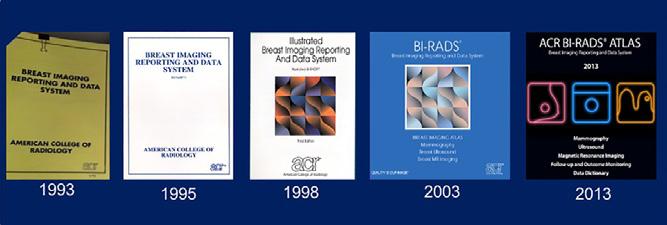
As envisioned, BI-RADS has undergone four revisions since its first edition in 1993 (1995, 1998, 2003, and 2013), with each carefully crafted to improve clarity, patient management, and quality assurance (Fig. 2.1). In 1998, the third edition included the first BI-RADS atlas with firstrate illustrations depicting each lexicon descriptor.
The fourth edition introduced many new lexicon descriptors such as the original asymmetry family (asymmetry, focal asymmetry, global asymmetry), division of suspicious calcifications into “intermediate risk” and
“higher probability of malignancy,” and the option to subclassify BI-RADS final assessment category 4 into subcategories (4 A, 4B, and 4 C) to better communicate the risk of malignancy to both the patient’s health care provider and the pathologist interpreting a woman’s biopsy tissue specimens. Furthermore, the fourth edition (2003) introduced the first BI-RADS ultrasound (US) and BI-RADS magnetic resonance imaging (MRI) lexicons. These new US and MRI lexicons were designed to mirror the same lexicon descriptors used in mammography whenever possible (e.g., mass, shape, and margin) but also allow for new modality-specific descriptors such as orientation and echo pattern in US or foci/focus and kinetic curve assessment in MRI. The current fifth edition (2013) further refines the lexicon using consistent terminology across mammography, US, and MRI sections with evidence-based justification and better quality images for all sections including mostly digital images for mammography. This edition also defines auditing rules that apply to all three sections and allows uncoupling of the final assessment category from its management recommendation as discussed in the section below.
THE BI-RADS LEXICON
The BI-RADS lexicon is a carefully constructed dictionary of descriptive terms for breast imaging findings, which has been published in numerous languages. It allows all radiologists to speak the same language and generate standard reports that are easily understood by referring providers and ancillary medical staff. The over 700-page BI-RADS Atlas provides detailed, annotated example images that illustrate the proper use of the lexicon terminology. Pathology of described findings is provided whenever possible. In addition to mammography, the most recent edition of the BI-RADS Atlas (2013) also includes comprehensive lexicons for breast US and MRI, plus a supplement with specific guidance for digital breast tomosynthesis (DBT).9 Specifics of lexicon and lesion descriptors are described in the relevant modality-specific chapters to follow.
The BI-RADS lexicon is systematically organized in a uniform branching format for each imaging modality. The organization is first divided at the highest level by imaging modality (mammography, US, MRI) followed by breast tissue and findings. Breast tissue is classified by its composition on mammography and by the amount of fibroglandular tissue and background parenchymal enhancement (BPE) on MRI. Breast composition in mammography refers to a woman’s individual admixture of dense fibroglandular tissue and fat within her breast (i.e., her breast density) and ranges from almost entirely fatty to extremely dense (Fig. 2.2). Similar terms are used to describe the amount of fibroglandular tissue on MRI. Sonographic tissue composition can also be described as homogeneous (fatty or fibroglandular) versus heterogeneous echotexture; however, these terms may only be used for screening and/or whole breast diagnostic US when the breast tissue can be assessed in its entirety. Further details on breast composition are outlined in the section titled
Fig. 2.1 Covers of the five editions of Breast Imaging Reporting and Data System (BI-RADS) beginning in 1993. (BI-RADS Atlas images obtained with permission from the American College of Radiology.)
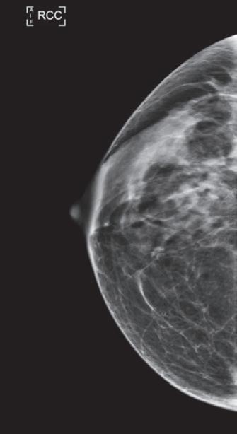
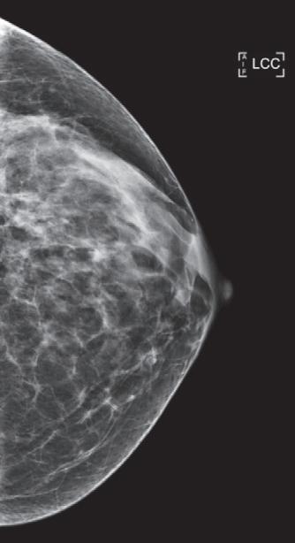
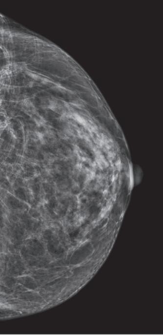
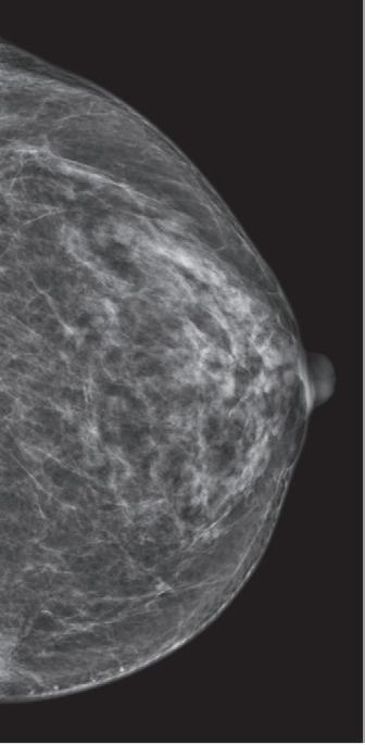
Fig. 2.2 Four synthetic mediolateral oblique mammograms depict the four Breast Imaging Reporting and Data System (BI-RADS) breast densities: (A) almost entirely fatty (type A); (B) scattered areas of fibroglandular density (type B); (C) heterogeneously dense, which may obscure small masses (type C); and (D) extremely dense, which lowers the sensitivity of mammography (type D).