No part of this publication may be reproduced or transmitted in any form or by any means, electronic or mechanical, including photocopying, recording, or any information storage and retrieval system, without permission in writing from the publisher. Details on how to seek permission, further information about the Publisher’s permissions policies and our arrangements with organizations such as the Copyright Clearance Center and the Copyright Licensing Agency, can be found at our website: www.elsevier.com/permissions
This book and the individual contributions contained in it are protected under copyright by the Publisher (other than as may be noted herein).
Notices
Knowledge and best practice in this eld are constantly changing. As new research and experience broaden our understanding, changes in research methods, professional practices, or medical treatment may become necessary.
Practitioners and researchers must always rely on their own experience and knowledge in evaluating and using any information, methods, compounds, or experiments described herein. In using such information or methods they should be mindful of their own safety and the safety of others, including parties for whom they have a professional responsibility.
With respect to any drug or pharmaceutical products identi ed, readers are advised to check the most current information provided (i) on procedures featured or (ii) by the manufacturer of each product to be administered, to verify the recommended dose or formula, the method and duration of administration, and contraindications. It is the responsibility of practitioners, relying on their own experience and knowledge of their patients, to make diagnoses, to determine dosages and the best treatment for each individual patient, and to take all appropriate safety precautions.
To the fullest extent of the law, neither the Publisher nor the authors, contributors, or editors, assume any liability for any injury and/or damage to persons or property as a matter of products liability, negligence or otherwise, or from any use or operation of any methods, products, instructions, or ideas contained in the material herein.
Previous edition copyrighted 2014 by Mosby, an imprint of Elsevier Inc.
Previous edition copyrighted 2010, 2005, 2001, 1997, 1993, 1987, 1982 by Mosby, Inc., an af liate of Elsevier Inc.
International Standard Book Number: 978-0-323-39966-1
Executive Content Strategist: Sonya Seigafuse
Content Development Manager: Lisa P. Newton
Senior Content Development Specialist: Tina Kaemmerer
Publishing Services Manager: Julie Eddy
Senior Project Manager: Mary G. Stueck
Design Direction: Renée Duenow
vi ACKNOWLEDGMENTS AND DEDICATION
Mary Stueck is the Senior Project Manager who led us through the production phase. We couldn’t have produced this edition without her expertise.
Most importantly, a thank you to Elsevier Publishing for allowing us to continue to be part of this wonderful reference for the past 44 years.
Finally, my thanks to my family for their ongoing support. My wife Deborah, son Daniel, daughter Molly, and granddaughter, Tatum. I’m especially proud that Daniel and Molly have entered the medical profession. They are both excellent professionals and they understand the importance of treating their patients with dignity and compassion. They have always been important to me even though I don’t express it adequately. My true inspiration is my granddaughter, Tatum, who makes me smile daily. What a beautiful and kind person she has become. When things got dif cult and
overwhelming, I only needed to see her picture or spend a few minutes with her and my spirit was renewed. Tatum will always own my heart. Finally, to Buddy, the wonder dog, for sitting in the of ce (ok…sleeping) while I wrote. The Bishop, Daniel’s dog, was a hero to those he found over the many years on search and rescue missions.
Deborah has been at my side for over 39 years. She has been the compassionate anchor that provides our family with the stability and encouragement to be successful in all of our professional and personal endeavors. My life changed in so many positive ways since I rst met her. Meeting the demands of a new edition of the text would not have been possible if it wasn’t for her enduring love and support. I dedicate this edition to my family.
Contributors
Joie Burns, MS, RT(R)(S), RDMS, RVT
Chapter 20
Diagnostic Medical Sonography Program Director
Radiologic Sciences
Boise State University Boise, Idaho
Mary J. Carrillo, MBA/HCM, RT(R)(M), CDT
Chapter 20
Medical Radiography Program Director
Health Sciences
GateWay Community College Phoenix, Arizona; Mammographer
SimonMed Imaging Mesa, Arizona
Jeanne Dial, MEd, CNMT, RSO
Chapter 20
Nuclear Medicine Technology Program Director
Health Sciences
GateWay Community College Phoenix, Arizona
Cheryl DuBose, EdD, RT(R)(MR)(CT) (QM)
Chapter 20
CT/MRI Program Director Arkansas State University Jonesboro, Arkansas
Frank Goerner, PhD, DABR
Chapter 1
Medical Physicist
Medical Physics
The Queens Medical Center Honolulu, Hawaii
Michele L. Gray-Murphy, BSRS, RT(R) (M)(ARRT)
Chapter 11
Associate of Science in Radiography Program Faculty
Allen College–UnityPoint Health Waterloo, Iowa
Kelli Welch Haynes, MSRS, RT(R)
Chapter 3
Program Director and Associate Professor Allied Health
Northwestern State University Shreveport, Louisiana
Chad Hensley, MEd, RT(R)(MR)
Chapter 16
Clinical Coordinator
Radiography
University of Nevada–Las Vegas Las Vegas, Nevada
Nicolle M. Hightower, MEd, RT(R)(VI)
Chapter 17
Diagnostic Medical Imaging Faculty and Clinical Coordinator
University of North Carolina at Chapel Hill Chapel Hill, North Carolina
How to Use the Positioning Pages
1 PROJECTION TITLE BARS describe the speci c position/ projection to be radiographed, including the proper name of the position, if such applies.
2 CLINICAL INDICATIONS section summarizes conditions or pathologies that may be demonstrated by the examination and/or projection. This brief review helps the technologist understand the purpose of the examination and which structures or tissues should be most clearly demonstrated.
3 PROJECTION SUMMARY BOXES list all the speci c routine or special projections most commonly performed for that body part.
4 TECHNICAL FACTORS section includes the image receptor (IR) size recommended for the average adult; whether the IR should be placed portrait or landscape in relation to the patient; a grid, if one is needed; and the kV range for analog and digital systems. The minimum SID (source-to-image receptor distance) is listed.
5 IMAGE RECEPTOR ICONS give a visual display of the IR relative size (cm) and orientation (portrait or landscape), relative collimated eld size, location of R and L markers, and the recommended AEC cell location (if AEC is used).
6 SHIELDING section describes shielding that should be used for the projection.
7 PATIENT POSITION section indicates the general body position required for the projection.
8 PART POSITION section gives a clear, step-by-step description of how the body part should be positioned in relation to the IR and/or tabletop. The CR icon is included for all those projections in which the CR is of primary importance to remind the technologist to pay special attention to the CR during the positioning process for that projection.
9 CENTRAL RAY (CR) section describes the precise location of the CR in relation to both the IR and the body part.
10 RECOMMENDED COLLIMATION section describes the collimation of the x-ray eld recommended for that projection.
11 RESPIRATION section lists the breathing requirements for that projection.
12 EVALUATION CRITERIA boxes describe evaluation/critique process that should be completed for each processed radiographic image. This process is divided into the following three broad categories: (1) anatomy demonstrated, (2) position, (3) exposure.
13 POSITIONING PHOTOGRAPHS shows a correctly positioned patient and part in relation to the CR and IR.
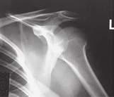
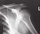
14 RADIOGRAPHIC IMAGES provide an example of a correctly positioned and correctly exposed radiographic image of the featured projection.
15 ANATOMY LABELED IMAGES identify speci c anatomy that should be demonstrated on the radiographic image shown. The labeled image, in most cases, matches the radiographic image example on the same page.
ParT n ■ T r n and P T n n
GENERAL, SYSTEMIC, AND SKELETAL ANATOMY AND ARTHROLOGY
General Anatomy
Anatomy is the study, classi cation, and description of the structure and organs of the human body, whereas physiology deals with the processes and functions of the body, or how the body parts work. In the living subject, it is almost impossible to study anatomy without also studying some physiology. However, radiographic study of the human body is primarily a study of the anatomy of the various systems, with less emphasis on the physiology. Consequently, anatomy of the human system is emphasized in this radiographic anatomy and positioning textbook.
n T : Phonetic respelling1 of anatomic and positioning terms is included throughout this text to facilitate correct pronunciation of the terms commonly used in medical radiography.
TrU TUra r an ZaT n
Several levels of structural organization make up the human body. The lowest level of organization is the chemic l level. All chemicals necessary for maintaining life are composed of toms, which are joined in various ways to form molecules. Various chemicals in the form of molecules are organized to form cells.
Cells
The cell is the basic structural and functional unit of all living tissue. Every single part of the body, whether muscle, bone, cartilage, fat, nerve, skin, or blood, is composed of cells.
Tissues
Tissues are cohesive groups of similar cells that, together with their intercellular material, perform a speci c function. The four basic types of tissue are as follows:
1. Epithelial (ep ″-i-the ′ le-al): Tissues that cover internal and external surfaces of the body, including the lining of vessels and organs, such as the stomach and the intestines
2. Connective: Supportive tissues that bind together and support various structures
3. Muscular: Tissues that make up the substance of a muscle
4. Nervous: Tissues that make up the substance of nerves and nerve centers
Organs
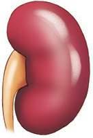
When complex assemblies of tissues are joined to perform a speci c function, the result is an organ. Organs usually have a speci c shape. Examples of organs of the human body are the kidneys, heart, liver, lungs, stomach, and brain.
System
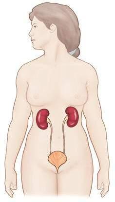
A system consists of a group or an association of organs that have a similar or common function. The urinary system, consisting of the kidneys, ureters, bladder, and urethra, is an example of a body system. The total body comprises 10 i ivi u l bo y systems.
Organism
The 10 systems of the body when functioning together make up the total organism—one living being ( Fig. 1.1 ).
Organism (10 systems)
Fig. 1.1 Levels of human structural organization.
Molecule
Systemic Anatomy
B d T
The human body is a structural and functional unit made up of 10 lesser units called systems. These 10 systems include (1) skeletal, (2) circulatory, (3) digestive, (4) respiratory, (5) urinary, (6) reproductive, (7) nervous, (8) muscular, (9) endocrine, and (10) integumentary (in-teg ″-u-men ′ -tar-e).
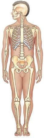
Skeletal System
The skeletal system ( Fig. 1.2 ) is important for the technologist to learn. The skeletal system includes the 206 sep te bo es of the body and their associated cartilages and joints. The study of bones is termed osteology, whereas the study of joints is called th ology.
The four functions of the skeletal system are as follows:
1.Support and protect many soft tissues of the body
2.Allow movement through interaction with the muscles to form a system of levers
3.Produce blood cells
4.Store calcium
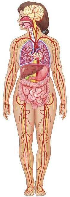
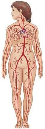
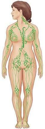
Circulatory System
The circulatory system ( Fig. 1.3 ) is composed of the following:
•The c iov scul o g s —heart, blood, and blood vessels
•The lymph tic system —lymph nodes, lymph vessels, lymph glands, and spleen
The six functions of the circulatory system are as follows:
1.Distribute oxygen and nutrients to the cells of the body
2.Transport cell waste and carbon dioxide from the cells
3.Transport water, electrolytes, hormones, and enzymes
4.Protect against disease
5.Prevent hemorrhage by forming blood clots
6.Assist in regulating body temperature
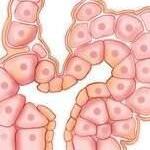
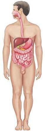
Digestive System
The digestive system includes the alimentary canal and certain accessory organs ( Fig. 1.4 ). The alimentary canal is made up of the mouth, pharynx, esophagus, stomach, small intestine, large intestine, and anus. Accessory organs of digestion include the salivary glands, liver, gallbladder, and pancreas.
The twofold function of the digestive system is as follows:
1.Prepare food for absorption by the cells through numerous physical and chemical breakdown processes