Bone 2nd edition g. petur nielsen - Get the ebook instantly with just one click

Page 1


https://ebookmass.com/product/bone-2nd-edition-g-peturnielsen/

Instant digital products (PDF, ePub, MOBI) ready for you

Download now and discover formats that fit your needs...

Diagnostic Pathology: Bone, 3rd Edition Gunnlaugur Petur

Nielsen

https://ebookmass.com/product/diagnostic-pathology-bone-3rd-editiongunnlaugur-petur-nielsen/

ebookmass.com

A New Ecology: Systems Perspective 2nd Edition Soeren Nors

Nielsen

https://ebookmass.com/product/a-new-ecology-systems-perspective-2ndedition-soeren-nors-nielsen/

ebookmass.com

Community Ecology 2nd Edition Gary G. Mittelbach

https://ebookmass.com/product/community-ecology-2nd-edition-gary-gmittelbach/

ebookmass.com

Fortran for Scientists and Engineers 4th Edition Stephen

https://ebookmass.com/product/fortran-for-scientists-andengineers-4th-edition-stephen-j-chapman/

ebookmass.com

SECOND EDITION

G. Petur Nielsen, MD

Pathologist, Department of Pathology

Director of Electron Microscopy

Director of Bone & Soft Tissue Pathology

Massachusetts General Hospital Professor of Pathology

Harvard Medical School Boston, Massachusetts

Andrew E. Rosenberg, MD

Professor and Vice Chair

Director of Bone & Soft Tissue Pathology

Department of Pathology

Miller School of Medicine

University of Miami Miami, Florida

Vikram Deshpande, MD

Associate Pathologist

Department of Pathology

Massachusetts General Hospital

Associate Professor of Pathology

Harvard Medical School

Boston, Massachusetts

Francis J. Hornicek, MD, PhD

Chief, Orthopaedic Oncology Service

Co-Director, Center for Sarcoma and Connective Tissue Oncology

Massachusetts General Hospital

Director, Stephan L. Harris Chordoma Center

The Henry J. Mankin, MD, Endowed Scholar Professor

Harvard Medical School

Co-Leader, Dana Farber/Harvard Cancer Center Sarcoma Program

Boston, Massachusetts

Susan V. Kattapuram, MD

Associate Radiologist

Massachusetts General Hospital

Associate Professor of Radiology

Harvard Medical School

Boston, Massachusetts

Daniel I. Rosenthal, MD

Associate Radiologist-in-Chief

Massachusetts General Hospital Professor of Radiology

Harvard Medical School

Boston, Massachusetts

1600 John F. Kennedy Blvd.

Ste 1800 Philadelphia, PA 19103-2899

DIAGNOSTIC PATHOLOGY: BONE, SECOND EDITION

Copyright © 2017 by Elsevier. All rights reserved.

ISBN: 978-0-323-47777-2

No part of this publication may be reproduced or transmitted in any form or by any means, electronic or mechanical, including photocopying, recording, or any information storage and retrieval system, without permission in writing from the publisher. Details on how to seek permission, further information about the Publisher’s permissions policies and our arrangements with organizations such as the Copyright Clearance Center and the Copyright Licensing Agency, can be found at our website: www.elsevier.com/ permissions.

This book and the individual contributions contained in it are protected under copyright by the Publisher (other than as may be noted herein).

Notices

Knowledge and best practice in this field are constantly changing. As new research and experience broaden our understanding, changes in research methods, professional practices, or medical treatment may become necessary.

Practitioners and researchers must always rely on their own experience and knowledge in evaluating and using any information, methods, compounds, or experiments described herein. In using such information or methods they should be mindful of their own safety and the safety of others, including parties for whom they have a professional responsibility.

With respect to any drug or pharmaceutical products identified, readers are advised to check the most current information provided (i) on procedures featured or (ii) by the manufacturer of each product to be administered, to verify the recommended dose or formula, the method and duration of administration, and contraindications. It is the responsibility of practitioners, relying on their own experience and knowledge of their patients, to make diagnoses, to determine dosages and the best treatment for each individual patient, and to take all appropriate safety precautions.

To the fullest extent of the law, neither the Publisher nor the authors, contributors, or editors, assume any liability for any injury and/or damage to persons or property as a matter of products liability, negligence or otherwise, or from any use or operation of any methods, products, instructions, or ideas contained in the material herein.

Publisher Cataloging-in-Publication Data

Names: Nielsen, G. Petur (Gunnlaugur Petur) | Rosenberg, Andrew, 1953-

Title: Diagnostic pathology. Bone / [edited by] G. Petur Nielsen and Andrew E. Rosenberg. Other titles: Bone.

Description: Second edition. | Salt Lake City, UT : Elsevier, Inc., [2017] | Includes bibliographical references and index.

Identifiers: ISBN 978-0-323-47777-2

Subjects: LCSH: Bones--Tumors--Handbooks, manuals, etc. | MESH: Bone Neoplasms--pathology--Atlases. | Bone Neoplasms--diagnosis--Atlases.

Classification: LCC RC280.B6 N54 2017 | NLM WZ 17 | DDC 616.8’4--dc23

International Standard Book Number: 978-0-323-47777-2

Cover Designer: Tom M. Olson, BA

Printed in Canada by Friesens, Altona, Manitoba, Canada

Last digit is the print number: 9 8 7 6 5 4 3 2 1

Preface

The pathology of the skeleton is complex and is the morphologic expression of a broad spectrum of diseases, including those caused by genetic (sporadic and inherited), malformative, inflammatory, metabolic, circulatory, traumatic, iatrogenic, and neoplastic disorders. Bone tumors, including both neoplasms and various conditions that may simulate them, are the focus of our book. This topic is one of the most challenging areas in surgical pathology for several reasons: Bone tumors are uncommon, making it difficult to acquire the necessary experience with their histological variants and mimics; the correct diagnosis usually requires the careful integration of radiological imaging studies and clinical findings; the implications of a diagnosis on a patient can be life changing; and medical schools and pathology training programs often have insufficient expertise to provide medical students and young pathologists with the skills needed to diagnose these lesions accurately and precisely.

This book reflects the philosophy and high standards practiced by the truly multidisciplinary team of physicians at the Massachusetts General Hospital and University of Miami, who have diagnosed and surgically treated tens of thousands of patients with bone tumors for many decades. Also important to acknowledge are the contributions of the many fellows and residents who participated in the efforts of patient care.

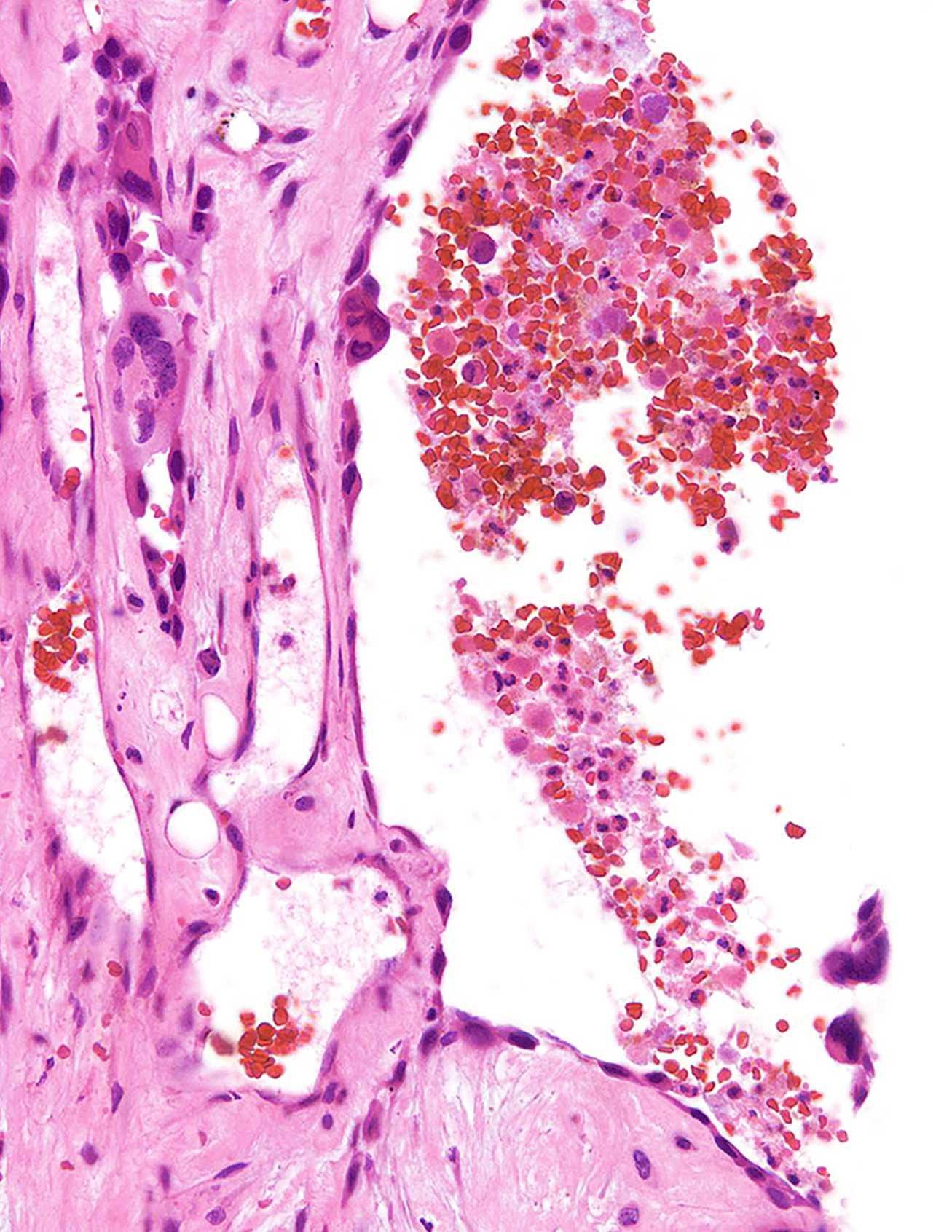
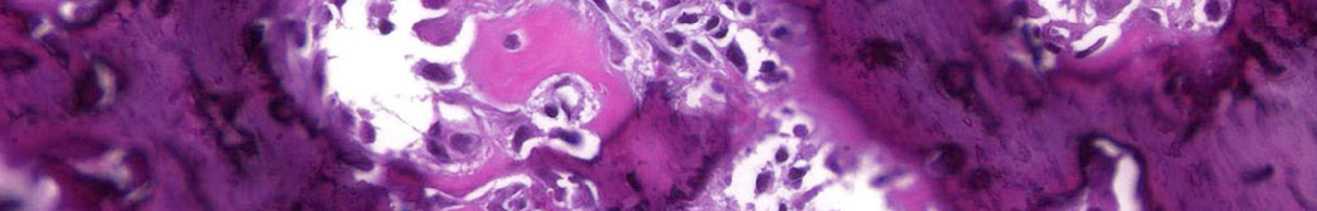
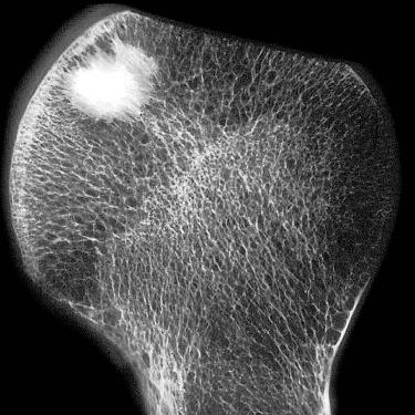
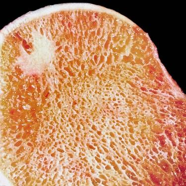
The authors are subspecialized physicians who have dedicated their professional lives to the diagnosis and surgical management of bone tumors. As a result, the figures include beautiful and classic examples and unusual variants of many of the diseases discussed and are the product of painstaking correlations between the clinical, imaging, macroscopic, histological, immunohistochemical, and molecular characteristics of bone tumors. The text synthesizes the literature and our combined extensive experience, and the images have been selectively culled from the patient files of the Massachusetts General Hospital, the University of Miami Miller School of Medicine, and the private consultations of the authors. The book is constructed in a thematic format with sections representing groups of related diseases and the chapters discussing individual entities and their differential diagnosis.

Accordingly, this textbook serves as an excellent resource for medical students, residents, fellows, and practicing physicians in the disciplines of pathology, radiology, and orthopedics. Medical and radiation oncologists who treat bone tumors will also find it valuable. Our opportunity to participate in the care of patients with bone tumors has been our call and honor, and we hope to do it justice by sharing our experience with the medical community—our goal is to enhance diagnostic accuracy and to provide the biological basis for optimal treatment.

G. Petur Nielsen, MD

Pathologist, Department of Pathology

Director of Electron Microscopy

Director of Bone & Soft Tissue Pathology

Massachusetts General Hospital

Professor of Pathology

Harvard Medical School

Boston, Massachusetts

Andrew E. Rosenberg, MD

Professor and Vice Chair

Director of Bone & Soft Tissue Pathology

Department of Pathology

Miller School of Medicine

University of Miami Hospital

Miami, Florida

Acknowledgments

Text Editors

Arthur G. Gelsinger, MA

Nina I. Bennett, BA

Lisa A. Gervais, BS

Karen E. Concannon, MA, PhD

Matt W. Hoecherl, BS

Megg Morin, BA

Image Editors

Jeffrey J. Marmorstone, BS

Lisa A. M. Steadman, BS

Illustrations

Laura C. Sesto, MA

Richard Coombs, MS

Lane R. Bennion, MS

Art Direction and Design

Tom M. Olson, BA

Laura C. Sesto, MA

Lead Editor

Terry W. Ferrell, MS

Production Coordinators

Angela M. G. Terry, BA

Rebecca L. Bluth, BA

Emily C. Fassett, BA

TABLE OF CONTENTS

SECTION9:GIANTCELL-RICHTUMORS

256GiantCellTumor

G.PeturNielsen,MDandAndrewE.Rosenberg,MD

270BrownTumor

G.PeturNielsen,MDandAndrewE.Rosenberg,MD

274GiantCellReparativeGranuloma

G.PeturNielsen,MDandAndrewE.Rosenberg,MD

SECTION10:CYSTICLESIONSOFBONE

282IntraosseousGanglion

G.PeturNielsen,MDandAndrewE.Rosenberg,MD

286UnicameralBoneCyst

G.PeturNielsen,MDandAndrewE.Rosenberg,MD

294AneurysmalBoneCyst

G.PeturNielsen,MDandAndrewE.Rosenberg,MD

306EpidermoidInclusionCyst

G.PeturNielsen,MDandAndrewE.Rosenberg,MD

SECTION11:VASCULARTUMORS

310ConventionalHemangioma

G.PeturNielsen,MDandAndrewE.Rosenberg,MD

318Lymphangioma/Lymphangiomatosis

G.PeturNielsen,MDandAndrewE.Rosenberg,MD

320EpithelioidHemangioma

G.PeturNielsen,MDandAndrewE.Rosenberg,MD

328PseudomyogenicHemangioendothelioma

G.PeturNielsen,MDandAndrewE.Rosenberg,MD

334EpithelioidHemangioendothelioma

G.PeturNielsen,MDandAndrewE.Rosenberg,MD

340Angiosarcoma

G.PeturNielsen,MDandAndrewE.Rosenberg,MD

SECTION12:HEMATOPOIETICTUMORS

346LangerhansCellHistiocytosis(Eosinophilic Granuloma)

G.PeturNielsen,MDandAndrewE.Rosenberg,MD

352PrimaryLymphoma

G.PeturNielsen,MDandAndrewE.Rosenberg,MD

360PlasmaCellMyeloma

VikramDeshpande,MD,G.PeturNielsen,MD,and AndrewE.Rosenberg,MD

368MastCellDisease

G.PeturNielsen,MDandAndrewE.Rosenberg,MD

370Erdheim-ChesterDisease

G.PeturNielsen,MDandAndrewE.Rosenberg,MD

376Rosai-DorfmanDisease

G.PeturNielsen,MDandAndrewE.Rosenberg,MD

SECTION13:MISCELLANEOUS MESENCHYMALTUMORS

382OsteofibrousDysplasia

VikramDeshpande,MD,G.PeturNielsen,MD,and AndrewE.Rosenberg,MD

386Adamantinoma

G.PeturNielsen,MDandAndrewE.Rosenberg,MD

392AdipocyticTumors

G.PeturNielsen,MDandAndrewE.Rosenberg,MD

400Leiomyosarcoma

G.PeturNielsen,MDandAndrewE.Rosenberg,MD

404Myoepithelioma

G.PeturNielsen,MDandAndrewE.Rosenberg,MD

410Schwannoma

G.PeturNielsen,MDandAndrewE.Rosenberg,MD

414MyxopapillaryEpendymoma

VikramDeshpande,MD,G.PeturNielsen,MD,and AndrewE.Rosenberg,MD

416PhosphaturicMesenchymalTumor

G.PeturNielsen,MDandAndrewE.Rosenberg,MD

SECTION14:METASTATICTUMORS

420MetastaticTumors

VikramDeshpande,MD,AndrewE.Rosenberg,MD,and G.PeturNielsen,MD

SECTION15:BONETUMORMIMICS

428BizarreParostealOsteochondromatous ProliferationandRelatedLesions

G.PeturNielsen,MDandAndrewE.Rosenberg,MD

434Melorheostosis

G.PeturNielsen,MDandAndrewE.Rosenberg,MD

438Amyloidoma

G.PeturNielsen,MD,AndrewE.Rosenberg,MD,and VikramDeshpande,MD

440GaucherDisease

G.PeturNielsen,MDandAndrewE.Rosenberg,MD

This page intentionally left blank

SECTION 1

Benign Bone-Forming Tumors

Bone Island/Osteopoikilosis

KEYFACTS

TERMINOLOGY

• Enostosis

CLINICALISSUES

• Incidentalradiographicfinding

• Largeboneislandsinchildrenmaybeconcerningfor osteosarcoma

• Rarelyneedtobebiopsied

• Multipleboneislandsrepresentosteopoikilosis

• Observationforsmallsolitarycharacteristiclesions,but thosethatarelargeorhaveunusualfeaturesmayrequire biopsy

IMAGING

• Mostfrequentsitesincludepelvis,proximalfemur,andribs

• Inosteopoikilosis,usuallyinvolveepiphysesofshorttubular bones

• Generallynotmuchlargerthan1cmindiameter

• Homogeneouslyradiodenselesionswithspiculated margins,whichmergewithsurroundingcancellousbone

MACROSCOPIC

• Hard,solid,tan-white

• Peripheryblendswithsurroundingcancellousbone

MICROSCOPIC

• Consistofcortical-typebonecontaininghaversian-like canals

• Predominatelylamellarbutmaybefocallywoven

• Osteopoikilosisboneislandtumorsareidenticalto sporadic,solitaryboneislands

TOPDIFFERENTIALDIAGNOSES

• Well-differentiatedosteosarcoma

• Scleroticmetastases

(Left)Specimenradiograph showsexcisedfemoralhead containingaboneisland.The boneislandisovaland radiodense.Theperipheryhas astellatemarginmergingwith theneighboringcancellous bone.(Right)Gross photographoffemoralhead showsboneislandbeneaththe articularsurface.Thebone islandisdense,tan-white,and hasaspiculatedborderasit mergeswiththeadjacentbony trabeculae.

(Left)Radiographofthepelvis demonstratesauniformly denseboneisland﬈.The spiculatedmarginsrepresent extensionsfromthelesion, whichmergewiththe surroundingcancellousbone. (Right)CTofaboneisland﬈ demonstratesthatitisofthe samedensityascorticalbone. Theabsenceofalytic componentcanbedifficultto confirmwithoutadditional cross-sectionalimaging,an importantfeatureto distinguishboneislandfrom other,moresinisterboneforminglesions.

SpecimenRadiographofBoneIsland
GrossPhotographofBoneIsland
BoneIslandInvolvingPelvis
CTScanofPelvicBoneIsland

Bone Island/Osteopoikilosis

TERMINOLOGY

Synonyms

• Enostosis

• Spottedbonedisease

Definitions

• Benignbone-formingtumorcomposedofcortical-type bonethatdevelopswithinmedullarycavity

• Osteopoikilosis

○ Syndromecharacterizedbypresenceofmultiple(usually many)boneislands

○ CanbeassociatedwithBuschke-Ollendorffsyndrome andmelorheostosis-likelesions

ETIOLOGY/PATHOGENESIS

Neoplasm

• Causeofboneislandisunknown

• Osteopoikilosismaybeinheritedinautosomaldominant fashion

○ AssociatedwithmutationsandlossoffunctioninLEMD3 locatedon12q14

CLINICALISSUES

Presentation

• Incidentalradiographicfindingandasymptomatic

○ Largeboneislandsmaybepainful

• Uncommoninchildren

• Lesionsinosteopoikilosismayundergoslow,progressive enlargementorinvolution

Treatment

• Observationforsmallsolitarylesionswithclassic radiographicfeatures

• Largervariantsorcasesinadolescentsmayrequirebiopsy toexcludemoreaggressivelesions,suchaswelldifferentiatedosteosarcomaandscleroticmetastasesin adults

Prognosis

• Excellent

• Malignanttransformationdoesnotoccur

IMAGING

GeneralFeatures

• Location

○ Mostfrequentsitesarepelvis,ribs,andproximalfemur

○ Inadults,incidenceinpelvicbones(1.0%)andribs(0.5%)

○ Epiphysealintubularbones

○ Osteopoikilosisisbilateralandsymmetricalin distributionandinmetaphysealandepiphysealregions oftubularbones

– Anybonemaybeaffected,includingtarsalandcarpal bones

• Size

○ Usually<1cmindiameter

○ Infrequently,"giant"boneislandsseveralcmindiameter occur

RadiographicFindings

• Small

• Oval ○ Longaxisofovalisusuallyparalleltomechanicalstresses onbone,representingadaptationtoWolfflaw

• Singleormultiple

• Largervariantsmayabutorbebasedonendostealsurface

○ Donotinvolvecortexanddonotelicitperiosteal reaction

• Homogeneouslyradiodenselesionswithspiculated marginsthatmergewithsurroundingcancellousbone

MRFindings

• DarkonT1WIandT2WI,similartonormalcorticalbone

CTFindings

• Smallstellatemedullarylesionwithcharacteristicsof cortical-typebone

BoneScan

• Canshowsomeuptakeofradionuclide

MACROSCOPIC

GeneralFeatures

• Hard,solid,andtan-white;peripheryblendsinto surroundingtrabeculae,whichaccountsforirregular spiculatedmargins

MICROSCOPIC

HistologicFeatures

• Consistofcortical-typebonecontaininghaversian-like canals

• Predominatelylamellarbutmaybefocallywoven

• Osteoblastsliningsurfacesareflatandquiescent

• Osteocytesaresmallandcytologicallybanal

• Boneislandsinosteopoikilosisareidenticaltosporadic, solitaryboneislands

DIFFERENTIALDIAGNOSIS

Well-DifferentiatedOsteosarcoma

• Infiltrative,composedofproliferatingmildlyatypical spindlecellsandwovenbone

ScleroticMetastases

• Usuallyinadultsandcontainhistologicallymalignantcells

DIAGNOSTICCHECKLIST

ClinicallyRelevantPathologicFeatures

• Multiplelesionsraisedifferentialdiagnosisofblastic metastases;multipleboneislandsseeninosteopoikilosis

PathologicInterpretationPearls

• Lesionisintramedullary,corticalintype,andpredominately lamellarinarchitecture

SELECTEDREFERENCES

1. KorkmazMFetal:Osteopoikilosis:reportofafamilialcaseandreviewofthe literature.RheumatolInt.ePub,2014

(Left)APradiograph demonstratesosteopoikilosis withmultipleboneislands involvingtheendsoftheshort tubularbones.Thesmall lesionsclusterattheendsof thebones.(Right)AP radiographofthekneesshows thetypicalfeaturesof osteopoikilosis.Alarge numberofsmallboneislands aresymmetricallydistributed inaprimarilyjuxtaarticular andmetaphysealdistribution. Despitetheirmultiplicity,each lesionindividuallyhasthe featurestypicalofabone island.

(Left)ReformattedcoronalCT scanofthehipshowsmultiple smallboneislandsthatare juxtaarticularand metaphysealindistribution. Eachlesionisindividually indistinguishablefroma solitaryboneisland.(Right) T1-weightedMRofthekneein apatientwithosteopoikilosis demonstratesthatthebone islandsaresmall,rather uniforminsize,ovalinshape, anduniformlydense.Each lesionisindividually indistinguishablefroma solitaryboneisland.

(Left)CoronalCT demonstratesan intramedullaryboneisland involvingtherightmandible ſt.Thelesionisscleroticwith thesamedensityasthe surroundingcortex.(Right) Grossphotographshowsrib andadjacentcostalcartilage. Anelongate,dense,andtanwhiteboneislandfillsthe involvedsegmentofthe medullarycavityſt.The adjacentcortexandcostal cartilageareunremarkable.

Osteopoikilosis
MRofOsteopoikilosis
BoneIslandofMandible
GrossPhotoofBoneIslandArisinginRib

GrossPhotograph

Whole-MountSection

LightMicroscopy LightMicroscopy

(Left)Grossphotographshows anexcisedfemoralheadwith anincidentalboneisland﬈. Thetumorislocatedbeneath thearticularcartilageand mergeswiththesurrounding trabecularbone.The neighboringmarrowisfatty andunremarkable.(Right) Low-powerviewshowsabone islandinvolvingthemedullary cavityoftheproximalfemur ﬈.Thelesioniscomposedof cortical-typebonethatblends imperceptivelywiththe surroundingtrabecularbone.

(Left)Histologicsectionofa solitaryboneislandisshown. Theboneislandiscomposed ofcortical-typebonewith haversian-likecanalsand transitionsintotheadjacent cancellousbone.The surroundingmarrowis predominatelyfattywith scatteredislandsof hematopoieticcells.(Right) Boneislandwithnumerous haversian-likesystems scatteredthroughoutthe lesionisshown.Theboneof thetumorissharply demarcatedfromtheadjacent marrow.

ofBoneIsland

(Left)Closeviewofthe transitionbetweenthe peripheryoftheboneislandis shownasitmergeswithan adjacentbonetrabeculum. Theboneiswovenand lamellarandhasasharp borderwiththesurrounding fattyandhematopoietic marrow.(Right)Resected femoralheadfromapatient withosteopoikilosisshows thatnumerousboneislands arepresentwithinthe medullarycavity.Thelesions areroundtoovalandvaryin size.Themarginsareirregular andspiculated.

Periphery
Osteopoikilosis

Turn static files into dynamic content formats.

Create a flipbook
Issuu converts static files into: digital portfolios, online yearbooks, online catalogs, digital photo albums and more. Sign up and create your flipbook.