https://ebookmass.com/product/biology-of-cardiovascular-and-
Instant digital products (PDF, ePub, MOBI) ready for you
Download now and discover formats that fit your needs...
The impact of nutrition and statins on cardiovascular diseases Lordan
https://ebookmass.com/product/the-impact-of-nutrition-and-statins-oncardiovascular-diseases-lordan/
ebookmass.com
Molecular, Cellular, and Metabolic Fundamentals of Human Aging Evandro Fei Fang
https://ebookmass.com/product/molecular-cellular-and-metabolicfundamentals-of-human-aging-evandro-fei-fang/
ebookmass.com
Medicinal Spices and Vegetables from Africa: Therapeutic Potential against Metabolic, Inflammatory, Infectious and Systemic Diseases Victor Kuete
https://ebookmass.com/product/medicinal-spices-and-vegetables-fromafrica-therapeutic-potential-against-metabolic-inflammatoryinfectious-and-systemic-diseases-victor-kuete/ ebookmass.com
Humans in the Making In the Beginning was Technique_ Volume 4 Michael J.F.Dubios
https://ebookmass.com/product/humans-in-the-making-in-the-beginningwas-technique_-volume-4-michael-j-f-dubios/
ebookmass.com
Nanotoxicity: Prevention and Antibacterial Applications of Nanomaterials (Micro and Nano Technologies) 1st Edition
Susai Rajendran (Editor)
https://ebookmass.com/product/nanotoxicity-prevention-andantibacterial-applications-of-nanomaterials-micro-and-nanotechnologies-1st-edition-susai-rajendran-editor/
ebookmass.com
Blackwood Institute: The Complete Trilogy Rose
https://ebookmass.com/product/blackwood-institute-the-completetrilogy-rose/
ebookmass.com
Weight Training for Beginners: A Complete Illustrated Guide to Strenght Training at Home for Men and Women. Easy and Effective Exercises and Workouts with dumbbells to Burn Fat and Build Muscle John Mcdillon
https://ebookmass.com/product/weight-training-for-beginners-acomplete-illustrated-guide-to-strenght-training-at-home-for-men-andwomen-easy-and-effective-exercises-and-workouts-with-dumbbells-toburn-fat-and-build-muscle-john-m/ ebookmass.com
L’attachement : Approche Théorique 4th Edition Guédeney
https://ebookmass.com/product/lattachement-approche-theorique-4thedition-guedeney/
ebookmass.com
Equine Reproductive Procedures 2nd Edition John Dascanio
https://ebookmass.com/product/equine-reproductive-procedures-2ndedition-john-dascanio/
ebookmass.com
Over the Hills and Far Away Lama Lindblom
https://ebookmass.com/product/over-the-hills-and-far-away-lamalindblom/
ebookmass.com
BIOLOGYOFCARDIOVASCULAR ANDMETABOLICDISEASES BIOLOGYOF CARDIOVASCULAR ANDMETABOLIC DISEASES CHAYA GOPALAN,PH.D.,FAPS
DepartmentofAppliedHealth,SchoolofEducation,Health&HumanBehavior; DepartmentofNurseAnesthesiology,SchoolofNursing,SouthernIllinoisUniversityEdwardsville, Edwardsville,IL,UnitedStates
ERIK KIRK,PH.D.
DepartmentofAppliedHealth,SchoolofEducation,Health&HumanBehavior, SouthernIllinoisUniversityEdwardsville,Edwardsville,IL,UnitedStates
AcademicPressisanimprintofElsevier
125LondonWall,LondonEC2Y5AS,UnitedKingdom
525BStreet,Suite1650,SanDiego,CA92101,UnitedStates
50HampshireStreet,5thFloor,Cambridge,MA02139,UnitedStates
TheBoulevard,LangfordLane,Kidlington,OxfordOX51GB,UnitedKingdom
Copyright©2022ElsevierInc.Allrightsreserved.
Nopartofthispublicationmaybereproducedortransmittedinanyformorbyanymeans,electronicor mechanical,includingphotocopying,recording,oranyinformationstorageandretrievalsystem,without permissioninwritingfromthepublisher.Detailsonhowtoseekpermission,furtherinformationaboutthe Publisher’spermissionspoliciesandourarrangementswithorganizationssuchastheCopyrightClearance CenterandtheCopyrightLicensingAgency,canbefoundatourwebsite: www.elsevier.com/permissions.
ThisbookandtheindividualcontributionscontainedinitareprotectedundercopyrightbythePublisher(other thanasmaybenotedherein).
Notices
Knowledgeandbestpracticeinthisfieldareconstantlychanging.Asnewresearchandexperiencebroadenourunderstanding, changesinresearchmethods,professionalpractices,ormedicaltreatmentmaybecomenecessary.
Practitionersandresearchersmustalwaysrelyontheirownexperienceandknowledgeinevaluatingandusingany information,methods,compounds,orexperimentsdescribedherein.Inusingsuchinformationormethodstheyshouldbe mindfuloftheirownsafetyandthesafetyofothers,includingpartiesforwhomtheyhaveaprofessionalresponsibility.
Tothefullestextentofthelaw,neitherthePublishernortheauthors,contributors,oreditors,assumeanyliabilityfor anyinjuryand/ordamagetopersonsorpropertyasamatterofproductsliability,negligenceorotherwise,orfromanyuse oroperationofanymethods,products,instructions,orideascontainedinthematerialherein.
LibraryofCongressCataloging-in-PublicationData
AcatalogrecordforthisbookisavailablefromtheLibraryofCongress
BritishLibraryCataloguing-in-PublicationData
AcataloguerecordforthisbookisavailablefromtheBritishLibrary
ISBN978-0-12-823421-1
ForinformationonallAcademicPresspublications visitourwebsiteat https://www.elsevier.com/books-and-journals
Publisher: StacyMasucci
AcquisitionsEditor: KatieChan
EditorialProjectManager: BillieJeanFernandez
ProductionProjectManager: OmerMukthar
CoverDesigner: ChristianJ.Bilbow
TypesetbySTRAIVE,India
Dedication Tomyfamilyandstudentswhohaveinspiredmeinmyteachingandwriting.
– Dr.ChayaGopalan
Tomyfamily,mentors,andthemanystudentsIhavehadtheprivilegetoteach.
– Dr.ErikKirk
Preface Teachinghealthsciencemajorsaboutthe biologyofcardiovascularandmetabolic diseaseinastraightforwardyetconcise wayhasalwaysbeenourpassion.Itisessentialforstudentswhowouldworkinhealth caretolearnaboutthesignificantcardiovascularandmetabolicdiseasestheywouldencounterintheircareers.Inaddition,students musthaveagoodunderstandingofthe manymetabolicpathways/mechanismsassociatedwiththeseconditionstoprovide qualitysupport.Forexample,onlyrecently havewelearnedabouttheroleofinflammationandtheinteractionwithspecificgenesin contributingtovariousdiseases.Itisalsovitalforstudentstounderstandhowdietand exercisecouldpreventortreattheseconditions.Thesearejustsomeofthecriticalareas forhealthsciencestudentstolearnasthey willsomedayworkinvariousareassuchas exercisescience,nutrition,dietetics,medicine,nursing,physicaltherapy,occupational therapy,andathletictraining,amongothers. Whiletheamountandtypeoftreatmentvary byprofession,allhealthprofessionalshave patientswithoneormorecommonconditions.Westronglyfeelthathealthscience studentswouldbenefitbyhavingabasicunderstandingofthemorecommoncardiovascularandmetabolicdiseases.Eveniftheyare notdirectlytreatingthecondition(s),practitionersshouldstillunderstandhowand whytheydevelopedsincemanyoftheirpatientswillhavetheseconditions.However, uponresearchingthebesttextbookfora basicunderstandingofthemorecommon cardiovascularandmetabolicdiseases,we
foundthatthechoiceswereminimal.Some excellentpathophysiologybooksgointo greatdetailovervariousdiseases,butthese textbooksaretooadvancedforanintroductorycourse.Wealsofoundthattheycontain manydetailsoverconditionsthat,although necessary,werenotverycommon.Therefore wedecidedtodevelopanintroductorytextbookfocusedonthemorecommoncardiovascularandmetabolicdiseasesinthe healthsciences.Manyconditionsarenotincluded,butthegoalinthisintroductorybook istofocusonthemorecommondiseasesand beasclearyetconciseintheapproach.We feelwehaveaccomplishedthatgoalwith thistextbook.Wehopeyouandyourstudentsdotoo!
Organization Thebookisorganizedintotwounits:Cardiovasculardiseasesandmetabolicdiseases. Eachcontainschaptersrelevanttothesetwo units.Tohelpwiththecomprehensionofthe material,theauthorshaveincludedvarious featuresandaidstoreinforceconceptsand enhancelearning.Thefollowingisanoverviewofeachfeatureandaid:
Chapterobjectives:Objectivesarelistedat thebeginningofeachchaptertohelp identifykeyconcepts. Illustrationsandfigures:Numerous illustrations,figures,charts,and tablesclarifyandenhancetheessential conceptsandinformation.
Abouttheauthors Dr.ChayaGopalan receivedherbachelor’sandmaster’sdegreesfromBangalore University,India,andPhDfromtheUniversityofGlasgow,Scotland,forherresearchon opioidpeptidesintheregulationofthereleaseofluteinizinghormone.Shecontinued herworkongalaninandotherneuropeptidesasapostdoctoralresearchfellowat MichiganStateUniversity.Dr.Gopalan wantedtofollowherpassionforteaching. ItstartedasanadjunctpositionatMaryville UniversityinSt.Louis,whichledtotenuretrackpositionsatSt.LouisCommunity CollegeandSt.LouisCollegeofPharmacy, andnowatSouthernIllinoisUniversity Edwardsville(SIUE).Shehasbeenteaching intheareasofanatomy,physiology,and pathophysiologyatbothgraduateandundergraduatelevelsforhealthprofessional programs.Dr.Gopalanhasbeenpracticing evidence-basedteachingusingteam-based learning,case-basedlearning,andmostrecently,theflippedclassroommethods.Besidesherpassionforteaching,Dr.Gopalan haskeptupwithlabresearchinneuroendocrinephysiology.Currently,sheisworking ontworesearchprojects:oneontheroleof gonadalsteroidhormonesinthesexualdimorphismofthebrainandtheotheronobesity,intermittentfasting,andphysicaland mentalexhaustion.Dr.Gopalanhasreceived manyteachingawards,includingtheArthur C.GuytonEducatoroftheYearAwardfrom theAmericanPhysiologicalSociety(APS), OutstandingTwo-YearCollegeTeaching AwardbytheNationalAssociationofBiologyTeachers,andExcellenceinUndergraduateEducationAwardbySIUE.Shehasalso
receivedseveralgrants,includingan NSF-IUSE,anNSF-STEMTalentExpansion Program,andtheAPSTeachingCareerEnhancementawards.
Besidesteachingandresearch,Dr. Gopalanenjoysmentoringnotonlyherstudentsbutalsoherpeers.Sheregularlyconductsworkshopsandparticipatesinpanel discussionsrelatedtohighereducation.Dr. Gopalanisveryactiveintheteachingsection oftheAPSandhasservedonmanycommittees.Shehaspublishednumerousmanuscriptsandcasestudiesandcontributed severaltextbookchaptersandquestion banksfortextbooksandboardexams.
Dr.ErikKirk receivedhisbachelorsfrom DruryUniversityandhisPhDfromtheUniversityofKansas.Hisresearchandclinical experienceareintheareasofexercise,metabolism,andobesity.Dr.KirkisCertifiedClinicalExercisePhysiologistthroughthe AmericanCollegeofSportsMedicineanda CertifiedStrengthandConditioningSpecialistsandCertifiedPersonalTrainerthrough theNationalStrengthandConditioningAssociation.Dr.Kirkteachescoursesinadvancedexercisephysiology,cardiovascular andrespiratoryphysiology,biologyofcardiovascularandmetabolicdiseases,and pathophysiologyandtreatmentofobesity. Dr.Kirk’sresearchinterestinvolvesevaluatinghowobesitycontributestocardiovascular andmetabolicdiseases.Hisworkfocuseson understandingthenormaladiposetissue physiology,thealterationsinfatmetabolism associatedwithobesityanddiabetes,and howweightlossimprovesthemetabolic problemscausedbyobesity.Heisalso
OBJECTIVES •Reviewthelocation,size,andexternala anatomyoftheheart.
•Locatetheinternaldetailsoftheheart includingthechambers,valves,andlayers withinthewall.
•Followtheflowofbloodthatreachesthe heartinastep-by-stepmanneruntilitejects blood.
•Summarizethecharacteristicsofthecardiac musclefiber.
•Comparetheelectricalactivitywithina cardiacconductive(pacemaker)celltothatof acardiaccontractilecell.
•Locatethepartsofthecardiacconduction system.Namethenormalpacemakerofthe heart.
•DefinethePwave,QRScomplex,Twave,PR andSTsegments,andPRandQTintervalsin anormalelectrocardiogram.
•Highlighttheeventsthatoccurinanormal cardiaccyclebeginningwiththe depolarizationofthepacemakercellsand endingwiththenextroundofpacemaker depolarization.Correlatetheelectricalevents tothemechanical,pressure,andblood volumechangesthatoccurinonecomplete cardiaccycle.
•Explainhowcardiacoutputismeasured. Describehoweachcomponentofcardiac outputisregulated.
•StatetheFrank-Starlinglawoftheheart.
•Definepreloadandafterload.
•Comparethemechanismsbywhichthe parasympatheticandsympatheticnervous systemsaffectcardiacoutput.
1.1Introduction Ahumanheartpumpsapproximately108,000timesperday,morethan39milliontimesin 1year,andnearly3billiontimesduringa75-yearlifespanatthenormalheartrate(HR)of 75beatsperminute(bpm).Eachchamberoftheheartejectsapproximately70mLofbloodper contractioninarestingadult.Thiswouldequalto5.25Lofbloodperminuteandapproximately14,000Lperday.Over1year,thiswouldequalto10,000,000Lor2.6milliongallons ofbloodsentthroughroughly60,000milesofvessels.
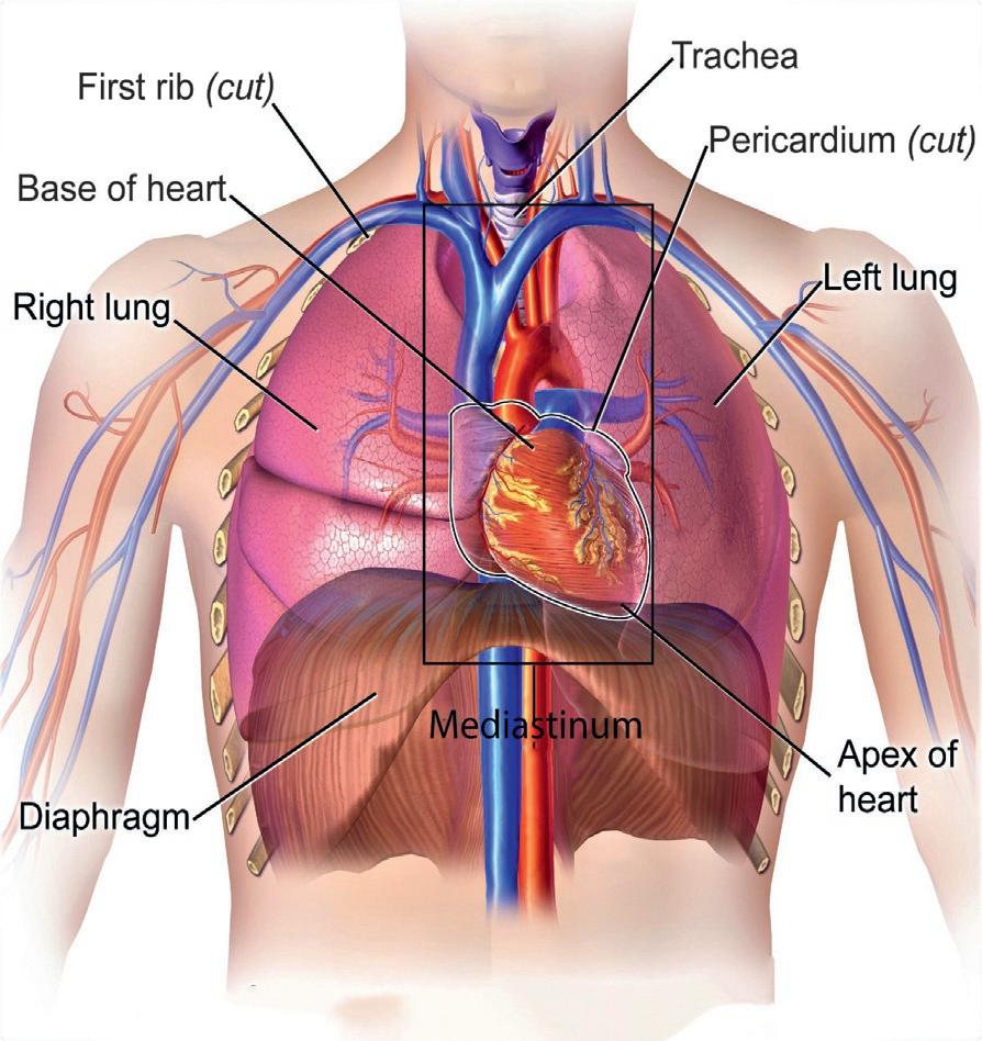
1.2Locationoftheheart Theheartissituatedwithinthethoraciccavity,mediallybetweenthelungs,inadedicated spaceknownasthe mediastinum (Fig.1.1).Theheartisseparatedfromtheotherstructures withinthemediastinumbyatoughmembraneknownasthe pericardium,or pericardialsac, whichsitsinitsownspaceknownasthe pericardialcavity.
1.3Thepericardium The pericardium,whichtranslatesas“aroundtheheart,”isadouble-layeredconnective tissuemembranethatsurroundstheheartlikeasacand,therefore,isalsocalledasa
1.4Layersoftheheartwall
FIG.1.1 Locationoftheheartinthethoraciccavity [1].Theheartissituatedwithinthethoraciccavity,mediallybetweenthelungsinthemediastinum.
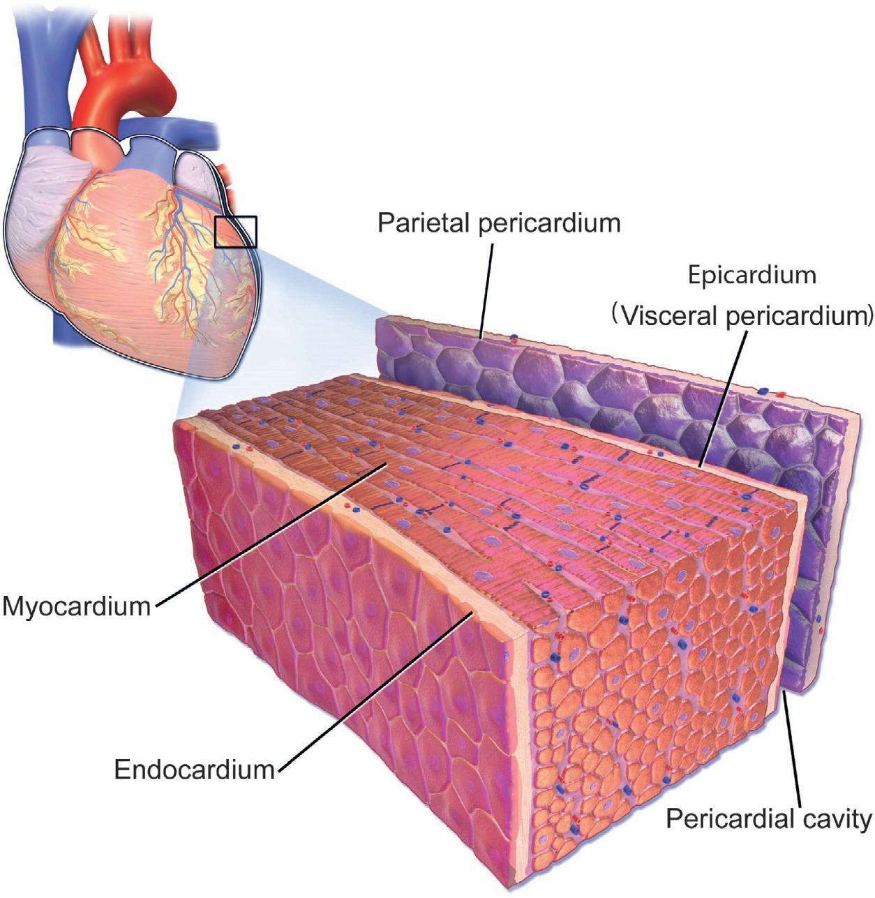
pericardialsac (Fig.1.2).Theoutersturdy parietalpericardium consistsofdenseconnective tissuethatprotectstheheartandmaintainsitspositionwithinthethoraciccavity.Theinner visceralpericardium ,or epicardium,isattachedtotheheartandispartoftheheartwall. Thereisaspacebetweenthevisceralpericardiumandtheparietalpericardiumandisreferredtoasthepericardialcavity.Thepericardiumsecretesasmallamountofserousfluid thatfillsthepericardialcavityandcoatsthetwopericardiallayerswhichservesasalubricant toreducefrictionastheheartexpandsandcontracts.
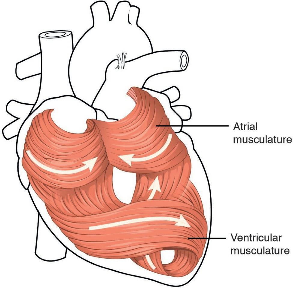
1.4Layersoftheheartwall Thewalloftheheartiscomposedofthreelayersofunequalthickness.Fromsuperficialto deep,theselayersarethe epicardium, myocardium,and endocardium (see Fig.1.2).Theoutermostlayer,theepicardium,isalsotheinnermostlayerofthepericardium,thevisceralpericardium.Thethickestlayeroftheheartisthemyocardium.Asthenamesuggests,the myocardiumconsistsofcardiacmusclefibersalongwithitssupplyofbloodvesselsand nervefiberstohelptheheartpumpblood.Itisthecontractionofthemyocardiumthatpumps bloodthroughtheheartandintothemajorarteriestobedistributedthroughoutthebody.As showninthefigure(Fig.1.3),thepatternisuniquetocardiacmusclewherethemusclefibers swirlandspiralaroundthechambersoftheheartandformafigure8patternbetweenthe atriaandtheventricles.Thisswirlingpatternallowsthehearttowithstandstressandpump bloodeffectively.
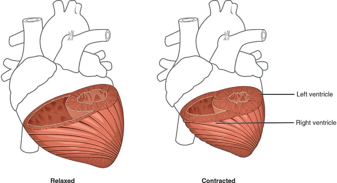
Althoughtheventriclesontherightandleftsidespumpthesameamountofbloodper contraction,themuscleoftheleftventricleismuchthickercomparedtotherightbecause theleftventriclemustgenerateaveryhighpressuretoovercomeresistancerequiredtopump bloodintothelong systemiccircuit.Ontheotherhand,therightventricledoesnotneedto generateasmuchpressuresincethe pulmonarycircuit isshorterandprovideslessresistance.
Fig.1.4 illustratesthedifferencesinmuscularthicknessbetweenthetwoventricles.Theinnermostlayeroftheheartwall,the endocardium,isathinlayerofconnectivetissue.Theendocardiumlinesthechambersandcoverstheheartvalves.
1.5Externalanatomyoftheheart Theadultheartweighsapproximately250–350g(9–12oz)andmeasures12cm(5in.)in length,8cm(3.5in.)wide,and6cm(2.5in.)inthickness.Awell-trainedathletemayhavea considerablylargerheartsinceexerciseresultsintheadditionofmuscleproteinstopump moreblood.Itisimportanttonote,however,thatthethickenedheartisnotalwaysdueto
FIG.1.2 Pericardialmembranesandlayersoftheheartwall [2]
FIG.1.3 Musculatureoftheheart [3].The swirlingpatternofcardiacmuscletissuecontributessignificantlytotheheart’sabilityto pumpbloodeffectively.
exercise.Itcanresultfromabnormalconditionssuchas hypertrophiccardiomyopathy, whichmaycausesuddendeathinapparentlyotherwisehealthyyoungpeople.
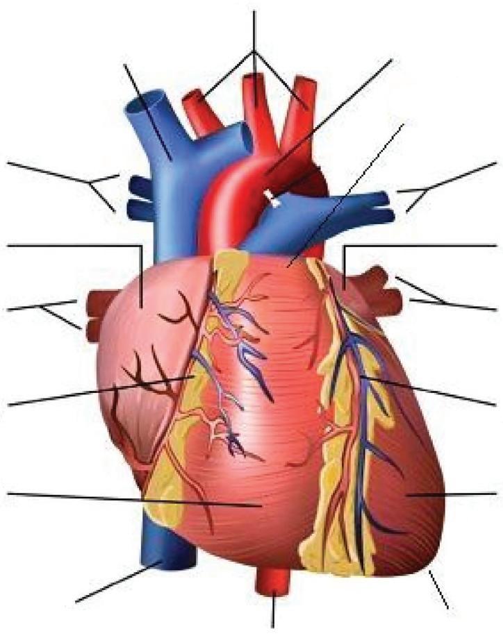
Theheartisbroaderatthesuperiorsurfaceandisreferredtoasthe base whichtapersoffat the apex (see Fig.1.5).Thebaseisatthelevelofthethirdcostalcartilagewhereastheapexis betweenthefourthandfifthribs.Cardiacmuscle(myocardium)isnourishedfromtheright andleftcoronaryarteries.Thesearteriesbranchoffintosmallerandsmallerarteries,deliveringoxygenatedbloodandnutrientstothemyocardium.
FIG.1.4 Differencesintherightandleftventricularmusclethickness [4]
Ascending Aorta (to head and arms)
Superior vena cava
Pulmonary artery (to right lung)
Right atrium
Right pulmonary veins (from right lung)
Right coronary artery
Right ventricle
Inferior vena cava
Descending aorta (to lower body)
FIG.1.5 Externalanatomyoftheanteriorpartoftheheart [5]
Aorta
Base
Pulmonary artery (to left lung)
Left atrium
Left pulmonary veins (from left lung)
Left coronary artery
Apex Left ventricle
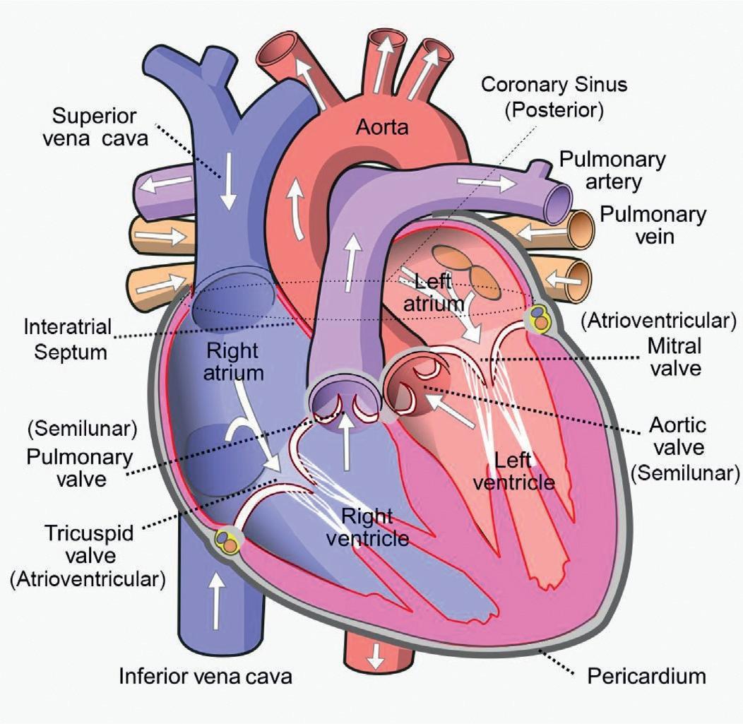
1.6Internalanatomyoftheheart Theheartisdividedintofourchambersbythesepta,orwalls.Locatedbetweenthetwo atriaisthe interatrialseptum.Theseptumsituatedbetweenthetwoventriclesiscalled the interventricularseptum.Theinterventricularseptumissubstantiallythickercompared totheinteratrialseptumtoallowtheventriclestogeneratehighpressurewhentheycontract. The atrioventricularseptum,asthenamesuggests,isfoundbetweentheatriaandtheventricles.Ithousesfouropeningsthatallowbloodtomovefromtheatriaintotheventriclesand fromtheventriclesintothepulmonarytrunkandaorta.Locatedineachoftheseopenings betweentheatriaandventriclesisa valve,aspecializedstructurethatensuresone-wayflow ofblood.Thevalvesbetweentheatriaandventriclesareknownasthe atrioventricular (AV) valves (Fig.1.6).TherightAVvalveisknownasthe tricuspidvalve andtheleftAVvalveis knownasthe mitralvalve.
1.6.1Chambersoftheheart
1.6.1.1Rightatrium
Therightatriumreceivesdeoxygenatedbloodfromthesystemiccirculation.Thetwomajorsystemicveins,the superior and inferiorvenaecavae,andthelargecoronaryveincalled the coronarysinus thatdrainsthemyocardium,emptyintotherightatrium(Figs.1.5and
FIG.1.6 Internalanatomyoftheheart [6].Thisanteriorviewoftheheartshows thefourchambers,themajorvessels,and theirearlybranches,aswellasthevalves. Thepresenceofthepulmonarytrunkand aortacoverstheinteratrialseptum,and theatrioventricularseptumiscutaway toshowtheatrioventricularvalves. Arrows representflowofbloodthroughthe heart.
1.6).Thesuperiorvenacavadrainsbloodfromregionsabovethediaphragmandtheinferior venacavadrainsbloodfromareasbelowthediaphragm.Thecoronarysinusdrainsmostof thecoronaryveinsthatreturnsystemicbloodfromtheheart.
1.6.1.2Rightventricle Therightventriclereceivesbloodfromtherightatriumthroughthetricuspidvalve,avalve foundintheopeningoftheatrioventricularseptum.Whentherightventriclecontracts,the pressurewithintheventricularchamberincreasescausingthebloodtoflowtowardthepulmonarytrunkandtherightatrium(Figs.1.7and1.8).Thetricuspidvalveclosestoprevent anypotentialbackflowintotherightatrium.
1.6.1.3Leftatrium Aftertheexchangeofgasesbetweenthealveoliandthepulmonarycapillaries,oxygenated bloodreturnstotheleftatriumviafourpulmonaryveins(Figs.1.7and1.8).Mostofthefilling occurswhiletheatriaarerelaxed.
1.6.1.4Leftventricle Thebloodfromtheleftatriumflowsintotheleftventriclethroughthebicuspid(mitral) valvewhichissituatedintheopeningoftheatrioventricularseptumontheleftsideofthe heart.Theleftventricleejectsbloodintotheaortathroughtheaorticsemilunarvalve (Figs.1.7and1.8).
FIG.1.7 Aflowchartshowingthecirculationofbloodthroughtheheartaswellasbetweentheheartandthe lungs. Whitearrows withintheheartand blackarrowsbetweenboxes representflowofbloodthroughtheheart. Blue boxes indicatedeoxygenatedblood, redboxes indicateoxygenatedblood,and purpleboxes indicatesitesofgas exchange.
FIG.1.8 Heartvalves [7].Theatrioventricularvalvesincludethetricuspidvalveandmitralvalve.Thesemilunar valvesarethepulmonaryandmitralvalves, https://www.heartandstroke.ca/heart-disease/conditions/valvularheart-disease
1.6.1.5Valvesoftheheart
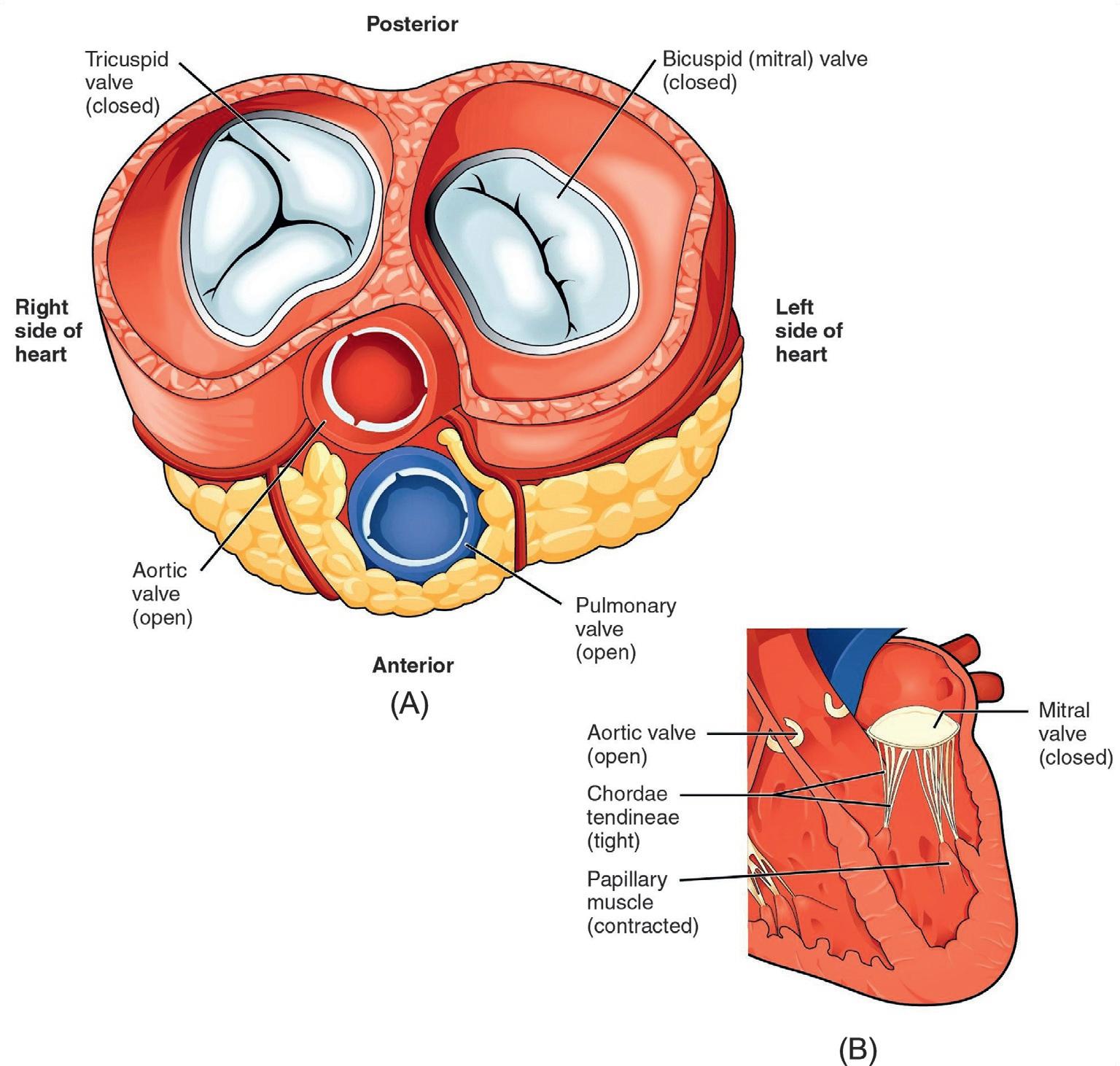
Valvesarespecializedstructuresthatensureone-wayflowofblood.Therearefourvalves intotalwithintheheart.Theopeningbetweentherightatriumandrightventricleisguarded byatricuspidvalveortherightAVvalve.Itconsistsofthreeflaps,orleaflets,madeofendocardiumalongwithadditionalconnectivetissue.Eachflapofthevalveisattachedtostrong strandsofconnectivetissue,the chordaetendineae (Figs.1.9Band 1.10B).Thereareseveral
FIG.1.9 Bloodflowfromtheleftatriumtotheleftventricle [7].(A)Atransversesectionthroughtheheartillustratesthefourheartvalves.Thetwoatrioventricularvalvesareopen(TricuspidandBicuspidorMitral);thetwosemilunarvalves(AorticandPulmonary)areclosed.Theatriaandvesselshavebeenremoved.(B)Afrontalsection throughtheheartillustratesbloodflowthroughthemitralvalve.Whenthemitralvalveisopen,itallowsblood tomovefromtheleftatriumtotheleftventricle.Theaorticsemilunarvalveisclosedtopreventthebackflowofblood fromtheaortatotheleftventricle.
chordaetendineaeassociatedwitheachoftheflapswhichinturnareattachedtoa papillary muscle (Figs.1.9Band 1.10B)thatextendsfromtheinferiorventricularsurface.Thereare threepapillarymusclesintherightventriclewhichcorrespondtothethreesectionsofthe valves.
Locatedattheopeningbetweentheleftatriumandleftventricleisthemitralvalve,also calledtheleftAVvalve.Structurally,thisvalveconsistsoftwocuspswhichareattachedby chordaetendineaetotwopapillarymusclesthatprojectfromtheinferiorwalloftheleft ventricle.
FIG.1.10 Bloodflowfromtheleftventricleintotheaortaandpulmonaryartery [8].(A)Atransversesection throughtheheartillustratesthefourheartvalvesduringventricularcontraction.Thetwoatrioventricularvalves areopen(TricuspidandMitral);thetwosemilunarvalves(AorticandPulmonary)areclosed.Theatriaandvessels havebeenremoved.(B)Afrontalviewshowstheclosedmitral(bi-leaflet)valvethatpreventsthebackflowofblood intotheleftatrium.Theaorticsemilunarvalveisopentoallowbloodtobeejectedintotheaorta.
Emergingfromtherightventricleatthebaseofthepulmonarytrunkisthe pulmonary semilunarvalve orthe pulmonaryvalve;itisalsoknownasthe pulmonicvalve orthe right semilunarvalve whichpreventsbackflowofbloodfromthepulmonarytrunkintotheright ventricle.Thepulmonaryvalveismadeofthreesmallflapsofendocardiumreinforcedwith connectivetissue.Whentheventriclerelaxes,thepressuredifferencecausesbloodtoflow backintotherightventriclefromthepulmonarytrunk.Thisflowofbloodfillsthepocket-like flapsofthepulmonaryvalve,causingthepulmonaryvalvetocloseproducinganaudible sound.
Atthebaseoftheaortaisthe aorticsemilunarvalve,orthe aorticvalve,whichprevents backflowfromtheaortaintotheleftventricle.Itisalsocomposedofthreeflaps.Whenthe ventriclerelaxesandbloodattemptstoflowbackintotheleftventriclefromtheaorta,blood willfillthecuspsofthevalve,causingittocloseandproducinganaudiblesound.
TheAVvalvesareclosedwhilethetwosemilunarvalvesareopen(Fig.1.10).Thisoccurs whentheventriclescontracttoejectbloodintothepulmonarytrunkandaorta.Closureofthe twoAVvalvespreventsbloodfrombeingforcedbackintotheatria.In Fig.1.9A,thetwoAV valvesareopenandthetwosemilunarvalvesareclosed.Thisoccurswhenbothatriaand ventriclesarerelaxedandfillingoftheventriclesfromtheatriaoccurs.
Whentheventriclesbegintocontract,thepressurewithintheventriclesrises,andblood flowstowardtheareaoflowerpressure,whichisinitiallyintheatria.Thisbackflowcauses thecuspsofthetricuspidandmitralvalvestoclose(Fig.1.9A).Duringtherelaxationphase ofthecardiaccycle,thepapillarymusclesare alsorelaxedandthetensiononthechordae tendineaeisslight(see Fig.1.9B).However,astheventriclescontract,sodothepapillary muscles.Thiscreatestensiononthechordaetendineae(see Fig.1.9 B),helpingtohold thecuspsoftheAVvalvesinplaceandpreventingthemfrombeingblownbackintothe atria.
UnliketheAVvalves,therearenopapillarymusclesorchordaetendineaeassociatedwith thesemilunarvalves.Whentheventriclesrelaxcausingachangeofpressure,itforcesthe bloodtowardtheventricles,thebloodpressesagainstthesecuspsandsealstheopenings. Visit http://openstaxcollege.org/l/heartvalve toobserveanechocardiogramofactualheart valvesopeningandclosing.
1.7Cardiacmuscleandelectricalactivity 1.7.1Structureofcardiacmuscle
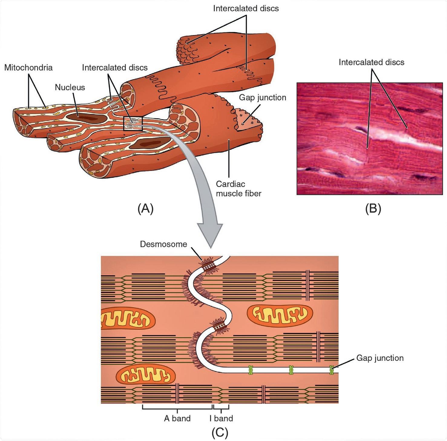
Cardiacmusclefibers,orcardiomyocytes,likeskeletalmusclefibers,arestriatedandhave asingle,centralnucleus.However,twoormorenucleimaybefoundoccasionally. Transverse(or T) tubules,thefoldsofthe sarcolemma (plasmamembrane),penetratedeepinto theinteriorofthecell,allowingelectricalimpulsestoreachtheinnerpartsofthecell. Sarcoplasmicreticulum (endoplasmicreticulum)storesCa2+ butisnotsufficientformusclecontraction.AdditionalCa2+ mustcomefromtheextracellularfluid.Mitochondriaarepresentin largenumbersinthecardiacmusclefiberstoprovideenergyformusclecontraction (Fig.1.11A).
Unlikeskeletalmusclefibers,cardiacmusclefibersbranchfreely.Ajunctionbetweentwo adjoiningcellsismarkedbyaspecializedstructurecalledan intercalateddisc (Fig.1.11A), whichhelpsspreaddepolarizationbetweenadjacentcells(Fig.1.11B).Thesarcolemmasfrom adjacentcellsbindtogetherattheintercalateddiscsandpossessgapjunctionstoallowpassageofionsbetweenthecells(Fig.1.11C). Desmosomes arespecialcell-to-celljunctions foundattheintercalateddiscstoprovideadditionalsupportandstabilityforthecardiacmusclefibers.
FIG.1.11 Cardiacmuscle [9].(A)Cardiacmusclecellshavemyofibrilscomposedofmyofilamentsarrangedin sarcomeres,Ttubulestotransmittheimpulsefromthesarcolemmatotheinteriorofthecell,numerousmitochondria forenergy,andintercalateddiscsthatarefoundatthejunctionofdifferentcardiacmusclecells. (B)Aphotomicrographofcardiacmusclecellsshowsthenucleiandintercalateddiscs.(C)Anintercalateddiscconnectscardiacmusclecellsandconsistsofdesmosomesandgapjunctions.
Therearetwomajortypesofcardiacmusclefibers: myocardialcontractilecells and myocardialconductingcells (pacemakercells).Themyocardialcontractilecellsconstitutethe bulk(99%)ofthecellsintheatriaandventriclesandareresponsibleforcontractionsthat pumpbloodthroughthebody.Themyocardialconductingcellsmakeuptheremaining 1%toformthe conductionsystem oftheheart.Themyocardialconductingcellsaregenerally muchsmallerthanthecontractilecellsandhavefewofthemyofibrilsorfilamentsneededfor contraction.
1.8Cardiacmusclemetabolism Fattyacidsandglucosefromthebloodsupplyarebrokendownwithinthemitochondriato releaseenergyintheformofATP.Bothfattyaciddropletsandglycogen,thestorageformof glucose,arestoredwithinthesarcoplasmtoprovideadditionalnutrientsupply.
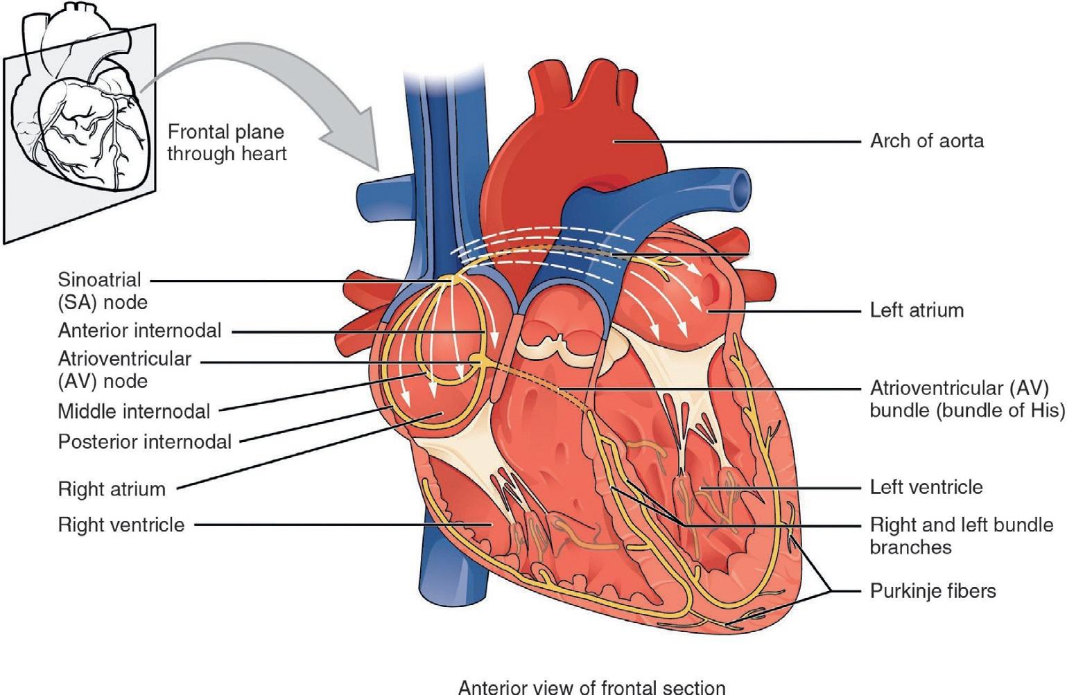
1.9Conductionsystemoftheheart Cardiacmusclehastheabilitytoinitiateitselectricalpotentialthatspreadsrapidlyfrom celltocelltotriggerthecontractilemechanism.Thispropertyisknownas autorhythmicity. Everycardiacmusclefiberiscapableofgeneratingitsownelectricalimpulse.Thecellwith thehigherinherentrateofdepolarizationsetsthepace,andtheimpulsespreadsfromthe fastertotheslowercelltotriggeracontraction.Thefivekeycomponentsofthecardiacconductionsystemincludethe sinoatrial (SA) node,the AVnode,the AVbundle or bundleof His,the AVbundlebranches,andthe Purkinjefibers (Fig.1.12).
FIG.1.12 Conductionsystemoftheheart [10].Theinitiationofanactionpotentialatthesinoatrial(SA)node spreadsthroughouttheatriareachingtheatrioventricular(AV)node,AVbundle(bundleofHis),bundlebranches, andultimatelyPurkinjefibers.Thecontractilecellsthenbegincontractionfromthesuperiortotheinferiorportionsof theatria,efficientlypumpingbloodintotheventricles.
1.9.1Sinoatrialnode Sinoatrialnodeisaspecializedgroupofmyocardialconductingcellslocatedinthesuperiorandposteriorwallsoftherightatriumclosetotheopeningofthesuperiorvenacava.The SAnodehasthehighestinherentrateofdepolarizationandthereforereferredtoasthe pacemaker oftheheart.Itinitiatesthesinusrhythmornormalelectricalpattern.Thisimpulse spreadsfromtheSAnodetotheatrialmyocardialcontractilecellsandtheAVnode.Theinternodalpathwaysthatconnectcontractilecellsconsistofthreebands(anterior,middle,and posterior; Fig.1.12).Theimpulsetakesapproximately50milliseconds(ms)totravelbetween theSAnodeandtheAVnode.
1.9.2Atrioventricularnode TheAVnodeisasecondsetofmyocardialconductivecells,locatedintheinferiorportion oftherightatriumaspartoftheatrioventricularseptum.Astheimpulsereachestheatrioventricularseptum,theconnectivetissuesurroundingtheheart,referredtoas fibrousskeleton,preventstheimpulsefromspreadingintothemyocardialcellsintheventriclesexcept theAVnode.
ThereisadelayinthespreadingofdepolarizationfromtheAVnodetotheAVbundle(see Fig.1.13,step3).Thisdelayintransmissionispartiallyduetothesmalldiameterofthecellsof thenode,whichslowstheimpulse.Thus,ittakestheimpulseapproximately100mstopass throughthenode.Thispausehelpsatrialmyocardiumtocompletetheircontractionthat pumpsbloodintotheventriclesbeforetheimpulseistransmittedtothecellsoftheventricle.
1.9.3Atrioventricularbundle(bundleofHis),bundlebranches,andPurkinje fibers
AVbundle,or bundleofHis,originatesattheAVnodeandproceedsthroughthe interventricularseptumbeforedividingintotwo AVbundlebranches,commonlycalled the left and rightbundlebranches.Accordingtotheirlocations,theleftbundlebranchsuppliestheleftventricle,andtherightbundlebranchsuppliestherightventricle.Sincetheleft ventricleismuchlargercomparedtotheright,theleftbundlebranchisalsoconsiderably largercomparedtotheright.Bothbundlebranchesdescendandreachtheapexoftheheart wheretheyconnectwiththePurkinjefibers(see Figs.1.12and1.13,step4).Thispassagetakes approximately25ms.
ThePurkinjefibersaremyocardialconductivecellsthatspreadtheimpulsetothemyocardialcontractilecellsintheventricles.Theyextendthroughoutthemyocardiumfromtheapex ofthehearttowardtheatrioventricularseptumandthebaseoftheheart.ThePurkinjefibers haveafastconductionrate,andtheelectricalimpulsereachesalloftheventricularmuscle cellsinabout75ms(see Fig.1.13,step5and6).Sincetheelectricalimpulsearrivesattheapex throughthebundlebranches,thecontractionalsobeginsattheapexandtravelstowardthe baseoftheheart,similartosqueezingatubeoftoothpastefromthebottom.Thisallowsthe bloodtobepumpedoutoftheventriclesintotheaortaandpulmonarytrunk.Thetotaltime elapsedfromtheinitiationoftheimpulseintheSAnodeuntildepolarizationoftheventricles isapproximately225ms.
1.10Membranepotentialsincardiacconductivecells
FIG.1.13 Cardiacconduction [11].(1)Thesinoatrial(SA)nodeandtheremainderoftheconductionsystemareat rest.(2)TheSAnodeinitiatestheactionpotential,whichsweepsacrosstheatria.(3)Afterreachingtheatrioventricularnode(AV),thereisadelayofapproximately100msthatallowstheatriatocompletepumpingbloodbeforethe impulseistransmittedtotheatrioventricularbundle.(4)Followingthedelay,theimpulsetravelsthroughtheatrioventricularbundleandbundlebranchestothePurkinjefibers(5and6).
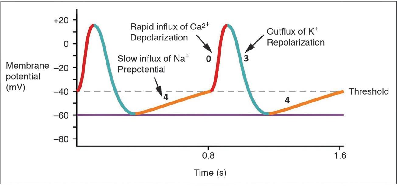
1.10Membranepotentialsincardiacconductivecells Therestingmembranepotentialofconductivefibersintheheartrangesfrom 50to 60mV.Actionpotentialsareconsiderablydifferentbetweencardiacconductivecells(pacemakercells)suchastheSAnodeandAVnodeandcardiaccontractilecells(nonpacemaker) suchasthePurkinjecells.Cardiacconductivecellsdonothaveastablerestingmembrane potentialbecausetheyconsistofaseriesofNa+ leakchannels(referredtoasfunnychannels) whichallowaslowinfluxofNa+ andthereforeraisemembranepotentialslowlyfromaninitialvalueof 60mVtoabout 40mV(orangeline).SlowCa2+ channelsarealsoopenduring thisperiodresultingina spontaneousdepolarization (or prepotentialdepolarization).The shiftinmembranepotentialtoitsthresholdlevelof 40mVcausesvoltage-gatedfastCa2+ channelstoopenandCa2+ enterthecellinlargenumbers,furtherdepolarizingitatamore
FIG.1.14 Actionpotentialinthesinoatrialnode [12].Theprepotential(phase4)isduetoaslowinfluxofNa+ until thethresholdisreachedfollowedbyarapiddepolarization(phase0)andrepolarization(phase3).Theprepotential (phase4)accountsforthemembranereachingthresholdandinitiatesthespontaneousdepolarizationandcontraction ofthecell.Notethelackofarestingmembranepotential.
rapidrateuntilthemembranepotentialreachesavalueofapproximately+5mV(redline). Thisphaseofrapiddepolarizationisreferredtoas phase0.Suchsuddenchangeinmembrane potentialallowsCa2+ channelstocloseandvoltage-gatedK+ channelstoopen,allowingan outfluxofK+ bringingarepolarizationstate.Thisparticularphaseisreferredtoas phase3. Whenthemembranepotentialreachesapproximately 60mV(restingmembranepotential), K+ channelscloseandNa+ channelsopenonceagain,andtheprepotentialphasebeginsasin thepreviouscycleandisreferredtoas phase4.Thisphenomenonexplainsthe autorhythmicity propertiesofcardiacmuscle(Fig.1.14).
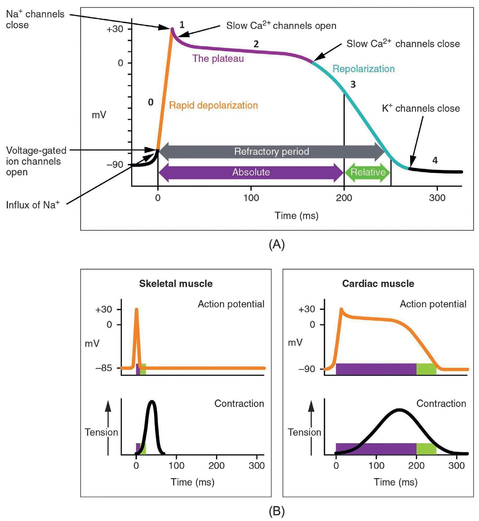
1.11Membranepotentialsincardiaccontractilecells Theactionpotentialpatternisdifferentincardiaccontractilemusclefiberswhichdemonstrateamuchmorestablerestingphasethanconductivecells.Therestingmembranepotential isapproximately 80mVforatrialmyofibersand 90mVfortheventricularmusclefibers. Thenatureoftheactionpotentialisverydifferentinthecardiaccontractilecells;thereisa rapiddepolarization,followedbya plateauphase andthenrepolarization.Thisphenomenon accountsforthelongrefractoryperiodsrequiredforthecardiacmusclecellstopumpblood effectivelybeforetheyarecapableoffiringforasecondtime.Thesecardiacmyocytesdonot normallyinitiatetheirownelectricalpotential,althoughtheyarecapableofdoingso,butare triggeredbyanimpulsefromaconductivecell(SAnode)toinitiateanactionpotential. Whenstimulatedbyanactionpotentialinthepacemakercell,voltage-gatedNa+ channels openinapositive-feedbackmannerallowingarapidinfluxofNa+ toraisethemembranepotentialtoapproximately+30mV,atwhichpointthevoltage-gatedNa+ channelsclose.The rapiddepolarizationperiodtypicallylasts3–5ms(orangeline; phase0).Depolarizationis followedbytheplateauphase.Duringtheinitialperiodoftheplateauphase,onlytheK+
channelsareopenandmembranepotentialdeclinesrelativelyslowly(phase1).Theopening oftheslowCa2+ channels,allowingCa2+ toenterthecellmaintainsamembranepotentialof approximately0mV.Duringthisperiod,K+ channelsarealsoopen,allowingK+ toexitthe cellwhileCa2+ aremovinginward.Thus,thisperiodconsistsofthemovementsofCa2+ inwardwhereasK+ outwardanditlastsapproximately175ms(purpleline; phase2).Next,the Ca2+ channelscloseandK+ channelsremainopen,allowingmoreK+ toexitthecellinitiatinga repolarizationphase(phase3).Therepolarizationphaselastsapproximately75ms.Atthis point,membranepotentialdropsuntilitreachesrestinglevelsandK+channelsclose (phase4)oncemoreandthecyclerepeats.Theentireeventlastsbetween250and300ms (Fig.1.15).
Afteranactionpotentialisinitiated,thecardiacmusclecellisunabletoinitiateanother actionpotentialforsometime,andthisperiodoftimeisreferredtoasthe refractoryperiod, whichlasts250msindurationandhelpsprotecttheheart.Thecardiacrefractoryperiodis separatedintoan absoluterefractoryperiod anda relativerefractoryperiod.Duringtheabsoluterefractoryperiod,anewactionpotentialcannotbeelicited.Ontheotherhand,during therelativerefractoryperiod,anewactionpotentialcanbegeneratedgivenasituationsuch asastrongstimulus.
Theactionpotentialinthecontractilecells ofthecardiacmuscleiscomparedwiththat oftheskeletalmusclefiberin Fig.1.15B.Boththedurationofanactionpotentialandrefractoryperiodaremuchshorterintheskele talmusclefibercomparedtothecontractile cellsofthecardiacmuscle. Fig.1.15 Balsocomparesthenatureofcontractionthatfollows eachactionpotential.Sincetherefractoryperiodismuchlongerinthecardiacmusclefiber,thecontractioncycleissuchthattheentirecyclemustbecompletedpriortoanew contractioncycleunlikeintheskeletalmusclewhereanewcontractionmaybeinitiated beforethecompletionofthepreviousoneduet otheshorterrefractoryperiod.Thisphenomenonisreferredtoastetanuswhichiscommonlyseenintheskeletalmusclebutnotin thecardiacmuscle.Cardiacmusclecellsunde rgotwitchtypeofcontractionswithlong refractoryperiodsfollowedbybriefrelaxationperiods.Therelaxationisessentialso theheartcanfillwithbloodforthenextcycle.Therefractoryperiodisverylongtoprevent thepossibilityof tetany,aconditioninwhichmuscleremainsinvoluntarilycontracted.In theheart,tetanyisnotcompatiblewithlife,sinceitwouldpreventtheheartfrom pumpingblood.
1.11.1Comparativeratesofconductionsystemfiring Thepatternofslowdepolarization,followedbyrapiddepolarizationandrepolarization,is seenintheSAnodeandafewotherconductivecellsintheheart.TheSAnodeservesasthe pacemakersinceitreachesthethresholdfasterthananyothercomponentoftheconduction system.Itinitiatestheimpulsesspreadingtotheotherconductingcells.TheSAnode,without nervousorendocrinecontrol,initiatesaheartimpulseapproximately80–100timesperminute.Althougheachcomponentoftheconductionsystemiscapableofgeneratingitsown impulse,therateprogressivelyslowsfromtheSAnodetothePurkinjefibers.
WithouttheSAnode,theAVnodewouldgenerateaheartrateof40–60bpm.IftheAV nodealsowasincapableofservingasapacemaker,theAVbundlewouldfireatarateof
FIG.1.15 Actionpotentialinthecardiaccontractilecells [13].(A)Notethelongplateauphaseduetotheinfluxof Ca2+.Theextendedrefractoryperiodallowsthecelltofullycontractbeforeanotherelectricaleventcanoccur.The numbers0,1,2,3,and4indicatethephasesoftheactionpotential.(B)Theactionpotentialforheartmuscleiscomparedtothatofskeletalmuscle.
approximately30–40impulsesperminute.Thebundlebrancheswouldhaveaninherentrate of20–30impulsesperminute,andthePurkinjefiberswouldfireatabout15–30impulsesper minute.WhileafewexceptionallytrainedaerobicathletesdemonstraterestingHRinthe rangeof30–40bpm(thelowestrecordedfigureis28bpmforMiguelIndurain,acyclist), formostindividuals,rateslowerthan50bpmwouldindicate bradycardia,aslowHR.As
1.12Electrocardiogram
ratesfallmuchbelowthislevel,theheartwouldbeunabletomaintainadequateperfusion ofbloodtovitaltissues,resultinginorganfailureormultisystemfailureandultimately death.
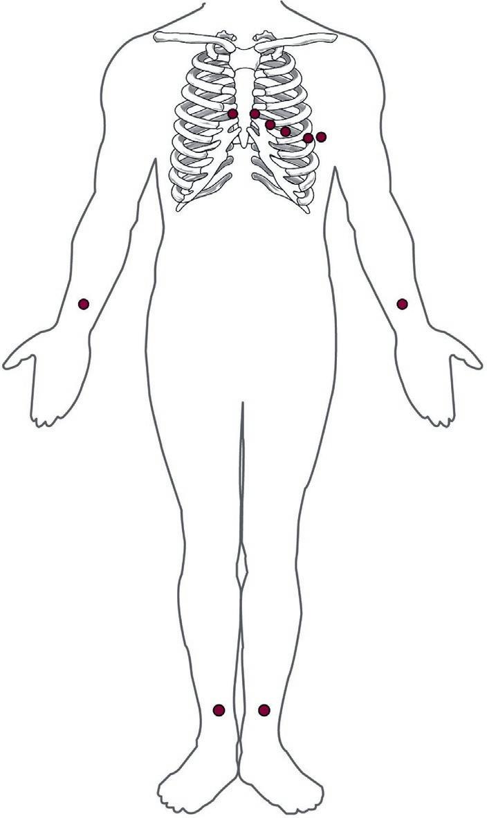
1.12Electrocardiogram Onecanrecordtheelectricalsignalsoftheheartbytheplacementofelectrodesincertain partsofthebody.Thetracingoftheelectricalsignalfromtheheartiscalledthe electrocardiogram (ECG),alsoabbreviatedas EKG (Kstandsfor kardiology,theGermantermforcardiology).AtypicalECGisnotarecordingfromasinglemusclefiberbuttheoverallchangein theflowofelectricalcurrentandrevealsadetailedpictureoftheheartfunctionthusserving asanimportantclinicaldiagnostictool.Theterm“lead”typicallydescribesthevoltagedifferencebetweentwooftheelectrodes.Thestandardelectrocardiographuses3,5,or12leads. Thegreaterthenumberofleadsanelectrocardiographuses,themoredetailedinformation theECGprovides.The12-leadelectrocardiographuses10electrodesplacedinthevarious locationsonthepatient’sbodyasshownin Fig.1.16
ThemajorpointsontheECGarethe Pwave,the QRScomplex,andthe Twave (Fig.1.17). ThesmallPwaverepresentsthedepolarizationoftheatriawhereasthelargeQRScomplex representsthedepolarizationoftheventricles.QRScomplexisamuchstrongerelectricalsignalbecauseofthelargersizeoftheventricularcardiacmuscle.TheTwaverepresents
FIG.1.16 StandardplacementofECGleadsina12-leadECG,6 electrodesareplacedonthechest,and4electrodesareplacedon thelimbs [14]

