
https://ebookmass.com/product/berne-levy-physiology-7th-ed-
https://ebookmass.com/product/social-problems-ebook-pdf-version/ ebookmass.com

https://ebookmass.com/product/berne-levy-physiology-7th-ed-
https://ebookmass.com/product/social-problems-ebook-pdf-version/ ebookmass.com
Editors
Bruce M. Koeppen, MD, PhD
Dean Frank H. Netter MD School of Medicine Quinnipiac University Hamden, Connecticut
Bruce A. Stanton, PhD
Andrew C. Vail Professor Microbiology, Immunology, and Physiology Director of the Lung Biology Center Geisel School of Medicine at Dartmouth Hanover, New Hampshire
Kim E. Barrett, PhD
Distinguished Professor of Medicine
University of California, San Diego, School of Medicine
La Jolla, California
Section 6: Gastrointestinal Physiology
Michelle M. Cloutier, MD
Professor
Department of Pediatrics
University of Connecticut School of Medicine
Farmington, Connecticut and Director
Asthma Center
Connecticut Children’s Medical Center Hartford, Connecticut
Section 5: The Respiratory System
John R. Harrison, PhD
Associate Professor
Department of Craniofacial Sciences
University of Connecticut Health Center
Farmington, Connecticut
Section 8: The Endocrine and Reproductive Systems
Bruce M. Koeppen, MD, PhD
Dean
Frank H. Netter MD School of Medicine
Quinnipiac University
Hamden, Connecticut
Section 1: Cellular Physiology
Section 7: The Renal System
Eric J. Lang, MD, PhD
Associate Professor
Department of Neuroscience and Physiology
New York University School of Medicine
New York, New York
Section 2: The Nervous System
Achilles J. Pappano, PhD
Professor Emeritus
Department of Cell Biology Calhoun Cardiology Center
University of Connecticut Health Center Farmington, Connecticut
Section 4: The Cardiovascular System
Helen E. Raybould, PhD Professor
Department of Anatomy, Physiology, and Cell Biology
University of California-Davis School of Veterinary Medicine Davis, California
Section 6: Gastrointestinal Physiology
Kalman Rubinson, PhD Emeritus Professor
Department of Neuroscience and Physiology
New York University School of Medicine
New York, New York
Section 2: The Nervous System
Bruce A. Stanton, PhD
Andrew C. Vail Professor Microbiology, Immunology, and Physiology Director of the Lung Biology Center Geisel School of Medicine at Dartmouth Hanover, New Hampshire
Section 1: Cellular Physiology
Section 7: The Renal System
Roger S. Thrall, PhD
Professor Emeritus
Immunology and Medicine
University of Connecticut Health Center Farmington, Connecticut and Director of Clinical Research
Department of Research Hospital for Special Care
New Britain, Connecticut
Section 5: The Respiratory System
James M. Watras, PhD
Associate Professor
Department of Cell Biology
University of Connecticut Health Center
Farmington, Connecticut
Section 3: Muscle
Bruce A. White, PhD
Professor
Department of Cell Biology
University of Connecticut Health Center
Farmington, Connecticut
Section 8: The Endocrine and Reproductive Systems
Withrow Gil Wier, PhD Professor
Department of Physiology
University of Maryland, Baltimore
Baltimore, Maryland
Section 4: The Cardiovascular System
We wish to express our appreciation to all of our colleagues and students who have provided constructive criticism during the revision of this book:
Hannah Carey, PhD
University of Wisconsin, Madison School of Veterinary Medicine
Madison, Wisconsin
Section 6: Gastrointestinal Physiology
Nathan Davis, PhD
Professor of Medical Sciences
Frank H. Netter MD School of Medicine
Quinnipiac University
Hamden, Connecticut
Section 8: The Endocrine and Reproductive Systems
L. Lee Hamm, MD
Senior Vice President and Dean
Tulane University School of Medicine
New Orleans, Louisiana
Chapter 37: Role of the Kidneys in the Regulation of Acid-Base Balance
Douglas McHugh, PhD
Associate Professor of Medical Sciences
Frank H. Netter MD School of Medicine
Quinnipiac University
Hamden, Connecticut
Section 1: Cellular Physiology
Orson Moe, MD
The Charles Pak Distinguished Chair in Mineral Metabolism
Donald W. Seldin Professorship in Clinical Investigation
University of Texas Southwestern Medical Center
Dallas, Texas
Section 7: The Renal System
R. Brooks Robey, MD, FASN FAHA
Associate Chief of Staff for Research
Chief of Nephrology at the White River Junction VA Medical Center
Geisel School of Medicine at Dartmouth
Hanover, New Hampshire
Section 7: The Renal System
Marion Siegman, PhD
Professor and Chair
Department of Molecular Physiology and Biophysics
Sidney Kimmel Medical College at Thomas Jefferson University
Philadelphia, Pennsylvania
Chapter 14: Smooth Muscle
Travis Solomon, MD, PhD
School of Medicine
University of Missouri
Kansas City, Missouri
Section 6: Gastrointestinal Physiology
Nancy Wills, PhD
Emeritus Professor of Medical Sciences
Frank H. Netter MD School of Medicine
Quinnipiac University
Hamden, Connecticut
Section 1: Cellular Physiology
We are pleased that the following section authors have continued as members of the seventh edition team: Drs. Kalman Rubinson and Eric Lang (nervous system), Dr. James Watras (muscle), Dr. Achilles Pappano (cardiovascular system), Drs. Michelle Cloutier and Roger Thrall (respiratory system), Drs. Kim Barrett and Helen Raybould (gastrointestinal system), and Dr. Bruce White (endocrine and reproductive systems). We also welcome the following authors: Dr. Withrow Gil Wier (cardiovascular system), and Dr. John Harrison (endocrine and reproduction systems).
As in the previous editions of this textbook, we have attempted to emphasize broad concepts and to minimize the compilation of isolated facts. Each chapter has been written to make the text as lucid, accurate, and current as possible. We have included both clinical and molecular information in each section, as feedback on these features has indicated that this information serves to provide clinical context and new insights into physiologic phenomena at the cellular and molecular levels. New to this edition is a list of sources that the reader can consult for further information on the topics covered in each chapter. We hope that you find this a valuable addition to the book.
The human body consists of billions of cells that are organized into tissues (e.g., muscle, epithelia, and nervous tissue) and organ systems (e.g., nervous, cardiovascular, respiratory, renal, gastrointestinal, endocrine, and reproductive). For these tissues and organ systems to function properly and thus allow humans to live and carry out daily activities, several general conditions must be met. First and foremost, the cells within the body must survive. Survival requires adequate cellular energy supplies, maintenance of an appropriate intracellular milieu, and defense against a hostile external environment. Once cell survival is ensured, the cell can then perform its designated or specialized function (e.g., contraction by skeletal muscle cells). Ultimately, the function of cells, tissues, and organs must be coordinated and regulated. All of these functions are the essence of the discipline of physiology and are presented throughout this book. What follows is a brief introduction to these general concepts.
Cells need a constant supply of energy. This energy is derived from the hydrolysis of adenosine triphosphate (ATP). If not replenished, the cellular ATP supply would
be depleted in most cells in less than 1 minute. Thus, ATP must be continuously synthesized. This in turn requires a steady supply of cellular fuels. However, the cellular fuels (e.g., glucose, fatty acids, and ketoacids) are present in the blood at levels that can support cellular metabolism only for a few minutes. The blood levels of these cellular fuels are maintained through the ingestion of precursors (i.e., carbohydrates, proteins, and fats). In addition, these fuels can be stored and then mobilized when ingestion of the precursors is not possible. The storage forms of these fuels are triglycerides (stored in adipose tissue), glycogen (stored in the liver and skeletal muscle), and protein. The maintenance of adequate levels of cellular fuels in the blood is a complex process involving the following tissues, organs, and organ systems:
• Liver: Converts precursors into fuel storage forms (e.g., glucose → glycogen) when food is ingested, and converts storage forms to cellular fuels during fasting (e.g., glycogen → glucose and amino acids → glucose).
• Skeletal muscle: Like the liver, stores fuel (glycogen and protein) and converts glycogen and protein to fuels (e.g., glucose) or fuel intermediates (e.g., protein → amino acids) during fasting.
• Gastrointestinal tract: Digests and absorbs fuel precursors.
• Adipose tissue: Stores fuel during feeding (e.g., fatty acids → triglycerides) and releases the fuels during fasting.
• Cardiovascular system: Delivers the fuels to the cells and to and from their storage sites.
• Endocrine system: Maintains the blood levels of the cellular fuels by controlling and regulating their storage and their release from storage (e.g., insulin and glucagons).
• Nervous system: Monitors oxygen levels and nutrient content in the blood and, in response, modulates the cardiovascular, pulmonary, and endocrine systems and induces feeding and drinking behaviors. In addition to energy metabolism, the cells of the body must maintain a relatively constant intracellular environment to survive. This includes the uptake of fuels needed to produce ATP, the export from the cell of cellular wastes, the maintenance of an appropriate intracellular ionic environment, the establishment of a resting membrane potential, and the maintenance of a constant cellular volume. All of these functions are carried out by specific membrane transport proteins.
Section 1: Cellular Physiology, 1
Bruce M. Koeppen and Bruce A. Stanton
1 Principles of Cell and Membrane Function, 2
2 Homeostasis: Volume and Composition of Body Fluid Compartments, 17
3 Signal Transduction, Membrane Receptors, Second Messengers, and Regulation of Gene Expression, 35
Section 2: The Nervous System, 51
Eric J. Lang and Kalman Rubinson
4 The Nervous System: Introduction to Cells and Systems, 52
5 Generation and Conduction of Action Potentials, 65
6 Synaptic Transmission, 84
7 The Somatosensory System, 108
8 The Special Senses, 127
9 Organization of Motor Function, 161
10 Integrative Functions of the Nervous System, 208
11 The Autonomic Nervous System and Its Central Control, 226
Section 3: Muscle, 241
James M. Watras
12 Skeletal Muscle Physiology, 242
13 Cardiac Muscle, 268
14 Smooth Muscle, 280
Section 4: The Cardiovascular System, 300
Achilles J. Pappano and Withrow Gil Wier
15 Overview of Circulation, 301
16 Elements of Cardiac Function, 304
17 Properties of the Vasculature, 345
18 Regulation of the Heart and Vasculature, 386
19 Integrated Control of the Cardiovascular System, 410
Section 5: The Respiratory System, 433
Michelle M. Cloutier and Roger S. Thrall
20 Introduction to the Respiratory System, 434
21 Static Lung and Chest Wall Mechanics, 447 22 Dynamic Lung and Chest Wall Mechanics, 456
23 Ventilation, Perfusion, and Ventilation/ Perfusion Relationships, 466 24 Oxygen and Carbon Dioxide Transport, 480
25 Control of Respiration, 489
26 Nonphysiological Functions of the Lung: Host Defense and Metabolism, 498
Section 6: Gastrointestinal Physiology, 510
Kim E. Barrett and Helen E. Raybould
27 Functional Anatomy and General Principles of Regulation in the Gastrointestinal Tract, 511
28 The Cephalic, Oral, and Esophageal Phases of the Integrated Response to a Meal, 520
29 The Gastric Phase of the Integrated Response to a Meal, 529
30 The Small Intestinal Phase of the Integrated Response to a Meal, 541
31 The Colonic Phase of the Integrated Response to a Meal, 559
32 Transport and Metabolic Functions of the Liver, 568
Section 7: The Renal System, 580
Bruce A. Stanton and Bruce M. Koeppen
33 Elements of Renal Function, 581
34 Solute and Water Transport along the Nephron: Tubular Function, 603
35 Control of Body Fluid Osmolality and Volume, 623
36 Potassium, Calcium, and Phosphate Homeostasis, 647
37 Role of the Kidneys in the Regulation of Acid-Base Balance, 670
Section 8: The Endocrine and Reproductive Systems, 685
Bruce A. White and John R. Harrison
38 Introduction to the Endocrine System, 686
39 Hormonal Regulation of Energy Metabolism, 698
40 Hormonal Regulation of Calcium and Phosphate Metabolism, 722
41 The Hypothalamus and Pituitary Gland, 733
42 The Thyroid Gland, 753
43 The Adrenal Gland, 766
44 The Male and Female Reproductive Systems, 787
TABLE 1.1
Golgi apparatusMitochondria
Nucleus
Rough endoplasmic reticulum
Lysosomes Endosomes
Plasma membrane
Smooth endoplasmic reticulum
• Fig. 1.1 Schematic drawing of a eukaryotic cell. The top portion of the cell is omitted to illustrate the nucleus and various intracellular organelles. See text for details.
Cytosol
Cytoskeleton
Nucleus
Mitochondria
Metabolism, protein synthesis (free ribosomes)
Cell shape and movement, intracellular transport
Genome (22 autosomes and 2 sex chromosomes), DNA and RNA synthesis
ATP synthesis by oxidative phosphorylation, Ca2+ storage
Smooth endoplasmic reticulum Synthesis of lipids, Ca2+ storage
Free ribosomes
Rough endoplasmic reticulum
Lysosome
Endosome
Golgi apparatus
Proteosome
Peroxisome
Translation of mRNA into cytosolic proteins
Translation of mRNA into membrane associated proteins or for secretion out of the cell
Intracellular degradation
Cellular uptake of cholesterol, removal of receptors from the plasma membrane, uptake of small molecules and water into the cell, internalization of large particles (e.g., bacteria, cell debris)
Modification, sorting, and packaging of proteins and lipids for delivery to other organelles within the cell or for secretion out of the cell
Degradation of intracellular proteins
Detoxification of substances
ATP, adenosine triphosphate; mRNA, messenger RNA.
inner or outer surfaces of the membrane, often by binding to the integral membrane proteins.
The major lipids of the plasma membrane are phospholipids and phosphoglycerides. Phospholipids are amphipathic
molecules that contain a charged (or polar) hydrophilic head and two (nonpolar) hydrophobic fatty acyl chains (Fig. 1.3). The amphipathic nature of the phospholipid molecule is critical for the formation of the bilayer: The hydrophobic fatty acyl chains form the core of the bilayer, and the polar head groups are exposed on the surface.
GPI-anchored membrane protein
Lipid-anchored membrane protein
Peripheral membrane protein
Integral membrane protein
Carbohydrate Cholesterol
Outer leaflet
Inner leaflet
Peripheral membrane protein
• Fig. 1.2 Schematic diagram of the cell plasma membrane. Not shown are lipid rafts. See text for details. (Modified from Cooper GM. The Cell—A Molecular Approach. 2nd ed. Washington, DC: Sinauer; 2000, Fig. 12.3.)
Phospholipid (e.g., phosphatidylcholine)
Hydrophilic region
Hydrophobic region
Glycolipid (e.g., galactosylceramide)
Sugar (e.g., galactose) Cholesterol
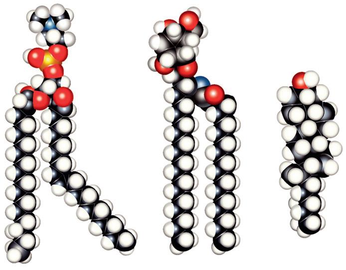
acid
OH group
Steroid region
• Fig. 1.3 Models of the major classes of plasma membrane lipids, depicting the hydrophilic and hydrophobic regions of the molecules. The molecules are arranged as they exist in one leaflet of the bilayer. The opposing leaflet is not shown. One of the fatty acyl chains in the phospholipid molecule is unsaturated. The presence of this double bond produces a “kink” in the fatty acyl chain, which prevents tight packing of membrane lipids and increases membrane fluidity. (Modified from Hansen JT, Koeppen BM: Netter’s Atlas of Human Physiology. Teterboro, NJ: Icon Learning Systems; 2002.)
The majority of membrane phospholipids have a glycerol “backbone” to which are attached the fatty acyl chains, and an alcohol is linked to glycerol via a phosphate group. The common alcohols are choline, ethanolamine, serine, inositol, and glycerol. Another important phospholipid, sphingomyelin, has the amino alcohol sphingosine as its “backbone” instead of glycerol. Table 1-2 lists these common phospholipids. The fatty acyl chains are usually 14 to 20
carbons in length and may be saturated or unsaturated (i.e., contain one or more double bonds).
The phospholipid composition of the membrane varies among different cell types and even between the bilayer leaflets. For example, in the erythrocyte plasma membrane, phosphatidylcholine and sphingomyelin are found predominantly in the outer leaflet of the membrane, whereas phosphatidylethanolamine, phosphatidylserine,
2 pA 1 second
• Fig. 1.4 Recording of current flow through a single ion channel. The channel spontaneously fluctuates between an open state and a closed state. The amplitude of the current is approximately 2 pA (2 × 10 12 amps); that is, 12.5 million ions/second cross the membrane.
monophosphate), and mechanical stretch of the plasma membrane. Ion channels can be regulated by a change in the number of channels in the membrane or by gating of the channels.
Solute carriers (denoted SLCs by the HUGO Gene Nomenclature Committee) represent a large group of membrane transporters categorized into more than 50 families; almost 400 specific transporters have been identified to date. These carriers can be divided into three groups according to their mode of transport. One group, uniporters (or facilitated transporters), transports a single molecule across the membrane. The transporter that brings glucose into the cell (glucose transporter 1 [GLUT-1], or SLC2A1) is an important member of this group. The second group, symporters (or cotransporters), couples the movement of two or more molecules/ions across the membrane. As the name implies, the molecules/ions are transported in the same direction. The Na+,K+,2Cl (NKCC) symporter found in the kidney (NKCC2, or SLC12A1), which is crucial for diluting and concentrating the urine (see Chapter 34), is a member of this group. The third group, antiporters (or exchange transporters), also couples the movement of two or more molecules/ions across the membrane; in this case, however, the molecules/ions are transported in opposite directions. The Na+-H+ antiporter is a member of this group
of solute carriers. One isoform of this antiporter (NHE-1, or SLC9A1) is found in all cells and plays an important role in regulating intracellular pH.
The ATP-dependent transporters, as their name implies, use the energy in ATP to drive the movement of molecules/ ions across the membrane. There are two groups of ATPdependent transporters: the ATPase ion transporters and the ATP-binding cassette (ABC) transporters. The ATPase ion transporters are subdivided into P-type ATPases and V-type ATPases.a The P-type ATPases are phosphorylated during the transport cycle. Na+,K+-ATPase is an important example of a P-type ATPase. With the hydrolysis of each ATP molecule, it transports three Na+ ions out of the cell and two K+ ions into the cell. Na+,K+-ATPase is present in all cells and plays a critical role in establishing cellular ion and electrical gradients, as well as maintaining cell volume (see Chapter 2).
V-type H+-ATPases are found in the membranes of several intracellular organelles (e.g., endosomes, lysosomes); as a result, they are also referred to as vacuolar H +-ATPases. The
aAnother type of ATPases, F-type ATPases, is found in the mitochondria, and they are responsible for ATP synthesis. They are not considered in this chapter.
Na+,K+-ATPase (also called the Na+,K+-pump or just the Na+pump) is found in all cells and is responsible for establishing the gradients of Na+ and K+ across the plasma membrane. These gradients in turn provide energy for several essential cell functions (see Chapter 2). Na+,K+-ATPase is composed of three subunits (α, β, and γ), and the protein exists in the membrane with a stoichiometric composition of 1α, 1β, 1γ
The α subunit contains binding sites for Na+,K+ and ATP. It is also the subunit that binds cardiac glycosides (e.g., ouabain), which specifically inhibit the enzyme. It has a transmembrane domain and three intracellular domains: phosphorylation (P-domain), nucleotide binding (N-domain), and actuator (A-domain). Although the α subunit is the functional subunit of the enzyme (i.e., it hydrolyzes ATP, binds Na+ and K+ , and translocates them across the membrane), it cannot function without the β subunit. The β subunit is responsible for targeting the α subunit to the membrane and also appears to modulate the affinity of the Na+,K+-ATPase for Na+ and K+
The α and β subunits can carry out Na+ and K+ transport in the absence of the γ subunit. However, the γ subunit appears to play a regulatory role. The γ subunit is a member of a family of proteins called FXYD proteins (so named for the FXYD amino acid sequence found in the protein).
H+-ATPase in the plasma membrane plays an important role in urinary acidification (see Chapter 37).
ABC transporters represent a large group of membrane transporters. They are found in both prokaryotic and eukaryotic cells, and they have amino acid domains that bind ATP (i.e., ABC domains). Seven subgroups of ABC transporters in humans and more than 40 specific transporters have been identified to date. They transport a diverse group of molecules/ions, including Cl , cholesterol, bile acids, drugs, iron, and organic anions.
Because biologically important molecules enter and leave cells through membrane transporters, membrane transport is specific and regulated. Although some membrane transporters are ubiquitously expressed in all cells (e.g., Na+,K+-ATPase), the expression of many other transporters is limited to specific cell types. This specificity of expression tailors the function of the cell to the organ system in which it is located (e.g., the sodium-glucose–linked transporters SGLT-1 and SGLT-2 in the epithelial cells of the intestines and renal proximal tubules). In addition, the amount of a molecule being transported across the membrane can be regulated. Such regulation can take place through altering the number of transporters in the membrane or altering the rate or kinetics of individual transporters (e.g., the time an ion channel stays in the open versus closed state), or both.
Solute and water can be brought into the cell through a process of endocytosis and released from the cell through the process of exocytosis. Endocytosis is the process whereby a piece of the plasma membrane pinches off and
Cystic fibrosis is an autosomal recessive disease characterized by chronic lung infections, pancreatic insufficiency, and infertility in boys and men. Death usually occurs because of respiratory failure. It is most prevalent in white people and is the most common lethal genetic disease in this population, occurring in 1 per 3000 live births. It is a result of mutations in a gene on chromosome 7 that codes for an ABC transporter. To date, more than 1000 mutations in the gene have been identified. The most common mutation is a deletion of a phenylalanine at position 508 (F508del). Because of this deletion, degradation of the protein by the endoplasmic reticulum in enhanced, and, as a result, the transporter does not reach the plasma membrane. This transporter, called cystic fibrosis transmembrane conductance regulator (CFTR), normally functions as a Cl channel and also regulates other membrane transporters (e.g., the epithelial Na+ channel [ENaC]). Thus in individuals with cystic fibrosis, epithelial transport is defective, which is responsible for the pathophysiologic process. For example, in patients not affected by cystic fibrosis, the epithelial cells that line the airway of the lung are covered with a layer of mucus that entraps inhaled particulates and bacteria. Cilia on the epithelial cells then transport the entrapped material out of the lung, a process termed mucociliary transport (see Chapter 26 for more details). In patients with cystic fibrosis, the inability to secrete Cl , Na+ , and H2O results in an increase in the viscosity of the airway surface mucus; thus the cilia cannot transport the entrapped bacteria and other pathogens out of the lung. This in turn leads to recurrent and chronic lung infections. The inflammatory process that accompanies these infections ultimately destroys the lung tissue, causing respiratory failure and death. In 2015, the U.S. Food and Drug Administration approved lumacaftor/ ivacaftor (Orkambi), a drug that increases the amount of F508del CFTR in the plasma membrane of lung epithelial cells.
is internalized into the cell interior, and exocytosis is the process whereby vesicles inside the cell fuse with the plasma membrane. In both of these processes, the integrity of the plasma membrane is maintained, and the vesicles allow for the transfer of the contents among cellular compartments. In some cells (e.g., the epithelial cells lining the gastrointestinal tract), endocytosis across one membrane of the cell is followed by exocytosis across the opposite membrane. This allows the transport of substances inside the vesicles across the epithelium, a process termed transcytosis.
Endocytosis occurs in three mechanisms. The first is pinocytosis, which consists of the nonspecific uptake of small molecules and water into the cell. Pinocytosis is a prominent feature of the endothelial cells that line capillaries and is responsible for a portion of the fluid exchange that occurs across these vessels. The second form of endocytosis, phagocytosis, allows for the cellular internalization of large particles (e.g., bacteria, cell debris). This process is an important characteristic of cells in the immune system (e.g., neutrophils and macrophages). Often, but not always, phagocytosis is a receptor-mediated process. For example,
As noted previously, the vast majority of biologically important molecules cross cell membranes via specific membrane transporters, rather than by diffusing through the lipid portion of the membrane. Nevertheless, eq. 1.3 can be and has been used to quantitate the diffusion of molecules across many biological membranes. When this is done, the value of the permeability coefficient (P) reflects the properties of the pathway (e.g., membrane transporter or, in some cases, multiple transporters) that the molecule uses to cross the membrane.
Despite the limitations of using diffusion to describe and understand the transport of molecules across cell membranes, it is also important for understanding gas exchange in the lungs (see Chapter 24), the movement of molecules through the cytoplasm of the cell, and the movement of molecules between cells in the extracellular fluid. For example, one of the physiological responses of skeletal muscle to exercise is the recruitment or opening of capillaries that are not perfused at rest. This opening of previously closed capillaries increases capillary density and thereby reduces the diffusion distance between the capillary and the muscle fiber so that oxygen and cellular fuels (e.g., fatty acids and glucose) can be delivered more quickly to the contracting muscle fiber. In resting muscle, the average distance of a muscle fiber from a capillary is estimated to be 40 µm. However, with exercise, this distance decreases to 20 µm or less.
The electrochemical gradient (also called the electrochemical potential difference) is used to quantitate the driving force acting on a molecule to cause it to move across a membrane. The electrochemical gradient for any molecule (Δµx) is calculated as follows:
Equation 1.4
∆µ x i o xm RT X X zFV =+ ln [] [] , where
R = the gas constant
T = temperature in degrees Kelvin
Ln = natural logarithm
[X]i = the concentration of X inside the cell
[X]o = the concentration of X outside the cell
zx = the valence of charged molecules
F = the Faraday constant
Vm = the membrane potential (Vm = Vi Vo)b
The electrochemical gradient is a measure of the free energy available to carry out the useful work of transporting the molecule across the membrane. It has two components: One component represents the energy in the concentration gradient for X across the membrane (chemical potential
bBy convention, membrane voltages are determined and reported with regard to the exterior of the cell. In a typical cell, the resting membrane potential (Vm) is negative. Positive Vm values can be observed in some excitable cells at the peak of an action potential.
difference). The second component (electrical potential difference) represents the energy associated with moving charged molecules (e.g., ions) across the membrane when a membrane potential exits (i.e., Vm ≠ 0 mV). Thus for the movement of glucose across a membrane, only the concentrations of glucose inside and outside of the cell need to be considered (Fig. 1.6A). However, the movement of K+ across the membrane, for example, would be determined both from the K+ concentrations inside and outside of the cell and from the membrane voltage (see Fig. 1.6B).
Eq. 1.4 can be used to derive the Nernst equation for the situation in which a molecule is at equilibrium across the membrane (i.e., Δµ = 0):
Equation 1.5a
Equation 1.5b
The value of Vm calculated with the Nernst equation represents the equilibrium condition and is referred to as the Nernst equilibrium potential (Ex, the Vm at which there is no net transport of the molecule across the membrane). It should be apparent that the Nernst equilibrium potential quantitates the energy in a concentration gradient and expresses that energy in millivolts. For example, for the cell depicted in Fig. 1.6B, the energy in the K+ gradient (derived from the Nernst equilibrium potential for K+ [ E K + ]) is proportional to 90.8 mV (causing K+ to move out of the cell). This is opposite to, and of greater magnitude than, the energy in the membrane voltage (Vm = 60 mV), which causes K+ to enter the cell. As a result, the electrochemical gradient is such that the net movement of K+ across the membrane will be out of the cell. Another way to state this is that the net driving force for K+ (Vm E K + ) is 30.8 mV (driving K+ out of the cell). This is described in more detail in Chapter 2.
The Nernst equation, at 37° C, can be written as follows by replacing the natural logarithm function with the base 10 logarithm function:
Equation 1.6a
Equation 1.6b