The main objective of this monograph is to provide the anatomical basis for neurologic abnormalities. Knowledge of basic clinical neuroanatomy will enable medical students to answer the first question asked when examining a patient with an injured or a diseased nervous system: “Where is the lesion located?” Knowledge of basic clinical neuroanatomy will enable students in health-related fields, such as nursing, physical therapy, occupational therapy, and physician assistants, to understand the anatomical basis of the neurologic abnormalities in their patients. To accomplish these objectives, the anatomical relationships and functions of the clinically important structures are emphasized. Effort is exerted to simplify as much as possible the anatomical features of the brain and spinal cord.
This monograph is neither a reference book nor a textbook of neuroanatomy. Most neuroanatomy textbooks include much information about anatomical structures that aids in the understanding of a particular system or mechanism, but when these structures are damaged, clinical signs or symptoms do not result. Such superfluous information is kept to a minimum in this book.
This basic clinical anatomy book is presented in three main sections: (1) the basic plan, (2) the functional systems, and (3) the associated structures. The basic plan includes the organization of the nervous system, its histologic features and supporting structures, distinguishing anatomical characteristics of the subdivisions of the brain and spinal cord, and an introduction to clinically important brain and spinal cord functional levels. Only those structures needed to identify the subdivisions and their levels are included in this part.
The second section deals with the functional systems and their clinically relevant features. This section is arranged so that the motor and somatosensory systems, of paramount importance because they include structures located in every subdivision of the brain and spinal cord,
are described first. The remainder of this section includes the pathways associated with the special senses, higher mental functions, and the behavioral and visceral systems.
In the third section, the vascular supply and the ventricular cerebrospinal fluid system are presented.
The visualization of three-dimensional anatomical relationships plays a key role in localizing lesions and understanding the anatomical basis of neurologic disorders. Every effort has been made to include illustrations that enhance this visualization of three-dimensional images of the clinically important structures. In addition to the threedimensional illustrations, schematic diagrams of the functional systems and drawings of myelinstained sections from selected functional levels of the brain and spinal cord are used to provide the anatomical relationships that enhance the understanding of the anatomical basis for neurologic disorders and their syndromes. Clinical relevance is emphasized throughout this book and illustrations of some neurologic abnormalities are included.
Review questions are found at the end of each chapter, and an entire chapter is devoted to the principles of locating lesions and clinical illustrations. Answers to the chapter questions are found in the appendixes. Also in the appendixes are a section devoted to cranial nerve components and their clinical correlations, a glossary of terms, a list of suggested readings, and an atlas of the myelinstained sections used throughout the book.
The authors are most grateful to Mr. Larry Clifford for his artistic skills in creating the illustrations, all of which are an invaluable part of this book. Our deep appreciation is expressed to Ms. Susan Quinn for her superb assistance in preparing the manuscript and to Ms. Susan McClain for her computer expertise in preparing the charts and tables. Finally, the authors are much indebted to the publisher, Williams & Wilkins, and its editorial and marketing staff for their interest, support, and patience throughout the project.
Part I
Organization, Cellular Components, and Topography of the CNS
1. Introduction, Organization, and Cellular Components 2
2. Spinal Cord: Topography and Functional Levels 17
3. Brainstem: Topography and Functional Levels 27
4. Forebrain: Topography and Functional Levels 39
Part II
Motor Systems
5. Lower Motor Neurons: Flaccid Paralysis 50
6. The Pyramidal System: Spastic Paralysis 67
7. Spinal Motor Organization and Brainstem Supraspinal Paths: Postcapsular Lesion Recovery and Decerebrate Posturing 81
8. The Basal Ganglia: Dyskinesia 88
9. The Cerebellum: Ataxia 104
10. The Ocular Motor System: Gaze Disorders 123
Part III
Sensory Systems
11. The Somatosensory System: Anesthesia and Analgesia 132
12. The Auditory System: Deafness 160
13. The Vestibular System: Vertigo and Nystagmus 169
14. The Visual System: Anopsia 178
15. The Gustatory and Olfactory Systems: Ageusia and Anosmia 197
Part IV
The Cerebral Cortex and Limbic System
16. The Cerebral Cortex: Aphasia, Agnosia, and Apraxia 206
17. The Limbic System: Anterograde Amnesia and Inappropriate Social Behavior 225
Part V
The Visceral System
18. The Hypothalamus: Vegetative and Endocrine Imbalance 236
19. The Autonomic Nervous System: Visceral Abnormalities 242
Part VI
The Reticular Formation and Cranial Nerves
20. Reticular Formation: Modulation and Activation 260
21. Summary of the Cranial Nerves: Components and Abnormalities 271
Part VII
Accessory Components
22. The Blood Supply of the Central Nervous System: Stroke 286
23. The Cerebrospinal Fluid System: Hydrocephalus 306
Part
VIII
Development, Aging, and Response of Neurons to Injury
24. Development of the Nervous System: Congenital Anomalies 318
25. Aging of the Nervous System: Dementia 328
26. Recovery of Function of the Nervous System: Plasticity and Regeneration 332
Part IX
Where is the Lesion?
27. Principles for Locating Lesions and Clinical Illustrations 342
Appendices
A. Answers to Chapter Questions 362
B. Glossary 391
C. Suggested Readings 414
D. Atlas of Myelin-Stained Sections 415
Index 429
Part I
Organization, Cellular Components, and Topography of the CNS
1 Introduction, Organization, and Cellular Components
Two fundamental properties of animals, irritability and conductivity, reach their greatest development in the human nervous system. Irritability, the capability of responding to a stimulus, and conductivity, the capability of conveying signals, are specialized properties of the basic functional units of the nervous system: the nerve cells or neurons. Neurons respond to stimuli, convey signals, and process information that enables the awareness of self and surroundings; mental functions such as memory, learning, and speech; and the regulation of muscular contraction and glandular secretion.
ORGANIZATION OF THE NERVOUS SYSTEM
The basic functional unit of the nervous system is the neuron. Each neuron has a cell body that receives nerve impulses and an axon that conveys the nerve impulse away from the cell body. The nervous system comprises neurons arranged in longitudinal series. The serial arrangement forms two types of circuits: reflex and relay. A reflex circuit conveys the impulses that result in an involuntary response such as muscle contraction or gland secretion (Fig. 1-1A). A relay circuit conveys impulses from one part of the nervous system to another. For example, relay circuits convey impulses from sensory organs in the skin, eyes, ears, and so forth that become perceived by the brain as sensations (Fig. 1-1B). Relay circuits are categorized according to their functions and are called functional paths, for example, pain path, visual path, or motor or voluntary movement path. A functional path may consist of a series of only two or three neurons or as many as hundreds
of neurons. Reflex circuits may overlap with parts of relay circuits (Fig. 1-1C).
A functional path may contain thousands or even millions of nerve cell bodies and axons. The nerve cell bodies may form pools or clumps, in which cases they are called nuclei or ganglia, or the nerve cell bodies may be arranged in the form of layers or laminae. The axons in a functional path usually form bundles called tracts, fasciculi, or nerves. Therefore, the entire nervous system is composed of functional paths whose neuronal cell bodies are located in the nuclei, ganglia, or laminae and whose axons are located in the tracts or nerves.
The human nervous system is divided into central and peripheral parts. The brain and spinal cord form the central nervous system (CNS), and the cranial, spinal, and autonomic nerves and their ganglia form the peripheral nervous system (PNS). The CNS integrates and controls the entire nervous system, receiving information (input) about changes in the internal and external environments, interpreting and integrating this information, and providing signals (output)
Sensor y perception
Sensor y stimulus
Movement or secretion Response
Sensory perception
Figure 1-1 Simple reflex and relay circuits. A. Three-neuron reflex circuit. B. Three-neuron sensory relay circuit. C. Combined three-neuron relay and reflex circuits.
for the execution of activities, such as movement or secretion. The PNS connects the CNS to the tissues and organs of the body. Hence, the PNS is responsible for conveying input and output signals to and from the CNS. Signals passing to the CNS are called afferent, whereas those passing away from the CNS are called efferent.
NERVOUS SYSTEM SUPPORT AND PROTECTION
Nerve cells are extremely fragile and cannot survive without the protection of supporting cells. The brain and spinal cord, also very fragile, are protected from the surrounding bones of the cranial cavity and vertebral or spinal canal by three coverings or membranes, called the meninges.
The Meninges
The CNS is supported and protected by the meninges, three connective tissue membranes located between the brain and the cranial bones
and between the spinal cord and the vertebral column. The meninges are, from external to internal, the dura mater, the arachnoid, and the pia mater. The meninges around the brain and spinal cord are continuous at the foramen magnum, the large opening in the base of the skull where the brain and spinal cord are continuous.
Dura Mater
The dura mater is a strong, fibrous membrane that consists of two layers. In the cranial dura, which surrounds the brain, the two layers are fused and adhere to the inner surfaces of the cranial bones except in those regions where the layers split (Fig. 1-2) to form the venous sinuses that carry blood from the brain to the veins in the neck. The inner layer of the dura forms four folds that extend internally to partially partition various parts of the brain (Fig. 1-3). The sickle-shaped falx cerebri lies in the longitudinal groove between the upper parts of the brain, the cerebral hemispheres. The falx cerebelli, also oriented longitudinally, separates the upper parts of the
#1
#1
Cerebral cor tex
Arachnoid
Pia mater
Dura mater Venous sinus (superior sagittal)
Dural fold (falx cerebri)
Subarachnoid space
1-2 Coronal section of cranial meninges showing a venous sinus and dural fold.
Anterior
Diaphragma sellae
Aper ture for pituitar y stalk
Free margins of tentorium
Calvaria
Arachnoid trabeculae
Figure
Posterior
Tentorium cerebelli (right side)
Falx cerebelli
Tentorium cerebelli (left side)
cerebelli
Falx cerebri
Figure 1-3 The dural folds as viewed from the left side.
hemispheres of the cerebellum, or “little brain.” The tentorium cerebelli is a flat dural fold that separates the posterior parts of the cerebral hemispheres above from the cerebellum below. The diaphragma sellae is a circular, horizontal fold beneath the brain that covers the sella turcica, in which the pituitary gland is located. The stalk of the pituitary gland pierces the diaphragma sellae and attaches to the undersurface of the brain.
The spinal dura consists of two layers: the outer layer forms the periosteal lining of the vertebral foramina that form the vertebral or spinal canal; the inner layer loosely invests the spinal cord and forms a cuff around the spinal nerves as they emerge from the vertebral canal.
Arachnoid
The arachnoid is a thin, delicate membrane that loosely surrounds the brain and spinal cord. The outer part of the arachnoid adheres to the dura (Fig. 1-4). Extending internally from this outer
Epidural hematoma
part are numerous cobweb-like projections or trabeculae that attach to the pia mater.
Pia Mater
The pia mater is the thin membrane that closely invests the brain and spinal cord. The pia is highly vascular and contains the small blood vessels that supply the brain and spinal cord.
Meningeal Spaces
Several clinically important spaces are associated with the meninges (Fig. 1-4). The epidural space is located between the bone and the dura mater, and the subdural space is located between the dura and arachnoid. Normally, both the epidural and subdural spaces are potential spaces in the cranial cavity. Both may become actual spaces if blood accumulates because of epidural or subdural hemorrhages caused by traumatic tearing of blood vessels that pass through the spaces. In the spinal cord, the subdural space is also potential, but the
Subdural hematoma
1-4 Relation of meningeal spaces to blood vessels and hemorrhages.
Dura mater
Arachnoid membrane
Arachnoid trabecula
Emissary vein
Pia mater
Calvaria
Brain
Cerebral artery
Subarachnoid space
Figure
epidural space is actual and contains semifluid fat and thin-walled veins.
The subarachnoid space is located in the area between the arachnoid and pia mater and contains cerebrospinal fluid. The subarachnoid space communicates with the cavities or ventricles of the brain where cerebrospinal fluid is formed. Also located within the subarachnoid space are the initial parts of the cranial and spinal nerves and numerous blood vessels on the surfaces of the brain and spinal cord. Vascular accidents involving the vessels here result in subarachnoid hemorrhage.
Clinical Connection
Inflammation of the meningeal membranes surrounding the brain and spinal cord, due primarily to either a viral or bacterial infection of the meninges, may result in a life-threatening condition of meningitis. Less common causes include fungal, parasitic, and drug-mediated meningitis. In adults, neck stiffness and headache with fever, altered consciousness, vomiting, and aversion to bright light or loud noises are the primary symptoms of meningitis. In children, symptoms may be less apparent than in adults and consist of only irritability and drowsiness. Pathogen access to the meninges may be blood borne or as the result of direct entry from the nasal cavities. Diagnosis most commonly is by lumbar puncture if there is no indication of elevated intracranial pressure in the patient. Bacterial meningitis is treated by antibiotics.
Supporting Cells
Three basic types of supporting or glial cells exist: ependymal, microglial, and macroglial cells. The ependymal cells line the fluid-filled cavities or ventricles of the brain and the central canal of the spinal cord. The microglial cells are mesodermal in origin being derived from bone marrow, are formed in all parts of the brain and spinal cord, and play roles in immunological activities. They also become macrophages, phagocytizing the debris resulting from injury, infections, or diseases in the CNS. The macroglia are derived from neuroectoderm and consist of four cell types: astrocytes and oligodendrocytes in the CNS and Schwann cells and capsular cells in the PNS.
Astrocytes
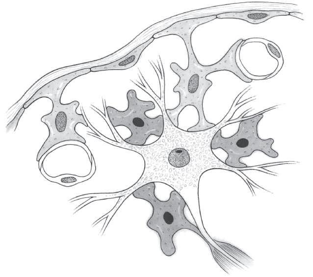
Astrocytes are the most numerous cells in the CNS (Fig. 1-5). Each astrocyte has a star-shaped cell body and numerous irregularly shaped processes, some of which may be extremely long. Processes of some astrocytes have end-feet on the surface of the brain or spinal cord. These end-feet form a protective covering called the external limiting membrane or glial membrane. Many astrocytic processes have vascular end-feet, which surround capillaries. The endothelial cells of CNS capillaries are interconnected by tight junctions and form the blood-brain barrier, which selectively governs the passage of materials, including many drugs, from the circulating blood into the CNS.
Astrocytes have other functions as well. They play a major role in the electrolyte balance of the CNS, produce neurotrophic factors necessary for neuronal survival, and remove certain neurotransmitters from synaptic clefts. Astrocytes are the first cells to undergo alterations in response to CNS insults such as ischemia, trauma, or radiation. Also, astrocytes form scars resulting from CNS injury. Astrocytes are highly susceptible to the formation of neoplasms.
Oligodendrocytes
The formation and maintenance of CNS myelin are the primary functions of the oligodendrocytes, small glial cells with relatively few processes (Fig. 1-5). The myelin sheath is formed by oligodendrocyte processes, which wrap around the axon to form a tight spiral. The myelin itself is located within the processes. Each oligodendrocyte envelops a variable number of axons depending on the thickness of the myelin sheaths. In the case of thin myelin sheaths, one oligodendrocyte may be related to 40 or 50 axons. Oligodendrocytes may also surround the cell bodies of neurons, but in this location, they do not contain myelin. Recent research suggests that oligodendrocytes also produce neurotrophic factors, the most important of which is a nerve growth factor that may promote the growth of damaged CNS axons. Autoimmune reactions to CNS myelin may be associated with multiple sclerosis.
Schwann Cells
The PNS counterpart of the oligodendrocyte is the Schwann cell. Unlike the oligodendrocyte, which envelops many myelinated axons,
Astrocyte
Exter nal limiting (glial) membrane
Astrocyte
Astrocyte
Perivascular end-foot
Oligodendrocyte
Oligodendrocyte
Dendrites
the Schwann cell envelops only part of one myelinated axon. During development of the myelin sheath, the Schwann cell first encircles and then spirals around the axon many times,
forming multiple layers or lamellae. The myelin is actually located within the Schwann cell lamellae (Fig. 1-6). The outermost layer of the Schwann cell lamellae is called the neurolemma
Schwann cell nucleus
Myelin lamellae
Axon
Axon
Myelin lamellae Node of Ranvier Neurolemma
Schwann cell nucleus
Neurolemma
Figure 1-6 Myelinated axon in the peripheral nervous system. A. Transverse view. B. Longitudinal view.
Pia mater
Capillar y endothelial cell
Myelin in oligodendrocyte process
Axon
Axon hillock Neuronal cell body
Figure 1-5 Relation of neurons, glia, and capillaries.
or sheath of Schwann. Because each Schwann cell myelinates only a small extent of the axon, myelination of the entire axon requires a long string of Schwann cells. Between each Schwann cell, the myelin is interrupted. These areas of myelin sheath interruption are called nodes of Ranvier (Figs. 1-6, 1-7). Similar interruptions of myelin sheaths occur in the CNS. In unmyelinated fibers, one Schwann cell envelops many axons. Autoimmune reactions to PNS myelin may be associated with Guillain-Barré syndrome.
Schwann cells not only form and maintain the myelin sheath but also are extremely important in the regeneration of damaged axons. When an axon is cut, the part of the axon separated from the cell body degenerates; however, the string of Schwann cells distal to the injury proliferates and forms a tube. Growth sprouts arising from the proximal end of the transected axon enter this tube and travel to the structures supplied by the axon before its injury. Such functional axonal regeneration is common in the PNS. Axonal regeneration has not occurred in the human CNS, and this lack of regeneration may be related, in part, to the absence of Schwann cells.
Capsular Cells
Capsular cells are the glial elements that surround the neuronal cell bodies in sensory and autonomic ganglia. The sensory ganglia of the spinal nerves and some cranial nerves contain large, round neurons whose cell bodies are surrounded by a nearly complete layer of flattened capsular or satellite cells, thereby separating the ganglion cell from the nonneural connective tissue and vascular structures. Although capsular cells are present in autonomic ganglia, because of the irregular shapes of these ganglion cells the capsules are less uniform and, hence, incomplete.
NEURONS
Morphologic Properties
A neuron consists of a cell body or soma and of protoplasmic processes called dendrites and axons (Fig. 1-7). The cell body is the metabolic
center of a neuron and contains the nucleus and the cytoplasm. The nucleus contains nucleoplasm, chromatin, a prominent nucleolus, and, in the female only, a nucleolar satellite. The cytoplasm contains the usual cellular organelles such as mitochondria, Golgi apparatus, and lysosomes. In addition, various-sized clumps of rough endoplasmic reticulum, called Nissl bodies, are prominent in the cytoplasm of neurons. However, the neuronal cytoplasm where the axon emerges is devoid of Nissl bodies; this area is called the axon hillock. Another cytoplasmic characteristic of neurons are neurofibrils, which are arranged longitudinally in the cell body, the axons, and the dendrites.
Neurons are classified morphologically as unipolar, bipolar, or multipolar according to their number of protoplasmic processes (Fig. 1-8). The single process of a unipolar neuron is the axon. Unipolar neurons are located almost exclusively in the ganglia of spinal nerves and some cranial nerves. Bipolar neurons have an axon and one dendrite and are limited to the visual, auditory, and vestibular pathways. All the remaining nerve cells are multipolar neurons and have an axon and between 2 and 12 or more dendrites.
Dendrites and Axons
Dendrites, cytologically similar to the neuronal cell body, are short and convey impulses toward the cell body (Table 1-1). Axons do not contain Nissl bodies, vary in length from microns to meters, and convey impulses away from the cell body.
The integrity of the axon, regardless of its length, is maintained by the cell body via two types of axoplasmic flow or axonal transport. In anterograde axonal transport, the cell body nutrients are carried in a forward direction from the cell body to the distal end or termination of the axon. Anterograde axonal transport is vital for axonal growth during development, for maintenance of axonal structure, and for the synthesis and release of neurotransmitters, the chemicals that assist in the transfer of nerve impulses from one cell to another.
Besides anterograde transport, retrograde axonal transport occurs from the distal end of the axon back to the cell body. The function
Unipolar
Anatomic axon
physiologic dendrite
Nucleolus
Cell body
Nissl body
Nucleus
Bipolar
Dendrite
Cell body
Nucleolus
Nucleus
Nissl body
Multipolar
Nissl body Cell body
Dendrites
Nucleolus
Nucleus
Figure 1-8 Morphologic types of neurons (arrows indicate direction of impulses).
of retrograde axonal transport is the return of used or worn out materials to the cell body for restoration.
Axons may be myelinated or unmyelinated. Myelinated axons are insulated by a sheath of myelin that starts near the cell body and stops just
Table
before the axon terminates (Fig. 1-7). Myelin is a multilayered phospholipid located within axonal supporting cells. The myelin sheath increases the conduction velocity of the nerve impulse along the axon. The thicker the myelin sheath, the faster the conduction velocity.
1-1 COMPARISON OF AXONS AND DENDRITES
Axons
Function Transport impulses from the cell body
Dendrites
Receive impulses and transport them toward the cell body
Length Vary from microns to meters Microns; seldom more than a millimeter
Branching pattern Limited to collaterals, preterminals, and terminals
Surface Smooth
Coverings Supporting cells and frequently myelin
Vary from simple to complex arborizations
Vary from smooth to spiny
Always naked
Axon
Axon hillock
Axon hillock
Axon
Axon
Axon hillock
Clinical Connection
Retrograde axonal transport is of clinical importance because it is the route by which toxins such as tetanus and viruses such as herpes simplex, rabies, and polio are transported into the CNS from the periphery.
Synapses
Axonal endings or terminals occur in relation to other neurons, muscle cells, or gland cells. The junction between the axonal ending and the neuron, muscle cell, or gland cell is called the synapse. An important anatomic characteristic of the synapse is that the axonal ending is separated from the surface of the other nerve, muscle, or gland cell by a space, the synaptic cleft. An important physiologic characteristic of a synapse is polarization; that is, the impulse always travels from the axon to the next neuron in the circuit or to the muscle or gland cells supplied by the axon. When a nerve impulse arrives at the synapse, chemicals called neurotransmitters are released into the synaptic cleft. Neurotransmitters, manufactured and released by the neurons, cross the synaptic cleft to affect the postsynaptic neuron, muscle, or gland cell. The transmitters at neuromuscular and neuroglandular synapses are excitatory; that is, they elicit muscle contraction or glandular secretion. However, the neurotransmitters at synapses between neurons may be excitatory, enhancing the production of an impulse in the postsynaptic neuron, or inhibitory, hindering impulse production in the postsynaptic neuron. All functions of the CNS, that is, awareness of sensations, control of movements or glandular secretions, and higher mental functions, occur as the result of the activity of excitatory and inhibitory synapses on neurons in various circuits.
Physiologic Properties
Resting Membrane Potential
Under steady-state conditions, neurons are electrically polarized to about –60 mV by the separation of extracellular cationic charges from intracellular anionic charges. This resting
membrane potential results from the differential distribution of ions and selective membrane permeability with four major cations and anions contributing to the resting membrane potential. Na+ and Cl ions are concentrated extracellularly, and K+ and organic anions (proteins and amino acids) are concentrated intracellularly. Transmembranous ion-selective channels or pores allow Na +, K+, and Cl ions to passively diffuse across the membrane as a result of concentration and electrical gradients. Proteins and amino acids do not move through the membrane as part of the resting membrane potential. The resting membrane potential is determined largely by Na+ influx and K+ efflux, and their active transport back across the membrane by an ATP-dependent Na+/K+ pump, thereby maintaining the membrane potential at about −60 mV.
Electrotonic Conductance in the Soma-dendritic Membrane
Electrotonic transients in the resting membrane potential can result in the interior of the cell becoming relatively more negative or hyperpolarized or less negative or depolarized. These potential shifts are electrotonically summated, temporally and spatially, as they are conducted passively from the soma and dendrites to the axon hillock-initial segment (Fig. 1-9).
Action Potential Initiation and Conductance
Depolarization of the axon hillock-initial segment region to about −45 mV results in the generation of an action potential. Unlike in the soma and dendrites where membrane transients are graded, membrane conductance at the axon hillock-initial segment becomes self-sustaining with the initiation of an action potential. The initiation or rising phase of an action potential is caused by the rapid influx of Na+ through voltage-sensitive channels. The subsequent falling phase of the action potential is slightly more prolonged and occurs by the efflux of K+. Starting at the initial axon segment and continuing through to its terminal branches, the propagation of the action potential occurs as a nondecremental voltage change. The velocity of propagation of an action potential is dependent on axonal diameter and myelination.
Temporal Summation of EPSPs
Mitochondrion
endoplasmic
Spatial Summation of EPSPs
Synaptic Integration and Action Potential Initiation
Figure 1-9 Electrotonic conductance in neuron, temporal and spatial summation, and action potential initiation. Synaptic interactions: A. Excitatory postsynaptic potentials (EPSPs) can spatially summate when they converge as they are electrotonically conducted from the dendrites to the soma. B. EPSPs can summate temporally when the same synaptic input is activated rapidly by multiple presynaptic action potentials. C. Excitatory and inhibitory inputs are integrated at the initial segment and sufficient depolarization generates an action potential (EPSP, excitatory postsynaptic potential; IPSP, inhibitory postsynaptic potential).
Saltatory Conduction
In unmyelinated, generally small-diameter (0.2–1.5 μm) axons (type IV motor or type C sensory), Na+ and K+ conductances and impulse propagation occur continuously between neighboring axonal membrane segments, resulting in slower impulse transmission (0.5–2 m/s). Conversely, in large-diameter (13–20 μm) myelinated axons (type I or Aα), impulse propagation is much faster (80–120 m/s) because Na+ and K+ conductance
changes occur discontinuously along the axonal membrane at small gaps (1 μm) between the edges of myelin sheaths, the nodes of Ranvier. In these nodal regions, Na+ channels are many times more numerous than in the internodal axonal membrane, whereas K+ channels are spread along the internodal axolemma. The low internodal capacitance and concentrated Na+ channels at the nodes allow the action potential to jump (saltatory conduction) between nodes, increasing the speed of conduction in myelinated axons (Fig. 1-10).
Rough
reticulum
Dendrite
Axon
Axon
Axon hillock
Neuron cell body
Golgi apparatus
Dendrites
Axosomatic synapse
Axodendritic synapse
Neuron cell body
Nucleus
Potential
Saltatory Conduction in Myelinated Axon
Nonsaltatory Conduction in Unmyelinated Axon
Impulse blockade
Figure 1-10 Normal and abnormal action potential propagation. A. In myelinated axons, action potential propagation is rapid because of saltatory current flow through the nodes of Ranvier where Na+ channels are concentrated. B. In unmyelinated axons, action potential propagation is slower because Na+ channels are uniformly distributed in the axolemma. C. Action potential propagation is blocked in demyelinated axons because current flow dissipates through the denuded membrane before reaching the next cluster of Na+ channels.
Action Potential Frequency Encodes Information
Information is transmitted between neurons or between neurons and effector structures by the propagation of action potentials. In many neurons, action potential frequency is linearly correlated with stimulus intensity and the resultant degree of depolarization of the soma-dendritic membrane. The more sustained the depolarization, the greater the frequency of action potentials. In other neurons, bursts of action potentials are generated by the superimposed action of Ca2+ currents that
result in the membrane remaining depolarized longer resulting in the repetitive Na+ influx and K+ efflux cycles. Yet other neurons associated with neuromodulatory and autonomic functions fire spontaneously at a relatively slow rate (1–10 Hz).
Synaptic Transmission
The synapse is the point of functional contact between neurons, and the neuromuscular junction is the point of functional contact between axons and skeletal muscle. Most synapses are electrochemical and mediated by
Axon
neurotransmitters. Some synapses are characterized as fast when the delay between presynaptic release and postsynaptic action is about 0.5 ms and involve neurotransmitters such as acetylcholine and amino acids stored in vesicles at or attached to the active zone of the presynaptic membrane. Other synapses are characterized as slow (delay is in terms of seconds) and occur when peptidergic and biogenic amines stored in dense core vesicles away from the terminal membrane are released later and for a longer time. Neurotransmitter release is sequentially triggered by the electrotonic invasion of the action potential into the terminal, the influx of Ca2+ ions through voltage-gated channels that trigger the binding of synaptic vesicles at presynaptic active zones, and the subsequent release of neurotransmitter by exocytosis into the synaptic cleft. Each synaptic vesicle contains a quantal amount of neurotransmitter, and the number of quanta released is directly correlated to the amount of Ca2+ entering the terminal. Neurotransmitters in the narrow synaptic cleft (approx. 100 nm) effect conformational changes in agent-specific postsynaptic receptors, leading to an opening or closing of ion channels. Transmembrane changes mediated by inotropic receptors that quickly depolarize the postsynaptic neuron generate excitatory postsynaptic potentials (EPSPs), whereas ionic changes that hyperpolarize the neuron are classified as inhibitory postsynaptic potentials. In the CNS, synaptic contacts can also be formed at en passant axonal swellings along axons.
PATHOPHYSIOLOGY OF DISEASES AFFECTING NEUROTRANSMISSION AND ACTION POTENTIAL PROPAGATION
Relatively common acquired hereditary disorders affect electrochemical transmission at the neuromuscular junction by either reducing the presynaptic release of acetylcholine or the postsynaptic action of acetylcholine.
Acquired autoimmune disorders affect transmission at the neuromuscular junction. Myasthenia gravis is an autoimmune disease affecting nicotinic acetylcholine receptors, leading to skeletal
muscle weakness and fatigability in orbital, oropharyngeal, and limb musculature. Muscle weakness and fatigability is generally variable in severity and progressive through active hours of the day. Nerve fibers are intact, and acetylcholine release at the nerve terminal is normal. Antibodies attack the acetylcholine receptor in the postjunctional folds, leading to a progressive decrement in amplitude of the evoked end-plate potentials and decreased muscle action potentials with repetitive stimulation. Structural changes of the postjunctional folds and diminished localization of the receptor at the crest of the folds also occur. Increasing the efficacy of the action of acetylcholine in the neuromuscular cleft with acetylcholinesterase inhibitors decreases the severity of the symptoms.
Muscle weakness and fatigability is predominantly in proximal limb and trunk musculature as seen in Lambert-Eaton myasthenic syndrome owing to diminished presynaptic release of acetylcholine from the nerve terminals. Muscle excitability remains normal.
Demyelinating diseases affect PNS Schwann cells or CNS oligodendroglia. Guillain-Barré syndrome is prototypical of an acquired, acuteonset inflammatory peripheral demyelinating neuropathy with axonal sparing. Multiple focal areas of demyelination of spinal roots and proximal nerve fibers result in very slow nerve conduction velocities and reduced compound action potential amplitude in electrophysiologic recordings from affected nerves. Symmetric and temporally progressive weakness in movements, first in the legs and then in the arms, gives the impression of an ascending paralysis. Difficulties in walking and rising from a chair are common complaints. Paralysis of respiratory muscles results in a high risk of respiratory failure. After treatment, functional recovery is possible by axonal remyelination. Charcot-Marie-Tooth disease (type 1A) is the most common hereditary polyneuropathy resulting in demyelination of sensory and motor axons.
Multiple sclerosis is the most common acquired demyelinating disease in the CNS with an immunologic cause. Symptomatology is dependent on the axonal tracts involved. Adjoining segments of myelin are lost (demyelinating plaques) in the white matter fiber tracts in the cerebrum, cerebellum, brainstem, and spinal cord. Normal impulse conduction
occurs proximal and distal to the plaques but is blocked or slowed at the plaques (Fig. 1-10C). Biophysical properties of the demyelinated axolemma are altered, thereby affecting impulse propagation. In demyelinated axons, depolarizing currents are no longer focused at the nodes, but rather are dissipated along the demyelinated axolemma owing to the paucity of Na+ channels in the internodal axolemma and the increased electrical capacitance of the affected segment of the axon. In axons with intact myelin, action potentials jump between nodes of Ranvier because of the high concentration of Na+ channels at the nodal region. Multiple sclerosis is characterized by chronically protracted cycles of relapse and remission. Remission with improvement of symptoms reflects partial remyelination of the affected axonal segments. Persistent deficits can reflect the failure to remyelinate or, more probably, axonal injury within the plaque and axonal degeneration.
Other common disorders that affect axons directly result from chronic nerve compression/ constriction (entrapment) or by degenerative diseases. The most common entrapment neuropathy involves the median nerve in the carpal tunnel syndrome. The median nerve is a mixed sensory and motor nerve that transmits sensory impulses from the palmer surface of the thumb and the first 2½ fingers (but not the little finger) and motor impulses to intrinsic hand muscles. As the median nerve passes from the forearm through the carpal tunnel in the wrist, it can be compressed in the carpal due to a number of factors. Highly repetitive hand movements may cause surrounding tendons to become irritated and swollen. Another contributing factor may be a genetic predisposition for a small carpal tunnel, which is consistent with the syndrome appearing three times more frequently in females than males. Constriction of median nerve axons causes the generation of abnormal impulses characterized initially as tingling or burning sensations, or mild numbness in the palmer surface of the thumb and index, middle, and lateral half of the ring fingers. Untreated, these abnormal sensations can become painful. Long-term compression will result in the degeneration of median nerve axons (see Chapter 26). A diagnosis of carpal tunnel syndrome is strongly supported when the physician taps the median nerve in the patient’s wrist and evokes tingling
or painful sensations on the palmer surface and fingers. This is known as a positive Tinel test. The mild dysesthesia experienced initially with carpal tunnel nerve compression can be treated with supported rest of the hand (a splint) or with injections of steroids into the tunnel. Moderate to severe cases require decompression of the nerve in the wrist by surgical incision of the retinaculum.
Disease-based neuropathies are diverse and bilateral and most commonly affect sensorimotor axons in the more distal lower and upper limbs. Burning sensations, tingling, numbness, and weakness progressively follow with the loss of sensations, decreased muscle bulk, abnormal reflexes, and muscle fasciculations. These are generally referred to as polyneuropathies. While diabetes is the most common cause for polyneuropathy, there are many other conditions, many with unknown etiology, that also contribute to the disorders.
Degeneration and Regeneration
All cells in the human body are able to reproduce, except nerve cells. As a result, the loss of neurons is irreparable; a neuron once destroyed can never be replaced. Conversely, axons can regenerate and regain their functions even after being completely transected or cut, as long as the cell body remains viable. This capacity to regenerate is limited, however, to axons in the PNS. Functional axonal regeneration has not occurred in the human CNS. Thus, the degeneration of neuronal cell bodies anywhere in the nervous system and the degeneration of CNS axons are irreparable.
1-1. What are the two main classes of cells in the central nervous system?
1-2. What is a synapse, and what are the chief characteristics of synapses in the central nervous system?
1-3. What is the significance of axoplasmic transport?
1-4. What are the chief differences between astrocytes and oligodendrocytes?
1-5. Between which cranial structures are the following located?
a. subdural hematoma
b. cerebrospinal fluid
c. epidural hematoma
1-6. Which of the following is most likely involved in a tumor originating from myelin-forming cells in the central nervous system?
a. neurons
b. oligodendrocytes
c. astrocytes
d. microglial cells
e. endothelial cells
1-7. A common route whereby viruses such as polio or rabies travel to central nervous system neuronal cell bodies is via:
a. blood-brain barrier transport
b. anterograde axonal transport
c. cerebrospinal fluid transport
d. retrograde axonal transport
e. transsynaptic transport
1-8. The cell most commonly associated with central nervous system tumors is the:
a. astrocyte
b. endothelial cell
c. microglial cell
d. neuron
e. oligodendrocyte
1-9. Hemorrhage of an artery on the surface of the brain will result in leakage of blood into the:
a. epidural space
b. ventricular system
c. subdural space
d. cerebral extracellular space
e. subarachnoid space
1-10. A patient complains of experiencing progressive weakness and fatigue during the day. Results from a nerve conduction study are normal. Repetitive nerve stimulation is followed by a progressive decrement in the amplitude of muscle contractions due to diminished muscle action potentials. This disorder is likely:
a. Lambert-Eaton syndrome
b. multiple sclerosis
c. Charcot-Marie-Tooth disease
d. myasthenia gravis
e. Guillain-Barré syndrome
1-11. The disorder identified in question 1-10 results from:
a. diminished action potential propagation to the axon terminal
b. abnormal presynaptic release of acetylcholine at the neuromuscular junction
c. abnormal postsynaptic response to acetylcholine
d. abnormal propagation of muscle action potentials
e. diminished contractile properties of muscle cells
2 Spinal Cord: Topography and Functional Levels
According to the U.S. Department of Health and Human Services, approximately 10,000 new spinal cord injuries occur in the United States each year, of which at least 50% result in permanent disabilities. Some 200,000 Americans must use wheelchairs because of spinal cord injuries. Most of these injuries result from trauma such as occurs in automobile or sports accidents. An estimated two-thirds of the victims are 30 years of age or younger; the majority are men.
The spinal cord connects with the spinal nerves and is the structure through which the brain communicates with all parts of the body below the head. Impulses for the general sensations such as touch and pain that arise in the limbs, neck, and trunk must pass through the spinal cord to reach the brain, where they are perceived. Likewise, commands for voluntary movements in the limbs, trunk, and neck originate in the brain and must pass through the spinal cord to reach the spinal nerves that innervate the appropriate muscles. Thus, damage to the spinal cord may result in the loss of general sensations and the paralysis of voluntary movements in parts of the body supplied by spinal nerves.
SPINAL CORD GROSS ANATOMY
The spinal cord is located within the vertebral canal, which is formed by the foramina of the 7 cervical (CV), 12 thoracic (TV), 5 lumbar (LV), and 5 sacral (SV) vertebrae that form the vertebral column, commonly called the spine. The spinal cord extends from the foramen magnum, the
large opening in the base of the skull, to the first lumbar vertebra (Fig. 2-1). Superiorly, the spinal cord is continuous with the brain, and, inferiorly, it ends by tapering abruptly into the conus medullaris (Fig. 2-1).
Clinical
Connection
The spinal cord is ordinarily protected by the strong bony ring formed by the vertebral column. However, highvelocity objects (e.g., bullets) or high-velocity impacts against immovable objects (e.g., trees, pavements, or automobile dashboards) can fracture vertebrae or dislocate them at the intervertebral articulations and compress or lacerate the spinal cord. The cervical vertebrae are the smallest and most fragile, and, hence, most fractures occur here. Dislocations are most apt to occur at the points of greatest mobility, which are (in descending order of occurrence) the articulations between CV5 and CV6, TV12 and LV1, and CV1 and CV2 (Fig. 2-1).
Spinal ner ves
Spinal cord segments Ver tebral levels
Common dislocation sites
2-1 Relations of vertebral column, spinal cord, and spinal nerves.
TV-12
Figure
There are 31 spinal cord segments (Fig. 2-1): 8 cervical (C), 12 thoracic (T), 5 lumbar (L), 5 sacral (S), and 1 coccygeal (Co). The segments are named and numbered according to the attachment of the spinal nerves. The spinal nerves are named and numbered according to their emergence from the vertebral canal. Spinal nerves C1 through C7 emerge through the intervertebral foramina above their respective vertebrae. Because there are only seven cervical vertebrae, spinal nerve C8 emerges between CV7 and TV1. The remaining spinal nerves emerge below their respective vertebrae (Fig. 2-1).
Until the third month of fetal development, the position of each segment of the developing spinal cord corresponds to the position of each developing vertebra. After this time, the vertebral column elongates more rapidly than does the spinal cord. At birth, the spinal cord ends at the disc between LV2 and LV3. Further growth of the vertebral column results in the inferior or caudal end of the spinal cord being located in adulthood usually at the middle third of LV1. However, variations from the middle third of TV11 to the middle third of LV3 may occur. The approximate relation between spinal levels and vertebral levels is shown in Figure 2-1.
Clinical Connection
The relation between spinal cord levels and vertebral levels is clinically important. The level of spinal cord lesions is always localized according to the spinal cord segment. Most spinal cord levels do not, however, correspond to vertebral levels. If neurosurgical procedures are to be performed, the spinal cord level must be correlated with the appropriate vertebral level.
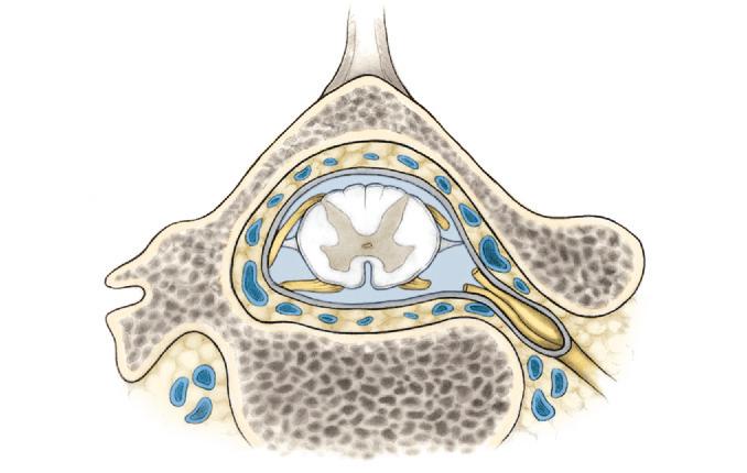
SPINAL MENINGES
The spinal cord is surrounded by three connective tissue membranes called the spinal meninges. From internal to external, the spinal meninges are called the pia mater, arachnoid, and dura mater (Fig. 2-2).
Pia Mater and Arachnoid
The pia mater completely surrounds and adheres to the spinal cord. The arachnoid loosely surrounds the spinal cord and is attached to the inner surface
Periosteum
Epidural space
Internal vertebral
venous plexus
Subarachnoid space
Dorsal root
Ventral root
Pia mater
Arachnoid
Dura mater
Denticulate ligament
Dorsal root ganglion
Spinal nerve
Intervertebral foramen
Figure 2-2 Relations of spinal meninges.
of the dura mater. The spinal cord is anchored to the dura by the denticulate ligaments and by the spinal nerve roots. The denticulate ligaments are 21 pairs of fibrous sheaths located at the sides of the spinal cord. Medially, the ligaments form a continuous longitudinal attachment to the pia mater. Laterally, they form triangular, toothlike processes that attach to the dura. Because of their pial attachments midway between the posterior and anterior surfaces of the spinal cord, the denticulate ligaments can be used as landmarks for surgical procedures. The spinal cord is also anchored by the roots of the spinal nerves, which are ensheathed by a cuff of dura where they perforate it near the intervertebral foramina.
Dura Mater
The spinal dura mater loosely surrounds the spinal cord. The area between the spinal dura and the periosteum lining the vertebral canal is the epidural
Spinal cord
space. Its contents include loose connective tissue, fat, and the internal vertebral venous plexus.
Clinical Connection
The internal vertebral venous plexus forms a valveless communication between the cranial dural sinuses, which collect blood from the veins of the brain, and the veins of the thoracic, abdominal, and pelvic cavities. It, therefore, provides a direct path for the spread of infections, emboli, or cancer cells from the viscera to the brain.
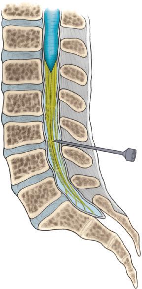
Inferior or caudal to the spinal cord, the dura mater forms the dural sac (Fig. 2-3), which extends inferiorly to the middle third of the second sacral vertebra. Caudal to this point, it surrounds the