No part of this publication may be reproduced or transmitted in any form or by any means, electronic or mechanical, including photocopying, recording, or any information storage and retrieval system, without permission in writing from the publisher. Details on how to seek permission, further information about the Publisher’s permissions policies and our arrangements with organizations such as the Copyright Clearance Center and the Copyright Licensing Agency, can be found at our website: www.elsevier.com/permissions
This book and the individual contributions contained in it are protected under copyright by the Publisher (other than as may be noted herein).
Notices
Knowledge and best practice in this field are constantly changing. As new research and experience broaden our understanding, changes in research methods, professional practices, or medical treatment may become necessary.
Practitioners and researchers must always rely on their own experience and knowledge in evaluating and using any information, methods, compounds, or experiments described herein. In using such information or methods they should be mindful of their own safety and the safety of others, including parties for whom they have a professional responsibility.
With respect to any drug or pharmaceutical products identified, readers are advised to check the most current information provided (i) on procedures featured or (ii) by the manufacturer of each product to be administered, to verify the recommended dose or formula, the method and duration of administration, and contraindications. It is the responsibility of practitioners, relying on their own experience and knowledge of their patients, to make diagnoses, to determine dosages and the best treatment for each individual patient, and to take all appropriate safety precautions.
To the fullest extent of the law, neither the Publisher nor the authors, contributors, or editors, assume any liability for any injury and/or damage to persons or property as a matter of products liability, negligence or otherwise, or from any use or operation of any methods, products, instructions, or ideas contained in the material herein.
Previous edition copyrighted 2007.
Library of Congress Cataloging-in-Publication Data
Names: Bishoff, Jay T., editor. | Kavoussi, Louis R., editor.
Title: Atlas of laparoscopic and robotic urologic surgery / [edited by] Jay T. Bishoff, Louis R. Kavoussi.
Other titles: Atlas of laparoscopic urologic surgery (Bishoff)
Description: Third edition. | Philadelphia, PA : Elsevier, [2017] | Preceded by: Atlas of laparoscopic urologic surgery / [edited by] Jay T. Bishoff, Louis R. Kavoussi. c2007. | Includes bibliographical references and index.
Identifiers: LCCN 2016030629 | ISBN 9780323393263 (hardcover : alk. paper)
Classification: LCC RD572 | NLM WJ 17 | DDC 617.4/60597—dc23 LC record available at https://lccn.loc.gov/2016030629
Senior Content Strategist: Charlotta Kryhl
Senior Content Development Specialist: Ann R. Anderson
Publishing Services Manager: Patricia Tannian
Senior Project Manager: Claire Kramer
Design Direction: Christian Bilbow
Printed in China.
Last digit is the print number: 9 8 7 6 5 4 3 2 1
This opus is dedicated to the courageous giants who took the risks that advanced our craft and created time to be incredible mentors and inspiration: Arthur Smith, Ralph Clayman, and Patrick Walsh.
Greensboro, North Carolina Continent Urinary Diversion
Sean McAdams, MD Department of Urology University of Minnesota Minneapolis, Minnesota Basic Instrumentation
Mani Menon, MD
The Raj and Padma Vattikuti Distinguished Chair Vattikuti Urology Institute
Henry Ford Hospital
Detroit, Michigan
Minimally Invasive Renal Recipient Surgery
Salvatore Micali, MD
Associate Professor of Urology Department of Urology
University of Modena and Reggio Emilia, Italy
Laparoscopic Denervation for Chronic Testicular Pain
Debora Moore, MD
Urology Clinic Site Director Charlotte VA Health Care Center Charlotte, North Carolina
Exiting the Abdomen and Closure Techniques
Robert Moore, MD
Urology Resident Site Director Salisbury VA Medical Center
Salisbury, North Carolina
Associate Professor of Urology
Wake Forest Baptist Health–Urology
Winston-Salem, North Carolina
Exiting the Abdomen and Closure Techniques
Monica S.C. Morgan, MD Department of Urology
Houston Methodist Hospital Houston, Texas
Laparoscopic and Robotic-Assisted Ureteral Reimplantation
Ravi Munver, MD, FACS
Vice Chairman
Chief of Minimally Invasive and Robotic Urologic Surgery
Department of Urology
Hackensack University Medical Center
Hackensack, New Jersey
Associate Professor of Surgery (Urology)
Department of Surgery
Division of Urology
Rutgers University–New Jersey Medical School
Newark, New Jersey
Laparoscopic and Robotic-Assisted Laparoscopic Pelvic Lymph Node Dissection
Stephen Y. Nakada, MD, FACS Professor and Chairman
The David T. Uehling Chair of Urology
Department of Urology
University of Wisconsin School of Medicine and Public Health
Professor and Chairman Department of Urology
University of Wisconsin Hospital and Clinics
Madison, Wisconsin
Stapling and Reconstruction
Yasser A. Noureldin, MD, MSc, PhD
Lecturer
Department of Urology
Benha University Hospital
Benha University
Benha, Egypt
Laparoscopic/Robotic Camera and Lens Systems
Michael C. Ost, MD
Associate Professor and Vice Chairman Department of Urology
University of Pittsburgh Medical Center
Chief of Division
Pediatric Urology
Children’s Hospital of Pittsburgh at the University of Pittsburgh Medical Center
Pittsburgh, Pennsylvania
Ureterolysis
Lane S. Palmer, MD
Professor and Chief
Pediatric Urology
Cohen Children’s Medical Center of New York
Hofstra Northwell School of Medicine
Hempstead, New York
Laparoscopic Orchiectomy
Jaspreet Singh Parihar, MD
Chief Resident
Department of Surgery
Division of Urology
Rutgers Robert Wood Johnson Medical School
New Brunswick, New Jersey
Insufflators and the Pneumoperitoneum
Jeffery E. Piacitelli, PA-C, MS
Robotics and Minimally Invasive Surgery–Urology
Intermountain Urological Institute
Intermountain Medical Center–Eccles
Outpatient Center
Murray, Utah
Considerations for the Assistant
Peter A. Pinto, MD
Head, Prostate Cancer Section
Fellowship Program Director
Urologic Oncology Branch
National Cancer Institute
National Institutes of Health
Bethesda, Maryland
Laparoscopic Partial Nephrectomy
Giacomo Maria Pirola, MD
Urology Resident
Department of Urology
University of Modena and Reggio
Emilia, Italy
Laparoscopic Denervation for Chronic Testicular Pain
James Porter, MD
Director, Robotic Surgery
Swedish Urology Group
Seattle, Washington
Laparoscopic and Robotic-Assisted Retroperitoneal Lymph Node Dissection
Aaron M. Potretzke, MD
Minimally Invasive/Robotic Surgery Fellow
Division of Urologic Surgery
Department of Surgery
Washington University School of Medicine
St. Louis, Missouri
Laparoscopic Pyeloplasty
Raj Pruthi, MD
Professor and Chair
Department of Urology
University of North Carolina
Chapel Hill, North Carolina
Robotic-Assisted Radical Cystectomy
Johar S. Raza, MD, MRCS, FCPS (urol)
Department of Urology
Roswell Park Cancer Institute
Buffalo, New York
Robotic-Assisted Intracorporeal Ileal Conduit
Jeremy N. Reese, MD, MPH, MEd Resident
Department of Urology
University of Pittsburgh Medical Center
Pittsburgh, Pennsylvania
Ureterolysis
Koon Ho Rha, MD, PhD, FACS
Professor
Department of Urology
Urological Science Institute
Yonsei University College of Medicine
Seoul, Republic of Korea
Laparoscopic/Robotic Boari Flap
Ureteral Reimplantation
Lee Richstone, MD
System Vice Chairman
The Arthur Smith Institute of Urology
Hofstra Northwell School of Medicine
Hempstead, New York
Chief
Department of Urology
The North Shore University Hospital Manhasset, New York
Robotic-Assisted and Laparoscopic Simple Prostatectomy
Bijan W. Salari, MD University of Toledo Medical Center Toledo, Ohio
Exiting the Abdomen and Closure Techniques
Jason M. Sandberg, MD
Department of Urology
Wake Forest School of Medicine and Baptist Hospital Winston-Salem, North Carolina
Continent Urinary Diversion
John Schomburg, MD
Department of Urology University of Minnesota Minneapolis, Minnesota
Basic Instrumentation
Michael J. Schwartz, MD
Associate Professor of Urology
The Arthur Smith Institute for Urology
Hofstra Northwell School of Medicine
Hempstead, New York
Laparoscopic Live Donor Nephrectomy
Casey A. Seideman, MD
Pediatric Urology Fellow
Cohen Children’s Medical Center of New York
Hofstra Northwell School of Medicine Hempstead, New York
Laparoscopic Orchiectomy
Paras H. Shah, MD
The Arthur Smith Institute for Urology Hofstra Northwell School of Medicine Hempstead, New York
Laparoscopic Live Donor Nephrectomy
Robotic-Assisted Laparoscopic Partial Cystectomy
Michael Siev, BA Research Fellow
The Arthur Smith Institute for Urology Hofstra Northwell School of Medicine
Hempstead, New York
Complications of Laparoscopic and Robotic-Assisted Surgery
Armine K. Smith, MD
Assistant Professor
Brady Urological Institute
Johns Hopkins University
Baltimore, Maryland
Assistant Professor
Department of Urology
George Washington University Washington, District of Columbia Nephroureterectomy
Akshay Sood, MD
Resident PGY-1
Vattikuti Urology Institute
Henry Ford Hospital Detroit, Michigan
Minimally Invasive Renal Recipient Surgery
Arun Srinivasan, MD
Attending Pediatric Urologist Division of Urology
The Children’s Hospital of Philadelphia Philadelphia, Pennsylvania
Laparoscopic Orchiopexy
Michael D. Stifelman, MD
Chairman Department of Urology
Hackensack University Medical Group
Hackensack, New Jersey
Buccal Mucosa Grafts for Ureteral Strictures
Necole M. Streeper, MD
Assistant Professor of Surgery Division of Urology
Penn State Milton S. Hershey Medical Center
Hershey, Pennsylvania
Stapling and Reconstruction
Li Ming Su, MD
David A. Cofrin Professor of Urology
Associate Chairman of Clinical Affairs Chief
Division of Robotic and Minimally Invasive Urologic Surgery
Department of Urology
University of Florida College of Medicine
Gainesville, Florida
Transperitoneal Technique Prostatectomy
Hassan G. Taan, MD
Clinical Instructor
Department of Urology
University of Pittsburgh Medical Center
Pittsburgh, Pennsylvania
Laparoscopic Simple Nephrectomy
Angelo Territo, MD
Urology Resident
Department of Urology
University of Modena and Reggio
Emilia, Italy
Laparoscopic Denervation for Chronic Testicular Pain
Manish A. Vira, MD
Assistant Professor of Urology
The Arthur Smith Institute for Urology
Hofstra Northwell School of Medicine
Hempstead, New York
Robotic-Assisted Laparoscopic Partial Cystectomy
Andrew A. Wagner, MD
Director of Minimally Invasive Urologic Surgery
Beth Israel Deaconess Medical Center
Assistant Professor of Surgery and Urology
Harvard Medical School
Boston, Massachusetts
Patient Preparation and Positioning for Laparoscopic and Robotic Urologic Surgery
Kyle J. Weld, MD
Director of Endourology
Wilford Hall Medical Center
Department of Urology
Lackland Air Force Base
San Antonio, Texas
Laparoscopic and Percutaneous Delivery of Renal Ablative Technology
Mary E. Westerman, MD
Department of Urology
Mayo Clinic Rochester, Minnesota
Laparoscopic Adrenalectomy
Michael Woods, MD
Associate Professor
Department of Urology
The University of North Carolina Chapel Hill, North Carolina
Robotic-Assisted Radical Cystectomy
Yuka Yamaguchi, MD
Division of Urology Department of Surgery
Alameda Health System
Oakland, California
Buccal Mucosa Graft for Ureteral Strictures
Akira Yamamoto, MD Resident of Urology Department Urology
University of Florida College of Medicine
Gainesville, Florida
Transperitoneal Radical Prostatectomy
Ramy Youssef, MD
Assistant Clinical Professor Department of Urology
University of California Irvine Orange, California
Laparoscopic and Percutaneous Delivery of Renal Ablative Technology
Contributors
Lee C. Zhao, MD, MS
Assistant Professor Department of Urology
New York University School of Medicine
New York, New York
Buccal Mucosa Graft for Ureteral Strictures
Philip T. Zhao, MD Endourology Fellow
The Arthur Smith Institute for Urology
Hofstra Northwell School of Medicine
Hempstead, New York
Robotic-Assisted and Laparoscopic Simple Prostatectomy
Matthew Ziegelmann, MD
Resident Physician Department of Urology
Mayo Clinic
Rochester, Minnesota
Laparoscopic Renal Cyst Decortication
Preface
Surgical technique is continuously evolving as physicians remain vigilant in their search for excellence. It has been 10 years since the last edition of this work. Much has changed in these years because of the collective efforts of those surgeons around the globe who are seeking ways to contribute to iterations that progressively make surgery safer, less invasive, and more successful. In addition, modern times have called for a focus on making surgical approaches cost effective. All these were the impetus for us to create a third edition of this text.
The role of minimally invasive surgery has continued to expand over the past decade. This text recognizes this reality through new and updated chapters. Indeed, most extirpative and reconstructive urologic procedures are now performed through keyhole incisions. Facilitating this trend has been the application of da Vinci surgical approaches to surgery. As such, specific sections and chapters have been added in recognition of this phenomenon.
This edition offers an expanded role for illustrative education. Teaching the art of surgery is so much more enhanced through visual lessons. The number of graphics has been
increased to help clarify the written word. Moreover, in this edition we have added David Leavitt as the video editor. His guidance has provided for an expanded library that allows enhanced understanding of the nuances of each surgical technique through detailed step-by-step instruction.
We are fortunate to have world experts contributing their experience as authors. This text has both well-recognized pioneers and recent innovators. They have painstakingly updated or added chapters that reflect the most up-to-date minimally invasive techniques to treat urologic disease. These authors selected key technical tips to help readers understand important nuances to successfully undertake described procedures.
Finally, we have to acknowledge the professional staff at Elsevier who truly helped convert our ideas into reality. Lotta Kryhl understood the importance of creating a third edition and demonstrated incredible leadership in helping with organization and crafting the proposal to upper management. Ann Ruzycka Anderson and Claire Kramer did a magnificent job in operationalizing the project and masterfully herding us editors and authors alike.
Section I Basic Techniques in Laparoscopic and Robotic Surgery
1 Patient Preparation and Positioning for Laparoscopic and Robotic Urologic Surgery, 1
Andrew A. Wagner, James S. Hwong
2 Laparoscopic/Robotic Camera and Lens Systems, 6
Yasser A. Noureldin, Sero Andonian
3 Basic Instrumentation, 18
John Schomburg, Sean McAdams, Kyle Anderson
4 Stapling and Reconstruction, 31
Stephen Y. Nakada, Necole M. Streeper
5 The da Vinci Surgical System, 39
Michael H. Johnson, Mohamad E. Allaf
6 Considerations for the Assistant, 43
Jeffery E. Piacitelli
7 Anesthetic Considerations for Laparoscopic and Robotic-Assisted Surgery, 47
Judith Aronsohn, Jin Jung
8 Insufflators and the Pneumoperitoneum, 54
Jaspreet Singh Parihar, Sammy E. Elsamra
9 Ports and Establishing Access into the Peritoneal Cavity, 58
Daoud Dajani, Mohamed A. Atalla
10 Retroperitoneal Access, 63
Peter A. Caputo, Jihad H. Kaouk
11 Exiting the Abdomen and Closure Techniques, 66
Bijan W. Salari, Debora Moore, Robert Moore
12 Complications of Laparoscopic and RoboticAssisted Surgery, 71
Michael Siev, Louis R. Kavoussi
Section II Lymphadenectomy
13 Laparoscopic and Robotic-Assisted Laparoscopic Pelvic Lymph Node Dissection, 81
Ravi Munver, Leonard Glickman
14 Laparoscopic and Robotic-Assisted Retroperitoneal Lymph Node Dissection, 89
Ashraf S. Haddad, James Porter
15 Endoscopic Subcutaneous Modified Inguinal Lymph Node Dissection for Squamous Cell Carcinoma of the Penis, 100
Jay T. Bishoff
Section III Renal Surgery
16 Laparoscopic Simple Nephrectomy, 105
Hassan G. Taan, Timothy D. Averch
17 Laparoscopic Radical Nephrectomy, 112
Aaron H. Lay, Jeffrey A. Cadeddu
18 Nephroureterectomy, 120
Armine K. Smith, Thomas W. Jarrett
19 Laparoscopic Partial Nephrectomy, 132
Sam J. Brancato, Steven F. Abboud, Peter A. Pinto
20 Laparoscopic Live Donor Nephrectomy, 143
Paras H. Shah, Michael J. Schwartz
21 Laparoscopic Renal Cyst Decortication, 152
Matthew Ziegelmann, Bohyun Kim, Matthew Gettman
22 Laparoscopic Renal Biopsy, 159
Jathin Bandari, Stephen V. Jackman
23 Laparoscopic and Percutaneous Delivery of Renal Ablative Technology, 167
Video 39-1 NOTES-Assisted Laparoscopic Transvesical Bladder Diverticulectomy
Ahmed Magdy, Günter Janetschek
Section VII Adrenal Surgery
40 Laparoscopic Adrenalectomy
Video 40-1 Laparoscopic Right Adrenalectomy
Paras H. Shah, Manaf Alom, David A. Leavitt, Louis R. Kavoussi
41 Partial Adrenalectomy
Video 41-1 Laparoscopic Partial Adrenalectomy
Daniela Colleselli, Ahmed Magdy, Günter Janetschek
Section VIII Testicular Surgery
42 Laparoscopic Orchiopexy
Video 42-1 Single-Stage Laparoscopic Orchiopexy
Arun Srinivasan, Mazyar Ghanaat
Video 42-2 Two-Stage Fowler-Stephens Laparoscopic Orchiopexy
Arun Srinivasan, Mazyar Ghanaat
44 Laparoscopic Varicocelectomy
Video 44-1 Laparoscopic Lymphatic-Sparing Varicocelectomy
Ronnie G. Fine, Haris S. Ahmed, David A. Leavitt, Israel Franco
45 Laparoscopic Denervation for Chronic Testicular Pain
Video 45-1 Laparoendoscopic Single-Site Spermatic Cord Denervation
Salvatore Micali, Giacomo Maria Pirola, Angelo Territo, Giampaolo Bianchi
Atlas of Laparoscopic and Robotic Urologic Surgery
SECTION I
Basic Techniques in Laparoscopic and Robotic Surgery
Patient Preparation and Positioning for Laparoscopic and Robotic Urologic Surgery
Andrew A. Wagner, James S. Hwong
“Before anything else, preparation is the key to success.”
Appropriate patient selection, thorough preparation, and careful patient positioning are essential in achieving a safe and successful outcome in laparoscopic surgery. No matter how prepared a surgeon may be for the technical exercise of laparoscopic surgery, inadequate execution of these important surgical preludes may result in unnecessary complications, extend operative time, and challenge the course of recovery. If the surgeon becomes entangled in a challenging situation, he or she must continuously evaluate for adequate progress to justify continuing laparoscopically versus converting to open surgery. Recognizing these situations during patient selection and proceeding with these cases with a healthy dose of surgical humility is fundamental to avoiding major complications and achieving a successful outcome.
PATIENT SELECTION
Preparation for laparoscopic surgery begins first and foremost with appropriate patient selection. The most experienced laparoscopic surgeons in the world are also experts at patient selection. Each case must be carefully considered prior to the patient reaching the operating room. Several aspects of surgery unique to laparoscopy must be considered before patient selection. The most significant of these include the altered physiology of pneumoperitoneum, the potential for prolonged procedure time during a team’s early learning curve, and the dangers of minimally invasive abdominal access.
Pneumoperitoneum of laparoscopy can significantly alter cardiopulmonary physiology, so an experienced anesthesia team is vitally important and should be involved in the preoperative planning of complicated cases. Several medical conditions are worthy of special mention and should prompt a careful review by both surgery and anesthesia teams. These include but are not limited to chronic obstructive pulmonary disease (COPD), restrictive lung disease, active cardiac disease, obesity, glaucoma, and cerebrovascular disease (Table 1-1).
Patients with pulmonary compromise present unique challenges during particularly long surgical cases. Insufflation of the peritoneum with CO2 can exacerbate hypercarbia in the COPD patient with a severe ventilation-perfusion mismatch. This hypercapnia (arterial CO2 >60 mm Hg) is cardiodepressive and can lead to acidosis and cardiac arrhythmias if left untreated. The typical treatment for hypercarbia is for the anesthesia team to increase ventilation rate, tidal volume, or both and for the surgical team to reduce intra-abdominal
Alexander Graham Bell
pressure (IAP). During surgery, the anesthesia team can easily monitor end-tidal CO2, which is proportional to arterial CO2. However, in patients with impaired pulmonary gas exchange (e.g., obstructive lung disease, low cardiac output, or pulmonary embolism), arterial CO2 can be significantly greater than end-tidal CO2. For these patients, regular measurement of arterial blood gas is recommended for more accurate monitoring. After laparoscopic surgery, patients with pulmonary compromise should be closely monitored for signs of hypercapnia.
Patients with cardiac disease are also at unique risk during laparoscopy. In particular, patients with cardiomyopathy, congestive heart failure, and ischemic heart disease require close monitoring as a result of the altered physiology of pneumoperitoneum. Increased intra-abdominal pressure from insufflation is exerted directly on the vasculature, decreasing venous return and preload, as well as systemic vascular resistance and afterload. These can be further exacerbated by decreased myocardial contractility induced by hypercapnia, ultimately leading to decreased stroke volume and cardiac output. Accordingly, careful fluid resuscitation by the anesthesiologist and attentive control of bleeding by the surgeon are warranted to prevent hypovolemia in these patients.
Other issues that warrant a thoughtful preoperative plan include obesity and central nervous system issues. Prolonged positioning for complex laparoscopy combined with an obese patient may increase the risk of rhabdomyolysis. If positioning is steep Trendelenburg (ST), increased intraocular pressure can lead to ischemic optic neuropathy and postoperative vision loss in the patient with glaucoma. Patients with cerebrovascular disease should be carefully selected because ST positioning can contribute to increased intracranial pressure. The astute urologist should not hesitate to seek specialty evaluation for any of these comorbidities before proceeding with surgery.
Patients with a previous history of abdominal surgery or peritonitis should be carefully considered for laparoscopy. These conditions can result in the formation of a significant amount of adhesions involving intra-abdominal viscera, presenting unique challenges and dangerous pitfalls for trocar placement. In general, for abdominal access, the surgeon should use the technique with which he or she has the most experience. Blind Veress needle placement for insufflation can be used away from the known surgical scars if the surgeon
TABLE 1-1 Comorbidities Exacerbated by Pneumoperitoneum and Robotic Surgery
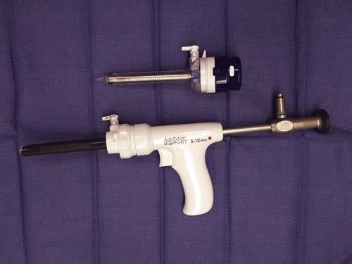
Figure 1-1. Trocars for initial port placement under direct vision. The Visiport Plus and Versaport (Covidien, Norwalk, Connecticut) trocars allow for placement of the initial trocar under direct vision. Both accommodate passage of a 0-degree laparoscope through the body of the trocars, allowing for visualization and identification of abdominal wall tissue through their clear tips during placement. A sharp crescent-shaped blade extends 1 mm through the tip of the Visiport Plus trocar (bottom) for sharp tissue dissection with each trigger pull. The sharpened tip of the Versaport trocar (top) dissects through the abdominal wall with a twisting motion and firm, steady pressure.
has experience with that technique. If not, then an open Hasson technique should be used for initial access. Regardless of insufflation method, no ports should ever be placed blindly, including the initial abdominal access port. Several varieties of “visual obturator” trocars are available and provide safer options for abdominal access (Fig. 1-1). Moreover, subsequent trocars should always be placed under direct vision after adhesions are cleared from the abdominal wall. Retroperitoneal or preperitoneal access can be considered in patients with a history of multiple complicated surgeries. Experience and additional training with these techniques are recommended. As stated previously, preoperative recognition of challenging situations such as a hostile abdomen is paramount in avoiding complications.
PREPARING THE PATIENT
Before surgery, all patients should be evaluated by the anesthesia team and obtain appropriate specialty clearance. Preoperative testing including electrocardiography, blood work, urinalysis, and cultures should be performed if appropriate. In addition, instructions for stopping anticoagulation
agents and antiplatelet agents should be conveyed to the patient. If an ostomy is planned, the patient should be evaluated by an ostomy nursing team, and potential ostomy sites should be marked bilaterally for placement. Preoperative ostomy education can be reviewed, and supplies such as ostomy pouches, thromboembolism-deterrent (TED) hose, and chlorhexidine body scrubs can be provided at this time. Prevention of surgical site infections begins preoperatively and includes skin treatment, bowel preparation when necessary, and antibiotic prophylaxis. On the evening before surgery, the patient should shower with a chlorhexidine body scrub and should refrain from waxing, shaving, or trimming the surgical site to prevent microtrauma to the skin. For the same reason, body hair should not be shaved with a blade but rather trimmed with mechanical clippers, which have been demonstrated to decrease the risk of surgical site infection. After the patient has been positioned, abdominal surgical sites should be sterilized with chlorhexidine, and genitalia with povidone-iodine solution.
If the bowel will be manipulated, mechanical bowel preparation with polyethylene glycol or sodium phosphate can be administered the evening before surgery. The constipated patient can be administered enemas or manually disimpacted. The rationale for mechanical bowel preparation includes reduction of fecal flora, easier manipulation of bowel, improved visualization, and easier anastomotic stapling. However, meta-analyses of colorectal surgery have not identified a clear statistical benefit to mechanical bowel preparation. Cochrane reviews were able to demonstrate trends toward decreased rates of anastomotic leakage with mechanical bowel preparation, although these did not reach statistical significance. Maneuvers for aggressive bowel preparation were further detracted by potentially morbid colonic mucosal changes, fluid shifts, and electrolyte derangements. Similar controversy exists surrounding administration of oral antibiotic bowel preparation (OABP) or selective decontamination of the digestive tract (SDD) with regimens such as tobramycin, polymyxin E, and amphotericin B. In general, parenteral antibiotic prophylaxis is used in lieu of these agents.
There is less controversy regarding parenteral antibiotic prophylaxis before incision. For laparoscopic procedures without entry into the digestive or urinary tract, the guidelines of the American Urological Association (AUA) recommend perioperative administration of a first-generation cephalosporin or clindamycin as an alternative in penicillin-allergic patients. If the urinary tract will be entered, a first- or second-generation cephalosporin or aztreonam with metronidazole or clindamycin is recommended. A fluoroquinolone or ampicillin-sulbactam is acceptable as an alternative regimen. For cases involving the intestine, AUA guidelines recommend a second- or thirdgeneration cephalosporin or aztreonam with metronidazole or clindamycin. Fluoroquinolones, ampicillin-sulbactam, ticarcillin and clavulanate potassium (Timentin), and piperacillin and tazobactam (Zosyn) can be used as alternative regimens. At our institution, a third-generation cephalosporin is combined with metronidazole for all cases involving bowel. All antibiotics should be administered 30 to 60 minutes before incision and should be continued for no more than 24 hours if there is no gross contamination during the procedure. Preoperative preparation should also include measures to prevent venous thromboembolism (VTE), a common cause of preventable death in surgical patients. The American College of Chest Physicians has developed evidence-based clinical guidelines for nonorthopedic surgical patients. Intermittent pneumatic compression (IPC) should be applied to all laparoscopy patients before induction of anesthesia. For patients at moderate and high risk for VTE without high risk of bleeding complications, subcutaneous heparin or
low-molecular-weight heparin (LMWH) should be administered. For high-risk cancer patients, extended-duration prophylaxis with LMWH for 4 weeks is recommended. Patients at high risk for bleeding complications can have pharmacologic prophylaxis withheld, although they should have mechanical prophylaxis with IPC preoperatively and pharmacologic prophylaxis should be initiated when the risk of bleeding diminishes. Pharmacologic prophylaxis should be administered 2 hours preoperatively, although LMWH appears to be effective 12 hours preoperatively.
PATIENT POSITIONING: BASIC CONSIDERATIONS
Prevention of positioning-related injuries should be of primary consideration when the anesthetized patient is manipulated. Pharmacologic paralysis required for laparoscopic surgery compounds the risk of injury as a result of decreased muscular tone and prolonged periods of immobility. These injuries can be broadly categorized into peripheral nerve injuries, vascularmediated injuries, and skin injuries, all of which can result in significant morbidity and mortality to the patient. Recognition of risk factors for positioning-related injuries and diligent prevention is key to avoiding these complications.
Injuries to peripheral nerves are a result of stretch or compression at susceptible nerve segments that can compromise neural blood supply, tear neural tissue, and disrupt axoplasmic flow. When the patient is positioned, care should be taken to ensure adequate padding at the elbow to avoid ulnar nerve compression at the cubital tunnel. If the arms are not tucked at the side, abduction at the shoulder should be limited to less than 90 degrees to prevent stretching of the brachial plexus over the humeral head. In the ST position, shoulder bracing should be avoided to prevent further loading of the brachial plexus. In the full-flank position, an axillary roll should be placed one handbreadth inferior to the axilla to support these important structures. When the patient’s lower extremities are positioned, close attention should be directed to the peroneal nerve, which can be compressed at the head of the fibula, and the median nerve, which can be injured at the medial tibial condyle.
Vascular-mediated injuries such as compartment syndrome and rhabdomyolysis are not unique to laparoscopic urology, but their risks may be exacerbated by insufflation, ST positioning, long operative times, and patient factors such as obesity. One possible contributing factor is ST positioning. With the legs elevated in the lithotomy position, perfusion pressure at the calf is reduced, which may increase the risk for compartment syndrome. Insufflation has also been theorized to contribute to decreased lower limb perfusion, and obesity may increase forces exerted at gluteal muscles, back muscles, and lower extremity supports. Long operative times (>4 to 5 hours) have also been associated with the development of rhabdomyolysis. Taken together, prevention of compartment syndrome and rhabdomyolysis should focus on limiting the degree of ST inversion and limiting operative time in morbidly obese patients.
The patient’s skin should be closely examined, and any preexisting lesions should be noted. Then all bony protuberances should be comfortably supported to distribute any forces that could lead to skin ischemia during a prolonged case. Similarly, any foreign bodies placed against the patient’s skin such as pulse oximeter connectors and intravenous access ports should also be padded. Gel pads, foam pads, egg crate foam, gauze, and towels can all serve in this capacity. For the patient’s skin to be protected from electrical burns, the electrocautery grounding pad should be well adhered across its entire surface. If necessary, body hair should be clipped to improve pad adherence. All patient jewelry should be removed, and the
1-2.
device for securing patients in steep Trendelenburg (ST) position. Safely securing a patient in ST position can be achieved with the TrenGuard system from D.A. Medical (Chagrin Falls, Ohio). This system uses a nuchal foam bolster secured to the operating table accessory rails, functioning like chocks for a wheel.
grounding pad should be placed as close to the operative field as possible to prevent alternate site burns.
PATIENT POSITIONING: LAPAROSCOPIC PELVIC SURGERY (SEE VIDEO 1-1)
Patient positioning for laparoscopic pelvic surgery has traditionally been the lithotomy position in ST. Although this allows the small bowel to fall away from the surgical site, affording increased working space and improving visualization, the position has numerous disadvantages. Chief among these are the risks to the patient as a result of the steep, inverted position, resulting in decreased perfusion pressure of the lower extremities and increased intracranial and intraocular pressures.
Keeping the patient safely secured to the operating table and preventing an intraoperative fall is also a major consideration. A number of devices and materials have been developed specifically for this application. Examples include vacuum bean bag immobilizers, high-friction gel or foam pads, and restraint systems such as the TrenGuard cervical bump (D.A. Surgical, Chagrin Falls, Ohio) (Fig. 1-2). In addition to these restraint methods, taping is often needed for extra support. Before skin preparation and draping, a full tilt test should be performed with the table in maximum Trendelenburg position to ensure the patient does not shift or slide. Familiarity with the patient securement system of choice is absolutely necessary to prevent slipping or falling.
In the lithotomy position, the legs should be well supported with heels firmly planted in surgical stirrups. Flexion at the hip and knees should be less than 90 degrees, and the lower leg should be pointed in line with the contralateral shoulder in the sagittal plane. The stirrups should not exert excessive pressure at the popliteal fossa, which could lead to compromise of popliteal vasculature. The stirrups should also be well padded at the fibular head to avoid peroneal nerve compression injury. “Candy cane” stirrups and knee crutches should not be used because these cannot safely position the legs for long robotic procedures. In ST position, the stirrups should be positioned as low as possible to prevent lower leg ischemia.
The challenges of ST positioning can be mitigated with some minor modifications and experience. At our institution,
Figure
TrenGuard
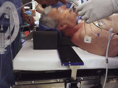
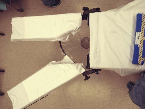
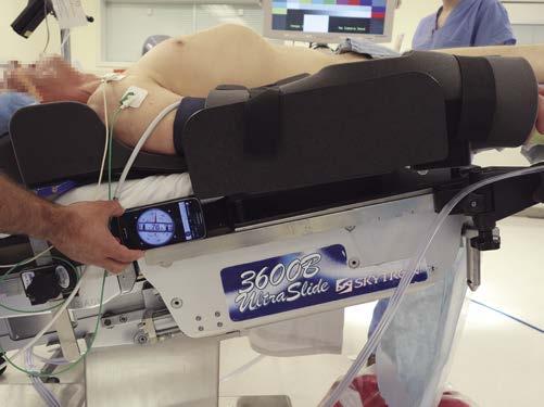
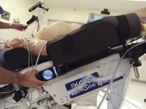
we use a split leg table during robotic-assisted laparoscopic prostatectomy. This avoids the risks of lithotomy positioning by keeping the legs straight on rotating bed attachments (Fig. 1-3). Moreover, we use a minimal Trendelenburg (MT) position—that is, just enough Trendelenburg inversion for the small bowel to fall out of the pelvis. Usually only 10 to 20 degrees of inversion is necessary (Fig. 1-4). In our experience, MT positioning is still a sufficient amount of inversion to clear the operative field while minimizing the deleterious physiologic effects of the ST position. Our method also requires less elaborate means of patient securement, decreasing time for operating room positioning and saving total operating room time.
PATIENT POSITIONING: LAPAROSCOPIC UPPER TRACT SURGERY (SEE VIDEO 1-2)
Minimally invasive kidney and adrenal surgery can be performed via a laparoscopic (transperitoneal) or retroperitoneoscopic approach. Either is acceptable, and the decision regarding approach should be based on surgeon training and
experience. There are no prospective perioperative or postoperative outcome data supporting one approach or the other. Of course, many other factors are important in determining surgical approach and should be carefully considered, including tumor size and location, potential for intra-abdominal adhesions, and patient body habitus.
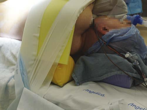
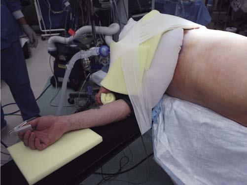
We use a modified lateral approach for all kidney and adrenal laparoscopic and robotic-assisted surgery (Fig. 1-5). This consists of the patient in a semisupine position, rotated laterally approximately 30 degrees. Rolled blankets or large gel rolls are used to support the patient’s back in this position by placing them behind the patient from the shoulder to buttocks. In contrast to lateral positioning for open retroperitoneal surgery, jackknife (flexed) positioning and the kidney rest are not necessary and can potentially reduce the actual laparoscopic working space.
Towels, pillows, or foam donuts are used to support the head and cervical spine in neutral position. The patient’s lower arm should be extended and supported on an arm board where it can be accessed as needed by the anesthesia team. The upper arm is slightly flexed and is supported with one folded pillow over the chest. Arm extension should be limited to 90 degrees or less to prevent a brachial plexus stretch injury. Foam-padded tape is used to secure the patient to the table, encircling the arms and securing the upper body and arms to the table. Pillows should be placed between the legs to keep the spine aligned. The dependent leg should be flexed at the hip and knee. The contralateral leg should remain extended with slight flexion at the knee and supported along its length with pillows. All bony protuberances such as the greater trochanter, head of the fibula, and lateral malleolus should be adequately padded with foam pads, gel pads, or egg crate foam.
Once appropriately positioned, the patient should be secured to the operating table and the table rotated a moderate amount to either side to ensure the body will not shift intraoperatively. If necessary during the case, the patient can still be rotated into a full flank position with movement of the operating table without undue stress on pressure points.
For retroperitoneoscopic surgery, the patient is typically placed in the full lateral flank position with the surgical site further rotated upward. Most surgeons choose to flex the table after positioning for retroperitoneoscopic surgery. With full flank position, an axillary roll should be positioned three fingerbreadths inferior to the axilla to reduce pressure on the axillary neurovasculature. The arms and legs can be secured in
Figure 1-3. Split table mechanism. Use of a split leg operating table instead of lithotomy fins simplifies patient positioning for laparoscopic pelvic surgery while mitigating the risk of injuries from prolonged positioning in the lithotomy position.
Figure 1-4. Minimal Trendelenburg positioning for laparoscopic pelvic surgery. A, Only 10 to 20 degrees of Trendelenburg are necessary to allow the small bowel to fall away from the pelvis. B, Steep Trendelenburg positioning confers additional risks while not significantly improving visualization.
ABFigure 1-5. Modified lateral positioning for laparoscopic surgery of the upper tract. A, In the modified lateral position, the patient is rotated approximately 30 degrees with the surgical target elevated. The body is supported with gel rolls or rolled towels. B, The dependent arm is extended and supported on an arm board; the contralateral arm is extended and supported with a folded pillow. The dependent leg is flexed at the hip and knee, and the contralateral leg is supported along its length with a slight bend at the knee. A generous amount of foam-padded tape is then used to secure the patient to the table.
the same manner as described earlier for the modified flank position.
CONCLUSION
Preoperative preparation for laparoscopic and robotic surgery remains of vital importance because it will set the stage for a safe and effective surgery. Careful consultation with the anesthesia team and specialists in pulmonology and cardiology
when appropriate remains crucial. The operating surgeon should understand physiologic changes associated with insufflation under these conditions. The surgeon should be present and guide preoperative positioning before laparoscopic and robotic cases. Early in one’s learning curve, positional injuries can be more common as a result of long operative times. With experience, pelvic surgery can be performed without ST positioning, and upper tract surgery can be performed using a modified lateral position.
Laparoscopic/Robotic Camera and Lens Systems
Yasser A. Noureldin, Sero Andonian
It has been said that exposure is key for open surgery. Similarly, the imaging platform used in endoscopic surgery, whether it is laparoscopic or robotic-assisted laparoscopic surgery, is a key for success. In this chapter, the history of laparoscope and imaging systems is reviewed. In addition, the difference between analog and digital image processing is explained. Three-dimensional imaging systems in addition to the da Vinci robotic system (Intuitive Surgical, Sunnyvale, Calif.) are described. Furthermore, advances in different scopes and cameras including high-definition (HD) and augmented reality (AR) imaging systems will be explained.
HISTORY OF THE LAPAROSCOPE
Surgical scopes are among the oldest surgical instruments. The first illuminated scope, dubbed the Lichtleiter or “Light Conductor,” consisted of a viewing tube, candle, and series of mirrors and was developed by Philipp Bozzini in 1804.1 Because of its impracticality, the device did not find favor among the surgeons of the day. However, it served as a source of inspiration to other inventors. Antonin Jean Desormeaux was the first urologist to view inside the bladder, in 1855.2 Using the principles of incandescent lighting, in 1867 Julius Bruck designed the first scope illuminated with an electrical light source. He used a platinum wire loop heated with electricity until it glowed. The main drawback to this design was the amount of heat generated by the light source, which could be conducted along the metal tubing of the scope to the tip. This heat represented a significant risk of burns to both the patient and the surgeon.3 In 1877, Maximilian Nitze used a lens system to widen the field of view (FOV) and succeeded in creating
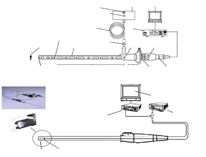
the first cystoscope as an instrument to visualize the urinary bladder through the urethra.4 The modern fiberoptic endoscope was invented by the British physicist Harold Hopkins in 1954.5 Hopkins used the term fiberscope to describe the bundle of glass or other transparent fibers used to transmit an image. The main advantage of the fiberscope was that the illumination source could be kept away from the scope with significant reduction in the amount of heat transmitted to the scope tip. However, the resolution of the fiberscope was limited by the number of fibers used. Therefore in the 1960s Hopkins invented the rod-lens system, which he patented in 1977.6 The rod-lens system used glass rods in place of air gaps, removing the need for lenses altogether, with resultant clarity and brightness that was up to 80 times greater than what was offered at the time (Fig. 2-1, top).6 The rod-lens system remains the standard for currently used rigid endoscopes when high image resolution is required.7 Over time, with advances in fiberoptics and magnifying lenses, sophisticated surgical scopes evolved. In the next two sections, developments in scopes and cameras are detailed.
SCOPES AND TECHNOLOGY
Since the 1960s, the classic laparoscope has been composed of an outer ring of fiberoptics used to transmit light into the body, and an inner core of rod lenses through which the illuminated visual scene is relayed back to the eye piece (Fig. 2-1, top).5 The different types of laparoscopes are defined in terms of the number of rods, size of laparoscope, and angle of view. With regard to size, laparoscopes are available in the range of 1.9 mm to 12 mm, but 5 mm is the most common size for
Figure 2-1. Top, Traditional rod-lens technology of Hopkins. Bottom, Videoscope technology. CCD, chargecoupled device. (Courtesy Olympus America, Melville, NY.