1. Vertebrate Central Nervous System Development
1.1 Introductory Remarks: The Order of the Universe
The individual universe each human being mentally carries around is a product of our brain, which most likely represents the most complex object in the world. How does the brain come into existence during development? The ontogeny of the vertebrate central nervous system (CNS: brain and spinal cord) itself is a highly complex yet strongly conserved morphogenetic process. The early vertebrate embryo exhibits three embryonic cellular sheets called germ layers, the most peripheral one being the ectoderm. Its dorsal portion represents the neuroectoderm that develops into the prospective nervous system. The neuroectoderm segregates early from the ventrolaterally lying general ectoderm, which forms the future epidermal skin and its derivatives. Subsequently, the central main portion of the neuroectoderm, the neural plate, is “swallowed” by the general ectoderm through the process of neurulation. As a result, the neural plate separates from the general ectoderm and descends into the deep of the embryo, where it develops into a hollow neural tube surrounding a cerebrospinal fluid- (liquor-) filled ventricle. Meanwhile, the general ectoderm closes the temporarily opened dorsal backside of the embryo. The apparent formation along the anteroposterior axis of initially three brain vesicles (forebrain, midbrain, hindbrain) that transform subsequently into five brain vesicles (telencephalon, diencephalon, mesencephalon, metencephalon, myelencephalon) somewhat hides the parallel establishment of finer transverse subdivisions called neuromeres (rhombomeres in the hindbrain, prosomeres in the forebrain, see later). What follows is the differentiation of functional subdivisions of the brain and spinal cord (for example, the optic tectum/ superior colliculus, a midbrain sensorimotor integration center, or the hypothalamus, the major control center of visceral processes). These events of vertebrate CNS formation are accompanied by the emigration of the most lateral portions of the neuroectoderm, the neural folds (Neuralwülste) and some directly adjacent neuroectodermal regions, which are not incorporated into the neural tube during neurulation. These neuroectodermal components comprise the neural crest of the head and trunk and the placodes that are located in the head only. Eventually neural crest and placodes give rise to cells that migrate to their final destination in the adult body, where they form almost all sensory organs and the sensory neurons in dorsal root and cranial nerve ganglia,
1. Vertebrate Central Nervous System
which innervate them, and the visceral ganglia of the autonomic nervous system. These somatic and visceral ganglia and their associated nerves represent the peripheral nervous system (PNS).
For many decades, scientists have investigated these complex developmental events leading to the formation of a nervous system in various vertebrates, especially in model animals, such as in the African clawed frog Xenopus, the chick, rodents like the mouse and rat, and most recently, the zebrafish, at molecular, cellular, histological, or morphological levels. Thus, a wealth of scientific literature is available, ready to satisfy almost any hunger for information.
Why then the present book? First of all, it integrates knowledge on CNS development coming from classical developmental studies with recent molecular data related to neuro- and gliogenesis. In particular, we will demonstrate the detailed spatiotemporal order of early postembryonic zebrafish brain development using molecular and cellular markers of neuro- and gliogenesis to visualize local differences, embedded in a holistic morphogenetic context. By combining old and new knowledge, we provide a neuroanatomically based molecular atlas of neural development in the zebrafish brain. We hope that such information—beyond delivering a refined molecular neuroanatomy—will prove to be enlightening in the future elucidation of neurogenetic pathways and their mechanisms.
1.2 Major Developmental Stages of the Vertebrate Neural Tube
Historically, scientists have always aimed to identify the fundamental building blocks of the vertebrate brain. In other words, they have tried to determine the basic developmental brain units and to answer the question of how these transform into functional structures of the mature brain. Before going into a more detailed discourse of the development of finer brain subdivisions (see Section 1.3), we will shortly examine the early events of brain morphogenesis relating to the emergence of vesicles and neuromeres, as well as of longitudinal zones, followed by an account on the neuromeric (prosomeric) model and on central nervous neurogenesis. An excellent in-depth historical account on the understanding of vertebrate brain morphogenesis has been given by Nieuwenhuys (1998a,b,c).
1.2.1 Vesicles, Neuromeres, and Longitudinal Zones
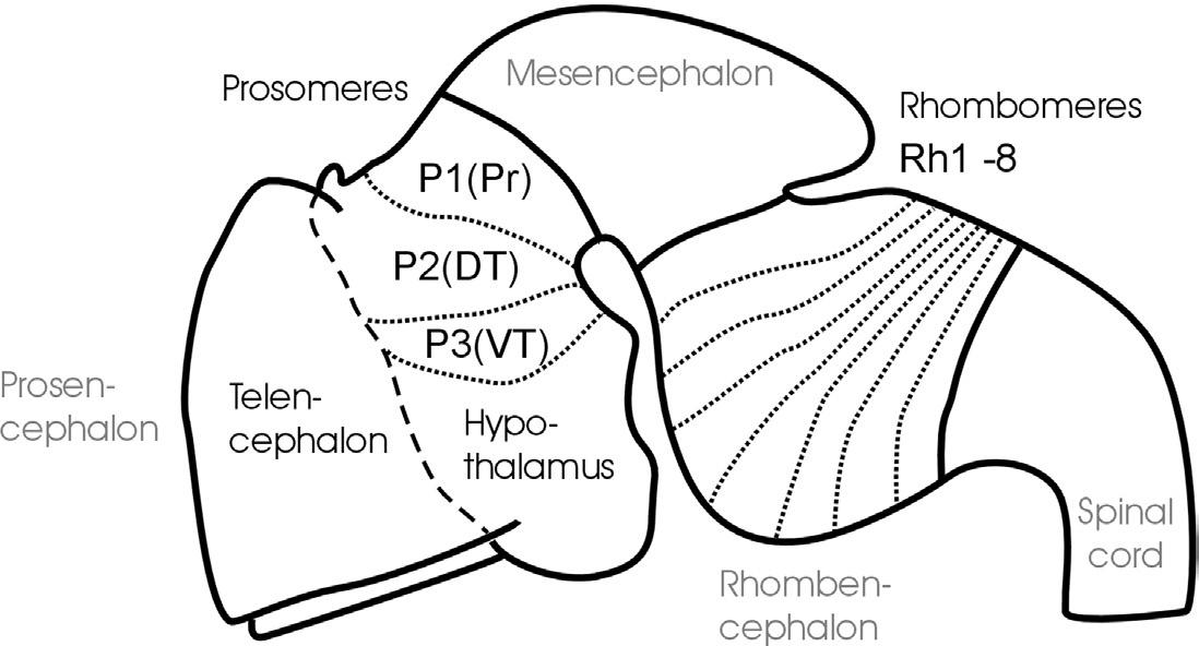
The classical description of a sequential appearance of two, three, and then five brain vesicles (i.e., transverse elements) along the anteroposterior vertebrate neural tube axis dates back to von Baer (1828). This view, however, gained popularity in the early twentieth century with the works of von Kupffer (1906) and Johnston (1909). The two-vesicle stage comprises a combined forebrain (prosencephalon) and midbrain (mesencephalon) vesicle, which is set apart by a vertical neural tube constriction from the hindbrain (rhombencephalon) vesicle. This is followed by the three-vesicle stage displaying forebrain, midbrain, and hindbrain vesicles (see Fig. 1) and, finally, by the five-vesicle stage, consisting of telencephalon and diencephalon (together forming the forebrain), mesencephalon, as well as metencephalon and myelencephalon (together forming the rhombencephalon).
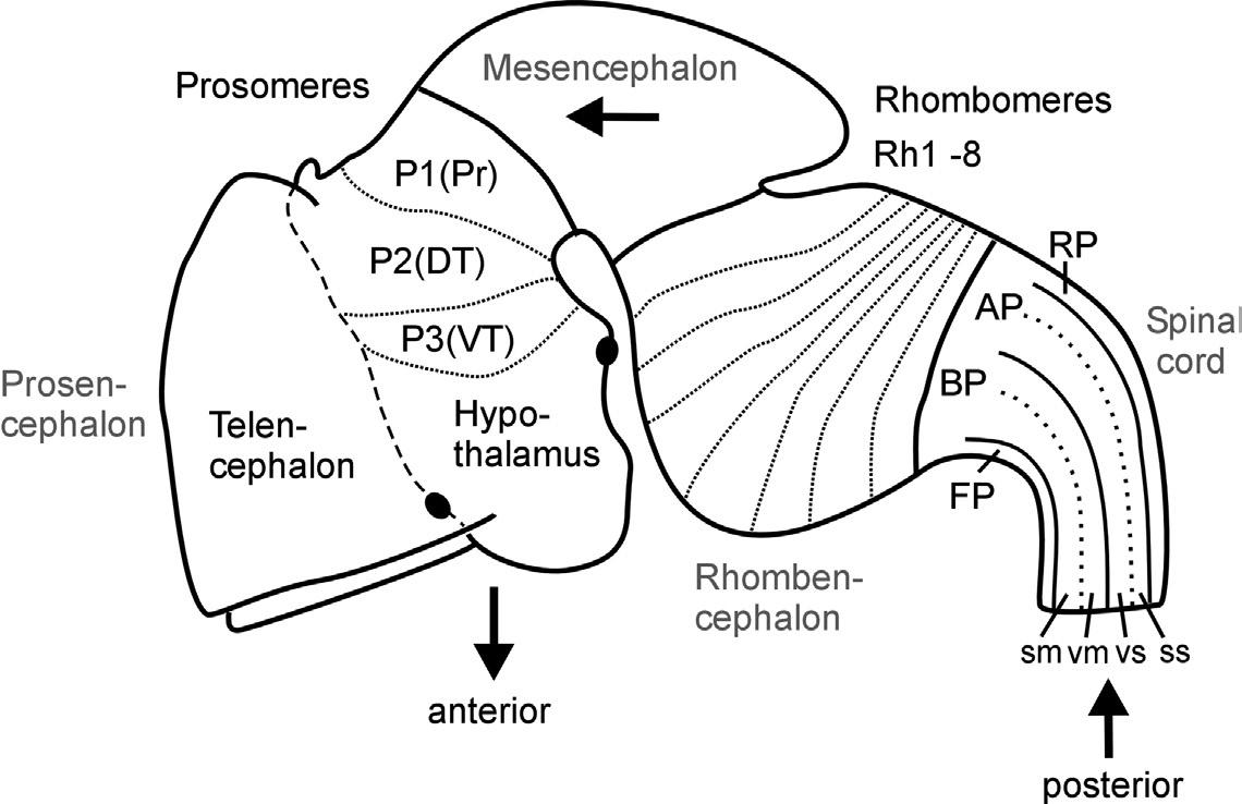
Fig. 1. Schematic lateral view of early mouse brain (E 12.5-13.5) shows prosomeric interpretation (Puelles and Rubenstein, 1993, 2003), including transverse (neuromeres) and longitudinal (columns) elements. Arrows designate anteroposterior axis of neural tube. Abbreviations: AP: alar plate; BP: basal plate; DT: dorsal thalamus (thalamus), FP: floor plate; P1-P3: prosomeres 1-3 (for more details see text); Pr: pretectum; Rh1-Rh8: rhombomeres 1-8; RP: roof plate; sm: somatomotor column; ss: somaotosensory column; T: telencephalon; vm: visceromotor column; vs: viscerosensory column; VT: ventral thalamus (prethalamus).
The subdivision of both forebrain and hindbrain into two separable (transverse) vesicles, each along the anteroposterior axis, arguably is an epiphenomenon, visible only in mammalian embryos that show an enormous early growth of the telencephalic hemispheres. This hypertrophy detracts from the fact that the
most rostral vertebrate neural tube in reality includes in addition to the dorsally lying telencephalon also ventrally the smaller hypothalamus. This renders the concept of transverse telencephalic and diencephalic vesicles obsolete because part of the diencephalon (the hypothalamus) is included in a transverse unit with the telencephalon (Puelles and Rubenstein, 1993, 2003). Also, the somewhat later, characteristic mammalian emergence of a large cerebellum and ventral pontine region led to the impression that the medulla oblongata develops from two separable vesicles, the metencephalic one exhibiting cerebellum and pons, and a myelencephalic vesicle posteriorly. Indeed, the dorsal portion of the anteriormost rhombomere specializes into a dorsal cerebellum. In contrast, the ventral pontine region, which relays cortical information to the cerebellum, develops from ventral and fused portions of rhombomeres 1-4 (Aroca and Puelles, 2005; Alonso et al., 2012). This complex development of the anterior hindbrain historically resulted in the misinterpretation that the two enlargements of cerebellum and pons form a true transverse unit, which was called the metencephalon to separate it from the posterior myelencephalon. However, the medulla oblongata does not show a real boundary between these two postulated vesicles; instead, it is rather divided into a higher number of early segmental entities, the rhombomeres (see later discussion and Fig. 1).
In contrast, the three-vesicle stage reflects on fundamental anteroposterior vertebrate brain divisions. Especially the midbrain-hindbrain boundary has been strongly corroborated in modern developmental neurobiology as a singularly definable boundary and signaling center in the vertebrate brain (Marín and Puelles, 1994; Bally-Cuif and Wassef, 1995; Brand et al., 1996; Lumsden and Krumlauf, 1996; Reifers et al., 1998; Wurst and Bally-Cuif, 2001). Similarly, there is clear evidence for cellular lineage restriction (Larsen et al., 2001) and signaling center function (Scholpp and Brand, 2003) at the forebrain-midbrain boundary.
Equally important for the understanding of the vertebrate CNS bauplan are two neural tube flexures, which were also historically noted early (His, 1888, 1893a). The cephalic flexure of the anteroposterior axis is between the mesencephalon and the prosencephalon, and the cervical flexure is between the rhombencephalon and the spinal cord (compare Fig. 1). The result of this bending of the neuraxis is that the forebrain floor is adjacent to the hindbrain floor. The correct recognition of the adequate course of the anteroposterior axis of the neural tube has important consequences for the interpretation of the topology of brain structures.
Whereas the vesicle story just outlined found its way easily into general textbook knowledge, the historical equally early description of neural tube segments or so-called neuromeres did not. Neuromeres represent a finer set of transitory transverse or segmental divisions of the vertebrate neural tube, and they remain, at least partially, controversial to the present day. Vertebrate hindbrain neuromeres (rhombomeres) are set apart from each other by transverse, vertical constrictions and were already described in the nineteenth century (von Baer, 1828; Orr, 1887). Later, similar morphological observations led to the interpretation of neuromeres to extend into midbrain (mesomeres) and forebrain as well (prosomeres; see Fig. 1; Rendahl, 1924; Bergquist, 1932; Vaage, 1969). However, in these early twentieth-century studies, the basis for recognizing transverse neuromeres was descriptive (and, even worse, transitory) morphology. Thus, serious opposition regarding the acceptance of neuromeres as the important building blocks of the CNS arose with its growing functional neuroanatomical understanding, especially by the so-called American school, which was based on the early work of Gaskell (1889) and later widely spread through the activities of Charles Judson Herrick (1868-1960), who is viewed as one of the founding fathers of modern comparative neurology. In this school's well corroborated view, longitudinal functional zones—embryonically represented dorsoventrally in the neural tube as roof, alar, basal, and floor plates (His, 1888, 1893a,b)—were considered the primary subdivisions of the CNS (see Fig. 1). Both the most dorsal (roof plate) and most ventral (floor plate) longitudinal zones exert critical developmental roles, for example, in mediating dorsoventral polarity of the neural tube or in guiding neuronal differentiation processes. In the adult vertebrate CNS, the roof plate gives rise to dorsal midline structures, such as the epiphysis or the choroid plexus, which covers the rhomboid opening of the medulla oblongata, whereas the floor plate develops, for example, into ventral midline glial cells.
The two intermediate longitudinal zones termed the alar and basal plates form pivotal elements of Herrick's concept of functional vertebrate brain and spinal cord organization. Alar and basal plate derivatives form the characteristic dorsoventral arrangement of four functional longitudinal subzones, that is, alar plate derived somato- and viscerosensory columns and basal plate derived viscero- and somatomotor columns as prototypically present in the spinal cord (see Fig. 1). This dorsoventral spinal cord organization of four sensory and motor zones has been used to consistently explain various local specializations (hypertrophies and reductions) in more anterior brain regions—at least into the midbrain. Thus, this
concept of longitudinal functional zones understandably gained much acceptance, and it is still discussed in recent textbooks.
A related question is how far anteriorly the vertebrate brain longitudinal zones extend (see Nieuwenhuys, 1998a). Considering alar and basal plates, there were two factions. Kingsbury (1922) believed that the basal plate tapers out at the mesencephalic-diencephalic boundary and that the alar plate alone forms the more anterior brain parts. Similarly, Herrick (1910) had proposed that the basal plate becomes thinner anteriorly and yet would only continue into the hypothalamus. Here, Herrick's original sin comes in: He believed that the more dorsal diencephalic divisions (ventral thalamus, dorsal thalamus, and epithalamus) all represent longitudinal zones (functionally elaborated alar plate subdivisions that he even saw to continue into the telencephalon), which turned out to be a false concept with long-lasting consequences (see later discussion). Kuhlenbeck (publications between 1926 and 1973; see Nieuwenhuys, 1998a) was supportive of Herrick's longitudinal zone concept (including the diencephalon), but highlighted its bauplan nature, disregarding the functional aspect. In contrast, Bergquist and Källén (1954)—similar to His (1888, 1893a,b)—proposed the continuation of both alar and basal plates into the general region of the optic chiasm/preoptic region. Thus, in line with their neuromeric concept, the forebrain prosomeres contained alar and basal plate derivatives. This has the important consequence that dorsal and ventral thalamus (and pretectum, for that matter) represent transverse—not longitudinal—elements along the anteroposterior neural tube axis, which is a now widely accepted concept (see Fig. 1 and following discussion).
Starting in the 1980s, vertebrate brain neuromery has been reconsidered and confirmed. However, a deep schisma exists between today's “segmentalists.”
Mostly dealing with rhombomeres (Hannemann et al., 1988; Holland and Hogan, 1988; Keynes and Stern, 1988; Lumsden and Keynes, 1989; Murphy et al., 1989; Lumsden, 1990; Trevarrow et al., 1990; Wilkinson and Krumlauf, 1990; Clarke and Lumsden, 1993; Lumsden and Krumlauf, 1996), many recent researchers stress that rigorous criteria for real transverse (neuromeric) neural tube elements (i.e., containing a piece of roof, alar, basal, and floor plates each) are exclusively met by rhombomeres. Among those criteria are glial intersegmental boundaries, repetitive (metameric) segmental generation of comparable neuronal classes and axonal routing in each neuromere, and most important, neuromere-specific cellular lineage restriction (Fraser et al., 1990). These criteria led to the most stringent definition available of a segment or neuromere as constituting a neural compartment
(Larsen et al., 2001). Strong complementary supporting evidence from molecular genetic studies showed that various regulatory genes, especially those of the Hox B cluster (Graham et al., 1989; Hunt and Krumlauf, 1992) or other genes, such as Krox20 (Wilkinson et al., 1989a,b; Oxtoby and Jowett, 1993) are hierarchically or specifically expressed respecting rhombomere boundaries, thereby confining positional information to the rhombomeres. Rhombomeric development finally leads to the different adult phenotypical identities of their respective derivatives along the anteroposterior hindbrain axis.
Also forebrain neuromeres have been rediscovered in vertebrates recently, and various numbers of such prosomeres were proposed based on new data (Puelles et al., 1987; Figdor and Stern, 1993; Puelles and Rubenstein, 1993, 2003). The most prominent contemporary, the so-called neuromeric or prosomeric model by Puelles and Rubenstein (1993, 2003) suggests an overall segmentation of the vertebrate brain and we will discuss this model in some more detail later. Generally, it may be stated that prosomeres (compare with Fig. 1) have not been corroborated equally well using the criteria applied to rhombomeres. However, cellular lineage restriction occurs at the forebrain-midbrain boundary, specifically between pretectum (synencephalon; P1) and mesencephalon (Larsen et al., 2001), and between dorsal and ventral thalamus mediated by the zona limitans intrathalamica (Zeltser et al., 2001). The zona limitans intrathalamica represents a transitory transitional region between dorsal and ventral thalamus, thereby separating and characterizing P2 (posterior parencephalon) and P3 (anterior parencephalon or prethalamus) as neuromeres. The diencephalon has alternatively been reported to contain four neuromeres (Figdor and Stern, 1993) characterized by lineage restriction, that is, two for the synencephalon (P1), and one each for the dorsal (P2) and ventral thalami (P3). Additional potential prosomere boundaries, especially those anterior to P3 (i.e., in the secondary prosencephalon) remain largely unexplored in the context of cellular lineage restriction.
Furthermore, as in the rhombencephalon, some early active regulatory genes are expressed in specific prosomeres (e.g., Prox in P1, Gbx2 in P2, Dlx2 in P3; Larsen et al., 2001) or respect at least partial prosomeric boundaries with their expression domains (Simeone et al., 1992a; Boncinelli et al., 1993, 1995; Bulfone et al., 1993a,b; Puelles and Rubenstein, 1993, 2003). Our own studies regarding the distribution of early proliferation zones support the presence of three posterior prosomeres, that is, P1 through P3 (Wullimann and Puelles, 1999; Mueller and Wullimann, 2002b), whereas no prosomeres are recognized based on proliferation
1. Vertebrate Central Nervous System Development
patterns in the more anterior forebrain (secondary prosencephalon). Also, neurogenic (Notch/Delta) and proneural (basic helix-loop-helix) gene expression closely parallels brain proliferation patterns, including the prosomeric pattern (P1/P2/P3) in the posterior forebrain (Mueller and Wullimann, 2003; Wullimann and Mueller, 2004a,b; see later).
In conclusion, there is strong evidence for three posterior prosomeres (P1 through P3) in the vertebrate forebrain, with the situation in the more anterior forebrain (secondary prosencephalon) being more elusive. Clearly, rhombomeres match better the criteria for neuromeres/compartments when compared to prosomeres. However, one must keep in mind that these criteria are only met transitionally during neuromere development and that the most critical criterion, that is, cell lineage restriction, is at no time completely met even in rhombomeres; there are reports on more than 5% of cells transgressing rhombomere boundaries (Birgbauer and Fraser, 1994). Furthermore, overt metameric organization (i.e., similar sets of anteroposteriorly repeated classes of neurons) as seen in the developing hindbrain does not necessarily require segmental neural tube organization during development. Metameric organization, as partially still present in the adult rhombencephalon, is even more pervasive in the spinal cord, which does not exhibit neuromeres (as defined earlier) during any period of development (Keynes and Stern, 1984). Therefore, metamery is apparently not necessarily strictly developmentally correlated with a neuromeric organization. Thus, the forebrain simply may be characterized by very early individual regulation of each of its transverse entities, resulting in a fast variable differentiation of neuromeres and their derivatives.
1.2.2 The Neuromeric or Prosomeric Model
Irrespective of the ongoing dispute about the identification and finite numbers of vertebrate neuromeres, Puelles and Rubenstein's neuromeric or prosomeric model (1993, 2003) integrates and puts into perspective much of the issues raised earlier, suggesting a vertebrate brain bauplan that has been of great heuristic value in many studies since. The correct topological identification of a particular brain structure, and, therefore, its comparative interpretation, critically depends on the concept of the basic vertebrate CNS bauplan one uses. Such a bauplan involves more than the recognition of transverse elements, it also requires to adequately indicate the course of the anteroposterior axis along the neural tube
and, in consequence, of the longitudinal zones. Following the neuromeric model, all central nervous longitudinal zones (floor, basal, alar, and roof plates) principally extend into the forebrain: The roof plate ends in the region of the anterior commissure, the floor plate at the anterior end of the mammillary hypothalamus (see two respective black dots in Fig. 1), whereas the basal and alar plate form a broad anterior end of the neural tube with the preoptic region (alar) and hypothalamus (basal), both united at the area of the optic chiasma (Puelles et al., 2013). The entire telencephalic hemispheres are thus lateral extensions of the alar plate. This is supported by studies on various longitudinally expressed genes (such as, for example, sonic hedgehog), which continue anteriorly to be expressed into the forebrain (Ericson et al., 1995; Shimamura et al., 1995, 1997; Hauptmann and Gerster, 2000). Such data clearly corroborate the old observation noted earlier that the anteroposterior axis of the rostral vertebrate neural tube is considerably deflected ventrally (compare with Fig. 1). Understanding the course of longitudinal zones is critical for the interpretation of transverse neural tube units (neuromeres), as the latter must lie perpendicular to the anteroposterior axis. In the neuromeric model of Puelles and Rubenstein (1993), the entire brain is principally divided into transverse zones (each containing a segment of all longitudinal zones), and neuromeres therefore exist not only in the rhombencephalon (rhombomeres), but also in the mesencephalon (mesomeres) and prosencephalon (prosomeres), very much following the ideas of Bergquist and Källén described earlier. In a revision of their original model, Puelles and Rubenstein (2003) have modified their initial claim of six prosomeres, suggesting only three posterior diencephalic prosomeres (P1/P2/P3), with the secondary prosencephalon possibly guided by different processes of regionalization, much like the conclusion reached in other recent studies discussed earlier (e.g., Wullimann and Puelles, 1999). However, we interpret the eminentia thalami as described here in the zebrafish as lying outside of the ventral thalamus, because some of the revisions (Puelles and Rubenstein, 2003) are based on information unavailable yet in the zebrafish (see also a later section: A Word Regarding Terminology and Ontology).
Despite some insecurity about the finite number of prosomeres, it is important to accept the longitudinal axis proposed in the neuromeric model to interpret adequately the topological transformation of longitudinal and transverse elements that occurs during the development of a theoretically straight vertebrate neural tube. A good example is the hypothalamus and the posteriorly adjacent basal diencephalic regions (i.e., retromammillary area in P3, posterior tuberculum
in P2, basal synencephalon in P1), which are recognized to represent basal (and floor) plate portions of the forebrain neural tube only when this deflected axis is respected (compare with Fig. 1). The corresponding forebrain alar (and roof) plate complements are represented by telencephalon/preoptic region for the hypothalamus and by the dorsal aspects of ventral and dorsal thalami (prethalamus and thalamus of Puelles and Rubenstein, 2003) and of pretectum for the three basal diencephalic regions just mentioned. A still widely accepted textbook opinion going back to C.J. Herrick (see earlier) holds that the diencephalon (i.e., hypothalamus, posterior tuberculum, thalamus, epithalamus, pretectum) is a transverse piece of the neural tube in which the hypothalamus represents the most ventral (i.e., basal) part, and the ventral thalamus, dorsal thalamus, and epithalamus are successively more dorsal parts of the diencephalic neural tube. In contrast, the neuromeric model considers the hypothalamus to represent the basal (ventral) part of the neural tube, associated with the dorsally located telencephalon (together representing the anterior forebrain or secondary prosencephalon; i.e., the most rostral piece of the neural tube). Accordingly, retromammillary region, posterior tuberculum (in the zebrafish corresponding to ventral and dorsal posterior tuberculum, respectively) and basal synencephalon represent three basal (ventral) forebrain portions associated with the corresponding alar plate portions (i.e., ventral thalamus, dorsal thalamus, pretectum) of the caudally adjoining piece of the neural tube (together forming the three prosomeres that constitute the posterior forebrain, i.e., anterior parencephalon, posterior parencephalon, and synencephalon). This example shows that it is far from trivial to disclose which CNS bauplan or model one uses to interpret the correct topological position of a particular area within the neural tube.
1.2.3
The Next Dimension: Understanding Central Nervous Proliferation and Neuro-/Gliogenesis
Along with the emergence of an increasingly adequate picture of relevant transverse and longitudinal elements of the vertebrate CNS, important concepts of cellular (neural) proliferation arose through the twentieth century. In principle, the neural tube could increase its size and develop various vesicles and neuromeres described earlier without cell proliferation; indeed, cell shape changes and transitory increase in liquor pressure have been implied in these processes. However, there is now ample evidence that cell proliferation (followed by migration and differentiation) is indeed involved in the shaping of the early CNS.
Historically, the Swedish comparative embryological school initiated by Nils Holmgren (and continued by Palmgren, Bergquist, and Källén) plays a crucial role in the understanding of how cellular proliferation relates to morphogenetic processes in the CNS, such as, for example, in the establishment of neuromeres. The central concept of the Swedish school is that the origin of adult brain structures from a particular location within the proliferative periventricular sheet of the neural tube is decisive for the final phenotypic appearance of those brain structures and, particularly, for their comparative interpretation, that is, for identifying homologies among brain structures in different vertebrate brains. Clearly at the core of this concept are local differences in the spatiotemporal proliferative behavior in the periventricular neural tube sheet and the subsequent migratory and differentiating behavior of brain cells originating there. This introduces the third dimension necessary to understand the complexity of brain morphogenesis in addition to those two discussed earlier (i.e., transverse and longitudinal neural tube elements), namely the developmental relationship of the proliferative ventricular zones (matrix) with the more peripheral (subpial) postmitotic cellular central nervous architecture deriving from these ventricular zones (Fig. 2).
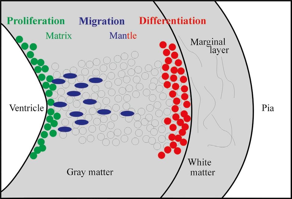
Fig. 2. Schematic transverse view of the developing vertebrate neural tube wall emphasizing distribution of proliferative, migrating, and differentiating neural cells from ventricle to pia.
Using descriptive histology, Bergquist (1932, 1954) and Källén (1951, 1952; see also Bergquist and Källén, 1954) brought this approach to fruition and established a grid of proliferating matrix zones (Grundgebiete) in the early vertebrate brain. These matrix zones were described to give off neurons in a locally specific spatiotemporal order, and—together with the subsequent migratory and differentiation behavior of fresh neurons leaving these matrix zones (migration areas)—to
determine the final morphology of every brain region (Fig. 3). This grid of matrix zones contained longitudinal as well as transverse zones and, ultimately, resulted in a neuromeric bauplan of the brain (see earlier). Because this work was comparative, the aspect of a common bauplan shared between all vertebrates was emphasized.
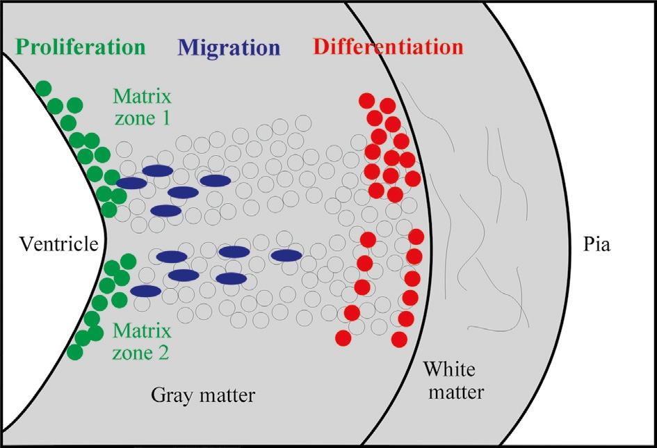
Fig. 3. Schematic transverse view of the developing vertebrate neural tube wall emphasizing local differences in neural proliferation and migration dynamics and their phenotypic results as viewed by H. Bergquist and B. Källén (see text).
As already mentioned in the context of neuromery, C.J. Herrick (and his followers, Ariëns-Kappers, Huber, and Crosby) vigorously attacked this concept, which seemed to him only to please detail-loving morphologists, deviating from the functional longitudinal zones considered truly relevant for understanding brain structure and comparative issues. With regard to searching for homology of brain structures in different vertebrates, the American school founded the long-lasting approach of studying functional adult neuronal connectivity using an increasing number of neuronal tracing substances. To this day, neurobiologists indeed successfully apply this strategy to the present day. Although powerful in revealing neuronal connectivity and, thus, delivering arguments for homology of central nervous structures among vertebrates, this approach must fail in cases of similar circuitry that evolved in parallel (homoplastic evolution) in different species. Another contemporary, L. Edinger, also placed himself against the ideas of the Swedish school. He instead proposed the sequential addition of forebrain parts during vertebrate evolution, which is incompatible with an ancestral common bauplan. Much in the vein of the Swedish school again was the “formanalytic approach” of H. Kuhlenbeck (publications between 1926 and 1973;
see Nieuwenhuys, 1998c), who stressed that only the origin from a topologically comparable ventricular site, is decisive for the identification of homologous brain structures and not the adult functional contexts.
What to make of this conflict today? Clearly, the Swedish school lacked a powerful methodology (such as fate mapping) for substantiating their claims. However, many subsequent developmental research lines using cellular and molecular methods eventually supported their concepts, as we shall see later. Except for the interpretation of longitudinal zones in the forebrain (see earlier), the concept of a bauplan of matrix zones of the Swedish school has never been fundamentally incompatible with that of the functional zones of the American school, but rather complements it from the developmental point of view.
Later, autoradiography using tritiated thymidine (pioneer studies: Angevine and Sidman, 1961; Fujita, 1963, 1964, 1966; Bayer and Altman, 1974; Rakic, 1974; reviews: Bayer and Altman, 1987, 1995a,b; Rakic, 2002, 2003a,b) allowed to directly demonstrate the location of proliferating neural cells (short survival time after incubation) or, alternatively, the birth dates (i.e., time of last mitosis) and adult fates of neural tube cells (long survival times after incubation). These studies revealed that a pseudostratified epithelium exists in the early neural tube, which had been suggested already in the 1930s by Sauer (1935). During this neural tube stage, every neural cell is proliferative and has both a ventricular and a pial attachment. The cell nucleus migrates periodically from ventricle to pia (interkinetic nuclear migration), always going through mitosis and cell division close to the ventricle and DNA replication close to the pia. Later, the developing neural tube shows real layers, that is, a ventricularly located proliferative cellular matrix layer, a more peripheral postmitotic cellular mantle layer, together forming the gray matter, and a marginal (largely noncellular) white matter layer, consisting of first neurites of differentiated cells as well as of incoming extrinsic axons (Fig. 4). It also became clear that the matrix layer is not uniform along the neural tube ventricular surface, but rather shows local differences in proliferation and migration dynamics. According to Bayer and Altman (1995b), the early neural proliferative matrix zones represent a “blueprint (bauplan) of the anatomy of the central nervous system.” Highly consistent with these views, the concept of histogenetic units (Puelles and Medina, 2002) adds the element of the genetic pathways that control differently developing neural tube units characterized by a matrix zone (see many examples in Section 3.2).
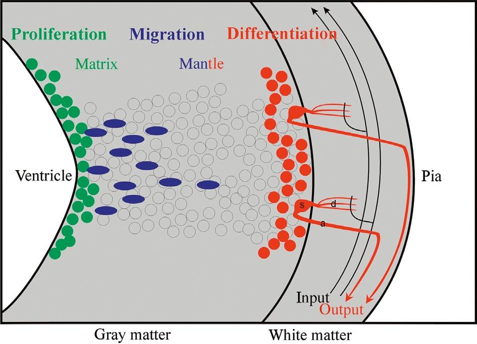
Fig. 4. Schematic transverse view of the developing vertebrate neural tube wall emphasizing differentiation into gray matter and white matter. The latter typically includes input and output tracts/ axons as well as dendritic arborizations of local neurons interacting with white matter fibers. a: axon, d: dendrite, s: neuronal soma.
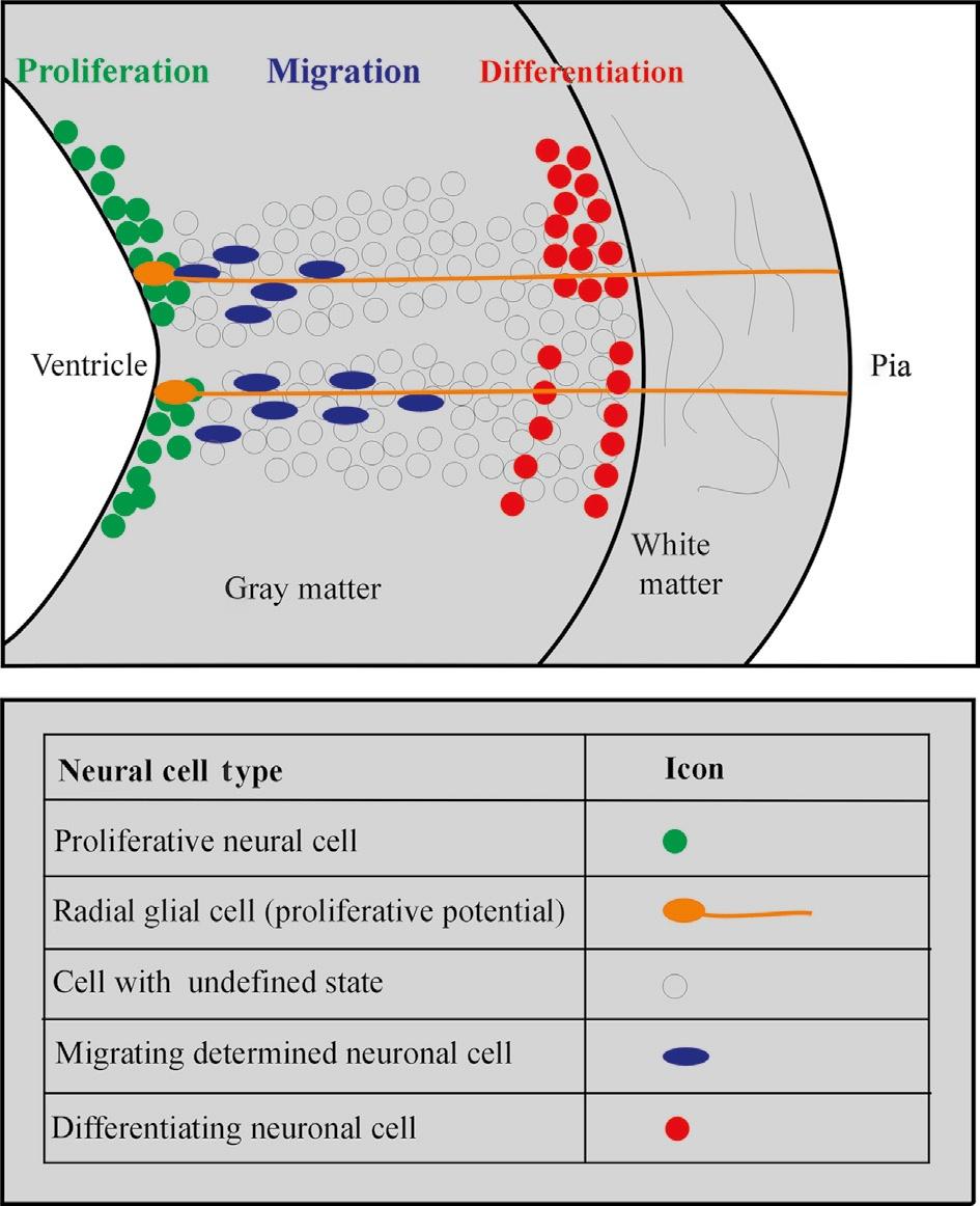
How do freshly postmitotic neural tube cells born at the ventricle manage to migrate peripherally once the neural tube goes beyond the stage of the ubiquitously proliferative pseudostratified epithelium? Already Ramón y Cajal (1890), among other contemporaries, documented the cytology and histology of an early differentiated neural cell type, the radial glia cell, which exhibits a periventricularly located cell body and a long peripheral (radial) process extending toward the pial surface (Fig. 5), therefore resembling somewhat the earlier proliferative neuroepithelial cells forming the pseudostratified epithelium. However, the role of radial glia in the guidance of peripherally migrating neural tube cells has been addressed only in the 1970s (Rakic, 1971, 1972, 1974), and its dynamics and molecular basis are best documented in mammalian isocortex (neocortex) and cerebellum (Rakic, 1988, 2002, 2003a,b; Hatten, 1990, 1993, 1999, 2002). Apparently, both the action of diffusible repellents/attractants and of surface-mediated interactions with radial glia processes are closely intertwined in the process of peripheral migration of neural cells (Rakic, 2003b). The latter have been mostly believed to originate from precursor cells different from radial glia cells.
Recently, the radial glia story has taken an unexpected and even more exciting turn. In addition to the meanwhile undisputed role of mammalian cortical radial glia as a guiding/scaffolding device for peripherally migrating neural cells, radial glia has been demonstrated to produce such migrating cells (Götz et al., 1998, 2002; Malatesta et al., 2000; Hartfuss et al., 2001; Noctor et al., 2001; Tamamaki et al., 2001). In the rodent cortex, differentiated radial glia cells—maintaining their peripheral radial process toward the pia—were discovered to remain proliferative
Fig. 5. Schematic transverse view of the developing vertebrate neural tube wall emphasizing the role of radial glial cells as a guiding device for migrating neural cells (see text).
and possess multipotency with regard to the generation of neurons and glia: They divide asymmetrically and give rise to most (i.e., projection) neurons in the cortical layers and, subsequently, to astroglial cells. This is not to exclude that there may be additional and/or more restricted neuronal and glial progenitor populations—possibly also deriving from radial glia cells—at certain developmental time points, especially in primates (Rakic, 2003b). An evolutionary novelty only seen in the developing mammalian isocortex and basal ganglia is the subventricular (or subependymal) zone of proliferative cells. The early basal ganglia consist of the lateral and medial ganglionic eminences, LGE and MGE (with the LGE representing the future striatum and the MGE giving rise to the future pallidum), which lie medioventrally to the cortex. The subventricular zone has been described to lie somewhat more remote from the ventricle than the proliferative ventricular zone and likely is an adaptation to the needs of large cell production (Smart, 1976; reviewed in Voogd et al., 1998). Whereas radial glia cells are abundant in the subventricular zone of the isocortex (Smart et al., 2002; Malatesta et al., 2003), they are absent in the subventricular zone of the ganglionic eminences (Malatesta et al., 2003; see later).
Although radial migration of newborn neural cells along radial glia fibers has been studied mostly in the mammalian cortex and cerebellum, it may be considered prototypical for the entire developing vertebrate neural tube because radial glia cells principally occur in every region of the vertebrate CNS, at least at early stages (Edwards et al., 1990; Hatten, 1999). In cases when newly generated neuronal and glial cells do not actively migrate, they remain oriented in their radial fiber domain when later-born cells at the ventricle are added at the ventricular side (for example in the basal ganglia; see later). Furthermore, also the multipotential role of radial glia in the generation of neural cells could be general for the entire CNS. This would be consistent with the fact that anamniotes exhibit a greater potential for adult neurogenesis than amniotes and retain radial glia in adult brain regions, whereas mammalian radial glia cells mostly do not persist into adulthood and are believed to at least partially transform into astrocytes (Götz et al., 2002; Rakic, 2003a). However, the proportion of radial glia derived neural cells varies greatly even within the mammalian telencephalon. In the ganglionic eminences, the radial glia (of the ventricular zone) generates only a small fraction of neural cells and an even smaller fraction of neurons, that is, striatal and some long-distance migrating olfactory bulb interneurons (Malatesta et al., 2003). Most of these (granule and periglomerular) olfactory bulb cells, as well as the striatal projection neurons (see later) are produced by nonradial glia progenitors of the subventricular zone of the lateral ganglionic eminence (Stenman et al., 2003; Campbell, 2003). Furthermore, the radial glia processes in the developing striatum lose their pial contact much earlier than in the developing isocortex (Smart, 1985). Therefore, striatal radial glia guided cellular migration appears insignificant compared to passive aggregation of postmitotic cells, making the presence of differentiated radial glia cells obsolete much earlier than in the cortex. In the ganglionic eminences, a thick embryonic proliferative subventricular zone of nonradial glia cells is formed, and it is the source of the overwhelming majority of striatal and pallidal projection neurons and interneurons (Malatesta et al., 2003), including those interneurons destined to invade the cortex (see later). However, the laterdeveloping, postnatal subventricular zone cells derive from radial glia cells and are gliogenic (Malatesta et al., 2003). Thus, the real question is how subpallial (and any other central nervous) glial and neuronal progenitors different from radial glia form out of the proliferative cells of the pseudostratified epithelium: Do these progenitors originate from very early radial glia and lose their glial characteristics, or alternatively, do they derive even earlier from the neuroepithelial cells directly?
Although radial glia guided migration may be considered the predominant mechanism of cellular translocation in the developing vertebrate neural tube (Rakic, 1988; Hatten, 1999), important exceptions to this general rule do exist. There are several well-established examples of nonradial glia guided, so-called tangential cellular migrations during CNS development because in these cases cells move perpendicular to the radial glia processes. For example, there is tangential migration of a minority of intrinsically generated (glutamatergic) isocortical projection neurons (O'Rourke et al., 1992), as well as long-distance tangential migration of GABAergic (destined for isocortex and striatum) and cholinergic (destined for striatum) interneurons originating mostly in the subpallial medial ganglionic eminence (Marín et al., 2000; see also discussion in the final, comparative chapter). The rhombic lip, which forms the crest of the V-shaped medulla oblongata, is the source of many tangentially migrating cells destined for basal brainstem structures, such as the inferior olive, the lateral reticular nucleus, or the pontine formation (Bayer and Altman, 1995b). The rhombic lip also gives off cells that invade the developing cerebellar plate and form the so-called external granular layer (EGL) lying directly below the pia (see later; Cerebellum). Finally, there is a rostral migratory stream of neural cells into the olfactory bulb (maintained into adulthood) consisting of cells produced in the subventricular and ventricular zones of the dorsal part of the lateral ganglionic eminence which turn into granule and periglomerular cells (Luskin, 1993; Wichterle et al., 2001; Stenman et al., 2003; Campbell, 2003; Malatesta et al., 2003; see also earlier discussion).
Somewhat unexpectedly, Altman and Bayer rejected both a strong role of radial glia guided migration and a close correspondence of their matrix zones to the bauplan of matrix zones suggested by Bergquist and Källén. But today, Altman and Bayer's results as outlined earlier appear highly compatible with the ideas described previously by the Swedish school (see earlier). Furthermore, radial (glia guided) cellular migration is clearly consistent with Bergquist and Källén's (1954) description of cellular migration waves originating in distinct neural proliferation matrix zones. The spatiotemporal regulation of these migration waves, together with subsequent differentiation processes, leads to various differing morphologies along the neural tube axis—with the various contributions by tangentially migrating neurons being exceptions to the rule to be integrated into the general picture of morphogenesis. Today, evidence concerning the genetic control of these processes of neurogenesis (some of which will be discussed in the final, comparative chapter) is increasingly emerging, complementing the picture designed by earlier active neuroscientists.
We have now laid the foundations to understand the most general principles of vertebrate CNS morphogenesis and are thus in a position to consider some cases of neural development of well-investigated mammalian brain parts in more detail.
1.3 Functional Brain Architecture: Mammalian Examples
In the following, some examples of neuro-/gliogenesis in mammalian brain development are discussed because the data presented later in this atlas will directly shed some light on comparable events in early zebrafish brain development.
1.3.1 Cortex
The mammalian isocortex represents a special case of a matrix or proliferation zone. Cortex development does not primarily depend on many different, separate matrix zones with great local spatiotemporal differences in proliferation, migration, and differentiation behavior as, for example, in the case of the dorsal thalamus where many different brain nuclei must be formed in a particular sequence (see later). The isocortex instead is of rather uniform thickness and histology (six layers, hence the name isocortex). Despite local differences in the emphasis of isocortical layers (layer 1 being most pial), they are unified by a common organizational theme: Layer 4 is always the recipient of input (intracortical or external), and its cells project up into layer 2, the pyramidal cells of layers 2/3 form the intracortical (long-distance) efferents, whereas layers 5/6 give rise to extracortical efferents (for example, to thalamus or spinal cord). Layer 1 predominantly plays a developmental role.
The isocortex is initially formed by a huge continuous sheet of proliferative units, and the crucial point is the maintenance of topological relationships between those proliferative units to allow for the correct development of local specializations of future cortical areas (e.g., visual or motor areas), that is, positional information in the horizontal and vertical direction must be maintained. According to the radial unit hypothesis of P. Rakic and coworkers (reviewed in Rakic, 1988, 2002, 2003a,b), the entirety of proliferative units close to the cortical ventricle form a protomap of future cytoarchitectonically different cortical areas. The species-specific mosaic of adult cortical aeralization is the result of both