The Oxford Linear Algebra for Scientists Andre Lukas
https://ebookmass.com/product/the-oxford-linear-algebra-forscientists-andre-lukas/
ebookmass.com
Classical Economics, Keynes and Money Essays in Honour of Carlo Panico John Eatwell
https://ebookmass.com/product/classical-economics-keynes-and-moneyessays-in-honour-of-carlo-panico-john-eatwell/
ebookmass.com
Aaron McDuffie Moore: An African American Physician, Educator, and Founder of Durham's Black Wall Street Blake Hill-Saya
https://ebookmass.com/product/aaron-mcduffie-moore-an-africanamerican-physician-educator-and-founder-of-durhams-black-wall-streetblake-hill-saya/ ebookmass.com
The Gambler and the Scholars Herbert Yardley, William & Elizebeth Friedman, and the Birth of Modern American Cryptology John F. Dooley
https://ebookmass.com/product/the-gambler-and-the-scholars-herbertyardley-william-elizebeth-friedman-and-the-birth-of-modern-americancryptology-john-f-dooley/ ebookmass.com
First Aid for the Family Medicine Boards, Third Edition (1st the
https://ebookmass.com/product/first-aid-for-the-family-medicineboards-third-edition-1st-the/
ebookmass.com
https://ebookmass.com/product/capm-certified-associate-in-projectmanagement-all-in-one-exam-guide-1st-edition-james-lee-haner/
ebookmass.com
Academic Press is an imprint of Elsevier
125 London Wall, London, EC2Y 5AS, UK
525 B Street, Suite 1800, San Diego, CA 92101-4495, USA
225 Wyman Street, Waltham, MA 02451, USA
The Boulevard, Langford Lane, Kidlington, Oxford OX5 1GB, UK
Copyright © 2016 Elsevier BV. All rights reserved.
No part of this publication may be reproduced or transmitted in any form or by any means, electronic or mechanical, including photocopying, recording, or any information storage and retrieval system, without permission in writing from the publisher. Details on how to seek permission, further information about the Publisher’s permissions policies and our arrangements with organizations such as the Copyright Clearance Center and the Copyright Licensing Agency, can be found at our website: www.elsevier.com/permissions
This book and the individual contributions contained in it are protected under copyright by the Publisher (other than as may be noted herein).
Notices
Knowledge and best practice in this field are constantly changing. As new research and experience broaden our understanding, changes in research methods, professional practices, or medical treatment may become necessary.
Practitioners and researchers must always rely on their own experience and knowledge in evaluating and using any information, methods, compounds, or experiments described herein. In using such information or methods they should be mindful of their own safety and the safety of others, including parties for whom they have a professional responsibility.
To the fullest extent of the law, neither the Publisher nor the authors, contributors, or editors, assume any liability for any injury and/or damage to persons or property as a matter of products liability, negligence or otherwise, or from any use or operation of any methods, products, instructions, or ideas contained in the material herein.
Library of Congress Cataloging-in-Publication Data
A catalog record for this book is available from the Library of Congress
British Library Cataloguing in Publication Data
A catalogue record for this book is available from the British Library
For information on all Academic Press publications visit our website at http://store.elsevier.com/
ISBN: 978-0-12-418669-9
Publisher: Mica Haley
Acquisition Editor: Mara Conner
Editorial Project Manager: Kathy Padilla
Production Project Manager: Lucía Pérez
Designer: Maria Inês Cruz
Acknowledgments
One of the pleasures of finishing a book is to acknowledge the colleagues and friends who gave important feedback, supplied us with probes, and supported the project in various ways. Thanks go to Patrick Blader (Toulouse, France); Uwe Strähle (Karlsruhe, Germany); Michael Lardelli (Adelaide, Australia); Eric Weinberg (Philadelphia, PA, USA); and Rachel McDonald, Ingvild Mikkola, and Steve Wilson (London, UK) for providing probes and antibodies. A big merci goes to Philippe Vernier (Gif-sur-Yvette, France) for enabling us to embark on this project in the first place. Thomas Mueller also thanks Wolfgang Driever (University of Freiburg, Germany) for his support above and beyond what can be expected. In addition, Thomas Mueller is grateful for the generous support of Brian Spooner, Susan Brown, and Gary Conrad from the Division of Biology at Kansas State University. Mario Wullimann thanks Benedikt Grothe (LudwigMaximilians-Universität Munich, Germany) for continuing support. Last but not least, this second edition would not have been possible without support and understanding of our families and friends. Thomas Mueller thanks Steffi Dippold; without her presence, work on this book would not have been half as enjoyable.
1. Vertebrate Central Nervous System
which innervate them, and the visceral ganglia of the autonomic nervous system. These somatic and visceral ganglia and their associated nerves represent the peripheral nervous system (PNS).
For many decades, scientists have investigated these complex developmental events leading to the formation of a nervous system in various vertebrates, especially in model animals, such as in the African clawed frog Xenopus, the chick, rodents like the mouse and rat, and most recently, the zebrafish, at molecular, cellular, histological, or morphological levels. Thus, a wealth of scientific literature is available, ready to satisfy almost any hunger for information.
Why then the present book? First of all, it integrates knowledge on CNS development coming from classical developmental studies with recent molecular data related to neuro- and gliogenesis. In particular, we will demonstrate the detailed spatiotemporal order of early postembryonic zebrafish brain development using molecular and cellular markers of neuro- and gliogenesis to visualize local differences, embedded in a holistic morphogenetic context. By combining old and new knowledge, we provide a neuroanatomically based molecular atlas of neural development in the zebrafish brain. We hope that such information—beyond delivering a refined molecular neuroanatomy—will prove to be enlightening in the future elucidation of neurogenetic pathways and their mechanisms.
1.2 Major Developmental Stages of the Vertebrate Neural Tube
Historically, scientists have always aimed to identify the fundamental building blocks of the vertebrate brain. In other words, they have tried to determine the basic developmental brain units and to answer the question of how these transform into functional structures of the mature brain. Before going into a more detailed discourse of the development of finer brain subdivisions (see Section 1.3), we will shortly examine the early events of brain morphogenesis relating to the emergence of vesicles and neuromeres, as well as of longitudinal zones, followed by an account on the neuromeric (prosomeric) model and on central nervous neurogenesis. An excellent in-depth historical account on the understanding of vertebrate brain morphogenesis has been given by Nieuwenhuys (1998a,b,c).
1.2.1 Vesicles, Neuromeres, and Longitudinal Zones
The classical description of a sequential appearance of two, three, and then five brain vesicles (i.e., transverse elements) along the anteroposterior vertebrate neural tube axis dates back to von Baer (1828). This view, however, gained popularity in the early twentieth century with the works of von Kupffer (1906) and Johnston (1909). The two-vesicle stage comprises a combined forebrain (prosencephalon) and midbrain (mesencephalon) vesicle, which is set apart by a vertical neural tube constriction from the hindbrain (rhombencephalon) vesicle. This is followed by the three-vesicle stage displaying forebrain, midbrain, and hindbrain vesicles (see Fig. 1) and, finally, by the five-vesicle stage, consisting of telencephalon and diencephalon (together forming the forebrain), mesencephalon, as well as metencephalon and myelencephalon (together forming the rhombencephalon).
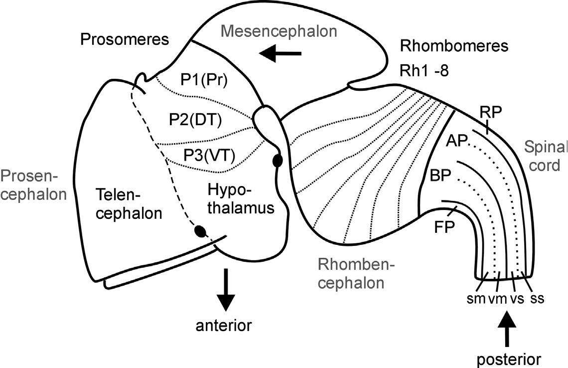
Fig. 1. Schematic lateral view of early mouse brain (E 12.5-13.5) shows prosomeric interpretation (Puelles and Rubenstein, 1993, 2003), including transverse (neuromeres) and longitudinal (columns) elements. Arrows designate anteroposterior axis of neural tube. Abbreviations: AP: alar plate; BP: basal plate; DT: dorsal thalamus (thalamus), FP: floor plate; P1-P3: prosomeres 1-3 (for more details see text); Pr: pretectum; Rh1-Rh8: rhombomeres 1-8; RP: roof plate; sm: somatomotor column; ss: somaotosensory column; T: telencephalon; vm: visceromotor column; vs: viscerosensory column; VT: ventral thalamus (prethalamus).
The subdivision of both forebrain and hindbrain into two separable (transverse) vesicles, each along the anteroposterior axis, arguably is an epiphenomenon, visible only in mammalian embryos that show an enormous early growth of the telencephalic hemispheres. This hypertrophy detracts from the fact that the
most rostral vertebrate neural tube in reality includes in addition to the dorsally lying telencephalon also ventrally the smaller hypothalamus. This renders the concept of transverse telencephalic and diencephalic vesicles obsolete because part of the diencephalon (the hypothalamus) is included in a transverse unit with the telencephalon (Puelles and Rubenstein, 1993, 2003). Also, the somewhat later, characteristic mammalian emergence of a large cerebellum and ventral pontine region led to the impression that the medulla oblongata develops from two separable vesicles, the metencephalic one exhibiting cerebellum and pons, and a myelencephalic vesicle posteriorly. Indeed, the dorsal portion of the anteriormost rhombomere specializes into a dorsal cerebellum. In contrast, the ventral pontine region, which relays cortical information to the cerebellum, develops from ventral and fused portions of rhombomeres 1-4 (Aroca and Puelles, 2005; Alonso et al., 2012). This complex development of the anterior hindbrain historically resulted in the misinterpretation that the two enlargements of cerebellum and pons form a true transverse unit, which was called the metencephalon to separate it from the posterior myelencephalon. However, the medulla oblongata does not show a real boundary between these two postulated vesicles; instead, it is rather divided into a higher number of early segmental entities, the rhombomeres (see later discussion and Fig. 1).
In contrast, the three-vesicle stage reflects on fundamental anteroposterior vertebrate brain divisions. Especially the midbrain-hindbrain boundary has been strongly corroborated in modern developmental neurobiology as a singularly definable boundary and signaling center in the vertebrate brain (Marín and Puelles, 1994; Bally-Cuif and Wassef, 1995; Brand et al., 1996; Lumsden and Krumlauf, 1996; Reifers et al., 1998; Wurst and Bally-Cuif, 2001). Similarly, there is clear evidence for cellular lineage restriction (Larsen et al., 2001) and signaling center function (Scholpp and Brand, 2003) at the forebrain-midbrain boundary.
Equally important for the understanding of the vertebrate CNS bauplan are two neural tube flexures, which were also historically noted early (His, 1888, 1893a). The cephalic flexure of the anteroposterior axis is between the mesencephalon and the prosencephalon, and the cervical flexure is between the rhombencephalon and the spinal cord (compare Fig. 1). The result of this bending of the neuraxis is that the forebrain floor is adjacent to the hindbrain floor. The correct recognition of the adequate course of the anteroposterior axis of the neural tube has important consequences for the interpretation of the topology of brain structures.
Whereas the vesicle story just outlined found its way easily into general textbook knowledge, the historical equally early description of neural tube segments or so-called neuromeres did not. Neuromeres represent a finer set of transitory transverse or segmental divisions of the vertebrate neural tube, and they remain, at least partially, controversial to the present day. Vertebrate hindbrain neuromeres (rhombomeres) are set apart from each other by transverse, vertical constrictions and were already described in the nineteenth century (von Baer, 1828; Orr, 1887). Later, similar morphological observations led to the interpretation of neuromeres to extend into midbrain (mesomeres) and forebrain as well (prosomeres; see Fig. 1; Rendahl, 1924; Bergquist, 1932; Vaage, 1969). However, in these early twentieth-century studies, the basis for recognizing transverse neuromeres was descriptive (and, even worse, transitory) morphology. Thus, serious opposition regarding the acceptance of neuromeres as the important building blocks of the CNS arose with its growing functional neuroanatomical understanding, especially by the so-called American school, which was based on the early work of Gaskell (1889) and later widely spread through the activities of Charles Judson Herrick (1868-1960), who is viewed as one of the founding fathers of modern comparative neurology. In this school's well corroborated view, longitudinal functional zones—embryonically represented dorsoventrally in the neural tube as roof, alar, basal, and floor plates (His, 1888, 1893a,b)—were considered the primary subdivisions of the CNS (see Fig. 1). Both the most dorsal (roof plate) and most ventral (floor plate) longitudinal zones exert critical developmental roles, for example, in mediating dorsoventral polarity of the neural tube or in guiding neuronal differentiation processes. In the adult vertebrate CNS, the roof plate gives rise to dorsal midline structures, such as the epiphysis or the choroid plexus, which covers the rhomboid opening of the medulla oblongata, whereas the floor plate develops, for example, into ventral midline glial cells.
The two intermediate longitudinal zones termed the alar and basal plates form pivotal elements of Herrick's concept of functional vertebrate brain and spinal cord organization. Alar and basal plate derivatives form the characteristic dorsoventral arrangement of four functional longitudinal subzones, that is, alar plate derived somato- and viscerosensory columns and basal plate derived viscero- and somatomotor columns as prototypically present in the spinal cord (see Fig. 1). This dorsoventral spinal cord organization of four sensory and motor zones has been used to consistently explain various local specializations (hypertrophies and reductions) in more anterior brain regions—at least into the midbrain. Thus, this
1. Vertebrate Central Nervous System Development
patterns in the more anterior forebrain (secondary prosencephalon). Also, neurogenic (Notch/Delta) and proneural (basic helix-loop-helix) gene expression closely parallels brain proliferation patterns, including the prosomeric pattern (P1/P2/P3) in the posterior forebrain (Mueller and Wullimann, 2003; Wullimann and Mueller, 2004a,b; see later).
In conclusion, there is strong evidence for three posterior prosomeres (P1 through P3) in the vertebrate forebrain, with the situation in the more anterior forebrain (secondary prosencephalon) being more elusive. Clearly, rhombomeres match better the criteria for neuromeres/compartments when compared to prosomeres. However, one must keep in mind that these criteria are only met transitionally during neuromere development and that the most critical criterion, that is, cell lineage restriction, is at no time completely met even in rhombomeres; there are reports on more than 5% of cells transgressing rhombomere boundaries (Birgbauer and Fraser, 1994). Furthermore, overt metameric organization (i.e., similar sets of anteroposteriorly repeated classes of neurons) as seen in the developing hindbrain does not necessarily require segmental neural tube organization during development. Metameric organization, as partially still present in the adult rhombencephalon, is even more pervasive in the spinal cord, which does not exhibit neuromeres (as defined earlier) during any period of development (Keynes and Stern, 1984). Therefore, metamery is apparently not necessarily strictly developmentally correlated with a neuromeric organization. Thus, the forebrain simply may be characterized by very early individual regulation of each of its transverse entities, resulting in a fast variable differentiation of neuromeres and their derivatives.
1.2.2 The Neuromeric or Prosomeric Model
Irrespective of the ongoing dispute about the identification and finite numbers of vertebrate neuromeres, Puelles and Rubenstein's neuromeric or prosomeric model (1993, 2003) integrates and puts into perspective much of the issues raised earlier, suggesting a vertebrate brain bauplan that has been of great heuristic value in many studies since. The correct topological identification of a particular brain structure, and, therefore, its comparative interpretation, critically depends on the concept of the basic vertebrate CNS bauplan one uses. Such a bauplan involves more than the recognition of transverse elements, it also requires to adequately indicate the course of the anteroposterior axis along the neural tube
and, in consequence, of the longitudinal zones. Following the neuromeric model, all central nervous longitudinal zones (floor, basal, alar, and roof plates) principally extend into the forebrain: The roof plate ends in the region of the anterior commissure, the floor plate at the anterior end of the mammillary hypothalamus (see two respective black dots in Fig. 1), whereas the basal and alar plate form a broad anterior end of the neural tube with the preoptic region (alar) and hypothalamus (basal), both united at the area of the optic chiasma (Puelles et al., 2013). The entire telencephalic hemispheres are thus lateral extensions of the alar plate. This is supported by studies on various longitudinally expressed genes (such as, for example, sonic hedgehog), which continue anteriorly to be expressed into the forebrain (Ericson et al., 1995; Shimamura et al., 1995, 1997; Hauptmann and Gerster, 2000). Such data clearly corroborate the old observation noted earlier that the anteroposterior axis of the rostral vertebrate neural tube is considerably deflected ventrally (compare with Fig. 1). Understanding the course of longitudinal zones is critical for the interpretation of transverse neural tube units (neuromeres), as the latter must lie perpendicular to the anteroposterior axis. In the neuromeric model of Puelles and Rubenstein (1993), the entire brain is principally divided into transverse zones (each containing a segment of all longitudinal zones), and neuromeres therefore exist not only in the rhombencephalon (rhombomeres), but also in the mesencephalon (mesomeres) and prosencephalon (prosomeres), very much following the ideas of Bergquist and Källén described earlier. In a revision of their original model, Puelles and Rubenstein (2003) have modified their initial claim of six prosomeres, suggesting only three posterior diencephalic prosomeres (P1/P2/P3), with the secondary prosencephalon possibly guided by different processes of regionalization, much like the conclusion reached in other recent studies discussed earlier (e.g., Wullimann and Puelles, 1999). However, we interpret the eminentia thalami as described here in the zebrafish as lying outside of the ventral thalamus, because some of the revisions (Puelles and Rubenstein, 2003) are based on information unavailable yet in the zebrafish (see also a later section: A Word Regarding Terminology and Ontology).
Despite some insecurity about the finite number of prosomeres, it is important to accept the longitudinal axis proposed in the neuromeric model to interpret adequately the topological transformation of longitudinal and transverse elements that occurs during the development of a theoretically straight vertebrate neural tube. A good example is the hypothalamus and the posteriorly adjacent basal diencephalic regions (i.e., retromammillary area in P3, posterior tuberculum
in P2, basal synencephalon in P1), which are recognized to represent basal (and floor) plate portions of the forebrain neural tube only when this deflected axis is respected (compare with Fig. 1). The corresponding forebrain alar (and roof) plate complements are represented by telencephalon/preoptic region for the hypothalamus and by the dorsal aspects of ventral and dorsal thalami (prethalamus and thalamus of Puelles and Rubenstein, 2003) and of pretectum for the three basal diencephalic regions just mentioned. A still widely accepted textbook opinion going back to C.J. Herrick (see earlier) holds that the diencephalon (i.e., hypothalamus, posterior tuberculum, thalamus, epithalamus, pretectum) is a transverse piece of the neural tube in which the hypothalamus represents the most ventral (i.e., basal) part, and the ventral thalamus, dorsal thalamus, and epithalamus are successively more dorsal parts of the diencephalic neural tube. In contrast, the neuromeric model considers the hypothalamus to represent the basal (ventral) part of the neural tube, associated with the dorsally located telencephalon (together representing the anterior forebrain or secondary prosencephalon; i.e., the most rostral piece of the neural tube). Accordingly, retromammillary region, posterior tuberculum (in the zebrafish corresponding to ventral and dorsal posterior tuberculum, respectively) and basal synencephalon represent three basal (ventral) forebrain portions associated with the corresponding alar plate portions (i.e., ventral thalamus, dorsal thalamus, pretectum) of the caudally adjoining piece of the neural tube (together forming the three prosomeres that constitute the posterior forebrain, i.e., anterior parencephalon, posterior parencephalon, and synencephalon). This example shows that it is far from trivial to disclose which CNS bauplan or model one uses to interpret the correct topological position of a particular area within the neural tube.
1.2.3
The Next Dimension: Understanding Central Nervous Proliferation and Neuro-/Gliogenesis
Along with the emergence of an increasingly adequate picture of relevant transverse and longitudinal elements of the vertebrate CNS, important concepts of cellular (neural) proliferation arose through the twentieth century. In principle, the neural tube could increase its size and develop various vesicles and neuromeres described earlier without cell proliferation; indeed, cell shape changes and transitory increase in liquor pressure have been implied in these processes. However, there is now ample evidence that cell proliferation (followed by migration and differentiation) is indeed involved in the shaping of the early CNS.
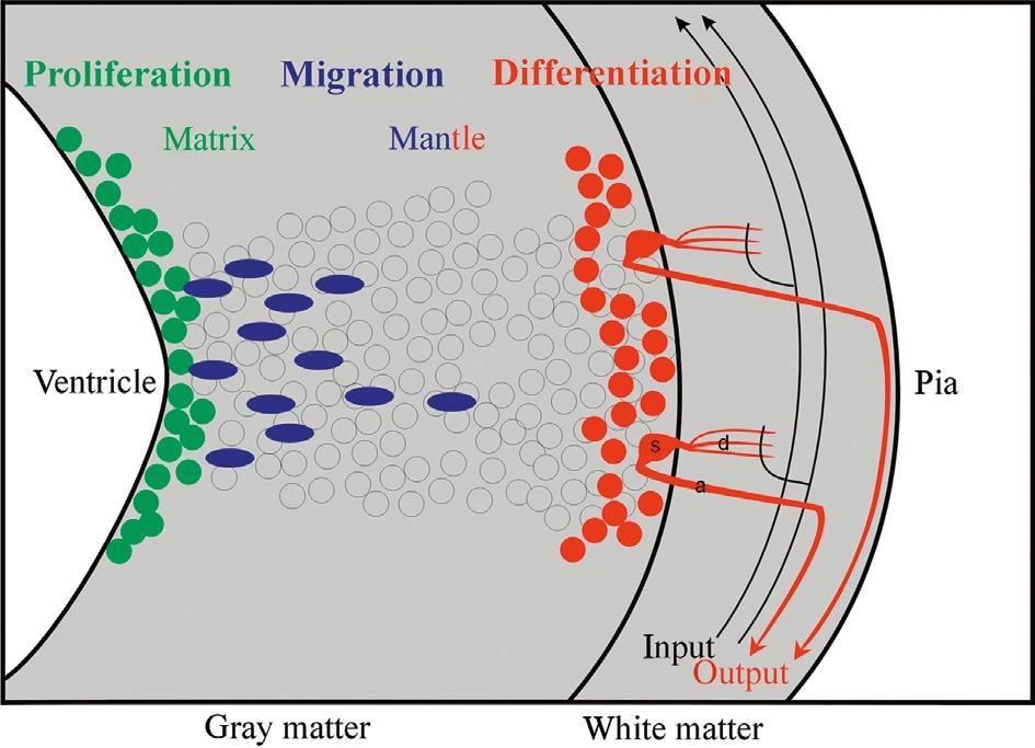
Historically, the Swedish comparative embryological school initiated by Nils Holmgren (and continued by Palmgren, Bergquist, and Källén) plays a crucial role in the understanding of how cellular proliferation relates to morphogenetic processes in the CNS, such as, for example, in the establishment of neuromeres. The central concept of the Swedish school is that the origin of adult brain structures from a particular location within the proliferative periventricular sheet of the neural tube is decisive for the final phenotypic appearance of those brain structures and, particularly, for their comparative interpretation, that is, for identifying homologies among brain structures in different vertebrate brains. Clearly at the core of this concept are local differences in the spatiotemporal proliferative behavior in the periventricular neural tube sheet and the subsequent migratory and differentiating behavior of brain cells originating there. This introduces the third dimension necessary to understand the complexity of brain morphogenesis in addition to those two discussed earlier (i.e., transverse and longitudinal neural tube elements), namely the developmental relationship of the proliferative ventricular zones (matrix) with the more peripheral (subpial) postmitotic cellular central nervous architecture deriving from these ventricular zones (Fig. 2).
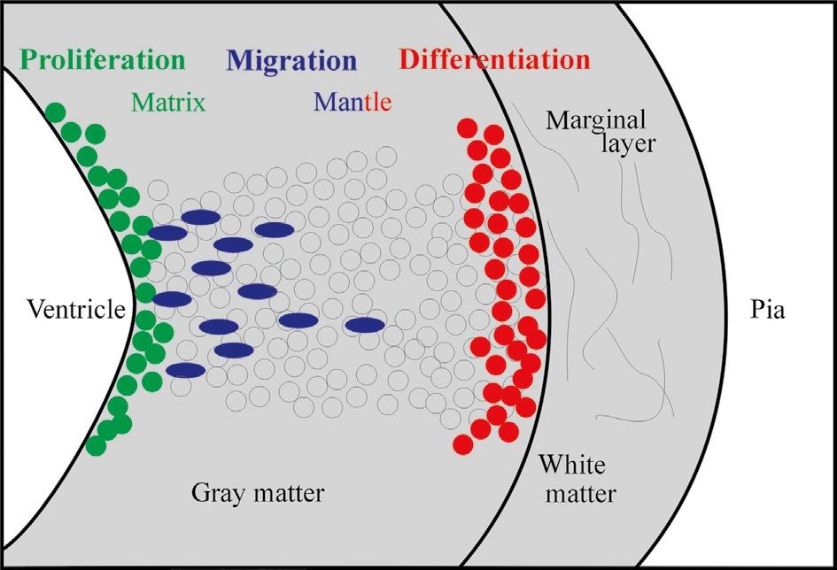
Fig. 2. Schematic transverse view of the developing vertebrate neural tube wall emphasizing distribution of proliferative, migrating, and differentiating neural cells from ventricle to pia.
Using descriptive histology, Bergquist (1932, 1954) and Källén (1951, 1952; see also Bergquist and Källén, 1954) brought this approach to fruition and established a grid of proliferating matrix zones (Grundgebiete) in the early vertebrate brain. These matrix zones were described to give off neurons in a locally specific spatiotemporal order, and—together with the subsequent migratory and differentiation behavior of fresh neurons leaving these matrix zones (migration areas)—to
Fig. 4. Schematic transverse view of the developing vertebrate neural tube wall emphasizing differentiation into gray matter and white matter. The latter typically includes input and output tracts/ axons as well as dendritic arborizations of local neurons interacting with white matter fibers. a: axon, d: dendrite, s: neuronal soma.
How do freshly postmitotic neural tube cells born at the ventricle manage to migrate peripherally once the neural tube goes beyond the stage of the ubiquitously proliferative pseudostratified epithelium? Already Ramón y Cajal (1890), among other contemporaries, documented the cytology and histology of an early differentiated neural cell type, the radial glia cell, which exhibits a periventricularly located cell body and a long peripheral (radial) process extending toward the pial surface (Fig. 5), therefore resembling somewhat the earlier proliferative neuroepithelial cells forming the pseudostratified epithelium. However, the role of radial glia in the guidance of peripherally migrating neural tube cells has been addressed only in the 1970s (Rakic, 1971, 1972, 1974), and its dynamics and molecular basis are best documented in mammalian isocortex (neocortex) and cerebellum (Rakic, 1988, 2002, 2003a,b; Hatten, 1990, 1993, 1999, 2002). Apparently, both the action of diffusible repellents/attractants and of surface-mediated interactions with radial glia processes are closely intertwined in the process of peripheral migration of neural cells (Rakic, 2003b). The latter have been mostly believed to originate from precursor cells different from radial glia cells.
Recently, the radial glia story has taken an unexpected and even more exciting turn. In addition to the meanwhile undisputed role of mammalian cortical radial glia as a guiding/scaffolding device for peripherally migrating neural cells, radial glia has been demonstrated to produce such migrating cells (Götz et al., 1998, 2002; Malatesta et al., 2000; Hartfuss et al., 2001; Noctor et al., 2001; Tamamaki et al., 2001). In the rodent cortex, differentiated radial glia cells—maintaining their peripheral radial process toward the pia—were discovered to remain proliferative
Although radial glia guided migration may be considered the predominant mechanism of cellular translocation in the developing vertebrate neural tube (Rakic, 1988; Hatten, 1999), important exceptions to this general rule do exist. There are several well-established examples of nonradial glia guided, so-called tangential cellular migrations during CNS development because in these cases cells move perpendicular to the radial glia processes. For example, there is tangential migration of a minority of intrinsically generated (glutamatergic) isocortical projection neurons (O'Rourke et al., 1992), as well as long-distance tangential migration of GABAergic (destined for isocortex and striatum) and cholinergic (destined for striatum) interneurons originating mostly in the subpallial medial ganglionic eminence (Marín et al., 2000; see also discussion in the final, comparative chapter). The rhombic lip, which forms the crest of the V-shaped medulla oblongata, is the source of many tangentially migrating cells destined for basal brainstem structures, such as the inferior olive, the lateral reticular nucleus, or the pontine formation (Bayer and Altman, 1995b). The rhombic lip also gives off cells that invade the developing cerebellar plate and form the so-called external granular layer (EGL) lying directly below the pia (see later; Cerebellum). Finally, there is a rostral migratory stream of neural cells into the olfactory bulb (maintained into adulthood) consisting of cells produced in the subventricular and ventricular zones of the dorsal part of the lateral ganglionic eminence which turn into granule and periglomerular cells (Luskin, 1993; Wichterle et al., 2001; Stenman et al., 2003; Campbell, 2003; Malatesta et al., 2003; see also earlier discussion).
Somewhat unexpectedly, Altman and Bayer rejected both a strong role of radial glia guided migration and a close correspondence of their matrix zones to the bauplan of matrix zones suggested by Bergquist and Källén. But today, Altman and Bayer's results as outlined earlier appear highly compatible with the ideas described previously by the Swedish school (see earlier). Furthermore, radial (glia guided) cellular migration is clearly consistent with Bergquist and Källén's (1954) description of cellular migration waves originating in distinct neural proliferation matrix zones. The spatiotemporal regulation of these migration waves, together with subsequent differentiation processes, leads to various differing morphologies along the neural tube axis—with the various contributions by tangentially migrating neurons being exceptions to the rule to be integrated into the general picture of morphogenesis. Today, evidence concerning the genetic control of these processes of neurogenesis (some of which will be discussed in the final, comparative chapter) is increasingly emerging, complementing the picture designed by earlier active neuroscientists.