No part of this publication may be reproduced or transmitted in any form or by any means, electronic or mechanical, including photocopying, recording, or any information storage and retrieval system, without permission in writing from the publisher. Details on how to seek permission, further information about the Publisher’s permissions policies and our arrangements with organizations such as the Copyright Clearance Center and the Copyright Licensing Agency, can be found at our website: www.elsevier.com/permissions.
This book and the individual contributions contained in it are protected under copyright by the Publisher (other than as may be noted herein).
Notices
Knowledge and best practice in this field are constantly changing. As new research and experience broaden our understanding, changes in research methods, professional practices, or medical treatment may become necessary.
Practitioners and researchers must always rely on their own experience and knowledge in evaluating and using any information, methods, compounds, or experiments described herein. In using such information or methods they should be mindful of their own safety and the safety of others, including parties for whom they have a professional responsibility.
With respect to any drug or pharmaceutical products identified, readers are advised to check the most current information provided (i) on procedures featured or (ii) by the manufacturer of each product to be administered, to verify the recommended dose or formula, the method and duration of administration, and contraindications. It is the responsibility of practitioners, relying on their own experience and knowledge of their patients, to make diagnoses, to determine dosages and the best treatment for each individual patient, and to take all appropriate safety precautions.
To the fullest extent of the law, neither the Publisher nor the authors, contributors, or editors, assume any liability for any injury and/or damage to persons or property as a matter of products liability, negligence or otherwise, or from any use or operation of any methods, products, instructions, or ideas contained in the material herein.
Previous editions copyrighted 2011, 2003, 1996, 1988, and 1981.
Library of Congress Cataloging-in-Publication Data
Names: Goldsmith, Jay P., editor. | Karotkin, Edward H., editor. | Keszler, Martin, editor. | Suresh, Gautham, editor.
Title: Assisted ventilation of the neonate : an evidence-based approach to newborn respiratory care / [edited by] Jay P. Goldsmith, MD, FAAP, Clinical Professor, Department of Pediatrics, Tulane University School of Medicine, New Orleans, Louisiana, Edward H. Karotkin, MD, FAAP, Professor of Pediatrics, Neonatal/ Perinatal Medicine, Eastern Virginia Medical School, Norfolk, Virginia, Martin Keszler, MD, FAAP, Professor of Pediatrics, Warren Alpert Medical School, Brown University, Director of Respiratory Services, Department of Pediatrics, Women and Infants Hospital, Providence, Rhode Island, Gautham K. Suresh, MD, DM, MS, FAAP, Section Head and Service Chief of Neonatology, Baylor College of Medicine, Texas Children’s Hospital, Houston, Texas.
Description: Sixth edition. | Philadelphia, PA : Elsevier, [2017]
Identifiers: LCCN 2016029284 | ISBN 9780323390064 (hardback : alk. paper)
Subjects: LCSH: Respiratory therapy for newborn infants. | Artificial respiration.
Classification: LCC RJ312 .A87 2017 | DDC 618.92/2004636--dc23 LC record available at https://lccn.loc.gov/2016029284
This book is dedicated to my wife, Terri, who has supported me through six editions of this text and my many nights away from home while caring for sick neonates.
JPG
I would like to dedicate this sixth edition of Assisted Ventilation of the Neonate to the numerous bedside NICU nurses, neonatal nurse practitioners, and respiratory therapists, and all of the other ancillary health care providers I have had the honor of working with over the past nearly 40 years at the Children’s Hospital of The King’s Daughters. Without your commitment to providing the best of care to our patients I could not have done my job.
EHK
I dedicate this book to my wife, Mary Lenore Keszler, MD, who has been my lifelong companion, inspiration, and best friend. Without her incredible patience and unwavering support, none of this work would have been possible. The book is also dedicated to the many tiny patients and their families who taught me many valuable lessons, and to the students, residents, and Fellows whose probing questions inspired me to seek a deeper understanding of the problems that face us every day.
MK
I dedicate this book to my teachers and mentors over the years, who taught me and guided me. I also thank my wife, Viju Padmanabhan, and my daughters, Diksha and Ila, for their support and patience with me over the years.
GKS
CONTRIBUTORS
Kabir Abubakar, MD Professor of Clinical Pediatrics Neonatology/Pediatrics Medstar Georgetown University Hospital Washington, DC
Namasivayam Ambalavanan, MBBS, MD Professor, Pediatrics University of Alabama at Birmingham Birmingham, AL
Robert M. Arensman, BS, MD Head, Division of Pediatric Surgery Department of Surgery University of Illinois at Chicago Chicago, IL
Eduardo Bancalari, MD
Professor of Pediatrics, Obstetrics, and Gynecology, Director, Division of Neonatology, Chief, Newborn Service Department of Pediatrics, Division of Neonatology University of Miami School of Medicine Miami, FL
Keith J. Barrington, MB, ChB Neonatologist and Clinical Researcher
Sainte Justine University Health Center, Professor of Paediatrics University of Montréal Montréal, Canada
Jonathan F. Bean, MD Chief Resident Department of General Surgery University of Illinois Hospital and Health Sciences Center Chicago, IL
Edward F. Bell, MD Professor of Pediatrics Department of Pediatrics University of Iowa Iowa City, IA
David M. Biko, MD Assistant Professor
The Children’s Hospital of Philadelphia, Pediatric Radiologist Pennsylvania Hospital The University of Pennsylvania Health System Philadelphia, PA
Laura D. Brown, MD Associate Professor Pediatrics University of Colorado School of Medicine Aurora, CO
Jessica Brunkhorst, MD Assistant Professor of Pediatrics Children’s Mercy Hospital University of Missouri - Kansas City Kansas City, Missouri
Waldemar A. Carlo, MD
Edwin M. Dixon Professor of Pediatrics University of Alabama at Birmingham, Director, Division of Neonatology University of Alabama at Birmingham Birmingham, AL
Robert L. Chatburn, MHHS, RRT-NPS, FAARC Clinical Research Manager
Respiratory Institute, Cleveland Clinic, Director, Simulation Fellowship Education Institute, Cleveland Clinic, Adjunct Professor of Medicine Lerner College of Medicine of Case Western Reserve University Cleveland, OH
Nelson Claure, MSc, PhD
Research Associate Professor of Pediatrics, Director, Neonatal Pulmonary Research Laboratory Department of Pediatrics, Division of Neonatology University of Miami School of Medicine Miami, FL
Clarice Clemmens, MD Assistant Professor of Pediatric Otolaryngology Medical University of South Carolina Charleston, SC
Christopher E. Colby, MD Associate Professor of Pediatrics Mayo Clinic Rochester, MN
Sherry E. Courtney, MD, MS Professor of Pediatrics Department of Pediatrics University of Arkansas for Medical Sciences Little Rock, AR
Peter G. Davis, MBBS, MD, FRACP Professor/Director of Neonatal Medicine
The University of Melbourne and The Royal Women’s Hospital Melbourne, Victoria, Australia
Eugene M. Dempsey, MBBCH BAO, FRCPI, MD, MSc
Clinical Professor
Paediatrics and Child Health
University College Cork, Department of Neonatology
Cork University Maternity Hospital Wilton, Cork, Ireland
Robert Diblasi, RRT-NPS, FAARC
Seattle Children’s Research Institute - Respiratory Care Center for Developmental Therapeutics Seattle, WA
Jennifer Duchon, MDCM, MPH
Clinical Fellow
Pediatric Infectious Disease
Columbia-Presbyterian Medical Center New York, NY
Jonathan M. Fanaroff, MD, JD
Associate Professor of Pediatrics
Case Western Reserve University School of Medicine, Co-Director, Neonatal Intensive Care Unit, Director, Rainbow Center for Pediatric Ethics
Rainbow Babies and Children’s Hospital Cleveland, OH
William W. Fox, MD
Attending Neonatologist
Division of Neonatology
Medical Director
Infant Breathing Disorder Center Children’s Hospital of Philadelphia, Professor of Pediatrics
University of Pennsylvania Perelman School of Medicine Philadelphia, PA
Debbie Fraser, MN, RNC-NIC
Associate Professor Faculty of Health Disciplines Athabasca University Athabasca, Alberta, Canada, Advanced Practice Nurse
NICU
St Boniface Hospital Winnipeg, Manitoba, Canada
John T. Gallagher, MPH, RRT-NPS, FAARC
Critical Care Coordinator
Pediatric Respiratory Care
University Hospitals, Rainbow Babies and Children’s Hospital Cleveland, OH
Jay P. Goldsmith, MD, FAAP
Clinical Professor
Pediatrics
Tulane University New Orleans, LA
Malinda N. Harris, MD
Assistant Professor of Pediatrics
Mayo Clinic Rochester, MN
William W. Hay, Jr., MD Professor
Pediatrics
University of Colorado School of Medicine Aurora, CO
Robert M. Insoft, MD
Chief Medical Officer and Attending Neonatologist
Women and Infants Hospital
Alpert Medical School of Brown University Providence, RI
Erik A. Jensen, MD Instructor of Pediatrics
The University of Pennsylvania, Attending Neonatologist
The Children’s Hospital of Philadelphia Philadelphia, PA
Jegen Kandasamy, MBBS, MD
Assistant Professor Pediatrics
University of Alabama at Birmingham Birmingham, AL
Edward H. Karotkin, MD, FAAP Professor of Pediatrics Neonatal/Perinatal Medicine
The Eastern Virginia Medical School Norfolk, VA
Martin Keszler, MD, FAAP Professor of Pediatrics
Alpert Medical School of Brown University, Director of Respiratory Services, Pediatrics Women and Infants Hospital Providence, RI
John P. Kinsella, MD Professor of Pediatrics Department of Pediatrics Section of Neonatology
University of Colorado School of Medicine and Children’s Hospital Colorado Aurora, CO
Haresh Kirpalani, BM, MRCP, FRCP, MSc
Professor
The University of Pennsylvania, Attending Neonatologist and Director Newborn and Infant Chronic Lung Disease Program
The Children’s Hospital of Philadelphia Philadelphia, PA; Emeritus Professor Clinical Epidemiology
McMaster University Hamilton, Ontario, Canada
Derek Kowal, RRT
Supervisor NICU, Respiratory Services Foothills Medical Centre
Alberta Health Services Calgary, Alberta, Canada
Satyan Lakshminrusimha, MBBS, MD
Professor of Pediatrics Director, Center for Developmental Biology of the Lung University at Buffalo, Chief of Neonatology
Women and Children’s Hospital of Buffalo Buffalo, NY
John D. Lantos, MD
Director of Bioethics
Children’s Mercy Hospital Professor Pediatrics University of Missouri - Kansas City Kansas City, MO
Krithika Lingappan, MD, MS, FAAP
Assistant Professor Section of Neonatology Department of Pediatrics
Texas Children’s Hospital Baylor College of Medicine Houston, TX
Akhil Maheshwari, MD
Professor of Pediatrics and Molecular Medicine
Pamela and Leslie Muma Endowed Chair in Neonatology, Chief, Division of Neonatology, Assistant Dean, Graduate Medical Education Pediatrics University of South Florida Tampa, FL
Mark C. Mammel, MD Professor of Pediatrics Department of Pediatrics University of Minnesota Minneapolis, MN
George T. Mandy, MD
Associate Professor of Pediatrics
Baylor College of Medicine Houston, TX
Richard J. Martin, MBBS Professor Pediatrics, Reproductive Biology, and Physiology and Biophysics
Case Western Reserve University School of Medicine, Drusinsky/Fanaroff Professor Pediatrics
Rainbow Babies and Children’s Hospital Cleveland, OH
Kathryn L. Maschhoff, MD, PhD
Assistant Professor of Clinical Pediatrics
The University of Pennsylvania, Attending Neonatologist
The Children’s Hospital of Philadelphia Philadelphia, PA
Bobby Mathew, MD
Associate Program Director
Assistant Professor of Pediatrics
University at Buffalo
Women and Children’s Hospital of Buffalo Buffalo, NY
Patrick Joseph McNamara, MD, MRCPCH, MSc
Associate Professor
Pediatrics and Physiology
University of Toronto, Staff Neonatologist
Pediatrics
Hospital for Sick Children Toronto, Ontario, Canada
D. Andrew Mong, MD
Assistant Professor
The University of Pennsylvania, Pediatric Radiologist
The Children’s Hospital of Philadelphia Philadelphia, PA
Colin J. Morley, DCH, MD, FRCPCH
Professor
Neonatal Research
Royal Women’s Hospital Melbourne, Cambridge, Great Britain
Leif D. Nelin, MD
Dean W. Jeffers Chair in Neonatology
Nationwide Children’s Hospital, Professor and Chief, Division of Neonatology
The Ohio State University and Nationwide Children’s Hospital Columbus, OH
Donald Morley Null Jr., MD Professor of Pediatrics Department of Pediatrics University of California Davis Sacramento, CA
Louise S. Owen, MBChB, MRCPCH, FRACP, MD
Neonatologist
Newborn Research
Royal Women’s Hospital, Honorary Fellow
Murdoch Childrens Research Institute Melbourne, Victoria, Australia
Allison H. Payne, MD, MSCR
Assistant Professor
Pediatrics
Division of Neonatology
UH Rainbow Babies and Children’s Hospital
Case Western Reserve University Cleveland, OH
Jeffrey M. Perlman, MBChB Professor of Pediatrics
Weill Cornell Medicine, Division Chief
Newborn Medicine
New York Presbyterian Hospital
Komansky Center for Children’s Health New York, NY
Joseph Piccione, DO, MS
Pulmonary Director Center for Pediatric Airway Disorders
The Children’s Hospital of Philadelphia, Assistant Professor of Clinical Pediatrics Division of Pediatric Pulmonary Medicine
University of Pennsylvania School of Medicine Philadelphia, PA
Richard Alan Polin, BA, MD
Director Division of Neonatology Department of Pediatrics
Morgan Stanley Children’s Hospital, William T Speck Professor of Pediatrics
Columbia University College of Physicians and Surgeons
New York, NY
Yacov Rabi, MD, FRCPC Assistant Professor Department of Pediatrics University of Calgary Calgary, Alberta, Canada
Aarti Raghavan, MD, FAAP
Assistant Professor Clinical Pediatrics Attending Neonatologist
Director Quality Improvement, Department of Pediatrics
Program Director, Neonatology Fellowship Program Department of Pediatrics
University of Illinois Hospital and Health Sciences System Chicago, Illinois
Matthew A. Rainaldi, MD
Assistant Professor of Pediatrics
Weill Cornell Medicine
New York Presbyterian Hospital
Komansky Center for Children’s Health New York, NY
Tara M. Randis, MD, MS
Assistant Professor of Pediatrics
Division of Neonatology
New York University School of Medicine
New York, NY
Lawrence Rhein, MD
Assistant Professor of Pediatrics
Newborn Medicine and Pediatric Pulmonology
Boston Children’s Hospital Boston, MA
Guilherme Sant’Anna, MD, PhD, FRCPC
Associate Professor of Pediatrics Department of Pediatrics, Neonatal Division, Associate Member of the Division of Experimental Medicine
McGill University Montreal, Quebec, Canada
Edward G. Shepherd, MD
Chief, Section of Neonatology Nationwide Children’s Hospital Associate Professor of Pediatrics
The Ohio State University Columbus, OH
Billie Lou Short, MD Chief, Neonatology
Children’s National Health System, Professor of Pediatrics
The George Washington University School of Medicine Washington, DC
Nalini Singhal, MBBS, MD, FRCPC Professor of Pediatrics Department of Pediatrics Cumming School of Medicine University of Calgary Calgary, Alberta, Canada
Roger F. Soll, MD Neonatologist
Wallace Professor of Neonatology University of Vermont College of Medicine Burlington, VT
Amuchou S. Soraisham, MBBS, MD, DM, MS, FRCPC, FAAP
Associate Professor of Pediatrics Department of Pediatrics Cumming School of Medicine University of Calgary Calgary, Alberta, Canada
Nishant Srinivasan, MD
Division of Pediatric Surgery, Department of Surgery Division of Neonatology, Department of Pediatrics University of Illinois Hospital and Health Sciences Center Chicago, IL
Daniel Stephens, MD General Surgery Chief Resident Department of Surgery University of Minnesota Minneapolis, MN
Gautham K. Suresh, MD, DM, MS, FAAP
Section Head and Service Chief of Neonatology
Baylor College of Medicine
Texas Children’s Hospital Houston, TX
Andrea N. Trembath, MD, MPH
Assistant Professor, Pediatrics Division of Neonatology
UH Rainbow Babies and Children’s Hospital Case Western Reserve University Cleveland, OH
Anton H. van Kaam, MD, PhD Professor of Neonatology
Emma Children’s Hospital Academic Medical Center Amsterdam, Netherland
Maximo Vento, MD, PhD Professor Division of Neonatology
University and Polytechnic Hospital La Fe, Professor Neonatal Research Group
Health Research Institute La Fe, Valencia, Spain
Michele C. Walsh, MD, MSEpi Professor, Pediatrics Division of Neonatology
UH Rainbow Babies and Children’s Hospital Case Western Reserve University Cleveland, OH
Julie Weiner, MD
Assistant Professor of Pediatrics Children’s Mercy Hospital University of Missouri - Kansas City Kansas City, MO
Gary M. Weiner, MD, FAAP Associate Professor/Director
Neonatal-Perinatal Fellowship Training Program
University of Michigan, C.S. Mott Children’s Hospital
Ann Arbor, MI
Dany E. Weisz, BSc, MD, MSc
Assistant Professor of Pediatrics
University of Toronto, Staff Neonatologist
Newborn and Developmental Paediatrics Sunnybrook Health Sciences Centre Toronto, Ontario, Canada
Bradley A. Yoder, MD Professor of Pediatrics
Medical Director, NICU University of Utah School of Medicine Salt Lake City, UT
Huayan Zhang, MD Attending Neonatologist, Medical Director
The Newborn and Infant Chronic Lung Disease Program Division of Neonatology Department of Pediatrics
Children’s Hospital of Philadelphia, Associate Professor of Clinical Pediatrics Department of Pediatrics
University of Pennsylvania Perelman School of Medicine Philadelphia, PA
Learn how to exhale, the inhale will take care of itself.
—Carla Melucci Ardito
I congratulate Drs. Goldsmith, Karotkin, Keszler, and Suresh on the publication of the sixth edition of their classic text, Assisted Ventilation of the Neonate. The first edition was published in 1981, when neonatal ventilation was in its infancy, and long before the availability of surfactant, generalized use of antenatal corticosteroids, and various modern modes of assisted ventilation. Indeed, in the 1970s many units did not have the benefit of neonatal ventilators and were forced to use adult machines that delivered far too great a tidal volume, even with a minimal turn of the knob controlling airflow. Not surprisingly, almost half the babies receiving mechanical ventilation developed air leaks, and the mortality was very high. Respiratory failure in preterm infants was the leading cause of neonatal mortality.
The term neonatology was coined in 1960 by Alexander Schaffer to designate the art and science of diagnosis and treatment of disorders of the newborn. Neonatal care was largely anecdote-based, and that era has been designated “the era of benign neglect and disastrous interventions.” The all-too-familiar stories of oxygen causing retrolental fibroplasia, prophylactic antibiotics causing death and kernicterus, diethylstilbestrol causing vaginal carcinoma, and the prolonged starvation of extremely preterm infants contributing to their dismal outcome are well documented.
Since 1975 we have witnessed dramatic increases in knowledge and the accumulation of evidence in randomized trials resulting in the transition to evidence-based medicine. This has been progressively documented in each successive edition of this text. There is now extensive science to support the various modalities of assisted ventilation.
The sixth edition documents the new science and the application of translational research from bench to bedside. There have been extensive changes in contributors as well as in the organization of the book. The wide array of authors, well-known
experts in their fields, represents many nationalities and points of view. Each mode of ventilation is discussed in detail, yet is easy to comprehend. There is a great balance between physiology, pathophysiology, diagnostic approaches, pulmonary imaging, and the techniques of mechanical ventilation, as well as the short- and long-term outcomes. This edition includes a thoughtful chapter on respiratory care in resource-limited countries and all the latest advances in delivery room management and resuscitation. There are also contributions on quality improvement and ethics and medicolegal aspects of respiratory care, in addition to a very informative chapter on pulmonary imaging. The sections on pharmacologic support provide the reader with all of the novel approaches to respiratory insufficiency and pulmonary hypertension, and the section on neurological outcomes and surgical interventions completes a comprehensive, yet easy-to-read textbook.
Assisted Ventilation of the Neonate, sixth edition, by Drs. Jay P. Goldsmith, Edward H. Karotkin, Martin Keszler, and Gautham K. Suresh, serves as a living, breathing companion, which guides you through the latest innovations in ventilatory assistance. It is a must read for neonatologists, neonatal fellows, neonatal respiratory therapists, and nurses working in the neonatal intensive care unit.
For breath is life, and if you breathe well you will live long on earth.
–Sanskrit Proverb
Avroy A. Fanaroff, MD
Emeritus Professor of Pediatrics
Case Western Reserve University
Emeritus Eliza Henry Barnes Professor of Neonatology Rainbow Babies and Children’s Hospital Cleveland, March 2016
PREFACE
Thirty-nine years ago, before there were exogenous surfactants, inhaled nitric oxide, high-frequency ventilators, and other modern therapies, two young neonatologists (JPG, EHK) were audacious enough to attempt to edit a primer on newborn assisted ventilation for physicians, nurses, and respiratory therapists entrusted with treating respiratory failure in fragile neonates. Because, even in the early days of neonatology, respiratory care was an essential part of neonatal intensive care unit (NICU) care, we thought that such a text could fill a void and provide a reference to the many caretakers in this new and exciting field. We called upon our teachers and mentors to write most of the chapters and they exceeded our expectations in producing a “how to” guide for successful ventilation of the distressed newborn. The first edition, published in 1981, was modeled after the iconic text of Marshall Klaus and Avroy Fanaroff, Care of the High-Risk Neonate, which was the “go to” reference for practicing neonatal caregivers at the time. Dr. Klaus wrote the foreword, and Assisted Ventilation of the Neonate was born.
The preface to the first edition started with a quotation from Dr. Sydney S. Gellis, then considered the Dean of Pediatrics in the United States:
As far as I am concerned, the whole area of ventilation of infants with respiratory distress syndrome is one of chaos. Claims and counterclaims about the best and least harmful method of ventilating the premature infant make me lightheaded. I can’t wait for the solution or solutions to premature birth, and I look forward to the day when this gadgetry will come to an end and the neonatologists will be retired.
Year Book of Pediatrics (1977)
Nearly four decades and five editions of the text later, we are still looking for the solutions to premature birth despite decades of research on how to prevent it, and neonatal respiratory support is still an important part of everyday practice in the modern NICU. No doubt, the practice has changed dramatically. Pharmacological, technological, and philosophical advances in the care of newborns, especially the extremely premature, have continued to refine the way we manage neonatal respiratory failure. Microprocessor-based machinery and information technology, the new emphasis on safety, quality improvement, and evidence-based medicine have affected our practice as they have all of medical care. Mere survival is no longer the only focus; the emphasis of neonatal critical care has changed to improving functional outcomes of even the smallest premature infant. While the threshold of viability has not changed significantly in the past decade, there certainly have been decreases in morbidities, even at the smallest weights and lowest gestational ages. The large institutional variation in morbidities such as bronchopulmonary dysplasia (BPD) can no longer be attributed solely to differences in the populations being treated. The uniform application of evidence-based therapies and quality improvement programs has shown significant improvements in outcomes, albeit not in all centers. We have recognized that much of neonatal lung injury is human-made and occurs predominantly in the most premature infants. Our perception of the ventilator has shifted from that of a lifesaving machine to a tool that can cause harm while it helps—a double-edged sword. However, the causes of this morbidity are multifactorial and its prevention remains controversial and elusive.
Specifically, attempts to decrease the incidence of BPD have concentrated on ventilatory approaches such as noninvasive ventilation, volume guarantee modes, and adjuncts such as caffeine and vitamin A. Yet some of these therapies remain unproven in large clinical trials and the incidence of BPD in national databases for very low birth-weight infants exceeds 30%. Thus, until there are social, pharmacological, and technical solutions to prematurity, neonatal caregivers will continue to be challenged to provide respiratory support to the smallest premature infants without causing lifelong pulmonary or central nervous system injury.
In this, the sixth edition, two new editors have graciously added their expertise to the task of providing the most up-to-date and evidence-based guidelines on providing ventilatory and supportive care to critically ill newborns. Dr. Martin Keszler, Professor of Pediatrics and Medical Director of Respiratory Care at Brown University, is internationally renowned for his work in neonatal ventilation. Dr. Gautham K. Suresh, now the Chief of Neonatology of the Newborn Center at Texas Children’s Hospital and a professor at Baylor University, is regarded as one of the foremost authorities on quality improvement in neonatal care. With an infusion of new ideas, the text has been completely rewritten and divided into five sections. The first section covers general principles and concepts and includes new chapters on respiratory diagnostic tests, medical legal aspects of respiratory care, and quality and safety. The second section reviews assessment, diagnosis, and monitoring methods of the newborn in respiratory distress. New chapters include imaging, noninvasive monitoring of gas exchange, and airway evaluation. Therapeutic respiratory interventions are covered in the greatly expanded third section, with all types of ventilator modalities and strategies reviewed in detail. Adjunctive interventions such as pulmonary and nursing care, nutritional support, and pharmacologic therapies are the subjects of the fourth section. Finally, the fifth section of the text reviews special situations and outcomes, including chapters on transport, BPD care, discharge, and transition to home as well as pulmonary and neurologic outcomes.
During the four-decade and six-edition life of this text, neonatology has grown and evolved in the nearly 1000 NICUs in the United States. The two young neonatologists are now near retirement and will be turning over the leadership of future editions of the text to the new editors. We have seen new and unproven therapies come and go, and despite our frustration at not being able to prevent death or morbidity in all of our patients, we continue to advocate for evidence-based care and good clinical trials before the application of new devices and therapies. We hope this text will stimulate its readers to continue to search for better therapies as they use the wisdom of these pages in their clinical practice. We have come full circle, as Dr. Klaus’s coeditor of Care of the High-Risk Neonate, Dr. Avroy Fanaroff, has favored us with the foreword to this edition. And as we wait for the solution(s) to prematurity, we should heed the wisdom of the old Lancet editorial: “The tedious argument about the virtues of respirators not invented over those readily available can be ended, now that it is abundantly clear that the success of such apparatus depends on the skill with which it is used” (Lancet 2: 1227, 1965).
Jay P. Goldsmith, MD, FAAP
Edward H. Karotkin, MD, FAAP
Martin Keszler, MD, FAAP
Gautham K. Suresh, MD, DM, MS, FAAP
Introduction and Historical Aspects
Edward H. Karotkin, MD, FAAP, and Jay P. Goldsmith, MD, FAAP
The past several decades have witnessed a significant reduction in neonatal mortality and morbidity in the industrialized world. A variety of societal changes, improvements in obstetric care, and advances in neonatal medical and surgical care are largely responsible for these dramatic improvements. Many of the advances, in particular those related to respiratory support and monitoring devices, nutrition, pharmacologic agents, and surgical management of congenital anomalies and the airway, which have contributed to improved neonatal outcomes, are discussed in this book.
The results of these advances have made death from respiratory failure relatively infrequent in the neonatal period unless there are significant underlying pathologies such as birth at the margins of viability, sepsis, necrotizing enterocolitis, intraventricular hemorrhage, or pulmonary hypoplasia. However, the consequences of respiratory support continue to be major issues in neonatal intensive care. Morbidities such as chronic lung disease (CLD), also known as bronchopulmonary dysplasia (BPD), oxygen toxicity, and ventilator-induced lung injury (VILI), continue to plague a significant number of babies, particularly those with birth weight less than 1500 g.
The focus today is not only to provide respiratory support, which will improve survival, but also to minimize the complications of these treatments. Quality improvement programs to reduce the unacceptably high rate of CLD are an important part of translating the improvements in our technology to the bedside. However, many key issues in neonatal respiratory support still need to be answered. These include the optimal ventilator strategy for those babies requiring respiratory support; the role of noninvasive ventilation; the best use of pharmacologic adjuncts such as surfactants, inhaled nitric oxide, xanthines, and others; the management of the ductus arteriosus; and many other controversial questions. The potential benefits and risks of many of these therapeutic dilemmas are discussed in subsequent chapters and it is hoped will assist clinicians in their bedside management of newborns requiring respiratory support.
The purpose of this chapter is to provide a brief history of neonatal assisted ventilation with special emphasis on the evolution of the methods devised to support the neonate with respiratory insufficiency. We hope that this introductory chapter will provide the reader with a perspective of how this field has evolved over the past several thousand years.
HISTORY OF NEONATAL VENTILATION: EARLIEST REPORTS
Respiratory failure was recognized as a cause of death in newborns in ancient times. Hwang Ti (2698-2599 BC), the Chinese philosopher and emperor, noted that this occurred more frequently in children born prematurely.1 Moreover, the medical literature of the past several thousand years contains many references to early attempts to resuscitate infants at birth.
The Old Testament contains the first written reference to providing assisted ventilation to a child (Kings 4:32-35). “And when Elisha was come into the house, behold the child was dead, and laid upon his bed . He went up, and lay upon the child and put his mouth upon his mouth, and his eyes upon his eyes, and his hands upon his hands: and he stretched himself upon the child; and the flesh of the child waxed warm … and the child opened his eyes.” This passage, describing the first reference to mouth-to-mouth resuscitation, suggests that we have been fascinated with resuscitation for millennia.
The Ebers Papyrus from sixteenth century BC Egypt reported increased mortality in premature infants and the observation that a crying newborn at birth is one who will probably survive but that one with expiratory grunting will die.2
Descriptions of artificial breathing for newly born infants and inserting a reed in the trachea of a newborn lamb can be found in the Jewish Talmud (200 BC to 400 AD).3 Hippocrates (c. 400 BC) was the first investigator to record his experience with intubation of the human trachea to support pulmonary ventilation.4 Soranus of Ephesus (98-138 AD) described signs to evaluate the vigor of the newborn (which were possibly a precursor to the Apgar score) and criticized the immersion of the newborn in cold water as a technique for resuscitation.
Galen, who lived between 129 and 199 AD, used a bellows to inflate the lungs of dead animals via the trachea and reported that air movement caused chest “arises.” The significance of Galen’s findings was not appreciated for many centuries thereafter.5
Around 1000 AD, the Muslim philosopher and physician Avicenna (980-1037 AD) described the intubation of the trachea with “a cannula of gold or silver.” Maimonides (1135-1204 AD), the famous Jewish rabbi and physician, wrote about how to detect respiratory arrest in the newborn infant and proposed
a method of manual resuscitation. In 1472 AD, Paulus Bagellardus published the first book on childhood diseases and described mouth-to-mouth resuscitation of newborns.1
During the Middle Ages, the care of the neonate rested largely with illiterate midwives and barber surgeons, delaying the next significant advances in respiratory care until 1513, when Eucharius Rosslin’s book first outlined standards for treating the newborn infant.2 Contemporaneous with this publication was the report by Paracelsus (1493-1541), who described using a bellows inserted into the nostrils of drowning victims to attempt lung inflation and using an oral tube in treating an infant requiring resuscitation.2
SIXTEENTH AND SEVENTEENTH CENTURIES
In the sixteenth and seventeenth centuries, advances in resuscitation and artificial ventilation proceeded sporadically with various publications of anecdotal short-term successes, especially in animals. Andreas Vesalius (1514-1564 AD), the famous Belgian anatomist, performed a tracheostomy, intubation, and ventilation on a pregnant sow. Perhaps the first documented trial of “long-term” ventilation was performed by the English scientist Robert Hooke, who kept a dog alive for over an hour using a fireside bellows attached to the trachea.
The scientific renaissance in the sixteenth and seventeenth centuries rekindled interest in the physiology of respiration and in techniques for tracheostomy and intubation. By 1667, simple forms of continuous and regular ventilation had been developed.4 A better understanding of the basic physiology of pulmonary ventilation emerged with the use of these new devices. Various descriptions of neonatal resuscitation during this period can be found in the medical literature. Unfortunately, these reports were anecdotal and not always appropriate by today’s standards. Many of the reports came from midwives who described various interventions to revive the depressed neonate such as giving a small spoonful of wine into the infant’s mouth in an attempt to stimulate respirations as well as some more detailed descriptions of mouth-to-mouth resuscitation.6
NINETEENTH CENTURY
In the early 1800s interest in resuscitation and mechanical ventilation of the newborn infant flourished. In 1800, the first report describing nasotracheal intubation as an adjunct to mechanical ventilation was published by Fine in Geneva.7 At about the same time, the principles for mechanical ventilation of adults were established; the rhythmic support of breathing was accomplished with mechanical devices, and on occasion, ventilatory support was carried out with tubes passed into the trachea. In 1806, Vide Chaussier, professor of obstetrics in the French Academy of Science, described his experiments with the intubation and mouth-to-mouth resuscitation of asphyxiated and stillborn infants.8 The work of his successors led to the development in 1879 of the Aerophore Pulmonaire (Fig. 1-1), the first device specifically designed for the resuscitation and shortterm ventilation of newborn infants.4 This device was a simple rubber bulb connected to a tube. The tube was inserted into the upper portion of the infant’s airway, and the bulb was alternately compressed and released to produce inspiration and passive expiration. Subsequent investigators refined these early attempts by designing devices that were used to ventilate laboratory animals.
Charles-Michel Billard (1800-1832) wrote one of the finest early medical texts dealing with clinical–pathologic correlations of pulmonary disease in newborn infants. His book, Traite des maladies des enfans nouveau-nes et a la mamelle, was published in 1828.9
Billard’s concern for the fetus and intrauterine injury is evident, as he writes: “During intrauterine life man often suffers many affectations, the fatal consequences of which are brought with him into the world … children may be born healthy, sick, convalescent, or entirely recovered from former diseases.”9
His understanding of the difficulty newborns may have in establishing normal respiration at delivery is well illustrated in the following passage: “ the air sometimes passes freely into the lungs at the period of birth, but the sanguineous congestion which occurs immediately expels it or hinders it from penetrating in sufficient quantity to effect a complete establishment of life. There exists, as is well known, between the circulation and respiration, an intimate and reciprocal relation, which is evident during life, but more particularly so at the time of birth
The symptoms of pulmonary engorgement in an infant are, in general, very obscure, and consequently difficult of observation; yet we may point out the following: the respiration is labored; the thoracic parietals are not perfectly develop(ed); the face is purple; the general color indicates a sanguineous plethora in all the organs; the cries are obscure, painful and short; percussion yields a dull sound.”9 It seems remarkable that these astute observations were made almost 200 years ago.
The advances made in the understanding of pulmonary physiology of the newborn and the devices designed to support a newborn’s respiration undoubtedly were stimulated by the interest shown in general newborn care that emerged in the latter part of the nineteenth century and continued into the first part of the twentieth century.10 The reader is directed to multiple references that document the advances made in newborn care in France by Dr. Étienne Tarnier and his colleague Pierre Budin. Budin may well be regarded as the “father of neonatology” because of his contributions to newborn care, including publishing survival data and establishing follow-up programs for high-risk newborn patients.10
In Edinburg, Scotland, Dr. John William Ballantyne, an obstetrician working in the latter part of the nineteenth and early twentieth centuries, emphasized the importance of prenatal care and recognized that syphilis, malaria, typhoid, tuberculosis, and maternal ingestion of toxins such as alcohol and opiates were detrimental to the development of the fetus.10
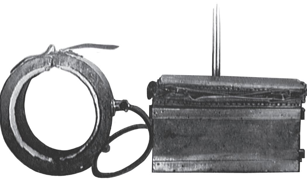
O’Dwyer 11 in 1887 reported the first use of long-term positive-pressure ventilation in a series of 50 children with croup. Shortly thereafter, Egon Braun and Alexander Graham Bell independently developed intermittent body-enclosing devices for the negative-pressure/positive-pressure resuscitation of newborns (Fig. 1-2).12,13 One might consider these seminal reports as the stimulus for the proliferation of work that
followed and the growing interest in mechanically ventilating newborn infants with respiratory failure.
TWENTIETH CENTURY
A variety of events occurred in the early twentieth century in the United States, including most notably the improvement of public health measures, the emergence of obstetrics as a full-fledged surgical specialty, and the assumption of care for all children by pediatricians.10 In 1914, the use of continuous positive airway pressure for neonatal resuscitation was described by Von Reuss.1 Henderson advocated positive-pressure ventilation via a mask with a T-piece in 1928.14 In the same year, Flagg recommended the use of an endotracheal tube with positive-pressure ventilation for neonatal resuscitation.15 The equipment he described was remarkably similar to that in use today.
Modern neonatology was born with the recognition that premature infants required particular attention with regard to temperature control, administration of fluids and nutrition, and protection from infection. In the 1930s and 1940s premature infants were given new stature, and it was acknowledged that of all of the causes of infant mortality, prematurity was the most common contributor.10
The years following World War II were marked by soaring birth rates, the proliferation of labor and delivery services in hospitals, the introduction of antibiotics, positive-pressure resuscitators, miniaturization of laboratory determinations, X-ray capability, and microtechnology that made intravenous therapy available for neonatal patients. These advances and a host of other discoveries heralded the modern era of neonatal medicine and set the groundwork for producing better methods of ventilating neonates with respiratory failure.
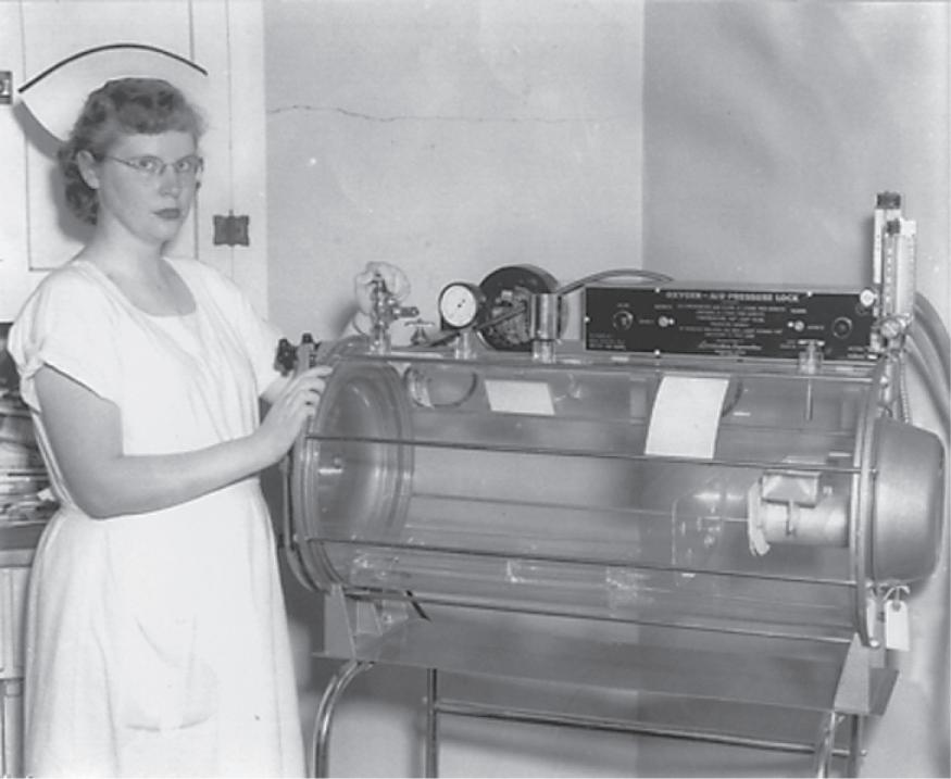
Improvements in intermittent negative-pressure and positive-pressure ventilation devices in the early twentieth century led to the development of a variety of techniques and machines for supporting ventilation in infants. In 1929, Drinker and Shaw16 reported the development of a technique for producing constant thoracic traction to produce an increase in endexpiratory lung volume. In the early 1950s, Bloxsom17 reported the use of a positive-pressure air lock for resuscitation of infants with respiratory distress in the delivery room. This device was similar to an iron lung; it alternately created positive and negative pressure of 1 to 3 psi at 1-min intervals in a tightly sealed cylindrical steel chamber that was infused with warmed humidified 60% oxygen.18 Clear plastic versions of the air lock quickly
1-3 Commercial Plexiglas version of the positive-pressure oxygen air lock. Arrival of the unit at the Dansville Memorial Hospital, Dansville, NY, June 1952. (Photo courtesy of James Gross and the Dansville Breeze. June 26, 1952.)
became commercially available in the United States in the early 1950s (Fig. 1-3). However, a study by Apgar and Kreiselman in 195319 on apneic dogs and another study by Townsend involving 150 premature infants20 demonstrated that the device could not adequately support the apneic newborn. The linkage of high oxygen administration to retinopathy of prematurity and a randomized controlled trial of the air lock versus care in an Isolette® incubator at Johns Hopkins University21 revealed no advantage to either study group and heralded the hasty decline in the use of the Bloxsom device.21
In the late 1950s, body-tilting devices were designed that shifted the abdominal contents to create more effective movement of the diaphragm. Phrenic nerve stimulation22 and the use of intragastric oxygen23 also were reported in the literature but had little clinical success. In the 1950s and early 1960s, many centers also used bag and tightly fitting face mask ventilation to support infants for relatively long periods of time.
The initial aspect of ventilator support for the neonate in respiratory failure was effective resuscitation. Varying techniques in the United States were published from the 1950s to the 1980s, but the first consensus approach was created by Bloom and Cropley in 1987 and adopted by the American Academy of Pediatrics as a standardized teaching program. A synopsis of the major events in the development of neonatal resuscitation is shown as a time line in Box 1-1.
The modern era of automated mechanical ventilation for infants can be dated back to the 1953 report of Donald and Lord,24 who described their experience with a patient-cycled, servo-controlled respirator in the treatment of several newborn infants with respiratory distress. They claimed that three or possibly four infants were successfully treated with their apparatus.
In the decades following Donald and Lord’s pioneering efforts, the field of mechanical ventilation made dramatic advances; however, the gains were accompanied by several temporary setbacks. Because of the epidemic of poliomyelitis in the 1950s, experience was gained with the use of the tanktype negative-pressure ventilators of the Drinker design.25 The success of these machines with children encouraged physicians to try modifications of them on neonates with some anecdotal success. However, initial efforts to apply intermittent positive-pressure ventilation (IPPV) to premature infants
FIG 1-2 Alexander Graham Bell’s negative-pressure ventilator, c. 1889. (From Stern L, et al. Can Med Am J. 1970.)
FIG
BOX 1-1 Neonatal Resuscitation Time Line
1300 BC: Hebrew midwives use mouth-to-mouth breathing to resuscitate newborns.
460-380 BC: Hippocrates describes intubation of trachea of humans to support respiration.
200 BC-500 AD: Hebrew text (Talmud) states, “we may hold the young so that it should not fall on the ground, blow into its nostrils and put the teat into its mouth that it should suck.”
98-138 AD: Greek physician Soranus describes evaluating neonates with system similar to present-day Apgar scoring, evaluating muscle tone, reflex or irritability, and respiratory effort. He believed that asphyxiated or premature infants and those with multiple congenital anomalies were “not worth saving.”
1135-1204: Maimonides describes how to detect respiratory arrest in newborns and describes a method of manual resuscitation.
1667: Robert Hooke presents to the Royal Society of London his experience using fireside bellows attached to the trachea of dogs to provide continuous ventilation.
1774: Joseph Priestley produces oxygen but fails to recognize that it is related to respiration. Royal Humane Society advocates mouth-tomouth resuscitation for stillborn infants.
1783-1788: Lavoisier terms oxygen “vital air” and shows that respiration is an oxidative process that produces water and carbon dioxide.
1806: Vide Chaussier describes intubation and mouth-to-mouth resuscitation of asphyxiated newborns.
1834: James Blundell describes neonatal intubation.
1874: Open chest cardiac massage reported in an adult.
1879: Report on the Aerophore Pulmonaire, a rubber bulb connected to a tube that is inserted into a neonate’s airway and then compressed and released to provide inspiration and passive expiration.
1889: Alexander Graham Bell designs and builds body-type respirator for newborns.
Late 1880s: Bonair administers oxygen to premature “blue baby.”
1949: Dr. Julius Hess and Evelyn C. Lundeen, RN, publish The Premature Infant and Nursing Care, which ushers in the modern era of neonatal medicine.
1953: Virginia Apgar reports on the system of neonatal assessment that bears her name.
1961: Dr. Jim Sutherland tests negative-pressure infant ventilator.
1971: Dr. George Gregory and colleagues publish results with continuous positive airway pressure in treating newborns with respiratory distress syndrome.
1987: American Academy of Pediatrics publishes the Neonatal Resuscitation Program based on an education program developed by Bloom and Cropley to teach a uniform method of neonatal resuscitation throughout the United States.
1999: The International Liaison Committee on Resuscitation (ILCOR) publishes the first neonatal advisory statement on resuscitation drawn from an evidence-based consensus of the available science. The ILCOR publishes an updated Consensus on Science and Treatment Recommendations for neonatal resuscitation every 5 years thereafter.
with respiratory distress syndrome (RDS) were disappointing overall. Mortality was not demonstrably decreased, and the incidence of complications, particularly that of pulmonary air leaks, seemed to increase.26 During this period, clinicians were hampered by the types of ventilators that were available and by the absence of proven standardized techniques for their use.
In accordance with the findings of Cournand et al.27 in adult studies conducted in the late 1940s, standard ventilatory technique often required that the inspiratory positive-pressure times be very short. Cournand et al. had demonstrated that the prolongation of the inspiratory phase of the ventilator cycle in patients with normal lung compliance could result in impairment of thoracic venous return, a decrease in cardiac output, and the unacceptable depression of blood pressure. To minimize
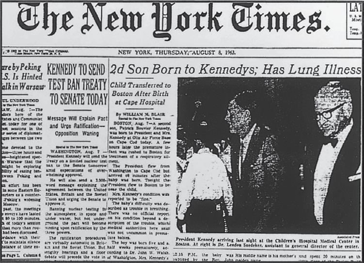
FIG 1-4 Front page of The New York Times. August 8, 1963. (Copyright 1963 by The New York Times Co. Reprinted by permission.)
cardiovascular effects, they advocated that the inspiratory phase of a mechanical cycle be limited to one-third of the entire cycle. Some ventilators manufactured in this period were even designed with the inspiratory-to-expiratory ratio fixed at 1:2.
Unfortunately, the findings of Cournand et al. were not applicable to patients with significant parenchymal disease, such as premature infants with RDS. Neonates with pulmonary disease characterized by poor lung compliance and complicated physiologically by increased chest wall compliance and terminal airway and alveolar collapse did not generally respond to IPPV techniques that had worked well in adults and older children. Clinicians were initially disappointed with the outcome of neonates treated with assisted ventilation using these techniques. The important observation of Avery and Mead in 1959 that babies who died from hyaline membrane disease (HMD) lacked a surface-active agent (surfactant), which increased surface tension in lung liquid samples and resulted in diffuse atelectasis, paved the way toward the modern treatment of respiratory failure in premature neonates by the constant maintenance of functional residual capacity and the eventual creation of surfactant replacement therapies.28
The birth of a premature son to President John F. Kennedy and Jacqueline Kennedy on August 7, 1963, focused the world’s attention on prematurity and the treatment of HMD, then the current appellation for RDS. Patrick Bouvier Kennedy was born by cesarean section at 34 weeks’ gestation at Otis Air Force Base Hospital. He weighed 2.1 kg and was transported to Boston’s Massachusetts General Hospital, where he died at 39 hours of age (Fig. 1-4). The Kennedy baby was treated with the most advanced therapy of the time, hyperbaric oxygen,29 but he died of progressive hypoxemia. There was no neonatal-specific ventilator in the United States to treat the young Kennedy at the time. In response to his death, The New York Times reported: “About all that can be done for a victim of hyaline membrane disease is to monitor the infant’s blood chemistry and try to keep it near normal levels.” The Kennedy tragedy, followed only 3 months later by the president’s assassination, stimulated further interest and research in neonatal respiratory diseases and resulted in increased federal funding in these areas.
Partially in response to the Kennedy baby’s death, several intensive care nurseries around the country (most notably at Yale, Children’s Hospital of Philadelphia, Vanderbilt, and the University of California at San Francisco) began programs focused on respiratory care of the premature neonate and the treatment of HMD. Initial success with ventilatory treatment
of HMD was reported by Delivoria-Papadopoulos and colleagues30 in Toronto, and as a result, modified adult ventilatory devices were soon in use in many medical centers across the United States. However, the initial anecdotal successes were also accompanied by the emergence of a new disease, BPD, first described in a seminal paper by Northway et al.31 in 1967. Northway initially attributed this disease to the use of high concentrations of inspired oxygen, but subsequent publications demonstrated that the cause of BPD was much more complex that and in addition to high inspired oxygen concentrations, intubation, barotrauma, volutrauma, infection, and other factors were involved. Chapter 35 discusses in great detail the current theories for the multiple causes of BPD or VILI.
BREAKTHROUGHS IN VENTILATION
A major breakthrough in neonatal ventilation occurred in 1971 when Gregory et al.32 reported on clinical trials with continuous positive airway pressure (CPAP) for the treatment of RDS. Recognizing that the major physiologic problem in RDS was the collapse of alveoli during expiration, they applied continuous positive pressure to the airway via an endotracheal tube or sealed head chamber (“the Gregory box”) during both expiration and inspiration; dramatic improvements in oxygenation and ventilation were achieved. Although infants receiving CPAP breathed spontaneously during the initial studies, later combinations of IPPV and CPAP in infants weighing less than 1500 g were not as successful.32 Nonetheless, the concept of CPAP was a major advance. It was later modified by Bancalari et al.33 for use in a constant distending negative-pressure chest cuirass and by Kattwinkel et al.,34 who developed nasal prongs for the application of CPAP without the use of an endotracheal tube.
The observation that administration of antenatal corticosteroids to mothers prior to premature delivery accelerated maturation of the fetal lung was made in 1972 by Liggins and Howie.35 Their randomized controlled trial demonstrated that the risks of HMD and death were significantly reduced in those premature infants whose mothers received antenatal steroid treatment.
Meanwhile, Reynolds and Taghizadeh,36,37 working independently in Great Britain, also recognized the unique pathophysiology of neonatal pulmonary disease. Having experienced difficulties with IPPV similar to those noted by clinicians in the United States, Reynolds and Taghizadeh suggested prolongation of the inspiratory phase of the ventilator cycle by delaying the opening of the exhalation valve. The “reversal” of the standard inspiratory-to-expiratory ratio, or “inflation hold,” allowed sufficient time for the recruitment of atelectatic alveoli in RDS with lower inflating pressures and gas flows, which, in turn, decreased turbulence and limited the effects on venous return to the heart. The excellent results of Reynolds and Taghizadeh could not be duplicated uniformly in the United States, perhaps because their American colleagues used different ventilators.
Until the early 1970s, ventilators used in neonatal intensive care units (NICUs) were modifications of adult devices; these devices delivered intermittent gas flows, thus generating IPPV. The ventilator initiated every mechanical breath, and clinicians tried to eliminate the infants’ attempts to breathe between IPPV breaths (“fighting the ventilator”), which led to rebreathing of dead air. In 1971, a new prototype neonatal ventilator was developed by Kirby and colleagues.38 This ventilator used continuous gas flow and a timing device to close
B A
To infant
Continuous gas flow
Continuous gas flow
FIG 1-5 Ayre’s T-piece forms the mechanical basis of most neonatal ventilators currently in use. A, Continuous gas flow from which an infant can breathe spontaneously. B, Occlusion of one end of the T-piece diverts gas flow under pressure into an infant’s lungs. The mechanical ventilator incorporates a pneumatically or electronically controlled time-cycling mechanism to occlude the expiratory limb of the patient circuit. Between sequential mechanical breaths, the infant can still breathe spontaneously. The combination of mechanical and spontaneous breaths is called intermittent mandatory ventilation. (From Kirby RR. Mechanical ventilation of the newborn. Perinatol Neonatol 5:47, 1981.)
the exhalation valve modeled after Ayre’s T-piece used in anesthesia (Fig. 1-5).24,36,38 Using the T-piece concept, the ventilator provided continuous gas flow and allowed the patient to breathe spontaneously between mechanical breaths. Occlusion of the distal end of the T-piece diverted gas flow under pressure to the infant. In addition, partial occlusion of the distal end generated positive end-expiratory pressure. This combination of mechanical and spontaneous breathing and continuous gas flow was called intermittent mandatory ventilation (IMV).
IMV became the standard method of neonatal ventilation and has been incorporated into all infant ventilators since then. One of its advantages was the facilitation of weaning by progressive reduction in the IMV rate, which allowed the patient to gradually increase spontaneous breathing against distending pressure. Clinicians no longer needed to paralyze or hyperventilate patients to prevent them from “fighting the ventilator.” Moreover, because patients continued to breathe spontaneously and lower cycling rates were used, mean intrapleural pressure was reduced and venous return was less compromised than with IPPV.39
Meanwhile, progress was also being made in the medical treatment and replacement of the cause of RDS, the absence or lack of adequate surfactant in the neonatal lung. Following the 1980 publication of a small series by Fujiwara et al. on the beneficial effect of exogenous surfactant in premature infants with HMD,40 several large randomized studies of the efficacy of surfactant were conducted. By the end of the decade the use of surfactant was well established. However, for decades there remained many controversies surrounding various treatment regimens (prophylactic vs rescue), types of surfactants, and dosing schedules.41
From 1971 to the mid-1990s, a myriad of new ventilators specifically designed for neonates were manufactured and sold.
The first generation of ventilators included the BABYbird 1®, the Bourns BP200®, and a volume ventilator, the Bourns LS 104/150®. All operated on the IMV principle and were capable of incorporating CPAP into the respiratory cycle (known as positive end-expiratory pressure [PEEP] when used with IMV).42
The BABYbird 1® and the Bourns BP200® used a solenoidactivated switch to occlude the exhalation limb of the gas circuit to deliver a breath. Pneumatic adjustments in the inspiratoryto-expiratory ratio and rate were controlled by inspiratory and expiratory times, which had to be timed with a stopwatch. A spring-loaded pressure manometer monitored peak inspiratory pressure and PEEP. These early mechanics created time delays within the ventilator, resulting in problems in obtaining short inspiratory times (less than 0.5 second).
In the next generation of ventilators, electronic controls, microprocessors, and microcircuitry allowed the addition of light-emitting diode monitors and provided clinicians with faster response times, greater sensitivity, and a wider range of ventilator parameter selection. These advances were incorporated into ventilators such as the Sechrist 100® and Bear Cub® to decrease inspiratory times to as short as 0.1 second and to increase ventilatory rates to 150 inflations per minute. Monitors incorporating microprocessors measured inspiratory and expiratory times and calculated inspiratory-to-expiratory ratios and mean airway pressure. Ventilator strategies abounded, and controversy regarding the best (i.e., least harmful) method for assisting neonatal ventilation arose. High-frequency positivepressure ventilation using conventional ventilators was also proposed as a beneficial treatment of RDS.43
Meanwhile, extracorporeal membrane oxygenation and true high-frequency ventilation (HFV) were being developed at a number of major medical centers.44,45 These techniques initially were offered as a rescue therapy for infants who did not respond to conventional mechanical ventilation. The favorable physiologic characteristics of HFV led some investigators to promote its use as an initial treatment of respiratory failure, especially when caused by RDS in very low birth-weight (VLBW) infants.46
A third generation of neonatal ventilators began to appear in the early 1990s. Advances in microcircuitry and microprocessors, developed as a result of the space program, allowed new dimensions in the development of neonatal assisted ventilation. The use of synchronized IMV, assist/control mode ventilation, and pressure support ventilation—previously used in the ventilation of only older children and adults—became possible in neonates because of the very fast ventilator response times. Although problems with sensing a patient’s inspiratory effort sometimes limited the usefulness of these new modalities, the advances gave hope that ventilator complications could be limited and that the need for sedation or paralysis during ventilation could be decreased. Direct measurement of some pulmonary functions at the bedside became a reality and allowed the clinician to make ventilatory adjustments based on physiologic data rather than on a “hunch.”
The mortality from HMD, now called RDS, decreased markedly from 1971 to 2007 owing to a multitude of reasons, some of which have been noted above. In the United States, the RDS mortality decreased from 268 per 100,000 live births in 1971 to 98 per 100,000 live births by 1985. From 1985 to 2007, the rate fell to 17 per 100,000 live births. Thus in a 36-year period, the mortality from RDS fell nearly 94%, owing in part to the improvements in ventilator technology, the development of medical adjuncts such as exogenous surfactant, and the skill of
the physicians, nurses, and respiratory therapists using these devices while caring for these fragile infants.47,48
Since 2005, an even newer generation of ventilators has been developed. These are microprocessor based, with a wide array of technological features including several forms of patient triggering, volume targeting, and pressure support modes and the ability to monitor many pulmonary functions at the bedside with ventilator graphics. As clinicians become more convinced that VILI is secondary to volutrauma more than barotrauma, the emphasis to control tidal volumes especially in the “micropremie” has resulted in some major changes in the technique of ventilation. Chapters 15 and 18-22 elaborate more fully on these advances.
Concurrent with these advances is an increased complexity related to controlling the ventilator and thus more opportunity for operator error. Some ventilators are extremely versatile and can function for patients of extremely low birth weight (less than 1000 g) to 70-kg adults. Although these ventilators are appealing to administrators who have to purchase these expensive machines for many different categories of patients in the hospital, they add increased complexity and patient safety issues in caring for neonates. Chapter 6 discusses some of these issues.
Respiratory support in the present-day NICU continues to change as new science and new technologies point the way to better outcomes with less morbidity, even for the smallest premature infants. However, as the technology of neonatal ventilators advanced, a concurrent movement away from intubation was gaining popularity in the United States. In 1987, a comparison of eight major centers in the National Institute of Child Health and Human Development group by Avery et al. reviewed oxygen dependency and death in VLBW babies at 28 days of age.49 Although all centers had comparable mortality, one center (Columbia Presbyterian Medical Center) had the lowest rate by far of CLD among the institutions. Columbia had adopted a unique approach to respiratory support of VLBW infants, emphasizing nasal CPAP as the first choice for respiratory support, whereas the other centers were using intubation and mechanical ventilation. Other centers were slow to adopt the Columbia approach, which used bubble nasal CPAP, but gradually institutions began using noninvasive techniques for at least the larger VLBW infants. A Cochrane review of multiple trials in 2012 concluded that the combined outcomes of death and BPD were lower in infants who had initial stabilization with nasal CPAP, and later rescue surfactant therapy if needed, compared to elective intubation and prophylactic surfactant administration (RR 1.12, 95% CI 1.02 to 1.24).50 In recent years, “noninvasive” respiratory support with the use of nasal CPAP, synchronized inspiratory positive airway pressure, RAM-assisted ventilation, and neuronally adjusted ventilatory assist has become a more widely used technique to support premature infants with respiratory distress in the hope of avoiding the trauma associated with intubation and VILI. Using a noninvasive approach as one potentially better practice, quality improvement programs to lower the rate of BPD have had mixed success. As of this writing, noninvasive ventilation has been supported by a number of retrospective and cohort studies, and there are some recent reports suggesting that the earlier use of noninvasive therapies has a role in treating neonates with respiratory disease and preventing the need for intubation to treat respiratory failure.
See Chapters 17, 19, and 21 for a more in-depth discussion of newer modes of neonatal assisted ventilation.
RECENT ADVANCES AND OUTCOMES
With the advances made in providing assisted ventilation to our most vulnerable patients, survival rates have improved dramatically. For babies born at less than 28 weeks’ gestation and less than 1000 g, survival reaches 85 to 90%. However, in recent years the emphasis has shifted from just survival to survival without significant neurologic deficit, CLD, or retinopathy of prematurity. Nonetheless, benchmarking groups such as the Vermont–Oxford Network have shown a wide variance in these untoward outcomes that cannot be explained by variances in the patient population alone. CLD in infants born at <1500 g birth weight (VLBW infants) varies from 6% to over 50% in various NICUs. Thus it appears that overall advances in the morbidity and mortality rates of the VLBW infant group as a whole will be made from more uniform application of technology already available rather than the creation of new devices or medications. Moreover, despite continued technological advances in respiratory support since 2007, there have been only minor improvements in morbidity and mortality rates in high-resource countries. Perhaps entirely new approaches are necessary to produce major leaps forward in the treatment of neonatal respiratory failure. However, in resource-limited areas throughout the world, the use of basic respiratory support technologies (i.e., CPAP, resuscitation techniques) has the potential to have a major impact on the outcomes of newborns (see Chapter 38).
Despite the wide array of technology now available to the clinician treating neonatal respiratory failure, there are still significant limitations and uncertainty about our care. We continue to research and discuss issues such as conventional vs high-frequency ventilation, noninvasive ventilation vs the early administration of surfactant, the best ventilator mode, the best rate, the optimum settings, and the most appropriate approach to weaning and extubation. There are very few randomized controlled trials that demonstrate significant differences in morbidity or mortality related to new ventilator technologies or strategies. This is due to the difficulty in enrolling neonates into clinical trials, the large number of patients needed to detect statistical differences in outcomes, the reluctance of device manufacturers to support expensive studies, and the rapidly changing software, which make it difficult for research to keep up with the technological advances. 51 It is the editors’ expectations that this book will provide some more food for thought in these areas and the necessary information for the physician, nurse, or respiratory therapist involved in the care of neonates to provide the best possible care based on the information available in 2016 and beyond.
REFERENCES
A complete reference list is available at https://expertconsult .inkling.com/
REFERENCES
1. Wiswell TE, Gibson AT: Historical Evolution of Neonatal Resuscitation. American Academy of Pediatrics, Neonatal Resuscitation Program, Instructor Resources, 2005.
2. Obladen M: History of neonatal resuscitation. Part 1: artificial ventilation. Neonatology 94:144, 2008.
3. O’Donnell CPF, Gibson AT, Davis PG: Pinching, electrocution, ravens’ beaks and positive pressure ventilation: a brief history of neonatal resuscitation. Arch Dis Child Fetal Neonatal Ed 91:F369-F373, 2006.
4. Stern L, Ramos AD, Outerbridge EW, et al: Negative pressure artificial respiration use in treatment of respiratory failure of the newborn. Can Med Assoc J 102:595, 1970.
5. Raju TNK: History of neonatal resuscitation: tales of heroism and desperation. Clin Perinatol 26:629-640, 1999.
6. Bourgeois L: Observations diverses sur la stérilité, perte de fruits, fécondité, accouchements, et maladies des femmes, et enfants nouveaux-nés, suivi de instructions à ma fille. Paris, Dehoury, 1609, Reprint Paris CoteFemmes Editions. 1992, p 95 (As reported in reference 1 A by Obladen).
7. Thatcher VS: History of Anesthesia, with Emphasis on the Nurse Specialists. Philadelphia, Lippincott, 1953.
8. Faulconer Jr A, Keys TE: Foundation of Anesthesiology. Springfield, Ill, Charles C Thomas, 1965.
9. Dunn PM: Charles-Michel Billard (1800-1832): pioneer of neonatal medicine. Arch Dis Child 65:711, 1990.
10. Desmond MM: A review of newborn medicine in America: European past and guiding ideology. Am J Perinatol 8:308, 1991.
11. O’Dwyer J: Fifty cases of croup in private practice treated by intubation of the larynx with a description of the method and of the danger incident thereto. Med Res 32:557, 1887.
12. Doe OW: Apparatus for resuscitating asphyxiated children. Boston Med Surg J 9:122, 1889.
13. Stern L, Ramos AD, Outerbridge EW, et al: Negative pressure artificial respiration use in treatment of respiratory failure of the newborn. Can Med Assoc J 102:595, 1970.
14. Henderson Y: The prevention and treatment of asphyxia in the newborn. JAMA 90:583-586, 1928.
15. Flagg PJ: Treatment of asphyxia in the newborn. JAMA 91:788-791, 1928.
16. Drinker P, Shaw LA: An apparatus of the prolonged administration of artificial respiration: 1. A design for adults and children. J Clin Invest 7:229, 1929.
17. Bloxsom A: Resuscitation of the newborn infant: use of positive pressure oxygen-air lock. J Pediatr 37:311, 1950.
18. Kendig JW, Maples PG, Maisels MJ: The Bloxsom air lock: a historical perspective. Pediatrics 108:e116, 2001.
19. Apgar V, Kreiselman J: Studies on resuscitation. An experimental evaluation of the Bloxsom air lock. Am J Obstet Gynecol 65:45, 1953.
20. Townsend Jr EH: The oxygen air pressure lock I. Clinical observations on its use during the neonatal period. Obstet Gynecol 4:184, 1954.
21. Reichelderfer TE, Nitowsky HM: A controlled study on the use of the Bloxsom air lock. Pediatrics 18:918-927, 1956.
22. Cross K, Roberts P: Asphyxia neonatorum treated by electrical stimulation of the phrenic nerve. BMJ 1:1043, 1951.
23. James LS, Apgar B, Burnard ED, et al: Intragastric oxygen and resuscitation of the newborn. Acta Pediatr Scand 52:245, 1963.
24. Donald I, Lord J: Augmented respiration: studies in atelectasis neonatorum. Lancet 1:9, 1953.
25. Stahlman MT: Assisted ventilation in newborn infants. In Smith GF, Vidyasagar D (eds): Historical Reviews and Recent Advances in Neonatal and Perinatal Medicine. vol. II. Evansville, IN, Mead Johnson Nutritional Division, 1984.
26. Kirby RR: Mechanical ventilation of the newborn. Perinatol Neonatal 5:47, 1981.
27. Cournand A, Motley HL, Werko L, et al: Physiological studies of the effects of intermittent positive pressure breathing on cardiac output in man. Am J Physiol 152:162, 1948.
28. Avery ME, Mead J: Surface properties in relation to atelectasis and hyaline membrane disease. AMA J Dis Child 97:517-527, 1959.
29. Cochran WD, Levison H, Muirhead DM, et al: A clinical trial on high oxygen pressure for the respiratory distress syndrome. N Engl J Med 212:347, 1965.
30. Delivoria-Papadopoulos M, Levison H, Swyer PR: Intermittent positive pressure respiration as a treatment in severe respiratory distress syndrome. Arch Dis Child 40:474-479, 1965.
31. Northway Jr WH, Rosan RC, Porter DY: Pulmonary disease following respiratory therapy of hyaline-membrane disease. Bronchopulmonary dysplasia. N Engl J Med 276:357, 1967.
32. Gregory GA, Kitterman JA, Phibbs RH, et al: Treatment of the idiopathic respiratory-distress syndrome with continuous positive airway pressure. N Engl J Med 284:1333, 1971.
33. Bancalari E, Garcia OL, Jesse MJ: Effects of continuous negative pressure on lung mechanics in idiopathic respiratory distress syndrome. Pediatrics 51:485, 1973.
34. Kattwinkel J, Fleming D, Cha CC, et al: A device for administration of continuous positive airway pressure by nasal route. Pediatrics 52:131, 1973.
35. Liggins GC, Howie RN: A controlled trial of antepartum glucocorticoid treatment for prevention of the respiratory distress syndrome in premature infants. Pediatrics 50:515-525, 1972.
36. Reynolds EOR: Pressure waveform and ventilator setting for mechanical ventilation in severe hyaline membrane disease. Int Anesthesiol Clin 12:269, 1974.
37. Reynolds EOR, Taghizadeh A: Improved prognosis of infants mechanically ventilated for hyaline membrane disease. Arch Dis Child 49:505, 1974.
38. Kirby R, Robison E, Schulz J, et al: Continuous flow ventilation as an alternative to assisted or controlled ventilation in infants. Anesth Analg 51:871, 1972.
39. Kirby RR: Intermittent mandatory ventilation in the neonate. Crit Care Med 5:18, 1977.
40. Fujiwara T, Maeta H, Chida S, et al: Artificial surfactant therapy in hyaline membrane disease. Lancet 1:55-59, 1980.
41. Soll RF, Blanco F: Natural surfactant extract versus synthetic surfactant for neonatal respiratory distress syndrome. Cochrane Database Syst Rev 2:CD000144, 2001.
42. Cassani III VL: We’ve come a long way baby! Mechanical ventilation of the newborn. Neonatal Netw 13:63, 1994.
43. Bland RD, Sedin EG: High frequency mechanical ventilation in the treatment of neonatal respiratory distress. Int Anesthesiol Clin 21:125, 1983.
44. Slutsky AS, Drazen FM, Ingram Jr RH, et al: Effective pulmonary ventilation with small-volume oscillations at high frequency. Science 209:609, 1980.
45. Pesenti A, Gottinoni L, Bombino L: Long-term extracorporeal respiratory support: 20 years of progress. Intern Ext Care Digest 12:15, 1993.
46. Frantz III ID, Werthammen J, Stark AR: High-frequency ventilation in premature infants with lung disease: adequate gas exchange at low tracheal pressure. Pediatrics 71:483, 1983.
47. Singh GK, Yu SM: Infant mortality in the United States: trends, differential, and projections 1950 through 2010. Am J Public Health 85(7):957964, 1995.
48. Heron M, Sutton PD, Xu J, et al: Annual review of vital statistics: 2007. Pediatrics 125(1):4-15, 2010.
49. Avery ME, Tooley WH, Keller JB, et al: Is chronic lung disease in low birth weight infants preventable? Pediatrics 79(1):26-30, 1987.
50. Rojas-Reyes MX, Mortley CJ, Soll R: Prophylactic versus selective use of surfactant in preventing morbidity and mortality in preterm infants. Cochrane Database Syst Rev 3:CD000510, 2012.
51. Brown MK, DiBlasi RM: Mechanical ventilation of the premature neonate. Respir Care 56(9):1298-1313, 2011.
Physiologic Principles*
Martin Keszler, MD, FAAP, and Kabir Abubakar, MD
To provide individualized care that optimizes pulmonary and neurodevelopmental outcomes, it is essential to have a good working knowledge of the unique physiology and pathophysiology of the newborn respiratory system.
It is the responsibility of those who care for critically ill infants to have a sound understanding of respiratory physiology, especially the functional limitations and the special vulnerabilities of the immature lung. The first tenet of the Hippocratic Oath states, “Primum non nocere” (“First do no harm”). That admonition cannot be followed without adequate knowledge of physiology. In daily practice, we are faced with the difficult task of supporting adequate gas exchange in an immature respiratory system, using powerful tools that by their very nature can inhibit ongoing developmental processes, often resulting in alterations in end-organ form and function.
In our efforts to provide ventilatory support, the infant’s lungs and airways are subjected to forces that may lead to acute and chronic tissue injury. This results in alterations in the way the lungs develop and the way they respond to subsequent noxious stimuli. Alterations in lung development result in alterations in lung function as the infant’s body attempts to heal and continue to develop. Superimposed on this is the fact that the ongoing development of the respiratory system is hampered by the healing process itself.
This complexity makes caring for infants with respiratory failure both interesting and challenging. To effectively provide support for these patients, the clinician must have an understanding not only of respiratory physiology but also of respiratory system development, growth, and healing.
Although the lung has a variety of functions, some of which include the immunologic and endocrine systems, the focus of this chapter is its primary function, that of gas exchange.
BASIC BIOCHEMISTRY OF RESPIRATION: OXYGEN AND ENERGY
The energy production required for a newborn infant to sustain his or her metabolic functions depends upon the availability of oxygen and its subsequent metabolism. During the breakdown of carbohydrates, oxygen is consumed and carbon dioxide and water are produced. The energy derived from this process is generated as electrons, which are transferred from electron donors to electron acceptors. Oxygen has a high electron affinity and therefore is a good electron acceptor. The energy produced during this process is stored as high-energy phosphate bonds,
* We wish to acknowledge gratefully the important contribution of Brian Wood, MD, who was the author of this chapter in the previous editions of this book.
primarily in the form of adenosine triphosphate (ATP). Enzyme systems within the mitochondria couple the transfer of energy to oxidation in a process known as oxidative phosphorylation. 1
For oxidative phosphorylation to occur, an adequate amount of oxygen must be available to the mitochondria. The transfer of oxygen from the air outside the infant to the mitochondria, within the infant’s cells, involves a series of steps: (1) convection of fresh air into the lung, (2) diffusion of oxygen into the blood, (3) convective flow of oxygenated blood to the tissues, (4) diffusion of oxygen into the cells, and finally, (5) diffusion into the mitochondria. The driving force for the diffusion processes is an oxygen partial pressure gradient, which, together with the convective processes of ventilation and perfusion, results in a cascade of oxygen tensions from the air outside the body to intracellular mitochondria (Fig. 2-1). The lungs of the newborn infant transfer oxygen to the blood by diffusion, driven by the oxygen partial pressure gradient. For gas exchange to occur efficiently, the infant’s lungs must remain expanded, the lungs must be both ventilated and perfused, and the ambient partial pressure of oxygen in the air must be greater than the partial pressure of the oxygen in the blood. The efficiency of the newborn infant’s respiratory system is determined by both structural and functional constraints; therefore, the clinician must be mindful of both aspects when caring for the infant.
The infant’s cells require energy to function. This energy is obtained from high-energy phosphate bonds (e.g., ATP) formed during oxidative phosphorylation. Only a small amount of ATP is stored within the cells. Muscle cells contain an additional store of ATP, but to meet metabolic needs beyond those that can be provided for by the stored ATP, new ATP must be made by phosphorylation of adenosine diphosphate (ADP).
Inspired gas: PO2 = 168 torr
Alveolar gas: PO2 = 100 torr
Arterial blood PO2 = 90 torr
Capillary blood PO2 = 40 torr
Extracellular fluid PO2 = 30 torr
Intracellular fluid PO2 = 10 torr
FIG 2-1 Transfer of oxygen from outside air to intracellular mitochondria via an oxygen pressure gradient: oxygen tension at various levels of the O2 transport chain.
This can be done anaerobically through glycolysis, but this is an inefficient process and leads to the formation of lactic acid. Long-term energy demands must be met aerobically, through ongoing oxidative phosphorylation within the mitochondria, which is a much more efficient process that results in the formation of carbon dioxide and water.
There is a hierarchy of how energy is used by the infant. During periods of high energy demand, tissues initially draw upon the limited stores of ATP, then use glycolysis to make more ATP from ADP, and then use oxidative phosphorylation to supply the infant’s ongoing energy requirements. Oxidative phosphorylation and oxygen consumption are so closely linked to the newborn infant’s energy requirements that total oxygen consumption is a reasonably good measure of the total energy needs of the infant. When the infant’s metabolic workload is in excess of that which can be sustained by oxidative phosphorylation (aerobic metabolism), the tissues will revert to anaerobic glycolysis to produce ATP. This anaerobic metabolism results in the formation of lactic acid, which accumulates in the blood and causes a decrease in pH (acidosis/acidemia). Lactic acid is therefore an important marker of inadequate tissue oxygen delivery.
ONTOGENY RECAPITULATES PHYLOGENY: A BRIEF OVERVIEW OF DEVELOPMENTAL ANATOMY
Lung Development
The tracheobronchial airway system begins as a ventral outpouching of the primitive foregut, which leads to the formation of the embryonic lung bud. The lung bud subsequently divides and branches, penetrating the mesenchyma and progressing toward the periphery. Lung development is divided into five sequential phases.2 The demarcation of these phases is somewhat arbitrary with some overlap between them. A variety of physical, hormonal, and other factors influence the pace of lung development and maturation. Adequate distending pressure of fetal lung fluid and normal fetal breathing movements are some of the more prominent factors known to affect lung growth and development.
Phases of Lung Development
• Embryonic phase (weeks 3 to 6)
• Pseudoglandular phase (weeks 6 to 16)
• Canalicular phase (weeks 16 to 26)
• Terminal sac phase (weeks 26 to 36)
• Alveolar phase (week 36 to 3 years)
Embryonic Phase (Weeks 3 to 6): Development of Proximal Airways
The lung bud arises from the foregut 21 to 26 days after fertilization.
Aberrant development during the embryonic phase may result in the following:
• Tracheal agenesis
• Tracheal stenosis
• Tracheoesophageal fistula
• Pulmonary sequestration (if an accessory lung bud develops during this period)
Pseudoglandular Phase (Weeks 6 to 16): Development of Lower Conducting Airways
During this phase the first 20 generations of conducting airways develop. The first 8 generations (the bronchi) ultimately acquire cartilaginous walls. Generations 9 to 20 comprise the
nonrespiratory bronchioles. Lymph vessels and bronchial capillaries accompany the airways as they grow and develop. Aberrant development during the pseudoglandular phase may result in the following:
• Bronchogenic cysts
• Congenital lobar emphysema
• Congenital diaphragmatic hernia
Canalicular Phase (Weeks 16 to 26): Formation of GasExchanging Units or Acini
The formation of respiratory bronchioles (generations 21 to 23) occurs during the canalicular phase. The relative proportion of parenchymal connective tissue diminishes. The development of pulmonary capillaries occurs. Gas exchange depends upon the adequacy of acinus–capillary coupling.
Terminal Sac Phase (Weeks 26 to 36): Refinement of Acini
The rudimentary primary saccules subdivide by formation of secondary crests into smaller saccules and alveoli during the terminal sac phase, thus greatly increasing the surface area available for gas exchange. The interstitium continues to thin out, decreasing the distance for diffusion. Capillary invasion leads to an increase in the alveolar–blood barrier surface area. The development and maturation of the surfactant system occurs during this phase. Birth and initiation of spontaneous or mechanical ventilation during the terminal sac phase may result in the following:
• Pulmonary insufficiency of prematurity (due to reduced surface area, increased diffusion distance, and unfavorable lung mechanics)
• Respiratory distress syndrome (due to surfactant deficiency and/or inactivation)
• Pulmonary interstitial emphysema (due to tissue stretching by uneven aeration, excessive inflating pressure, and increased interstitium that traps air in the perivascular sheath)
• Impairment of secondary crest formation and capillary development, leading to alveolar simplification, decreased surface area for gas exchange, and variable increase in interstitial cellularity and/or fibroproliferation (bronchopulmonary dysplasia [BPD]).
Alveolar Phase (Week 36 to 3 Years): Alveolar Proliferation and Development
Saccules become alveoli as a result of thinning of the acinar walls, dissipation of the interstitium, and invagination of the alveoli by pulmonary capillaries with secondary crest formation during the alveolar phase. The alveoli attain a polyhedral shape.
MECHANICS
The respiratory system is composed of millions of air sacs that are connected to the outside air via airways. The lung behaves like a balloon that is held in an expanded state by the intact thorax and will deflate if the integrity of the system becomes compromised. The interior of the lung is partitioned so as to provide a large surface area to facilitate efficient gas diffusion. The lung is expanded by forces generated by the diaphragm and the intercostal muscles. It recoils secondary to elastic and surface tension forces. This facilitates the inflow and outflow of respiratory gases required to allow the air volume contained within the lung to be ventilated. During inspiration the diaphragm contracts. The diaphragm is a dome-shaped muscle at rest. As it contracts, the diaphragm flattens, and the volume of
the chest cavity is enlarged. This causes the intrapleural pressure to decrease and results in gas flow into the lung.3 During unlabored breathing, the intercostal and accessory muscles serve primarily to stabilize the rib cage as the diaphragm contracts, countering the forces resulting from the decrease in intrapleural pressure during inspiration. This limits the extent to which the infant’s chest wall is deformed inward during inspiration.
Although the premature infant’s chest is very compliant, the rib cage offers some structural support, serves as an attachment point for the respiratory muscles, and limits lung deflation at end expiration. The elastic elements of the respiratory system—the connective tissue—are stretched during inspiration and recoil during expiration. The air–liquid interface in the terminal air spaces and respiratory bronchioles generates surface tension that opposes lung expansion and promotes lung deflation. The conducting airways, which connect the gas exchange units to the outside air, provide greater resistance during exhalation than during inspiration, because during inspiration, the tethering elements of the surrounding lung tissue increase the airway diameter, relative to expiration. The respiratory system is designed to be adaptable to a wide range of workloads; however, in the newborn infant, several structural and functional limitations make the newborn susceptible to respiratory failure. Differences between the shape of a newborn infant’s chest and that of an adult put the infant at a mechanical disadvantage. Unlike the adult’s thorax, which is ellipsoid in shape, the infant’s thorax is more cylindrical and the ribs are more horizontal, rather than oblique. Because of these anatomic differences, the intercostal muscles in infants have a shorter course and provide less mechanical advantage for elevating the ribs and increasing intrathoracic volume during inspiration than do those of adults. Also, because the insertion of the infant’s diaphragm is more horizontal than in the adult, the lower ribs tend to move inward rather than upward during inspiration. The compliant chest wall of the infant exacerbates this inward deflection with inspiration. This is particularly evident during rapid eye movement (REM) sleep, when phasic changes in intercostal muscle tone are inhibited. Therefore, instead of stabilizing the rib cage during inspiration, the intercostal muscles are relaxed. This results in inefficient respiratory effort, which may be manifested clinically by intercostal and substernal retractions associated with abdominal breathing, especially when lung compliance is decreased. The endurance capacity of the diaphragm is determined primarily by muscle mass and the oxidative capacity of muscle fibers. Infants have low muscle mass and a low percentage of type 1 (slow twitch) muscle fibers compared to those of adults.4 To sustain the work of breathing, the diaphragm must be provided with a continuous supply of oxygen. The infant with respiratory distress is thus prone to respiratory muscle fatigue leading to respiratory failure. During expiration the main driving force is elastic recoil, which depends on the surface tension produced by the air–liquid interface, the elastic elements of the lung tissue, and the bony development of the rib cage. Expiration is largely passive. The abdominal muscles can aid in exhalation by active contraction if required, but they make little contribution during unlabored breathing. Because the chest wall of premature infants is compliant, it offers little resistance against expansion upon inspiration and little opposition against collapse upon expiration.
This collapse at end expiration can lead to atelectasis. In premature infants the largest contributor to elastic recoil is surface tension. Pulmonary surfactant serves to reduce surface tension and stabilize the terminal airways. In circumstances in which
surfactant is deficient, the terminal air spaces have a tendency to collapse, leading to diffuse atelectasis. Distending airway pressure in the form of positive end-expiratory pressure (PEEP) or continuous positive airway pressure (CPAP) may be applied to the infant’s airway to counter the tendency toward collapse and the development of atelectasis. The application of airwaydistending pressure also serves to stabilize the chest wall.
Lung compliance and airway resistance are related to lung size. The smaller the lung, the lower the compliance and the greater the resistance. If, however, lung compliance is corrected to lung volume (specific compliance), the values are nearly identical for term infants and adults.5 In term infants, immediately after delivery, specific compliance is low but normalizes as fetal lung fluid is absorbed and a normal functional residual capacity (FRC) is established. In premature infants, specific compliance remains low, due in part to diffuse microatelectasis and failure to achieve a normal FRC, because the lung recoil forces are incompletely opposed by the excessively compliant chest wall.
The resistance within lung tissue during inflation and deflation is called viscous resistance. Viscous resistance is elevated in the newborn. In immature small lungs, there are relatively fewer terminal air spaces and relatively more stroma (cells and interstitial fluid). This is manifested by a low ratio of lung volume to lung weight. Although in absolute terms airway resistance is high in the newborn infant, when corrected to lung volume (specific conductance, which is the reciprocal of resistance per unit lung volume), the relative resistance is lower than in adults. It is important to remember that because of the small diameter of the airways in the lungs of the newborn infant, even a modest further narrowing, will result in a marked increase in resistance. That the newborn’s bronchial tree is short and the inspiratory flow velocities are low are teleologic advantages for the newborn because both of these factors decrease airway resistance.
Overcoming the elastic and resistive forces during ventilation requires energy expenditure and accounts for the work of breathing. The normal work of breathing is essentially the same for newborns and adults when corrected for metabolic rates.5 When the work of breathing increases in response to various disease states, the newborn is at a decided disadvantage. The newborn infant lacks the strength and endurance to cope with a significant increase in ventilatory workload. A large increase in ventilatory workload can lead to respiratory failure.
Elastic and resistive forces of the chest, lungs, abdomen, airways, and ventilator circuit oppose the forces exerted by the respiratory muscles and/or ventilator. The terms elastic recoil, flow resistance, viscous resistance, and work of breathing are used to describe these forces. Such forces may also be described as dissipative and nondissipative forces. The latter refers to the fact that the work needed to overcome elastic recoil is stored like the energy in a coiled spring and will be returned to the system upon exhalation. Resistive and frictional forces, on the other hand, are lost and converted to heat (dissipated). The terms elasticity, compliance, and conductance characterize the properties of the thorax, lungs, and airways. The static pressure–volume curve illustrates the relationships between these forces at various levels of lung expansion. Dynamic pressure–volume loops illustrate the pressure–volume relationship during inspiration and expiration (Figs. 2-2 to 2-4).
Elastic recoil refers to the tendency of stretched objects to return to their original shape. When the inspiratory muscles relax during exhalation, the elastic elements of the chest wall, diaphragm, and lungs, which were stretched during inspiration, recoil to their original shapes. These elastic elements behave like springs (Fig. 2-5). The surface tension forces at the air–liquid
FIG 2-2 Static pressure–volume curves for the chest wall, the lung, and the sum of the two for a normal newborn infant. Functional residual capacity (FRC) or rest volume (less than 20% of total lung capacity) is the point at which collapsing and distending pressures balance out to zero pressure. The lung would empty to residual volume if enough collapsing pressure (forced expiration) was generated to overcome the chest wall elastic recoil in the opposite direction. The premature infant has an even steeper chest wall compliance curve than that shown here, whereas his or her lung compliance curve tends to be flatter and shifted to the right, depending on the degree of surfactant deficiency.
FIG 2-4 Comparison of the pressure–volume curve of a normal infant (solid line) with that of a newborn with respiratory distress syndrome (dotted line). Note that very little hysteresis (i.e., the difference between the inspiratory and the expiratory limbs) is observed in the respiratory distress syndrome curve because of the lack of surfactant for stabilization of the alveoli after inflation. The wide hysteresis of the normal infant’s lung curve reflects changes (reduction) in surface tension once the alveoli are opened and stabilized. RDS, Respiratory distress syndrome.
FIG 2-3 Extended compliance or lung expansion curve with “flattened” areas (A and C ) at both ends. Area A represents the situation in disease states leading to atelectasis or lung collapse. Area C represents the situation in an overexpanded lung, as occurs in diseases involving significant air trapping (e.g., meconium aspiration) or in the excessive application of distending pressure during assisted ventilation. FRC, Functional residual capacity.
FIG 2-5 Elastic recoil is the tendency of elements in the chest wall and lungs that are stretched during inspiration to snap back or recoil (arrows) to their original state at the end of expiration. At this point (functional residual capacity or rest volume), the “springs” are relaxed and the structure of the rib cage allows no further collapse. Opposing forces of the chest wall and elastic recoil balance out, and intrathoracic and airway pressures become equal (this further defines functional residual capacity or rest volume; see also Fig. 2-2).
AB
FIG 2-6 Diagrammatic illustration of the Laplace relationship and the effects of (A) surfactant film and (B) alveolar radius on wall or surface tension. The degree (reflected in the size of the brown arrows) of airway or intra-alveolar pressure (P) needed to counteract the tendency of alveoli to collapse (represented by the black arrows) is directly proportional to double the wall or surface tension (ST) and inversely proportional to the size of the radius (r). Distending airway pressure applied during assisted ventilation can be likened to a “pneumatic splint.”
early airway closure and collapse, with resultant gas trapping in premature infants.
The respiratory system’s resting volume is very close to the closing volume of the lung (the volume at which dependent lung regions cease to ventilate because the airways leading to them have collapsed). In newborns, closing volume may occur even above FRC (see Fig. 2-2).6 Gas trapping related to airway closure has been demonstrated experimentally by showing situations in which the thoracic gas volume is greater than the FRC. For this to occur, the total gas volume measured in the chest at end expiration is greater than the amount of gas that is in communication with the upper airway (FRC).
The main contributor to lung elastic recoil in the newborn is surface tension. The pressure required to counteract the tendency of the bronchioles and terminal air spaces to collapse is described by the Laplace relationship:
FIG 2-7 Pressure–volume loop showing the compliance line (AC, joining points of no flow); work done in overcoming elastic resistance (ACEA), which incorporates the frictional resistance encountered during expiration (ACDA); work done in overcoming frictional resistance during inspiration (ABCA); and total work done during the respiratory cycle (ABCEA, or the entire shaded area).
interfaces in the distal bronchioles and terminal airways decrease the surface area of the air–liquid interfaces (Fig. 2-6).
At some point, the forces that tend to collapse are counterbalanced by those that resist further collapse. The point at which these opposing forces balance is called the resting state of the respiratory system and corresponds to FRC (Fig. 2-7; see also Fig. 2-2). Because the chest wall of the newborn infant is compliant, it offers little opposition to collapse at end expiration. Thus the newborn, especially the premature newborn, has a relatively low FRC and thoracic gas volume, even when the newborn does not suffer from primary surfactant deficiency. Clinically, this manifests as a mild degree of diffuse microatelectasis and is referred to as pulmonary insufficiency of prematurity. This low FRC and the relative underdevelopment of the conducting airway’s structural support explain the tendency for
Simply stated, this relationship illustrates that the pressure ( P ) needed to stabilize the system is directly proportional to twice the surface tension (2 ST ) and inversely proportional to the radius of curvature ( r ). In infants, the relationship should be modified, because, unlike in a soap bubble, there is an air–liquid interface on only one side of the terminal lung unit, so P = ST / r probably describes the situation more accurately in the lung.
In reality, alveoli are not spherical but polyhedral and share their walls with adjacent alveolar structures, making strict application of Laplace’s law suspect. Nonetheless, the basic concept of the law does apply to both terminal air sacs and small airways, and it provides a crucial framework for the understanding of respiratory physiology. The surface tension in the lung is primarily governed by the presence or absence of surfactant. Surfactant is a surface-active material released by type II pneumocytes. It is composed mainly of dipalmitoyl phosphatidylcholine but contains other essential components, such as surfactant-associated proteins A, B, C, and D, as well.
Surfactant has a variety of unique properties that enable it to decrease surface tension at end expiration and thereby prevent
further lung deflation below resting volume and allow an increase in surface tension upon lung expansion that facilitates elastic recoil at end inspiration. In addition, surfactant reduces surface tension when lung volume is decreased.7 A reduction in the quantity of surfactant results in an increase in surface tension and necessitates the application of more distending pressure to counter the tendency of the bronchioles and terminal air spaces to collapse (see Fig. 2-6, A).
As can be seen from the Laplace relationship, the larger the radius of curvature of the terminal bronchioles or air spaces, the less pressure is needed to hold them open or to expand them further (see Fig. 2-6, B). The smaller the radius of curvature (e.g., in premature infants), the more pressure is required to hold the airways open. Surfactant helps this situation throughout the respiratory cycle. As the radii of the air–liquid interfaces become smaller during exhalation, the effectiveness of surfactant in reducing surface tension increases; as the radii become larger, its effectiveness decreases.
Respiratory distress syndrome (RDS) imposes a significant amount of energy expenditure on the newborn infant, who must generate high negative intrapleural pressures to expand and stabilize his or her distal airways and alveoli (see Fig. 2-4). In untreated RDS, each breath requires significant energy expenditure because lung volumes achieved with the high opening pressures during inspiration are rapidly lost as the surfactant-deficient lung collapses to its original resting volume during expiration. The burden imposed by this large work of breathing may quickly outstrip the infant’s ability to maintain this level of output and lead to respiratory failure.
The infant with RDS may need relatively high inflation pressure to open atelectatic alveoli, and provision of adequate end-expiratory pressure will help keep the lung open. However, once the lung is expanded, the radii of the bronchioles and terminal air spaces are larger, and, therefore, less pressure is required to hold them open or to expand them further. Attention should be paid to tidal volume and overall lung volume after initial alveolar recruitment to avoid overdistention and volutrauma, which are major factors in the development of BPD.8 Failure to reduce inspiratory and distending pressures appropriately and thus avoid lung overdistention once normal lung volume has been achieved may lead to air-leak complications such as pulmonary interstitial emphysema (PIE) and pneumothorax (see Chapter 20).
Compliance
Compliance is a measure of the change in volume resulting from a given change in pressure: CL = ∆V/ ∆P
where CL is lung compliance, ΔV is change in volume, and ΔP is change in pressure.
Static Compliance
When measured under static conditions, compliance reflects only the elastic properties of the lung. Static compliance is the reciprocal of elastance, the tendency to recoil toward its original dimensions upon removal of the distending pressure required to stretch the system. Static compliance is measured by determining the transpulmonary pressure change after inflating the lungs with a known volume of gas. Transpulmonary pressure is the pressure difference between alveolar pressure and pleural
pressure. It is approximated by measuring pressure at the airway opening and in the esophagus. To generate a pressure–volume curve, pressure measurements are made during static conditions after each incremental volume of gas is introduced into the lungs (see lung curve in Fig. 2-2). If one measures the difference between pleural pressures (esophageal) and atmospheric pressures (transthoracic) at different levels of lung expansion, the plotted curve will be a chest wall compliance curve (see chest wall curve in Fig. 2-2). This kind of plot shows the elastic properties of the chest wall. In the newborn, the chest wall is very compliant; thus large volume changes are achieved with small pressure changes. Taking the lung and chest wall compliance curves together gives the total respiratory system compliance (see the total curve in Fig. 2-2).
Dynamic Compliance
If one measures compliance during continuous breathing, the result is called dynamic compliance. Dynamic compliance reflects not only the elastic properties of the lungs but, to some extent, also the resistive component. It measures the change in pressure from the end of exhalation to the end of inspiration for a given volume and is based on the assumption that at zero flow the pressure difference reflects compliance. The steeper the slope of the curve connecting the points of zero flow, the greater the compliance. Dynamic compliance is the compliance that is generally measured in the clinical setting, but its interpretation can be problematic.9
At the fairly rapid respiratory rates common in infants, the instant of zero flow may not coincide with the point of lowest pressure. This is because dynamic compliance is rate dependent. For this reason, dynamic compliance may underestimate static compliance, especially in infants who are breathing rapidly and those with obstructive airway disease. Two additional factors further complicate the interpretation of compliance measurements. In premature infants, REM sleep is associated with paradoxical chest wall motion, so pressure changes recorded from the esophagus may correlate poorly with intrathoracic or pleural pressure changes. Chest wall distortion generally results in underestimation of esophageal pressure changes.10 Also, because lung compliance is related to lung volume, measured compliance is greatly affected by the initial lung volume above which the compliance measurement is made. Ideally, comparisons should be normalized to the degree of lung expansion, for example, to FRC. Lung compliance divided by FRC is called specific lung compliance.
Dynamic pressure–volume relationships can be examined by simultaneous recording of pressure and volume changes. The pressure–volume loop allows one to quantify the work done to overcome airway resistance and to determine lung compliance (see Figs. 2-4 and 2-7). Figure 2-3 shows a static lung compliance curve upon which three pressure–volume loops are superimposed. Each of the loops shows a complete respiratory cycle, but each is taken at a different lung volume. The overall compliance curve is sigmoidal. At the lower end of the curve (at low lung volume), the compliance is low, that is, there is a small change in volume for a large change in pressure (see Fig. 2-3, A). This correlates with underinflation. Pressure is required to open up terminal airways and atelectatic terminal air spaces before gas can move into the lung. The lung volume is starting below critical opening pressure. At the center of the curve, the compliance is high; there is a large change in volume for a small change in pressure. This is where normal tidal
breathing should occur (see Fig. 2-3, B). This is the position of maximum efficiency in a mechanical sense, the best ventilation/ perfusion matching and lowest pulmonary vascular resistance. At the upper end of the curve (at high lung volume), the compliance is low; again, there is a small change in volume for a large change in pressure (see Fig. 2-3, C). This correlates with a lung that already is overinflated. Applying additional pressure yields little in terms of additional volume but may contribute significantly to airway injury and compromises venous return because of increased transmission of pressure to the pleural space. This is the result of the chest wall compliance rapidly falling with excessive lung inflation. Thus it is important to understand that compliance is reduced at both high and low lung volumes. Low lung volumes are seen in surfactant deficiency states (e.g., RDS), whereas high lung volumes are seen in obstructive lung diseases, such as BPD. Reductions in both specific compliance and thoracic gas volume have been measured in infants with RDS.11,12
The rapid respiratory rates of premature infants with surfactant deficiency can compensate for chest wall instability to a certain extent, because the short expiratory time results in gas trapping that tends to normalize their FRC. They also use expiratory grunting as a method of expiratory braking to help maintain FRC. In infants with RDS treated in the pre-surfactant era, serial measurements of FRC and compliance have been shown to be sensitive indicators of illness severity.13
Dynamic lung compliance has been shown to decrease as the clinical course worsens and to improve as the recovery phase begins. When mechanical ventilation is used in infants with noncompliant lungs resulting from surfactant deficiency, elevated distending pressures may be required initially to establish a reasonable FRC. Figure 2-4 shows the pressure–volume loop of a normal infant and that of an infant with RDS. A higher pressure is required to establish an appropriate lung volume in the infant with RDS than in the normal infant. However, this lung volume will be lost if the airway pressure is allowed to return to zero without the application of PEEP. Mechanical ventilation without PEEP leads to surfactant inactivation resulting in worsening lung compliance, and the repeated cycling of the terminal airways from below critical opening pressure leads to cellular injury and inflammation (atelectotrauma). This results in alveolar collapse, atelectasis, interstitial edema, and elaboration of inflammatory mediators.
Once atelectasis occurs, lung compliance deteriorates, surfactant turnover is increased, and ventilation/perfusion mismatch with increased intrapulmonary right-to-left shunting develops. A higher distending pressure and higher concentrations of inspired oxygen (FiO2) will be required to maintain lung volume and adequate gas exchange, resulting in further injury. Early establishment of an appropriate FRC, administration of surfactant, use of CPAP or PEEP to avoid the repeated collapse and reopening of small airways (atelectotrauma), avoidance of overinflation caused by using supraphysiologic tidal volumes (volutrauma), and avoidance of use of more oxygen than is required (oxidative injury) all are important in achieving the best possible outcome and long-term health of patients.14
The level of PEEP at which static lung compliance is maximized has been termed the best, or optimum, PEEP. This is the level of PEEP at which O2 transport (cardiac output and O2 content) is greatest. If the level of PEEP is raised above the optimal level, dynamic compliance decreases rather than increases.15 Additionally, venous return and cardiac output are
compromised by excessive PEEP. One hypothesis for this reduction in dynamic lung compliance is that some alveoli become overexpanded because of the increase in pressure, which puts them on the “flat” part of the compliance curve (see Fig. 2-3, C). Therefore, despite the additional pressure delivered, little additional volume is obtained. The contribution of this “population” of overexpanded alveoli may be sufficient to reduce the total lung compliance. It has been shown that dynamic lung compliance was reduced in patients with congenital diaphragmatic hernia (CDH) even though some of the infants had normal thoracic gas volumes.11 The reduction in dynamic lung compliance in patients with CDH is attributed to overdistention of the hypoplastic lung into the “empty” hemithorax after surgical repair of the defect. Because CDH infants have a reduced number of alveoli, they develop areas of pulmonary emphysema that persist at least into early childhood.16
Based on available evidence, it seems prudent to avoid rapid reexpansion of the lungs in the treatment of CDH. Clinicians must be alert to any sudden improvement in lung compliance in infants receiving assisted ventilation (i.e., immediately after administration of surfactant or recruitment of lung volume). If inspiratory pressure is not reduced as compliance improves, cardiovascular compromise may develop because proportionately more pressure is transmitted to the mediastinal structures as lung compliance improves. The distending pressure that was appropriate prior to the compliance change may become excessive and lead to alveolar overexpansion and ultimately air leak.17 The use of volume-targeted ventilation would be ideal in these circumstances, because in this mode the ventilator will decrease the inspiratory pressure as lung compliance improves to maintain a set tidal volume.18
Because the chest wall is compliant in the premature infant, use of paralytic agents to reduce chest wall impedance is rarely necessary. Little pressure is required to expand the chest wall of a premature infant (see chest wall curve in Fig. 2-2). In studies investigating the use of paralytic agents in premature infants at risk for pneumothoraces, no change in lung compliance or resistance was demonstrated after 24 or 48 hours of paralysis, and many of the infants studied required more rather than less ventilator support after paralysis.19,20
In the past, paralysis was often used in larger infants who were “fighting the ventilator” or who were actively expiring against it despite the use of sedation and/or analgesia.19 It should be noted that poor gas exchange (inadequate support) is usually the cause rather than the result of the infant’s “fighting” the ventilator, and heavy sedation or paralysis masks this important clinical sign. The use of synchronized mechanical ventilation modes such as assist/control will obviate the need to paralyze or heavily sedate infants because they will then be breathing in synchrony with the ventilator.21-23
During positive-pressure ventilation, the relative compliance of the chest wall and the lungs determines the amount of pressure transmitted to the pleural space. Increased intrapleural pressure leads to impedance of venous return and decreased cardiac output, a well-documented but largely ignored complication of positive-pressure ventilation. The relationship is described by the following equation:
PPL = Paw × CL /CL + CCW
where PPL is pleural pressure, Paw is mean airway pressure, CL is compliance of the lungs, and CCW is compliance of the chest wall.
Thus it can be seen that in situations of good lung compliance but poor chest wall compliance, transmission of pressure to the pleural space and hemodynamic impairment are increased. This situation commonly arises in cases of increased intra-abdominal pressure with upward pressure on the diaphragm, as may be seen in infants with necrotizing enterocolitis or after surgical reduction of viscera that had developed outside the abdominal cavity—for example, large omphalocele, gastroschisis, or CDH.
Resistance
Resistance is the result of friction. Viscous resistance is the resistance generated by tissue elements moving past one another. Airway resistance is the resistance that occurs between moving molecules in the gas stream and between these moving molecules and the wall of the respiratory system (e.g., trachea, bronchi, bronchioles). The clinician must be aware of both types of resistance, as well as the resistance to flow as gas passes through the ventilator circuit and the endotracheal tube. In infants, viscous resistance may account for as much as 40% of total pulmonary resistance.24 The relatively high viscous resistance in the newborn is due to relatively high tissue density (i.e., a low ratio of lung volume to lung weight) and the higher amount of pulmonary interstitial fluid. This increase in pulmonary interstitial fluid is especially prevalent after cesarean section delivery25 and in conditions such as transient tachypnea of the newborn or delayed absorption of fetal lung fluid.
A reduction in tissue and airway resistance has been shown after administration of furosemide.26 Airway resistance (R) is defined as the pressure gradient (P1 P2) required to move gas through the airways at a constant flow rate ( V or volume per unit of time). The standard formula is as follows:
R = P1 P2 / V
Airway resistance is determined by flow velocity, length of the conducting airways, viscosity and density of the gases, and especially the inside diameter of the airways. This is true for both laminar and turbulent flow conditions.
Although in absolute terms airway resistance is elevated in the newborn infant, when corrected to lung volume (specific conductance, which is the reciprocal of resistance per unit lung volume), the relative resistance is lower than in adults. It is important to remember that because of the small diameter of the airways in the lungs of the newborn infant, even a modest narrowing will result in a marked increase in resistance.
Resistance to flow depends on whether the flow is laminar or turbulent. Turbulent flow results in inefficient use of energy, because the turbulence leads to flow in random directions, unlike with laminar flow, in which molecules move in an orderly fashion parallel to the wall of the tube. Therefore, the pressure gradient necessary to drive a given flow is always greater for turbulent flow but cannot be easily calculated. The Reynolds number is used as an index to determine whether flow is laminar or turbulent.27 It is a unitless number that is defined as follows:
turbulent flow is likely if the tube has a large radius, a high velocity, a high density, or a low viscosity. When flow is laminar, resistance to flow of gas through a tube is described by Poiseuille’s law:
∝ L × η/r 4
where R is the resistance, L is the length of the tube, η is the viscosity of the gas, and r is the radius. In the following paragraphs we will consider each factor in more detail.
Flow Rate
Average values for airway resistance in normal, spontaneously breathing newborn infants are between 20 and 30 cm H2O/L/s,28 and these values can increase dramatically in disease states. Nasal airway resistance makes up approximately two-thirds of total upper airway resistance; the glottis and larynx contribute less than 10%; and the trachea and first four or five generations of bronchi account for the remainder.29 Average peak inspiratory and expiratory flow rates in spontaneously breathing term infants are approximately 2.9 and 2.2 L/min, respectively.28 Maximal peak inspiratory and expiratory flow rates average about 9.7 and 6.4 L/min, respectively.30 The range of flow rates generated by spontaneously breathing newborns (including term and premature infants) is approximately 0.6 to 9.9 L/min. Turbulent flow is produced in standard infant endotracheal tubes (ETTs) whenever flow rates exceed approximately 3 L/min through 2.5-mm internal diameter (ID) tubes or 7.5 L/min through 3.0-mm ID tubes.31 Flow rates that exceed these critical levels produce disproportionately large increases in airway resistance. For example, increasing the rate of flow through a 2.5-mm ID ETT from 5 to 10 L/min raises airway resistance from 32 to 84 cm H2O/L/s, more than twice its original value.31 Flow conditions are likely to be at least partially turbulent (“transitional”) when ventilator flow rates exceed 5 L/min in infants intubated with 2.5-mm ID ETTs or when rates exceed 10 L/min in infants with 3.0-mm ID ETTs. With turbulent flow, resistance increases exponentially. The resistance produced by infant ETTs is equal to or higher than that in the upper airway of a normal newborn infant breathing spontaneously. The increased resistance caused by the ETT poses little problem as long as the infant receives appropriate pressure support from the ventilator, because the machine can generate the additional pressure needed to overcome the resistance of the ETT. However, when the infant is being weaned from the ventilator or if the infant is disconnected from the ventilator with the ETT still in place, the infant may not be capable of generating sufficient effort to overcome the increase in upper airway resistance created by the ETT.32 LeSouef et al.33 measured a significant reduction in respiratory system expiratory resistance after extubation in premature newborn infants recovering from a variety of respiratory illnesses, including RDS, pneumonia, and transient tachypnea of the newborn.
Airway or Tube Length
where r is the radius, v is the velocity, d is the density, and η is the viscosity. If the Reynolds number is greater than 2000, then turbulent flow is very likely. According to this equation,
Resistance is linearly proportional to tube length. The shorter the tube, the lower the resistance; therefore it is good practice to cut ETTs to the shortest practical length. Shortening a 2.5mm ID ETT from 14.8 cm (full length) to half its length is feasible, because the depth of insertion in a small preemie is usually about 6 cm. This would reduce the resistance of the tube to half. Cutting the tube to 4.8 cm reduces the flow resistance in vitro to