AIDS TO THE EXAMINATION OF THE PERIPHERAL NERVOUS SYSTEM
First edition © 1942 Medical Research Council. Published by HM Stationary Office
Second edition © 1976 Medical Research Council. Published by HM Stationary Office
Third edition © 1986 The Guarantors of Brain. Published by Bailliere Tindall
Fourth edition © 2000 The Guarantors of Brain. Published by WB Saunders
Fifth edition © 2010 The Guarantors of Brain. Published by Elsevier Ltd
Sixth edition © 2023 The Guarantors of Brain. Published by Elsevier Ltd. All rights reserved.
No part of this publication may be reproduced or transmitted in any form or by any means, electronic or mechanical, including photocopying, recording, or any information storage and retrieval system, without permission in writing from the publisher. Details on how to seek permission, further information about the Publisher’s permissions policies and our arrangements with organizations such as the Copyright Clearance Center and the Copyright Licensing Agency, can be found at our website: www.elsevier.com/permissions
This book and the individual contributions contained in it are protected under copyright by the Publisher (other than as may be noted herein).
Some of the material in this work is © Crown copyright 1976. Reprinted by permission of the Controller of Her Majesty’s Stationery Office.
Notices
Practitioners and researchers must always rely on their own experience and knowledge in evaluating and using any information, methods, compounds or experiments described herein. Because of rapid advances in the medical sciences, in particular, independent verification of diagnoses and drug dosages should be made. To the fullest extent of the law, no responsibility is assumed by Elsevier, authors, editors or contributors for any injury and/or damage to persons or property as a matter of products liability, negligence or otherwise, or from any use or operation of any methods, products, instructions, or ideas contained in the material herein.
ISBN: 978-0-3238-7110-5
Publisher: Jeremy Bowes
Content Project Manager: Shubham Dixit
Design: Brian Salisbury
Illustration Coordinator: Narayanan Ramakrishnan
Marketing Manager: Deborah Watkins
Printed in India
Last digit is the print number: 9 8 7 6 5 4 3 2 1
In 1940 Dr George Riddoch was Consultant Neurologist to the Army. He realised the necessity of providing centres to deal with peripheral nerve injuries during the war. In collaboration with Professor J. R. Learmonth, Professor of Surgery at the University of Edinburgh, peripheral nerve injury centres were established in the neurosurgical units at Gogarburn near Edinburgh and at Killearn near Glasgow. Professor Learmonth suggested an illustrated guide on peripheral nerve injuries for the use of surgeons working in general hospitals. In collaboration with Dr Ritchie Russell, a few photographs demonstrating the testing of individual muscles were taken in 1941. Dr Russell returned to Oxford in 1942 and was replaced by Dr M. J. McArdle as Neurologist to Scottish Command. The photographs were completed by Dr McArdle at Gogarburn with the help of the Department of Medical Illustration at the University of Edinburgh. About 20 copies in loose-leaf form were circulated to surgeons in Scotland.
In 1942 Professor Learmonth and Dr Riddoch added the diagrams illustrating the innervation of muscles by various peripheral nerves modified from Pitres and Testut (Les Neufs en Schemas, Doin, Paris, 1925) and the diagrams of cutaneous sensory distributions and dermatomes. This was first published by the Medical Research Council in 1942 as Aids to the Investigation of Peripheral Nerve Injuries (War Memorandum No. 7) and revised in 1943. It became a standard work, and over the next 30 years many thousands of copies were printed.
It was thoroughly revised between 1972 and 1975 with new photographs and many new diagrams, including a coloured drawing of the brachial plexus, and was republished under the title Aids to the Examination of the Peripheral Nervous System (Memorandum No. 45), reflecting the wide use made of this booklet by students and practitioners and its more extensive use in clinical neurology, which was rather different from the wartime emphasis on nerve injuries.
In 1984 the Medical Research Council transferred responsibility for this publication to the Guarantors of Brain for whom a new edition was prepared. Modifications were made to some of the diagrams, and a new drawing of the lumbosacral plexus was included.
Most of the photographs for the 1943, 1976 and 1986 editions show Dr. McArdle, who died in 1989, as the examining physician. A new set of colour photographs was prepared for the fourth edition published in 2000 with Dr. M. D. O’Brien as the examining physician. The diagrams of the brachial plexus and lumbosacral plexus were retained, but all other diagrams were redrawn, including the cutaneous branches. The introduction for the fifth edition, published in 2010, was revised and new diagrams of the cutaneous distribution of the trigeminal nerve added. New to this edition are a diagram of the spine and spinal roots, a list of the common entrapment and compression palsies with arrows to show these sites in the peripheral nerve diagrams, and new diagrams of the dermatome and nerve distribution of the male inguinal region and the female perineum. The nerve diagrams are not intended to illustrate anatomic detail but to show the usual order of innervation so that the level of a lesion can be determined; the usual pattern is shown, but there is considerable variation in the branching patterns of peripheral nerves (Sunderland 1978). There have been a few minor changes to existing diagrams.
M.D. O’Brien for The Guarantors of Brain
Sunderland S. Nerve and Nerve Injuries. 2nd ed. London: Churchill Livingstone; 1978. See Compston A. From the archives. Brain 2010;133:2838–2844 for a detailed account of the history of this publication.
MRC Nerve Injuries Committee, 1942–1943
Brigadier G. Riddoch, md, frcp (Chairman)
Brigadier W. Rowley Bristow, mb, frcs
G. L. Brown, msc, mb (1942)
Brigadier H. W. B. Cairns, dm, frcs
E. A. Carmichael, cbe, mb, frcp
Surgeon Captain M. Critchley, md, frcp, rnvr
J. G. Greenfield, md, frcp
Professor J. R. Learmonth, cbe, chm, frcse
Professor H. Platt, md, frcs
Professor H. J. Seddon, dm, frcs (1942)
Air Commodore C. P. Symonds, md, frcp
J. Z. Young, ma
F. J. C. Herrald, mb, mrcpe (Secretary)
MRC Revision Subcommittee, 1972–1975
Sir Herbert Seddon, cmg, dm, frcs (Chairman until October 1973)
Professor J. N. Walton, td, md, dsc, frcp (Chairman from October 1973)
Professor R. W. Gilliatt, dm, frcp
M. J. F. McArdle, mb, frcp
M. D. O’Brien, md, mrcp
Professor P. K. Thomas, dsc, md, frcp
R. G. Willison, dm, frcpe
Editorial Committee for the Guarantors of Brain, 1984–1986
Sir John Walton, td, md, dsc, frcp (Chairman)
Professor R. W. Gilliatt, dm, frcp
M. Hutchinson, mb, bds
M. J. F. McArdle, mb, frcp
M. D. O’Brien, md, frcp
Professor P. K. Thomas, dsc, md, frcp
R. G. Willison, dm, frcpe
Fourth edition prepared for the Guarantors of Brain, 1999–2000
Fifth edition prepared for the Guarantors of Brain, 2009–2010
Sixth edition prepared for the Guarantors of Brain, 2020–2022
M. D. O’Brien, md, frcp
The Guarantors of Brain are very grateful to:
Patricia Archer, phd for the drawings of the brachial plexus and lumbosacral plexus
Ralph Hutchings for the photography
Paul Richardson, Richard Tibbitts and Joanna Cameron for the artwork and diagrams
Michael Hutchinson, mb, bds and Robert Whitaker, ma, md, mchir, frcs for advice on the neuroanatomy
Sarah Keer-Keer, (Harcourt Publishers) for her help and encouragement.
This atlas is intended as a guide to the examination of patients with lesions of peripheral nerves or nerve roots.
Examination should if possible be conducted in a warm and quiet room where patient and examiner will be free from distraction. Most patients will be unfamiliar with the procedures in a neurologic examination, so the nature and object of the tests should be explained in some detail to secure their interest and cooperation.
MOTOR TESTING
Inspection: Look for abnormal posture, wasting and fasciculation with the limb at rest.
Tone: In adults, the assessment of tone is only useful for upper motor neuron and extrapyramidal lesions.
Power: Muscle power is assessed by testing the strength of movement at a single joint, which is usually achieved by more than one muscle acting in different ways, and these may have different spinal root and peripheral nerve supplies.
A muscle may act as a prime mover, as a fixator, as an antagonist, or as a synergist. Thus the flexor carpi ulnaris acts as a prime mover when it flexes and adducts the wrist, a fixator when it immobilises the pisiform bone during contraction of the adductor digiti minimi, an antagonist when it resists extension of the wrist, and a synergist when the digits but not the wrists are extended.
CHOICE OF MOVEMENT
Ideally, movements should be chosen that help to differentiate upper from lower motor neuron lesions and be innervated by a single spinal root and peripheral nerve; and in peripheral nerve lesions, to identify the affected nerve and the site of the lesion. Therefore preference should be given to muscles that have a single root innervation and preferably an easily elicitable reflex, a single peripheral nerve innervation, be the main or only muscle effecting the movement, and one that can be seen and felt. This is not often possible, especially in the lower limb. The table on page 66 lists the commonly tested movements; indicates whether they are more obviously weaker in upper motor neuron lesions; and gives their principal root supply, relevant reflex if there is one, peripheral nerve, and main effector muscle.
TECHNIQUE
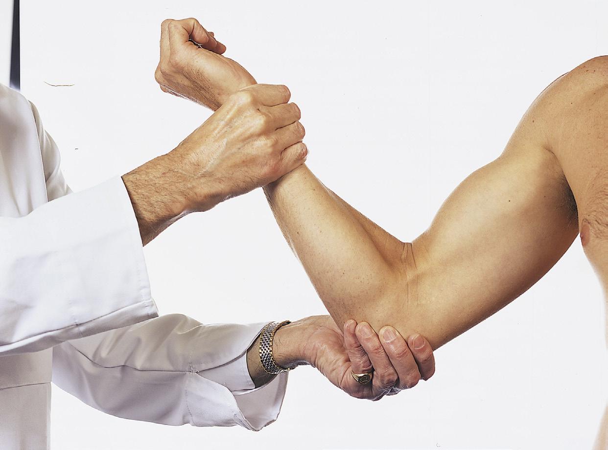
When testing a movement, the limb should be firmly supported proximal to the relevant joint so that the test is confined to the chosen muscle group and does not require the patient to fix the limb proximally by muscle contraction. In this book, this principle is illustrated in Figs 13, 20, 30, 33 among others. In some illustrations, the examiner’s supporting hand has been omitted for clarity (e.g. Figs 32, 34–36, 50, 55). The amount of leverage applied should be such that a patient with normal strength is evenly matched with the examiner, so that minimal weakness is more easily appreciated (e.g. Figs 23, 72). However, the same technique should be used for all patients so that the examiner can acquire experience of the variability in strength of different subjects. Optimal techniques are illustrated in the figures.
Muscle power may be recorded using the Medical Research Council (MRC) scale, but this is not a linear scale, and subdivisions of grade 4 are often necessary. Grades 4−, 4 and 4+ may be used to indicate movement against slight, moderate and strong resistance, respectively.
MRC SCALE FOR MUSCLE STRENGTH
0 No contraction
1 Flicker or trace of contraction
2 Full range of active movement, with gravity eliminated
3 Active movement against gravity
4 Active movement against gravity and resistance
5 Normal power
In the peripheral nerve diagrams, muscles are shown in the usual order of innervation; the origin of their motor supply from nerve trunks and the origin of the cutaneous branches are also shown, which are guides to the level of a lesion. In the figures showing methods of testing, the usual nerve supply to each muscle is stated along with the spinal segments from which they are derived, the more important of these are in bold type. Tables showing limb muscles arranged according to their supply by individual nerve roots and peripheral nerves are on pages 64 and 65.
SENSORY TESTING
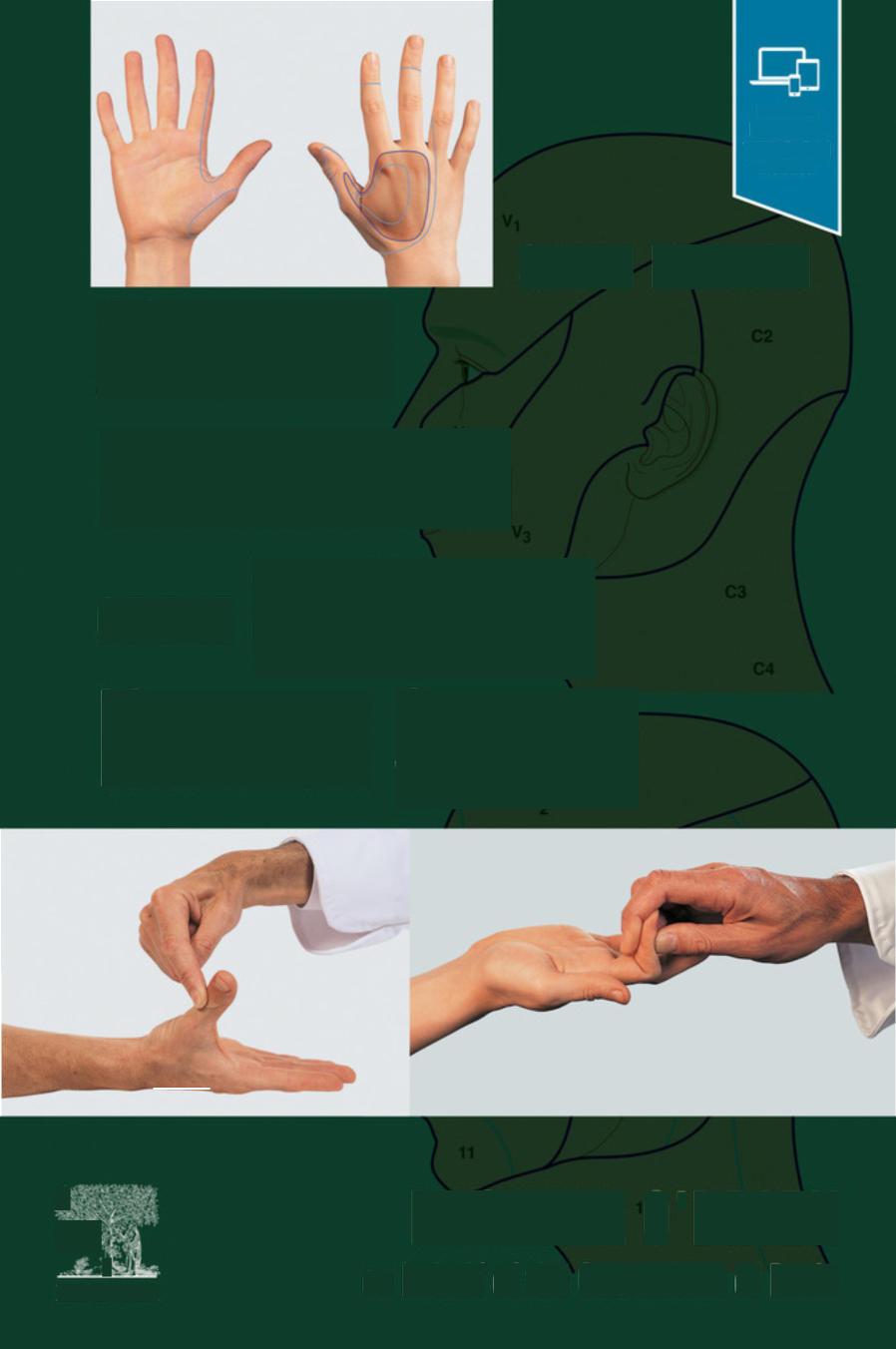
Asking the patient to outline the area of sensory abnormality can be a useful guide to the detailed examination. If this clearly indicates the distribution of a peripheral nerve (e.g. the lateral cutaneous nerve of the thigh, meralgia paresthetica, Fig. 61), the area can be mapped to light touch, tested with cotton wool or a light finger touch and to pain using a clean pin (not a needle, which is designed to cut the skin). Work from the insensitive area towards an area of normal sensation. If the area of sensory abnormality is hypersensitive (hyperpathia), the direction is reversed. Otherwise, it may be helpful to divide sensory testing into those modalities that travel in the ipsilateral posterior columns of the spinal cord (light touch, vibration and joint position sense) and those that travel in the crossed spinothalamic tracts (pain and temperature). Appreciation of vibration, a repetitive touch/pressure stimulus, is a sensitive test for demyelinating peripheral neuropathies. Two-point discrimination is a sensitive and quantifiable test of light touch, but it is only reliable on the face and fingertips. Always start with a stimulus at or below the normal threshold and coarsen the stimulus as required.
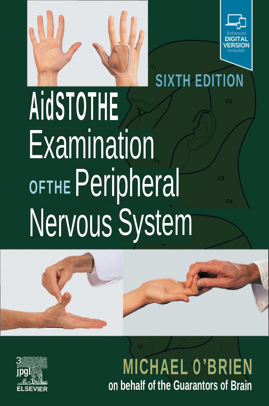
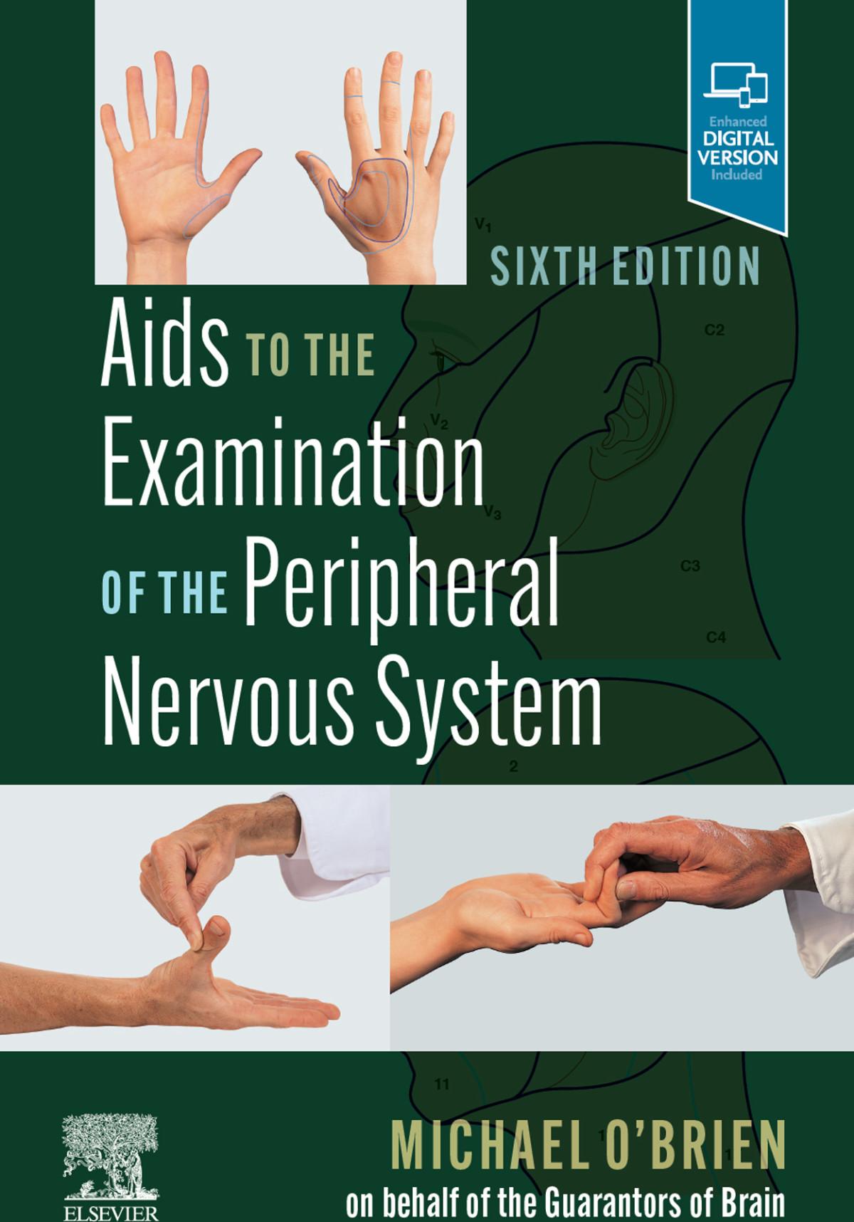
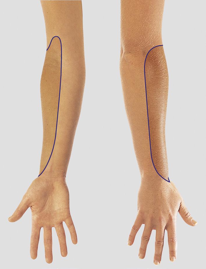
There is considerable overlap in the area of skin (dermatome) supplied by consecutive nerve roots, so that section of a single root may result in a very small area of sensory impairment. Conversely, the rash of herpes zoster may be quite extensive because it affects the whole area that has any supply from the affected root. The dermatome illustrations in Figs 90 through 97 are a compromise. The heavier axial lines, which separate nonconsecutive dermatomes, are more reliable as boundaries. The area of impairment with a peripheral nerve lesion is more reliable and consistent than that from a nerve root lesion. The areas shown in the figures are the usual ones. Some nerves show considerable variation between patients (see Figs 27, 61 and 64) and others are much more consistent (e.g. the ulnar nerve reliably splits at least part of the ring finger, see Fig. 48).
COMMON COMPRESSION AND ENTRAPMENT NEUROPATHIES
The two principal causes of a mononeuropathy are:
1. Entrapment by anatomic structures, where a nerve passes down a fibro-osseous tunnel or crosses a fascial plane
2. Compression by external forces, often where the nerve lies relatively unprotected between skin and bone
1. ENTRAPMENT NEUROPATHIES
Median nerve (page 26):
Pronator syndrome: the nerve is trapped between the two heads of pronator teres
Anterior interosseous nerve: the nerve may be trapped between the two heads of pronator teres with the median nerve or separately, in which case neuralgic amyotrophy is a possible diagnosis
Carpal tunnel syndrome: the nerve is trapped in the carpal tunnel beneath the flexor retinaculum
Ulnar nerve (page 32):
Cubital tunnel syndrome: the nerve is trapped between the two heads of flexor carpi ulnaris when the elbow is fully flexed
Posterior interosseous nerve: the nerve may be trapped between the two heads of supinator at the arcade of Frohse
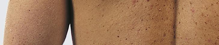
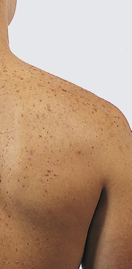
Dorsal cutaneous branches of the intercostal nerves from T3 to T6 (notalgia paresthetica, page 13): the nerves are trapped as they pass through the erector spinae muscles
Lateral cutaneous nerve of the thigh (meralgia paresthetica, page 42): the nerve is trapped as it passes through the inguinal ligament medial to the anterior superior iliac spine
Common fibular nerve (page 45): the nerve is trapped at the fibular head between the insertion of fibularis longus and the fibula
2. COMPRESSION NEUROPATHIES
Radial nerve (page 18):
‘Saturday night palsy’: the nerve is compressed against the shaft of the humerus
Superficial radial nerve: the nerve may be compressed against the radius at the lateral border of the forearm
Ulnar nerve (page 32):
‘Tardy ulnar palsy’: the nerve is compressed in the ulnar groove
Guyon’s canal: the nerve may be compressed in Guyon’s canal, usually by a ganglion
Deep motor branch: may be compressed at the hook of the hamate
Sciatic nerve (page 50): the nerve may be compressed as it enters the thigh below gluteus maximus
Posterior cutaneous nerve of the thigh (page 43): may be compressed as it enters the thigh below gluteus maximus
Common fibular nerve (page 45): may be compressed against the head of the fibula
These sites are indicated in the figures by a red arrow.
This section excludes obvious causes of trauma such as stab wounds, compound fractures, bullet wounds, and surgery where the site of the lesion is usually obvious. Note that a mononeuropathy may be the presenting feature of a mononeuritis multiplex, an underlying polyneuropathy, a systemic vasculitis, diabetes, or a familial liability to pressure palsies.
For a detailed discussion of the clinical features of the mononeuropathies, see Staal A, van Gijn J, Spaans F. Mononeuropathies, Examination, Diagnosis and Treatment. London: WB Saunders; 1999. ISBN 0-7020-1779-5.
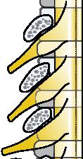
THE SPINE AND SPINAL ROOTS
VERTEBRA
1–8 CERVICAL NERVE ROOTS
CERVICAL NERVE ROOTS 1st THORACIC NERVE ROOT 1st Rib
6 7 8
1–12 THORACIC NERVE ROOTS
Note: The cervical spinal roots emerge above the corresponding vertebrae in close proximity to the disc, so that the C6 root is affected by a lateral disc protrusion at C5/6.
There are eight cervical roots but only seven cervical vertebrae, so that below the C7 vertebra the spinal roots emerge below the corresponding vertebrae, but in the lumbar spine the root emerges clear of the disc and would be affected by a lateral disc protrusion above the corresponding vertebra.
1–5 LUMBAR NERVE ROOTS
1–5 SACRAL NERVES
1 COCCYGEAL NERVE
3
4
5 LUMBAR NERVE ROOTS
Cocycx
Fig. 1 Diagram of the spine and spinal nerve roots.
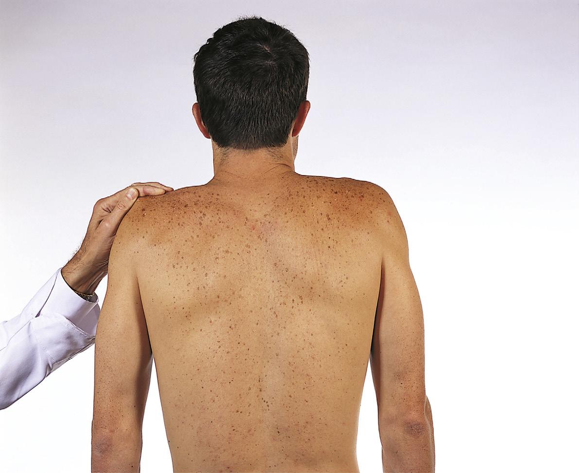
Fig. 2 Trapezius (Spinal accessory nerve and C3, The patient is elevating the shoulder against resistance.
Arrow: The thick upper part of the muscle can be seen and felt.
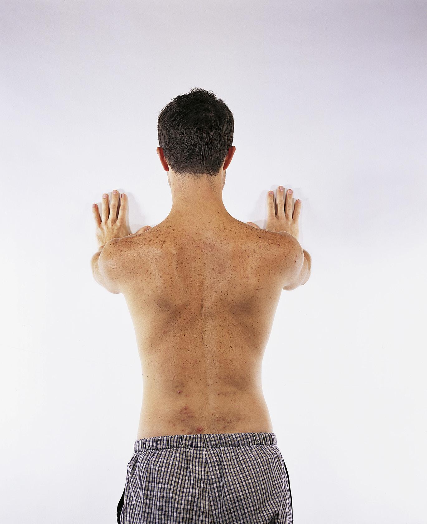
Fig. 3 Trapezius (Spinal accessory nerve and C3, C4). The patient is pushing the palms of the hands hard against a wall with the elbows fully extended.
Arrow: The lower fibres of the trapezius can be seen and felt.
they supply.
4
Fig.
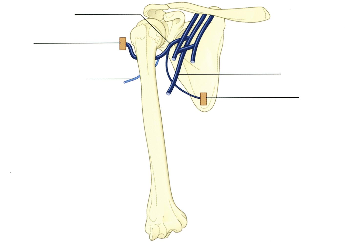
Diagram of the brachial plexus, its branches and the muscles
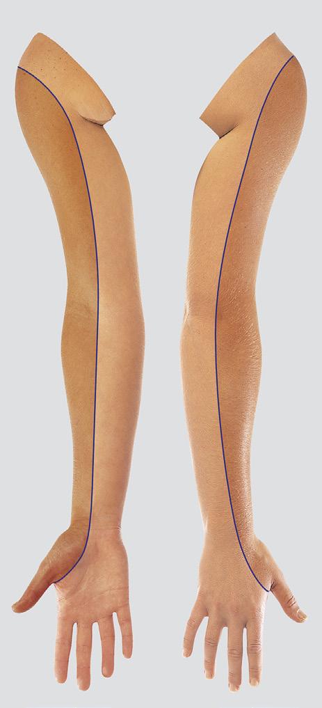
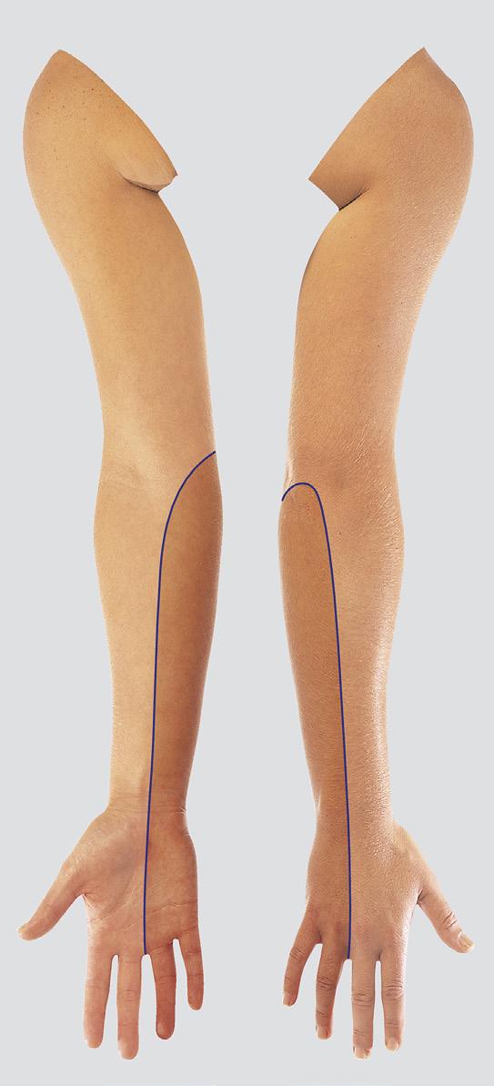
Fig. 5 The approximate area within which sensory changes may be found in complete lesions of the brachial plexus (C5, C6, C7, C8, T1).
Fig. 6 The approximate area within which sensory changes may be found in lesions of the upper roots (C5, C6) of the brachial plexus.
Fig. 7 The approximate area within which sensory changes may be found in lesions of the lower roots (C8, T1) of the brachial plexus.
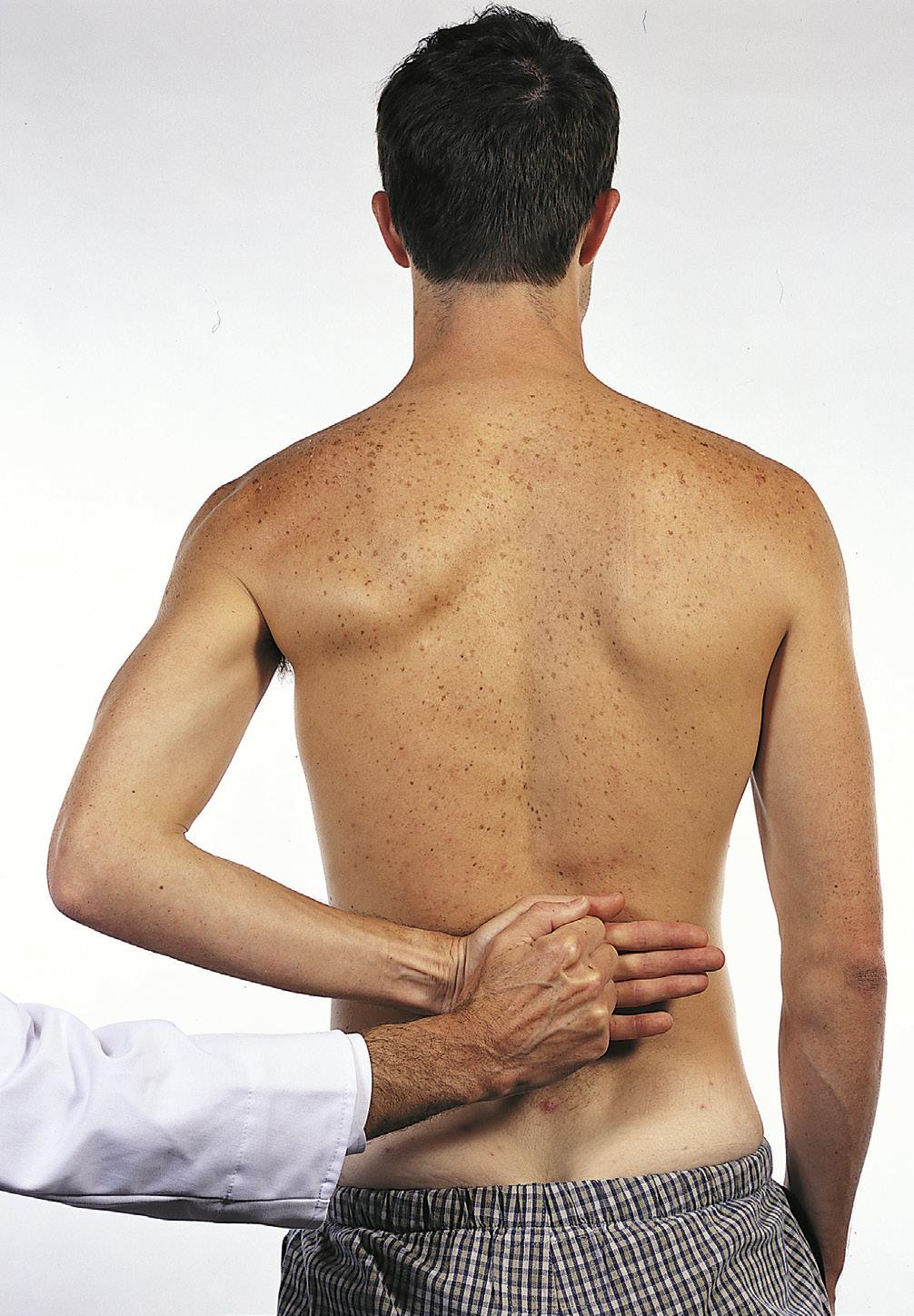
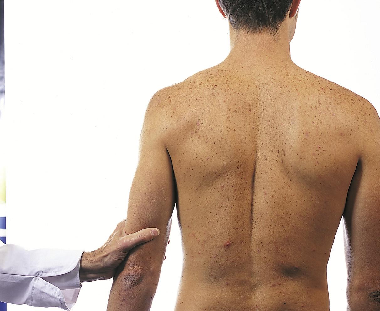
Fig. 8 Rhomboids (Dorsal scapular nerve; C4, C5).
The patient is pressing the palm of the hand backwards against the examiner’s hand. Arrow: The muscle bellies can be felt and sometimes seen.
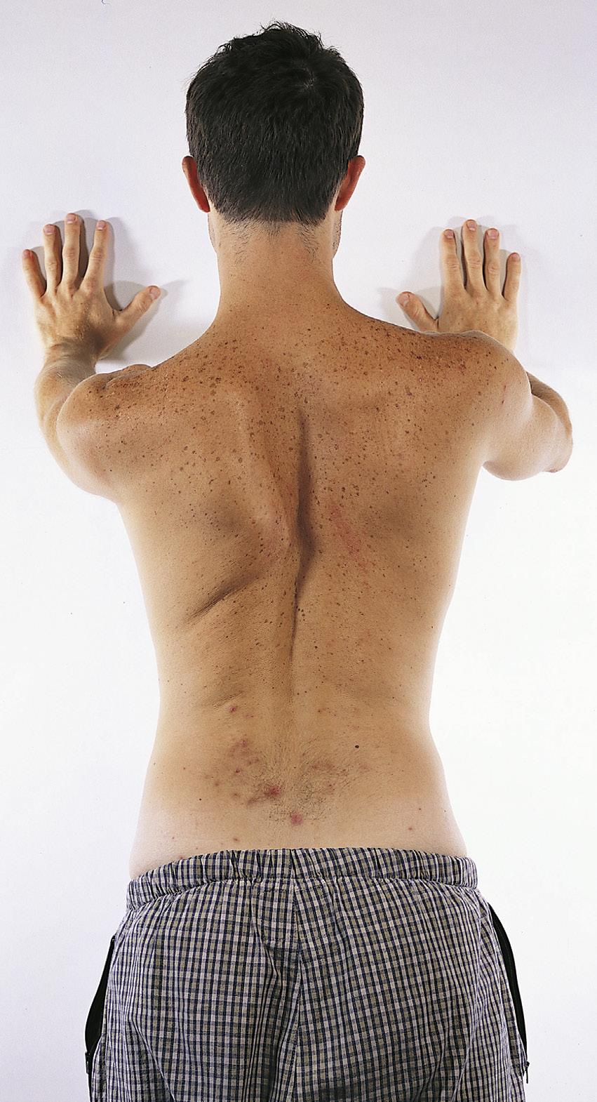
Fig. 9 Serratus Anterior (Long thoracic nerve; C5, C6, C7).
The patient is pushing against a wall. The left serratus anterior is weak and there is winging of the scapula.
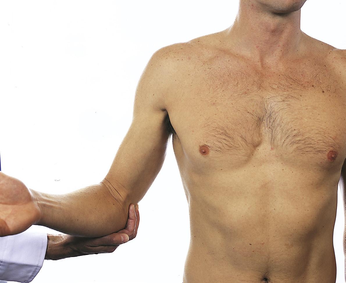
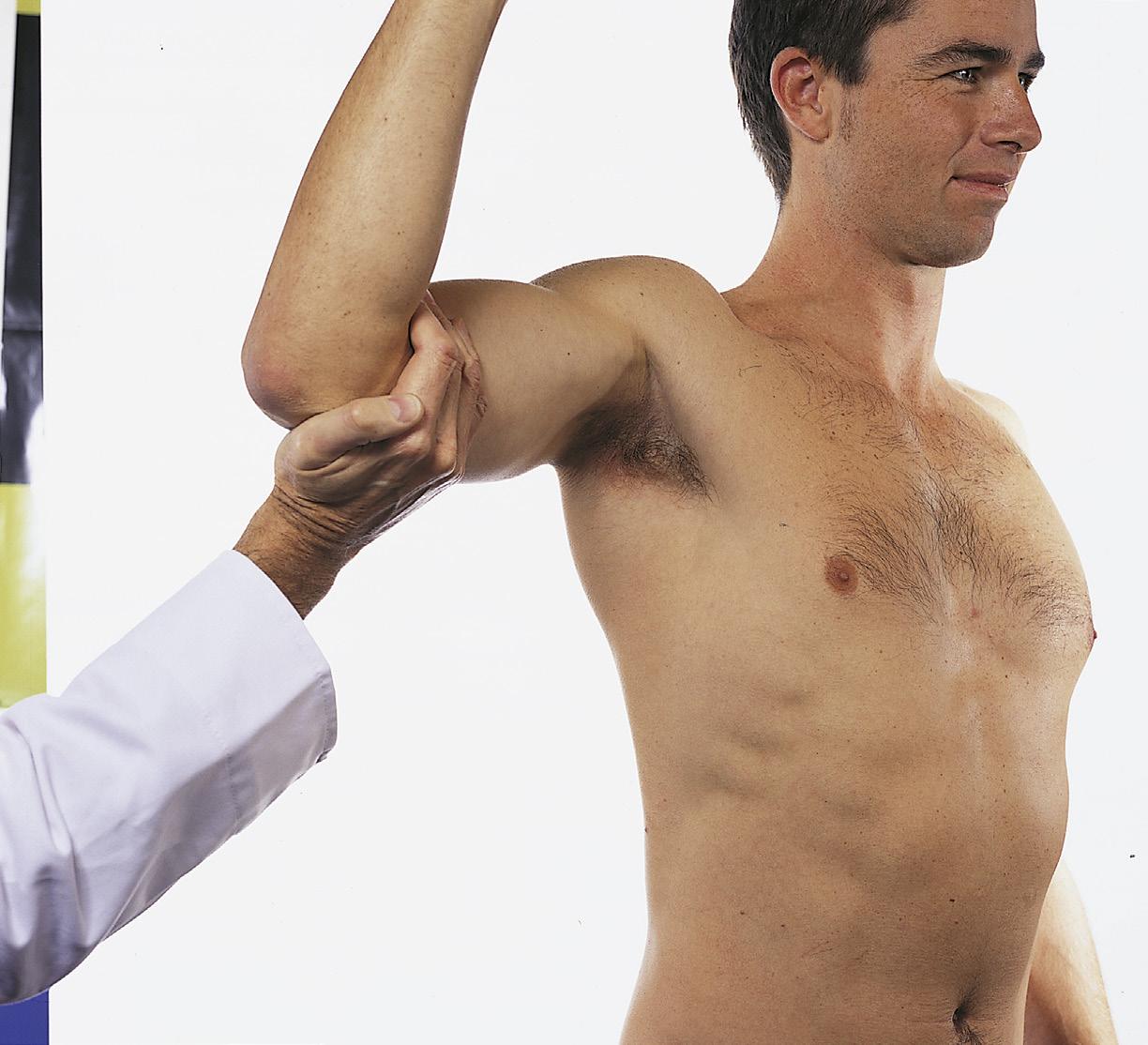
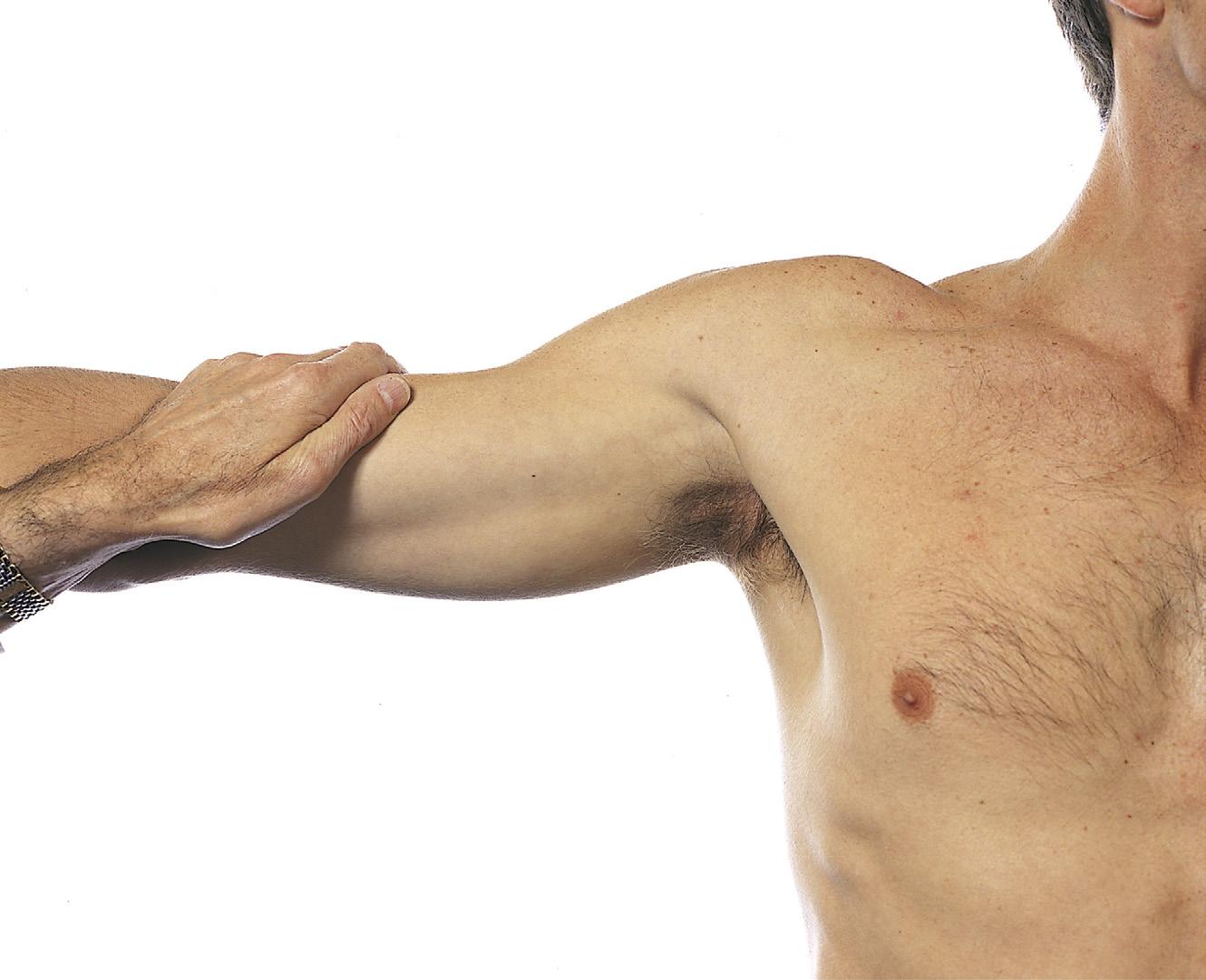
Fig. 10 Pectoralis Major: Clavicular head (Lateral pectoral nerve; C5, C6).
The upper arm is above the horizontal and the patient is pushing forward against the examiner’s hand.
Arrow: The clavicular head of the pectoralis major can be seen and felt.
Fig. 11 Pectoralis Major: Sternocostal head (Lateral and medial pectoral nerves; C6, C7, C8, T1).
The patient is adducting the upper arm against resistance.
Arrow: The sternocostal head can be seen and felt.
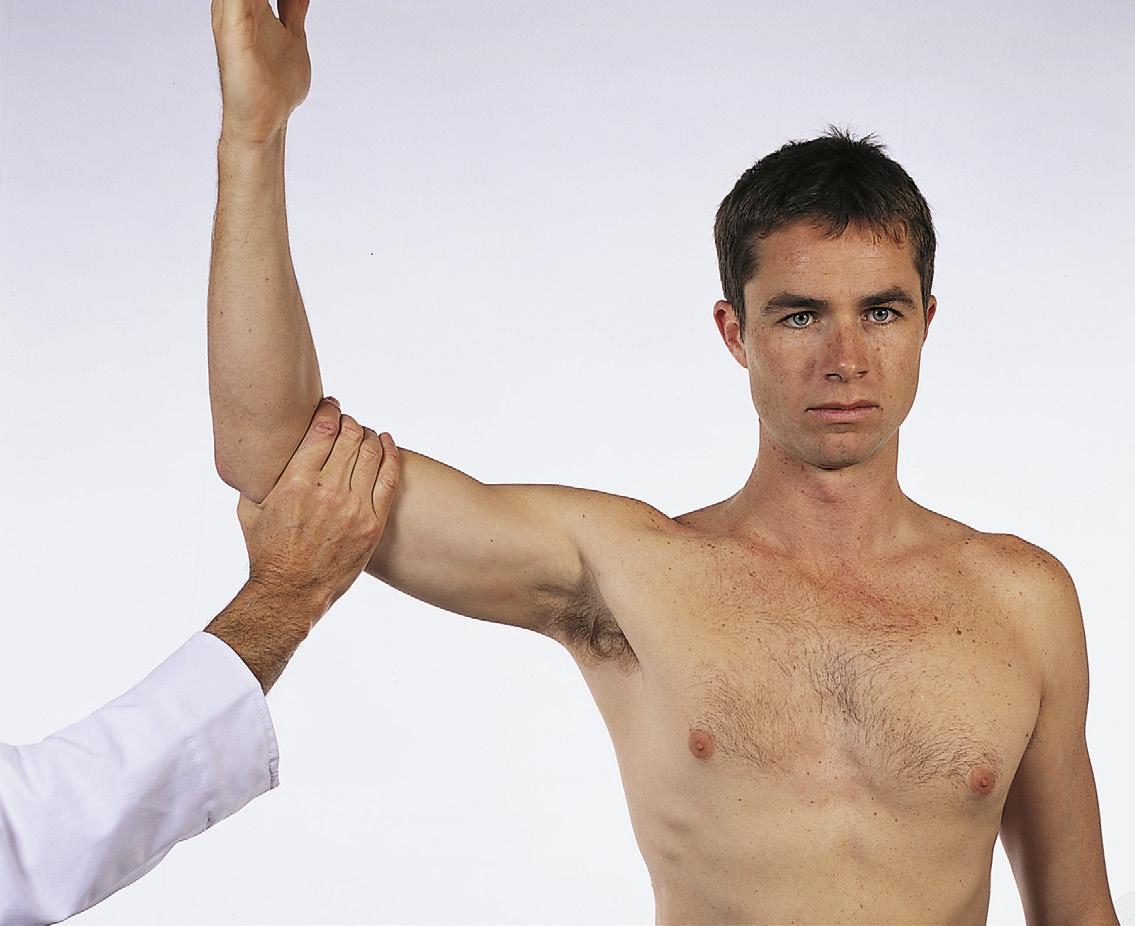
Fig. 12 Supraspinatus (Suprascapular nerve; C5, C6).
The patient is abducting the upper arm against resistance.
Arrow: The muscle belly can be felt and sometimes seen.
Fig. 13 Infraspinatus (Suprascapular nerve; C5, C6).
The patient is externally rotating the upper arm at the shoulder against resistance. The examiner’s right hand is resisting the movement and supporting the forearm with the elbow at a right angle; the examiner’s left hand is supporting the elbow and preventing abduction of the arm.
Arrow: The muscle belly can be seen and felt.
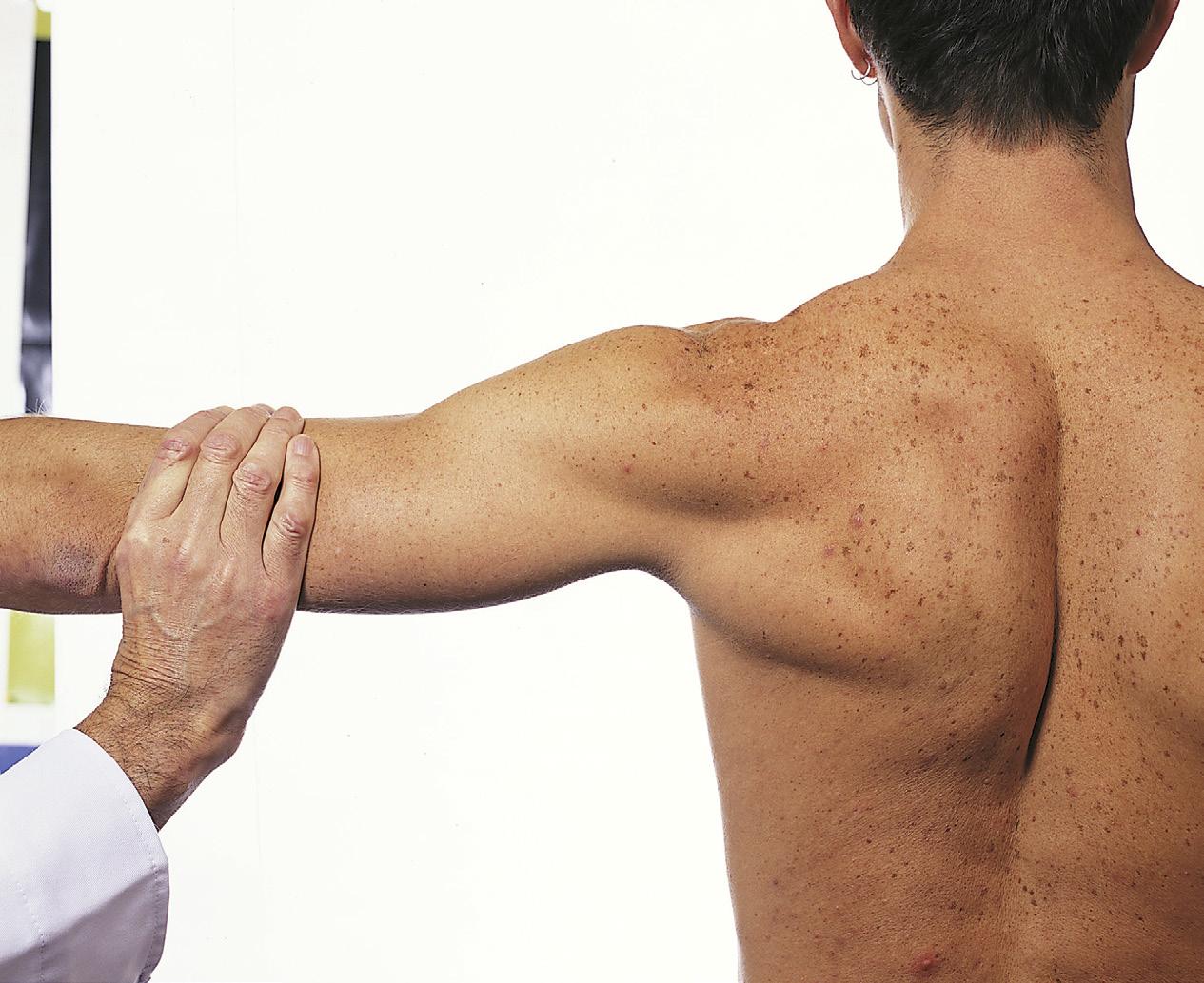
The upper arm is horizontal and the patient is adducting it against resistance.
Lower arrow: The muscle belly can be seen and felt. Upper arrow: Indicates teres major.
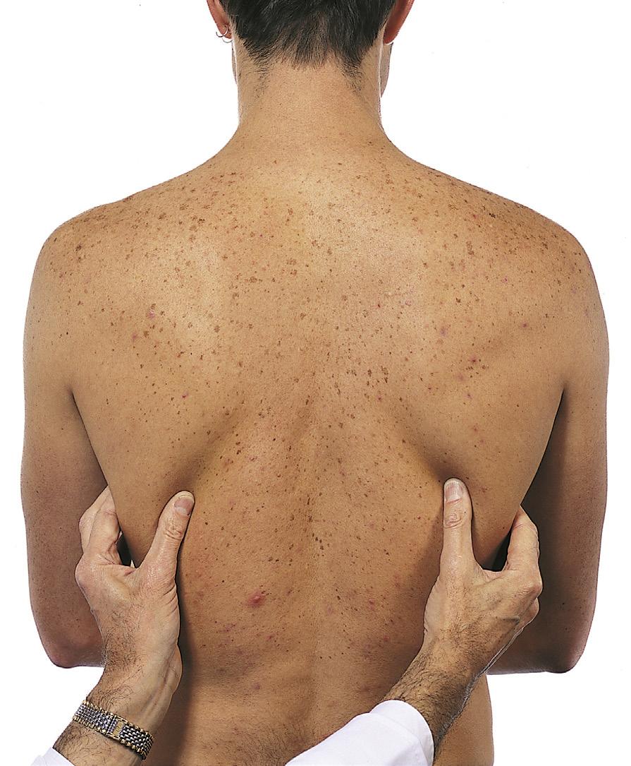
The muscle bellies can be felt to contract when the patient coughs.
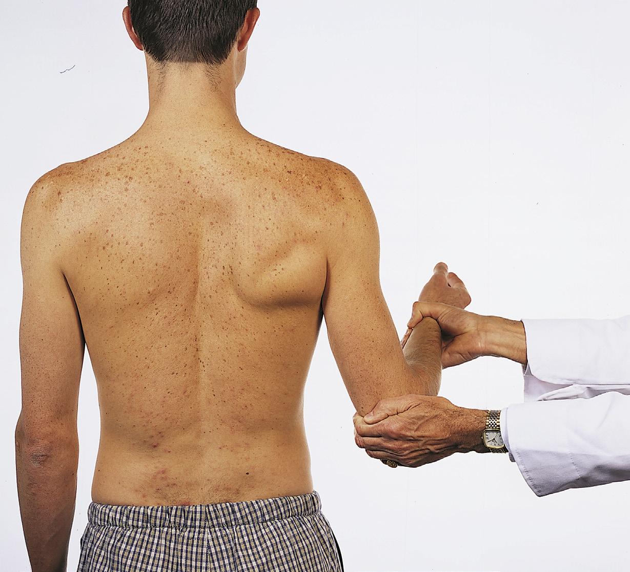
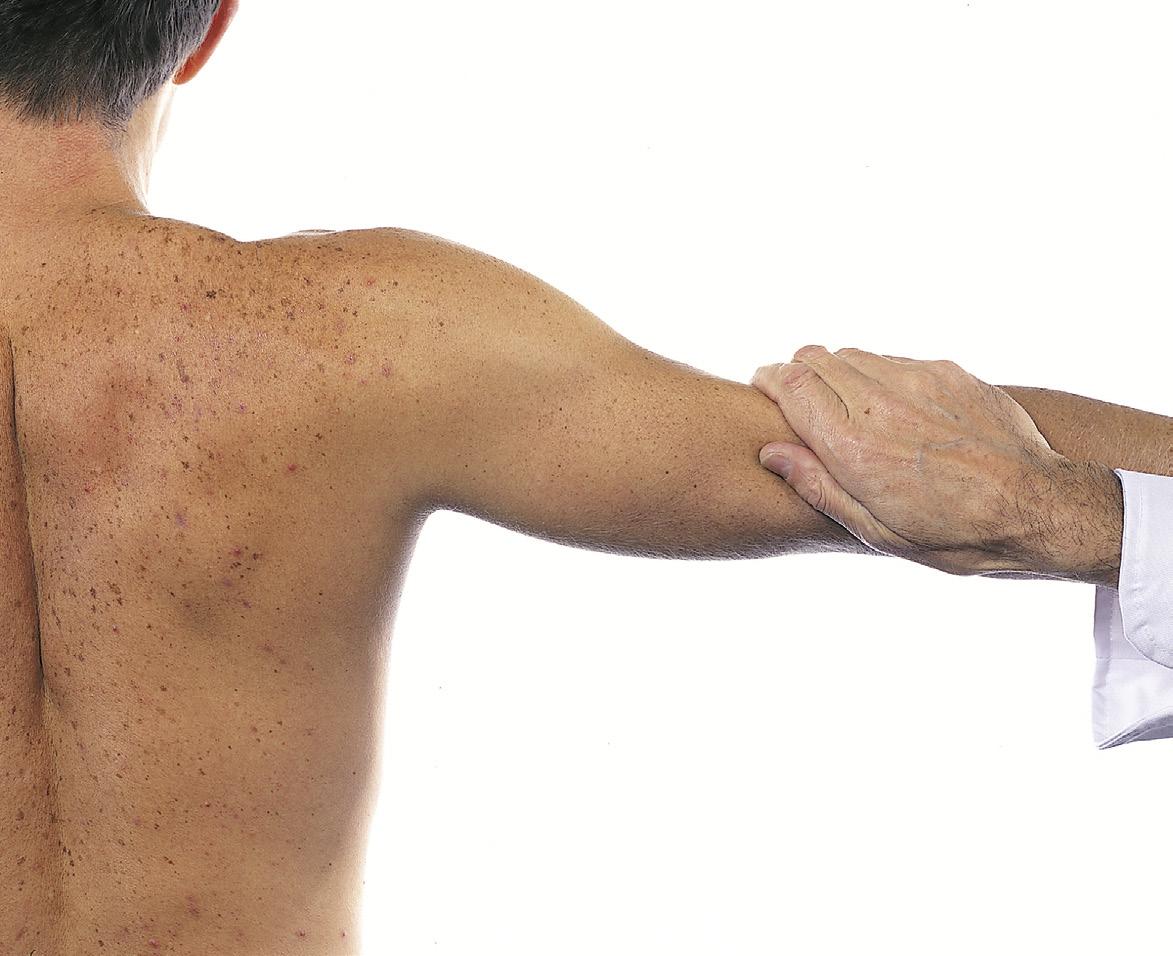
Fig. 14 Latissimus Dorsi (Thoracodorsal nerve; C6, C7, C8).
Fig. 15 Latissimus Dorsi (Thoracodorsal nerve; C6, C7, C8).
Fig. 16 Teres Major (Subscapular nerve; C5, C6, C7).
The patient is adducting the elevated upper arm against resistance.
Arrow: The muscle belly can be seen and felt.
Fig. 17 The approximate area within which sensory changes may be found in lesions of the posterior cutaneous branches of the intercostal nerves from T2 to T6 (notalgia paresthetica).