Activate the eBook version of this title at no additional charge. Unlock your eBook today.
1. Visit expertconsult.inkling.com/redeem
2. Scratch box below to reveal your code
3. Type code into “Enter Code” box
4. Click “Redeem”
5. Log in or Sign up
6. Go to “My Library” It’s that easy! Elsevier eBooks for Practicing Clinicians gives you the power to browse and search content, view enhanced images, highlight and take notes—both online and offline.
Place Peel Off Sticker Here
For technical assistance: email expertconsult.help@elsevier.com call 1-800-401-9962 (inside the US) call +1-314-447-8300 (outside the US)
Abdominal Imaging The Core Requisites
Joseph R. Grajo, MD
Chief of Abdominal Imaging
Vice Chair for Research
Associate Residency Program Director
Abdominal Imaging Fellowship Director
Department of Radiology
University of Florida College of Medicine
Gainesville, Florida
United States
Dushyant V. Sahani, MD
Associate Professor of Radiology
Department of Radiology
Massachusetts General Hospital
Boston, Massachusetts
United States
Anthony E. Samir, MD, MPH
Associate Director, Ultrasound
Radiology
Massachusetts General Hospital
Boston, Massachusetts
United States
Elsevier
1600 John F. Kennedy Blvd. Ste 1600 Philadelphia, PA 19103-2899
No part of this publication may be reproduced or transmitted in any form or by any means, electronic or mechanical, including photocopying, recording, or any information storage and retrieval system, without permission in writing from the publisher. Details on how to seek permission, further information about the Publisher’s permissions policies and our arrangements with organizations such as the Copyright Clearance Center and the Copyright Licensing Agency, can be found at our website: www.elsevier.com/permissions.
This book and the individual contributions contained in it are protected under copyright by the Publisher (other than as may be noted herein).
Notice
Practitioners and researchers must always rely on their own experience and knowledge in evaluating and using any information, methods, compounds or experiments described herein. Because of rapid advances in the medical sciences, in particular, independent verification of diagnoses and drug dosages should be made. To the fullest extent of the law, no responsibility is assumed by Elsevier, authors, editors or contributors for any injury and/or damage to persons or property as a matter of products liability, negligence or otherwise, or from any use or operation of any methods, products, instructions, or ideas contained in the material herein.
Library of Congress Control Number: 2021932208
Content Strategist: Kayla Wolfe
Content Development Specialist: Erika Ninsin
Publishing Services Manager: Deepthi Unni
Project Manager: Srividhya Vidhyashankar
Design Direction: Patrick C. Ferguson
To my wife and best friend, Nicolette, who inspires me and loves me unconditionally, and our daughter Brooklyn, who amazes me each day.
To my parents, Joseph and Ruth, who raised me to set no limits to my potential and my sister, Jennifer, who has always supported and appreciated me.
Joseph R. Grajo, MD
To my mentors, colleagues, and trainees, who challenge me and encourage my academic aspirations.
Contributors
Neha Agrawal, MD
Abdominal Imaging Fellow Department of Radiology
Massachusetts General Hospital Boston, Massachusetts United States
Ahmad Al-Samaraee, MD, MPH Department of Radiology
Massachusetts General Hospital Boston, Massachusetts
Department of Radiology University of Minnesota Minneapolis, Minnesota United States
Mark Anderson, MD
Abdominal Radiologist
Massachusetts General Hospital Instructor Harvard Medical School Boston, Massachusetts United States
Masoud Baikpour, MD
Diagnostic Radiology Resident Department of Radiology
Massachusetts General Hospital Boston, Massachusetts United States
Miguel Gosalbez, BS, MD Resident, Radiology Department of Radiology University of Florida Gainesville, Florida United States
Carolyn Hanna, BS, MD Resident, Radiology University of Florida Gainesville, Florida United States
Simon Ho, MD Resident, Radiology University of Florida Gainesville, Florida United States
Richard G. Kavanagh, MB, BCh, BAO, BSc, MCh, FFRRCSI
Abdominal Imaging Fellow
Massachusetts General Hospital Boston, Massachusetts United States
David Knipp, MD Fellow
Department of Radiology
Massachusetts General Hospital Boston, Massachusetts United States
Hamed Kordbacheh, MD
Abdominal Imaging Research Fellow Department of Radiology
Massachusetts General Hospital Boston, Massachusetts United States
Qian Li, MD
Instructor of Radiology
Department of Radiology
Massachusetts General Hospital Boston, Massachusetts United States
Weier Li, MD Fellow
Department of Radiology
Massachusetts General Hospital Boston, Massachusetts United States
Babak Maghdoori, MD, FRCPC
Abdominal imaging specialist
Cardiothoracic imaging specialist
Department of Medical Imaging
Georgian Radiology Consultant University of Toronto Toronto, Ontario Canada
Laura L. Magnelli, MD
Abdominal Imaging Fellow, PGY-6
Department of Radiology
Division of Abdominal Imaging
University of Florida College of Medicine
Gainesville, Florida United States
Craig Meiers, MD
Interventional Radiology Fellow Department of Radiology University of Florida Gainesville, Florida United States
Aileen O’Shea, MB, BAO, BCh, FFRRCSI
Abdominal Imaging Fellow Division of Abdominal Imaging Massachusetts General Hospital Boston, Massachusetts United States
Arinc Ozturk, MD
Clinical Research Fellow Department of Radiology Massachusetts General Hospital Boston, Massachusetts United States
Eric W. Pepin, MD, PhD Resident Department of Radiology University of Florida Gainesville, Florida United States
John Pham, MD
Radiology, Resident Department of Radiology University of Florida Gainesville, Florida United States
Theodore T. Pierce, MD Instructor Department of Radiology Massachusetts General Hospital Boston, Massachusetts United States
Vinay Prabhu, MD, MS
Clinical Assistant Professor Department of Radiology NYU Langone Health New York, New York United States
Jesse Rayan, MD
Abdominal Imaging Fellow Division of Abdominal Imaging Massachusetts General Hospital Boston, Massachusetts United States
Justin Ruoss, MD Physician Department of Radiology University of Florida Gainesville, Florida United States
Jehan L. Shah, MD
Diagnostic Radiology Resident Department of Radiology University of Florida Gainesville, Florida
Interventional Radiology Fellow Radiology Mayo Clinic Jacksonville, Florida United States
Boris Sinayuk, MD Assistant Professor Department of Diagnostic Imaging Warren Alpert Medical School Rhode Island Hospital Providence, Rhode Island United States
Joe Uricchio, MD Resident Department of Radiology University of Florida Gainesville, Florida United States
Series Foreword
Congratulations to Dr Grajo, lead editor and co-editors, Drs Sahani and Samir for producing Abdominal Imaging: The Core Requisites , the first book in the newly reimagined The Core Requisites series. Dr Grajo and colleagues assembled a stellar team of contributors and successfully pivoted from a traditional narrative or prose-based approach for knowledge sharing to an outline format that immediately brings forward and highlights the most important facts and concepts for each topic. The outline format makes the material covered readily accessible to readers, a key goal for anyone developing a textbook. Abdominal Imaging: The Core Requisites is outstanding and will serve as a model for subsequent books in the series as they are updated and revised.
Dr. Grajo and colleagues used the transition to the new “The Core Requisites” format to emphasize problem-based diagnostic scenarios to complement enduringly important material on anatomy, physiology, physics and imaging methods. Using a problem-based approach in Abdominal Imaging: The Core Requisites allows presentation of material in the way radiologists encounter diagnostic challenges in actual practice including their relative importance and challenges in differential diagnosis versus a traditional taxonomic approach that most often treats each disease or condition one at a time and at the same level of emphasis regardless of clinical prevalence.
While the format of The Core Requisites series differs substantially from the traditional Radiology Requisites series, the philosophy remains the same—the production of a series of books covering the core material required
across the spectrum of what radiologists need to know from their first encounters with subject material in different subspecialty areas, to studying for board exams and later for reference during clinical practice. The books in the The Core Requisites series will continue to be richly illustrated; Abdominal Imaging: The Core Requisites has over 500 illustrations. The books are intended to be practical, not encyclopedic.
Print and electronic formats will be produced simultaneously and included with each purchase. The electronic version will provide mobile access via multiple kinds of devices and is searchable, adding additional value. This approach is in keeping with our expectations in the internet age of access to knowledge at the time-of-need and point-of-care.
Congratulations again to Drs. Grajo, Sahani and Samir for launching this new The Core Requisites series on a terrific start. I hope that this and the following books in the series will become regarded with the same fondness as earlier books in the Radiology Requisites family that have been used by radiologists at all career stages now for over thirty years.
James H Thrall, MD
Chairman Emeritus Department of Radiology, Massachusetts General Hospital Distinguished Taveras Professor of Radiology Harvard Medical School Boston, Massachusetts
Preface
When Elsevier approached me about helping to relaunch the Requisites series, I was excited about the opportunity to contribute to the rebirth of a classic and fundamental radiology textbook. I was honored to be asked to spearhead a new abdominal imaging title, combining the contents of gastrointestinal and genitourinary radiology as seen in prior iterations. Furthermore, I was thrilled for another chance to work with two of my most influential mentors and prestigious colleagues, Dr. Anthony Samir and Dr. Dushyant Sahani.
This textbook serves as a launch point for a rebranding of the Requisites series as “The Core Requisites.” In this series, we aim to present high-yield information in a concise format with easy-to-read chapters, targeting a wide audience
but focused on the radiology trainee in preparation for clinical rotations and board examinations.
Our approach to Abdominal Imaging: The Core Requisites was to present focused “need to know” material in a format that addresses commonly encountered clinical scenarios, such as right upper quadrant pain, chronic liver disease, and postoperative imaging. In each chapter, we review important anatomy, discuss imaging techniques and protocol considerations, describe specific disease processes associated with the clinical scenario, summarize relevant tumor staging, and highlight key elements of a structured report.
We hope that you will enjoy this new venture in your journey of continuous learning.
Joseph R. Grajo,
MD
SECTION I
Gastrointestinal Tract, 1
1 Abdominal Radiography, 2
WEIER LI
2 Gastrointestinal Fluoroscopy, 7
WEIER LI
3 Gastric Wall Thickening/Masses, 12
DAVID KNIPP
4 Hollow Viscus Perforation, 17
DAVID KNIPP
5 Small Bowel Obstruction, 23
JOHN PHAM AND SIMON HO
6 Bowel Wall Thickening, 30
AHMAD AL-SAMARAEE
7 Imaging of the Postoperative Bowel, 37
JEHAN L. SHAH AND MIGUEL GOSALBEZ
8 Mesenteric Ischemia, 47
RICHARD G. KAVANAGH
9 Colorectal Cancer and Screening, 54
RICHARD G. KAVANAGH
10 Right Upper Quadrant Pain, 62
NEHA AGRAWAL
11 Right Lower Quadrant Pain, 72
NEHA AGRAWAL
12 Pancreatitis, 85
JOHN PHAM AND SIMON HO
13 Solid Pancreatic Masses, 92
JEHAN L. SHAH AND MIGUEL GOSALBEZ
14 Cystic Pancreatic Lesions, 103
JEHAN L. SHAH
SECTION II
Hepatobiliary, 114
15 Chronic Liver Disease, 115
ARINC OZTURK
16 Imaging of the Cirrhotic Liver, 124
ARINC OZTURK
17 Benign Focal Liver Lesions 135
CRAIG MEIERS
18 Cholecystitis, 146
BABAK MAGHDOORI AND HAMED KORDBACHEH
19 Jaundice, 156
BABAK MAGHDOORI AND HAMED KORDBACHEH
SECTION III
Lymphatic System, 165
20 Splenomegaly/Splenic Lesions, 166
ERIC W. PEPIN
21 Lymphadenopathy, 173 JUSTIN RUOSS
SECTION IV
Genitourinary, 182
22 Urolithiasis, 183 MARK A. ANDERSON
23 Cystic Renal Masses, 188 LAURA MAGNELLI
24 Solid Renal Lesions, 195 LAURA MAGNELLI AND CAROLYN HANNA
28 Adrenal Gland Enlargement and Nodules, 229 ERIC W. PEPIN AND JOE URICCHIO
SECTION V
Reproductive System, 243
29 Prostate Imaging, 244 QIAN LI AND JOSEPH R. GRAJO
30 Testicular Lesions, 251 QIAN LI
31 Endometrial/Junctional Zone Thickening, 259 MASOUD BAIKPOUR
32 Focal Uterine Lesions, 268
MASOUD BAIKPOUR
33 Cystic Adnexal Lesions, 281
AILEEN O’SHEA
34 Solid Adnexal Lesions, 299
AILEEN O’SHEA
35 Müllerian Abnormalities, 306
VINAY PRABHU
36 Pelvic Floor Disease, 314
VINAY PRABHU
SECTION VI Miscellaneous, 318
37 Postoperative Imaging, 319
JESSE RAYAN
38 Abdominal/Pelvic Trauma, 327 THEODORE T. PIERCE
39 Pitfalls, 337 THEODORE T. PIERCE Index, 345
SECTION I
Gastrointestinal Tract
1 Abdominal Radiography
WEIER LI
CHAPTER OUTLINE
Anatomy, Embryology, Pathophysiology
Techniques
Specific Disease Processes
Intraluminal Gas
Extraluminal Gas
Calcifications
Anatomy, Embryology, Pathophysiology
n There are five distinct densities in plain radiography, four of which are natural: gas (black), fat (dark gray), soft tissue (medium gray), calcifications (white), and metal (intense white).
n Dedicated assessment of each density is essential in any search pattern.
Techniques
n The standard abdominal radiograph is obtained in a supine projection, with x-rays passing from anteroposterior (AP). Field of view should span the inferior ribs to the inferior pubic rami, and include both lateral abdominal walls.
n Additional projections can be obtained to assess for free air or air-fluid levels. These include upright positioning (erect) or with the patient lying on his or her side (lateral decubitus). The erect view is typically preferred for ease of use and must include the diaphragm even at the expense of the pelvis. A lateral decubitus view can be alternatively performed in patients unable to tolerate upright imaging.
Specific Disease Processes
INTRALUMINAL GAS
There is wide variation in the appearance of a normal bowel gas pattern.
n Gas in the left upper quadrant (stomach) can produce a gastric bubble on erect imaging. If distended with air or ingested contrast, the normal gastric rugae can be a distinguishing feature.
n The small bowel is comprised of the duodenum, jejunum, and ileum. It is identified on radiography by the presence of circumferential folds called valvulae conniventes (or plicae circulares) that traverse the entire width of the small bowel. The jejunum begins at the ligament of Treitz and primarily
Soft Tissues and Bone
Foreign Bodies
High Yield Topics
KUB Emergencies
Physics Pearls
resides in the left upper quadrant. The increased folds of the jejunum give it a feathery pattern. In contrast, the ileum is usually larger in caliber and contains fewer folds, producing a more featureless mucosal pattern. In general, the small bowel should be less than 3 cm in diameter.
n The large bowel (or colon) extends from the ileocecal valve to the anus, and comprises the cecum, ascending colon, transverse colon, descending colon, sigmoid colon and rectum. The large bowel is distinguished by haustra, which are large folds that only cross portions of the bowel wall. Large bowel contains feculent material that often gives a mottled appearance, representing a mixture of gas/liquid/solid material. The cecum can be normal at up to 9 cm in diameter, whereas the remainder of the large bowel can distend normally up to 6 cm.
EXTRALUMINAL GAS
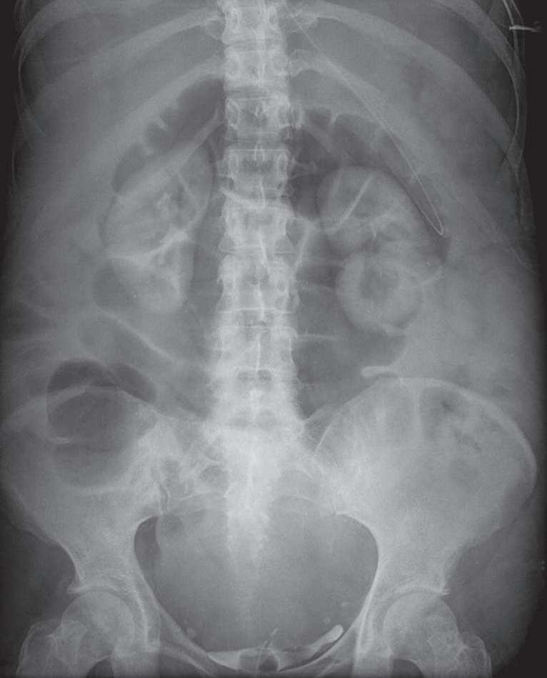
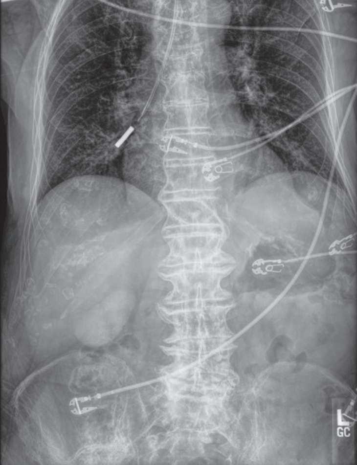
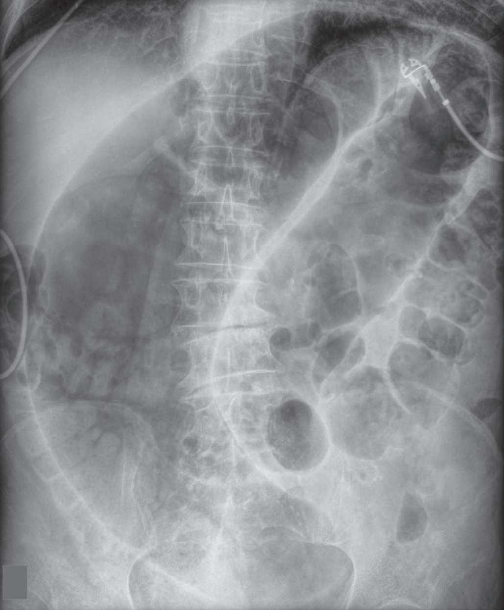
Extraluminal gas outside of the bowel lumen can range from atypical to grossly pathological (Fig. 1.1).
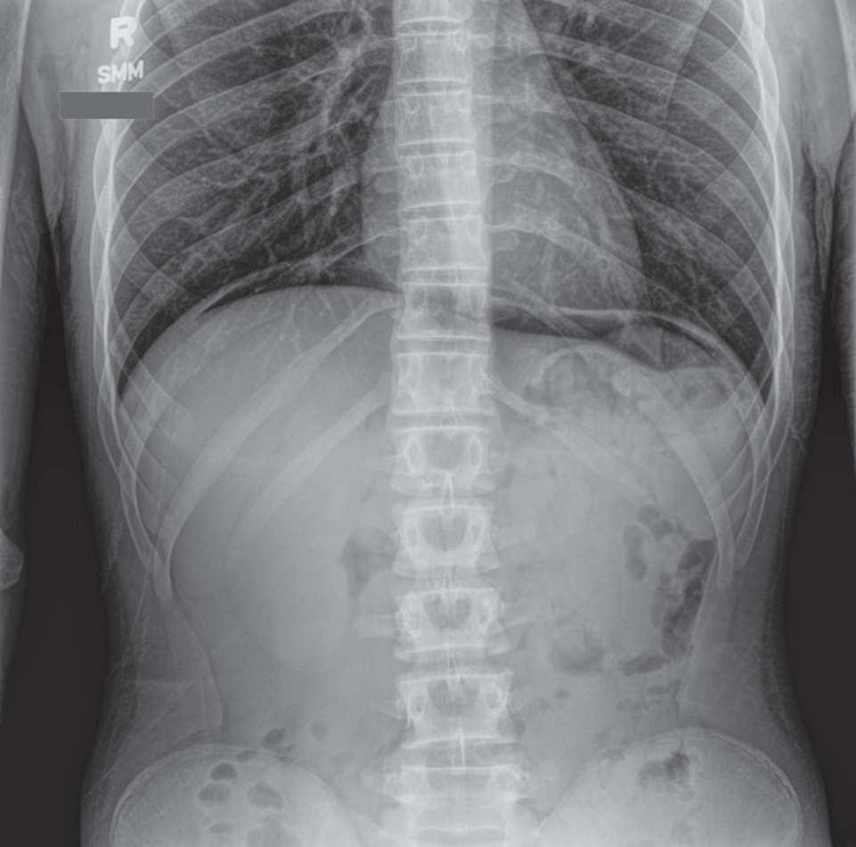
n Gas within the peritoneal cavity is called pneumoperitoneum. This is best identified on erect imaging, which will show free air under the diaphragm, particularly on the right (Fig. 1.2). Caution must be exercised to identify the normal left upper quadrant stomach bubble when evaluating for free air on the left. On decubitus imaging, free air will appear clearly outside the contours of the bowel lumen, rising to the antidependent portion of the radiograph.
n Small volumes of postoperative pneumoperitoneum can be an expected finding within 7 to 10 days after surgery. If there has been no history of recent intervention, or if the volume of pneumoperitoneum is larger than expected, there should be a concern for pathological hollow viscus perforation.
n In the absence of upright or decubitus projections, large volume pneumoperitoneum can be identified by a variety of signs, including Rigler’s sign, football sign and so on. (Fig. 1.3).
n Gas in the bowel wall (pneumatosis) can occur in a variety of benign or pathological processes. Correlation with history is essential as pneumatosis can be iatrogenic (e.g., related to recent G-tube placement), pathological
Fig. 1.1 Portable anteroposterior KUB demonstrates marked conspicuity of the bilateral kidneys because of pneumoretroperitoneum. A nasogastric tube is also noted, as well as a Foley catheter with excreted contrast in the bladder.
1.2 Upright KUB demonstrates free air under the diaphragm, the so called “continuous diaphragm sign.”
(related to bowel ischemia), medication-related (i.e., chemotherapy) or idiopathic.
n Air can also be seen overlying the liver. Gas within the bile ducts (pneumobilia) can be seen in the presence of a sphincterotomy, biliary stent, biliary bypass, or biliary fistula. Pneumobilia should overlie the central liver. Air that extends to the periphery of the liver may reflect portal venous gas, which is always pathological. Gas in the portal vein is typically a result of bowel ischemia and may be an ominous sign.
1.3 Portable anteroposterior KUB demonstrates pneumoperitoneum with Rigler’s sign. Note the conspicuity of the bowel walls because of air outlining both sides.
CALCIFICATIONS
Calcium deposits in a wide variety of normal and abnormal structures.
n Benign aortic calcifications are common and can help visualize the borders of the aorta and major branches. Large aortic and branch vessel aneurysms can be identified by the demarcation of their associated wall calcifications.
n Pancreatic calcifications overlying the mid-abdomen can be seen in chronic pancreatitis.
n Calcified renal or bladder calculi can be seen overlying the expected renal fossa and bladder. These should be distinguished from pelvic phleboliths, which are extremely common, especially in elderly adults. Phleboliths tend to be rounder and have a radiolucent center, as well as a more lateral position in the pelvis compared with urolithiasis.
n Densely calcified gallstones or a calcified (porcelain) gallbladder can often be seen in the right upper quadrant.
n Uterine fibroids can often become calcified. Ovarian dermoid cysts are also an uncommon cause of pelvic calcifications.
n Other unusual causes of calcifications include calcified lymphadenopathy, remote fat necrosis and “dropped gallstones.”
SOFT TISSUES AND BONE
The outlines of the major abdominal organs can often be distinguished by their surrounding fat planes. Patients with cachexia or extreme weight loss may lose these fat planes, making the identification of normal abdominal organs difficult. Similarly, disruption of the
Fig.
Fig.
normal fat planes can be a secondary clue for ascites or hemoperitoneum.
n The liver and gallbladder are located in the right upper quadrant. A Riedel lobe is a normal anatomic variant where the right liver projects inferiorly to the level of the pelvis.
n Splenomegaly can be noted with mass effect upon the normal bowel gas in the left hemiabdomen.
n Both kidneys can often be visualized around the T12-L2 vertebral body levels. The psoas muscle shadows are just medial to each kidney and mark the course of both ureters.
n Large abdominal soft tissue masses can produce mass effect on the adjacent structures, particularly the bowel gas.
n The imaged osseous structures include the lower spine, pelvis, and proximal hips and femurs. A wide variety of pathology includes fractures, sclerotic and lytic metastases; inflammatory disease and degenerative changes can be seen.
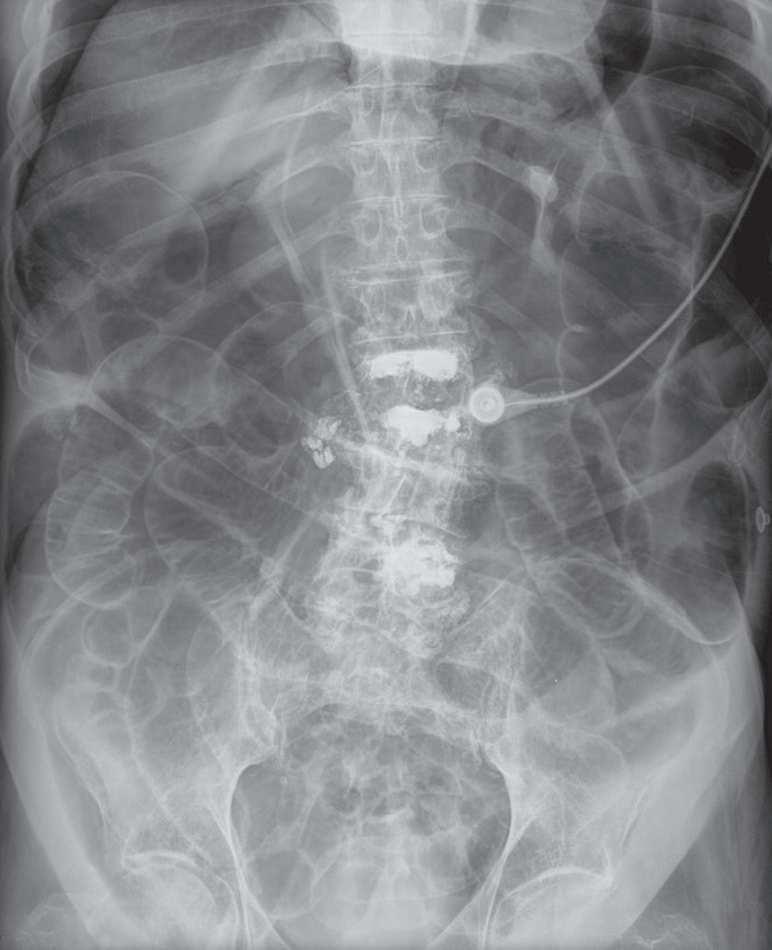
FOREIGN BODIES
Foreign bodies should be evaluated for appropriateness and positioning whenever possible.
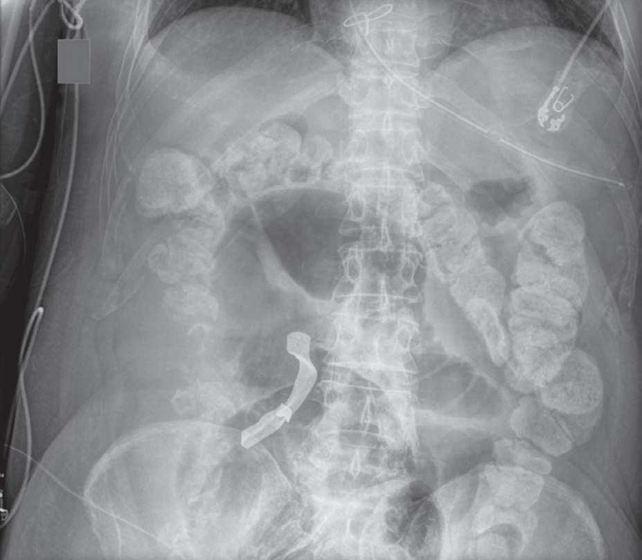
n Iatrogenic lines and tubes, such as nasogastric or percutaneous gastrostomy tubes, biliary tubes, nephrostomy tubes, ureteral stents, peritoneal drainage catheters and peritoneal dialysis catheters (Fig. 1.4).
n Surgical material, which can suggest prior procedures, such as cholecystectomy clips, gastric bypass clips, bowel sutures and nephrectomy clips.
n Implanted devices, such as an intrauterine contraceptive device, renal or biliary stents, inferior vena cava filter, endovascular aortic stent and hip prosthesis.
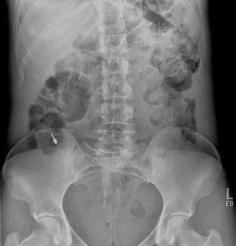
n Other foreign bodies, such as bullet fragments, ingested foreign body, retained surgical needle or sponge and rectal foreign body (Figs. 1.5 and 1.6).
High Yield Topics
KUB EMERGENCIES
Obstruction
Small Bowel Obstruction Ileus
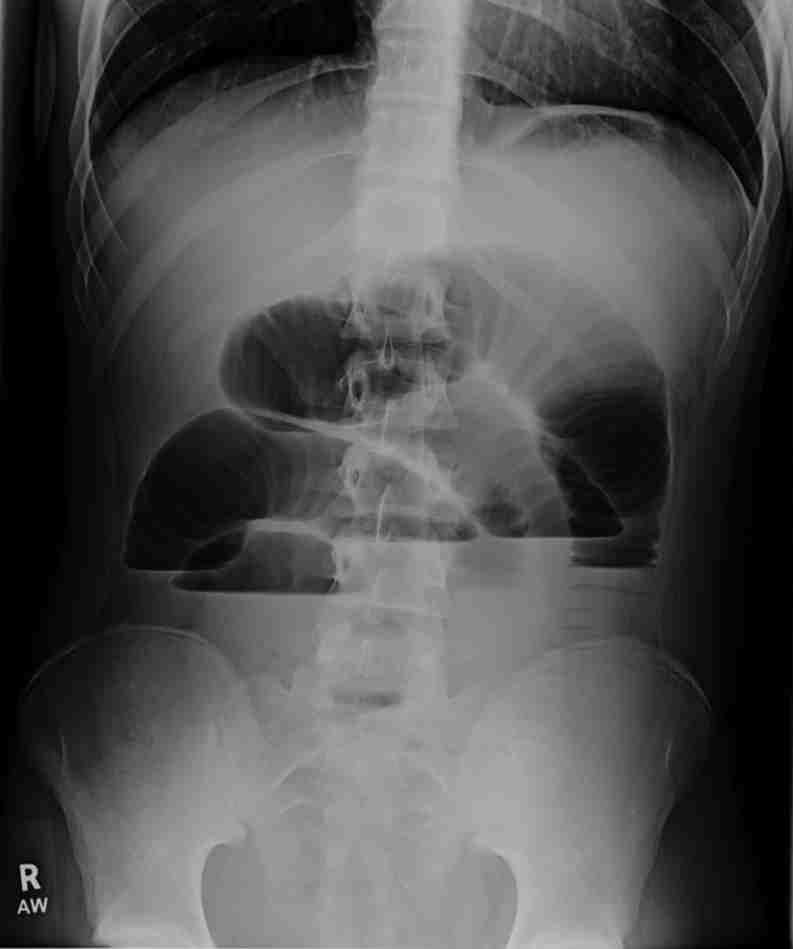
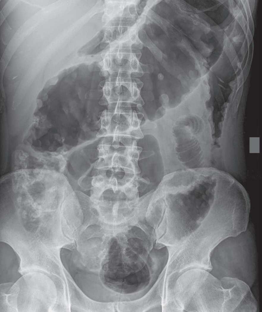
Disproportionate small bowel dilation over large bowel dilation (Fig. 1.7)
Air-fluid levels on upright radiograph
‘String of pearls sign’
Diffuse small and large bowel dilation
Concordant history (surgery, drugs, etc.)
Large Bowel Obstruction
Massively dilated cecum
Collapsed distal colon
NG TUBE
Fig. 1.4 Portable anteroposterior KUB demonstrates a weighted feeding tube tip above the diaphragm over the right hemithorax, within the right lower lobe bronchus.
NG TUBE
Fig. 1.5 Portable anteroposterior KUB demonstrates a normally positioned nasogastric tube in the stomach. However, there is a radiodense surgical pad marker in the right lower quadrant from a retained laparotomy pad.
Fig. 1.6 Portable anteroposterior KUB demonstrates a metallic foreign body overlying the cecum, consistent with dislodged dental implant during traumatic intubation.
Pneumoperitoneum (free air): suggestive of hollow viscus perforation. Signs include:
n Free air under the diaphragm on upright imaging.
n Rigler’s sign (gas outlining both walls of bowel).
n Outlining of liver edge, falciform ligament, urachus.
n Football sign: massive pneumoperitoneum.
Pneumatosis: can be a sign of ischemia, although sometimes benign.
n Associated with portal venous gas (air in the portal veins).
n Gas tracking as bubbly collections along the wall of small bowel (pneumatosis intestinalis), colon (pneumatosis coli), gallbladder (emphysematous cholecystitis), bladder (emphysematous cystitis), kidney (emphysematous pyelonephritis), or stomach (emphysematous gastritis).
n Associated dilation can be ominous.
Volvulus: twisting of bowel upon itself resulting in obstruction and impending ischemia.
n Sigmoid (more common): coffee bean sign, extremely dilated, extending from left lower quadrant to right upper quadrant (\).
n Cecal (less common): extends from right lower quadrant to left upper quadrant (/) (Fig. 1.8).
n Gastric volvulus and midgut volvulus are difficult to diagnose by plain radiograph and usually identified on fluoroscopy or computed tomography.
Megacolon: complication of inflammatory bowel disease and infectious colitis (Clostridium difficile) (Fig. 1.9).
n Marked colonic dilation (typically transverse colon): greater than 6 cm.
n Ahaustral markings: featureless bowel morphology.
n Pseudopolyps: related to bowel ulceration.
n Thumbprinting: mucosal edema.
Fig. 1.7 Upright KUB demonstrates multiple dilated loops of small bowel with prominent air fluid levels, consistent with small bowel obstruction. R
jmf supine 1 of 2
Fig. 1.8 Portable anteroposterior KUB demonstrates marked distention of a loop of large bowel extending to the left upper quadrant, consistent with cecal volvulus.
PJL
Fig. 1.9 Portable anteroposterior KUB demonstrates severe dilatation of the transverse colon with diffuse pseudopolyps along the colonic walls, consistent with ulcerative colitis and toxic megacolon.
Physics Pearls
n There is much more soft tissue to penetrate in abdominal radiography compared with the chest, resulting in 503 more radiation dose per examination.
n Kilovolt (kV) level is kept as low as possible to maintain tissue contrast, while still high enough to penetrate the soft tissues.
n Milliampere (mA) level is therefore maximized in abdominal radiography. Portable radiography machines often have a fixed mA; kV is often increased in portable exams, leading to degraded imaging.
n Minimizing patient motion also plays a key role.
n Scatter is reduced with collimation and a Bucky grid.
n Consider gonadal shielding in young males.
Suggested Reading
1. Levine MS. Plain film diagnosis of the acute abdomen. Emerg Med North Am. 1985;3:541-562.
2. Kellow ZS, MacInnes M, Kurzencwyg D, et al. The role of abdominal radiography in the evaluation of the non-trauma emergency patient. Radiology. 2008;248:887-893.
3. Ros PR, Huprich JE. ACR appropriateness criteria on suspected small bowel obstruction. J Am Coll Radiol. 2006;3:838-841.
4 Maglinte D, Balthazar E, Kelvin F, et al. The role of radiology in the diagnosis of small-bowel obstruction. AJR Am J Roentgenol. 1997;168:1171-1180.
5. Ahn S, Mayo-Smith W, Murphy B, et al. Acute nontraumatic abdominal pain in adult patients: abdominal radiography compared with CT evaluation. Radiology. 2002;225:159-164.
2 Gastrointestinal Fluoroscopy
WEIER LI
CHAPTER OUTLINE Techniques
Fluoroscopic Contrast Agents
Equipment Factors
Pearls for Maximizing Image Quality and Minimizing Radiation Exposure
Techniques
n Fluoroscopy is an imaging modality where continuous x-ray images are obtained to evaluate the body in real time, often with the aid of administered radiopaque contrast material (Fig. 2.1).
n Fluoroscopic examinations attempt to answer specific questions, and therefore understanding the indications and limitations of each examination is crucial.
Protocols
Barium Swallow (Esophagram)
Abnormalities on Esophagram
Physics Pearls
n Imaging with fluoroscopy is heavily operator controlled, allowing for wide customization and personalization of each study.
Types and indications of fluoroscopic procedures in the abdomen and pelvis include:
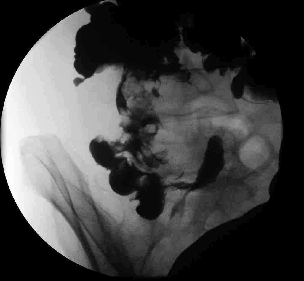
Patency of fallopian tubes, uterine abnormalities (Fig. 2.4)
FLUOROSCOPIC CONTRAST AGENTS
n Contrast agents are compounds that enable improved visualization of internal luminal structures, spaces, and tracts, and allow for controlled real time evaluation of targeted organs.
n Fluoroscopic contrast agents are divided into two types: positive and negative contrast. Positive contrast materials, such as barium and iodine compounds, absorb x-rays more strongly than surrounding tissues and appear radiopaque.
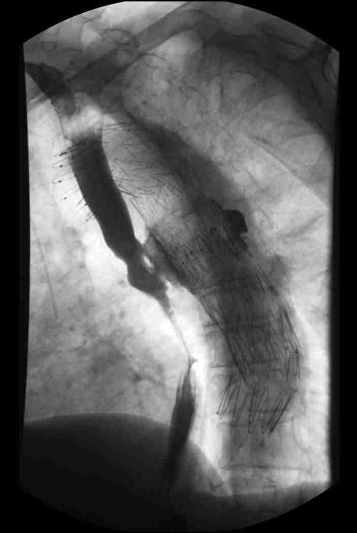
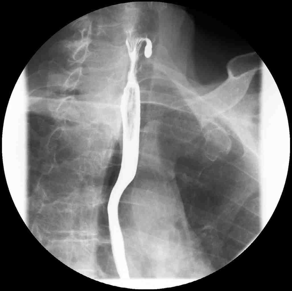
Fig. 2.2 Postoperative swallow study with water-soluble contrast demonstrates a posterior leak, which communicates with the aortic graft, consistent with aortoesophageal fistula.
Fig. 2.3 Small bowel follow through demonstrates narrowing and structuring of the terminal ileum consistent with Crohn disease.
Fig. 2.4 Hysterosalpingogram with two separate cervical catheters demonstrates uterine didelphys.
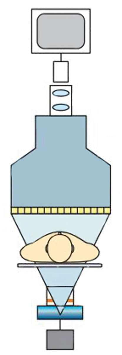
Fig. 2.1 Schematic diagram of a fluoroscopic imaging system.
Conversely, negative contrast materials, such as air or CO2, absorb x-rays less strongly and are radiolucent.
Barium Sulfate
n Barium sulfate is an element compound that is mixed with water and either ingested or instilled into the gastrointestinal tract. Differing concentrations of barium sulfate suspensions are used to evaluate the wall lining, size, shape, contour and patency of the hollow viscus.
n Barium is an inert element; allergic reactions are exceedingly rare. It is the contrast agent of choice when evaluating for aspiration because barium can be coughed out without major issue. Conversely, barium is contraindicated in the evaluation of possible bowel perforation, as barium within the abdominal cavity can cause peritonitis.
n Side effects of barium include: bloating, constipation, cramping, nausea or vomiting.
Water-Soluble Agents
n Water-soluble contrast agents can be divided into ionic or nonionic agents or, depending on the osmolarity, as high- and low-osmolar agents. Nonionic agents have lower osmolarity and demonstrate fewer side effects. Water-soluble agents are used when barium is contraindicated, specifically in the evaluation of perforation.
n Water-soluble agents have multiple disadvantages. These agents are less radiodense than barium and result in poorer opacification of the gastrointestinal tract. In addition, the high osmolar content of these agents causes rapid dilution of these agents as it travels in the distal small bowel.
n In patients with suspected aspiration, water-soluble agents are contraindicated. Aspiration or these agents can lead to pneumonitis and severe pulmonary edema because of their high osmolarity.
n Gastrografin (diatrizoate meglumine and diatrizoate sodium) is a noniodine based water-soluble contrast agent that can be used in patients with iodinated contrast allergy.
EQUIPMENT FACTORS
n Source-to-image distance (SID).
n Fluoroscopic kilovoltage peak (kVp).
n Fluoroscopic milliampere (mA).
n Focal spot.
n Field of view.
n Grid use.
n Fluoroscopic acquisition mode.
n Dose rate selection.
n Video frame rate.
PEARLS FOR MAXIMIZING IMAGE QUALITY AND MINIMIZING RADIATION EXPOSURE
n Collimate image to match axis of organ being evaluated.
n Keep image intensifier as close to patient as possible.
n Use rapid sequence option only to document motion abnormalities or to catch a rapidly changing segment that can only be evaluated with a single shot.
n A fluoroscopic store option is available for an image that one wishes to record.
Protocols
BARIUM SWALLOW (ESOPHAGRAM)
n Fluoroscopy is the main radiology modality for evaluation of the esophagus. Fluoroscopy is uniquely suited for the evaluation of dynamic oropharyngeal function, esophageal morphology, motility, mucosa, gastroesophageal (GE) junction, and reflux.
n Evaluation of the esophagus can be performed either with single-contrast or double-contrast technique. Singlecontrast examination is usually performed with ingestion of oral barium. A double-contrast technique involves first ingesting an effervescent agent to allow for gaseous distention of the esophagus before barium administration.
Anatomy
n The esophagus is approximately 20 to 35 cm long and divided into the cervical, thoracic and abdominal esophagus.
n The esophagus begins at the cricopharyngeus (upper esophageal sphincter) and ends at the lower esophageal sphincter (or ampulla/vestibule).
Technique
Patient should be nothing-by-mouth (NPO) for the examination.
n Scout with collimation for foreign body, metal, or in the evaluation of fistula or leak.
n Effervescent crystals for double-contrast examination.
n Dynamic evaluation of pharynx with lateral and anteroposterior (AP) projection for aspiration.
n Upright left posterior oblique (LPO) for distal esophagus.
n Horizontal right anterior oblique (RAO) for esophageal motility, mucosal abnormality, GE junction.
n Supine, slowly rolling toward the right (Schatzki maneuver) with provocative maneuvers for reflux (cough, Valsalva). Water siphon test can be considered.
n Brief examination of the stomach and proximal bowel for gross abnormalities.
n Upright swallow of barium tablet for functional obstruction.
Normal Esophageal Impressions
n Cervical impression because of the cricoid cartilage at C5–C6.
n Thoracic impression because of the aortic arch at T4–T5.
n Abdominal impression because of the diaphragm at T10–T11.
ABNORMALITIES ON ESOPHAGRAM
Esophageal Dysmotility
Fluoroscopy is uniquely suited for the evaluation of motility abnormalities, which include:
n Achalasia: failure of the lower esophageal sphincter to relax, with decreased peristalsis. “Birdbeak” appearance with smooth tapering.
n Pseudoachalasia: irregular narrowing of the lower esophageal sphincter because of infiltrative tumor with irregular lobulations.
n Diffuse esophageal spasm: intermittent, uncoordinated contractions, which cause a “corkscrew” appearance that can lead to chest pain or dysphagia.
n Scleroderma: absent peristalsis of the smooth muscle leading to a dilated distal esophagus.
Diverticula
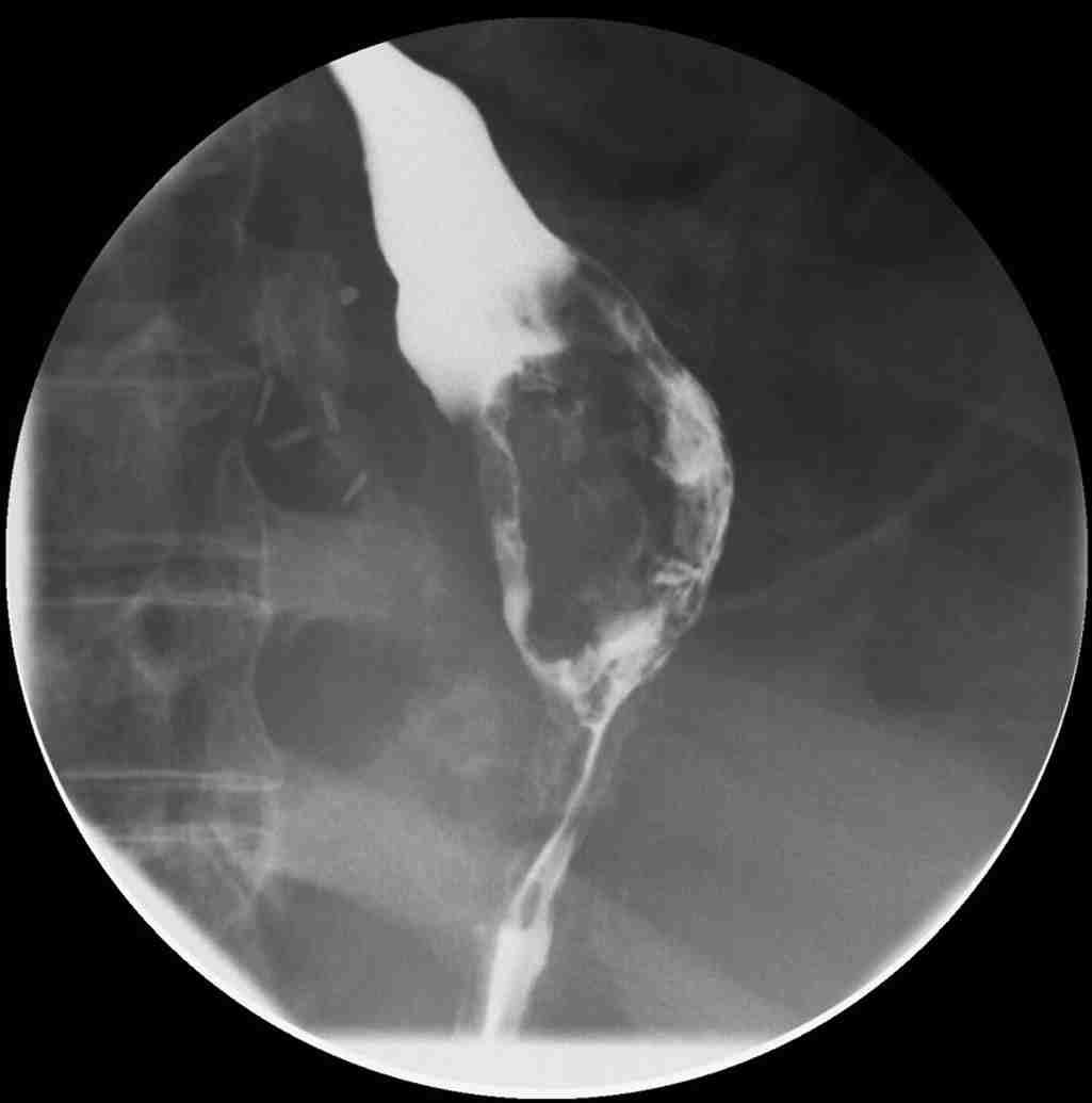
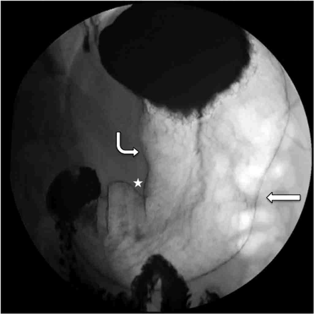
n Zenker’s: most common, arises at Killian’s dehiscence near the pharyngoesophageal junction; posterior.
n Killian-Jameson: just inferior to Zenker’s; lateral (Fig. 2.5).
n Traction: mediastinal inflammation.
n Epiphrenic: occur at the thoracic esophagus, pulsion diverticula related to motility disorder.
Rings and Webs
n A-ring: physiological contrast of esophageal smooth muscle above the esophageal vestibule.
n B-ring: concentric Schatzki ring located at the GE junction and often symptomatic.
n Web: thin mucosal fold, shelf-like.
Extrinsic Compression
n Cricopharyngeal bar: normal posterior compression at C5.
n Vascular: aorta, aberrant vascular anatomy, varices, aneurysm.
n Thyroid: cervical impression.
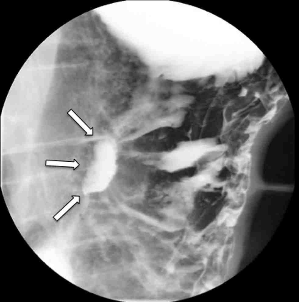
n Adenopathy or malignancy (Fig. 2.6).
Mucosal/Submucosal Masses
n Inflammatory polyp: smooth margins, intraluminal.
n Adenocarcinoma: irregular margins (Fig. 2.7).
n Fibrovascular polyp: benign tumor with large stalk.
n Leiomyoma: most common benign submucosal mass, smooth, and rounded.
Diffuse Mucosal Abnormalities
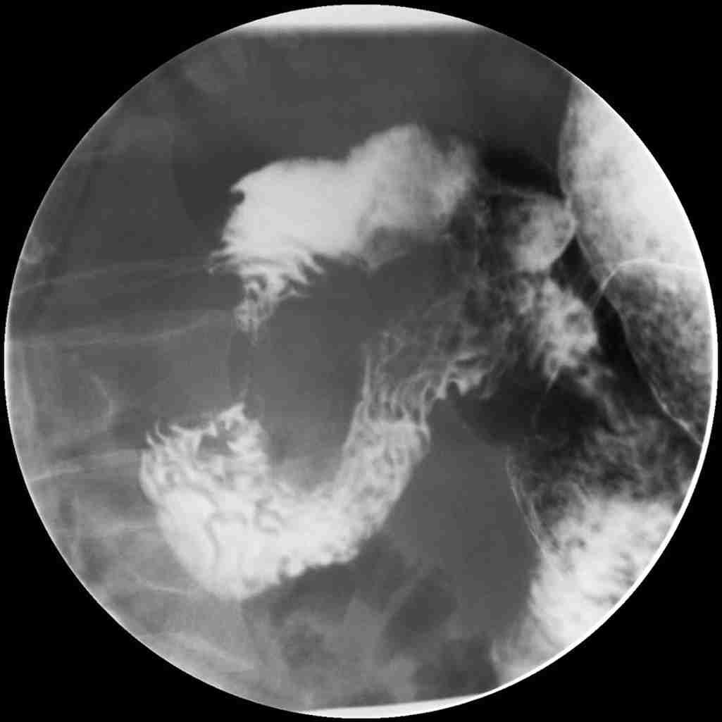
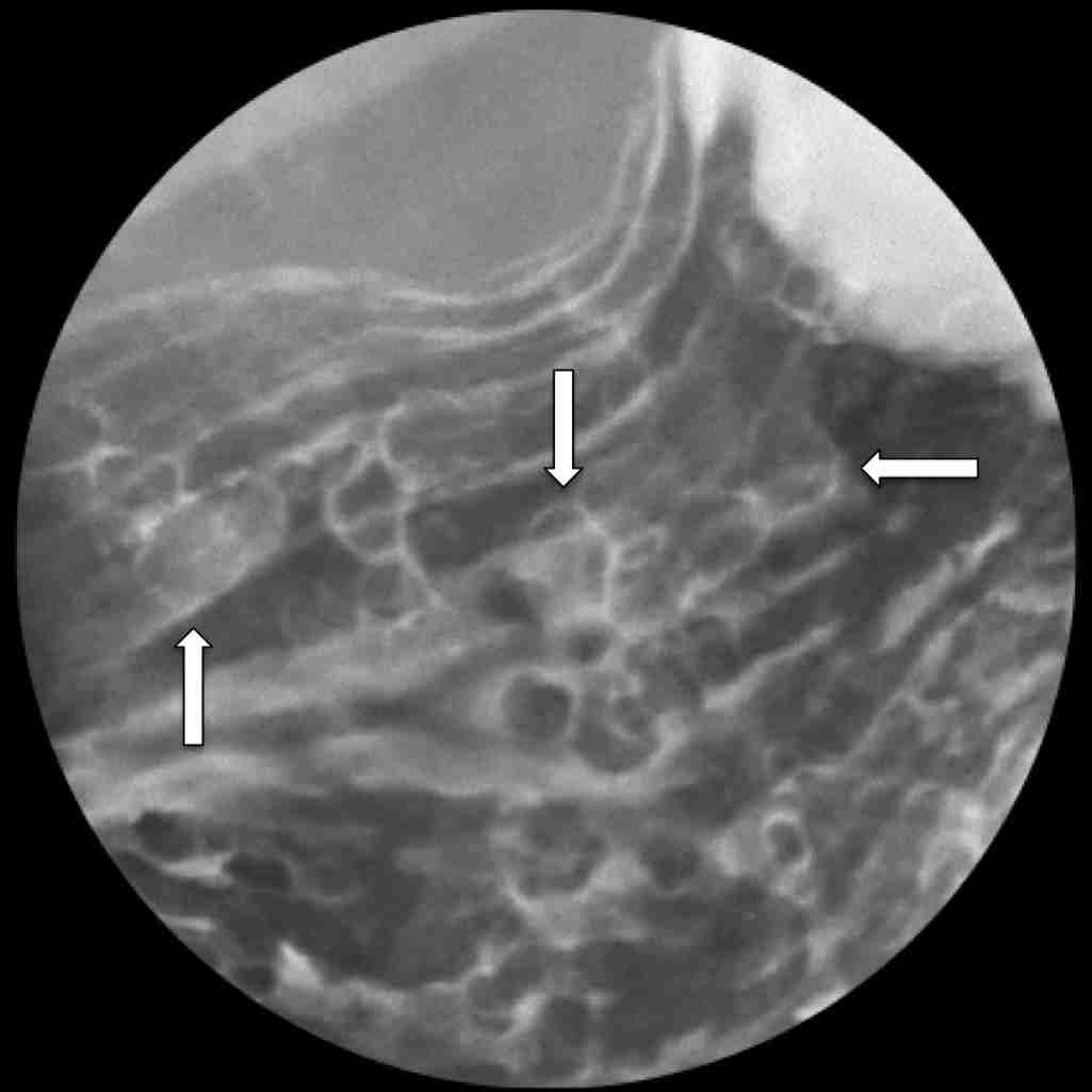
n Candida esophagitis: immunocompromised patient, ulcers, mucosal nodules in longitudinal columns (Fig. 2.8).
2.7 Barium swallow demonstrates a large mass-like filling defect in the distal esophagus, which was biopsied as an esophageal carcinoma.
n Herpes esophagitis: multiple small ulcers.
n Human immunodeficiency virus (HIV)/cytomegalovirus (CMV): large ulcers, typically greater than 1 cm.
n Glycogenic acanthosis: benign degenerative condition, discrete plaques and nodules.
n Reflux esophagitis: erosions, strictures, ulcerations associated with gastroesophageal reflux disease (GERD).
Fig. 2.6 Upper gastrointestinal study demonstrates an apple core lesion in the second portion of the duodenum because of mass effect from a pancreatic ductal adenocarcinoma.
Fig. 2.5 Initial swallow of barium demonstrates a small left lateral outpouching from the anteroinferior portion of the hypopharynx, consistent with Killian-Jameson diverticulum.
Fig.
Strictures
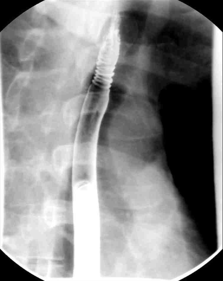
n Peptic stricture: related to GERD, distal esophagus, short segment with smooth tapering, possibly consequence of Barrett’s esophagus.
n Caustic ingestion: correlate with history (especially lye ingestion), long smooth stricture.
n Pill esophagitis.
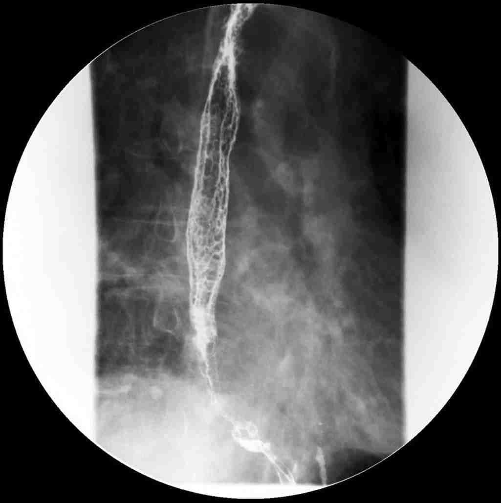
n Eosinophilic esophagitis: common in younger patients, large concentric rings, upper esophagus (Fig. 2.9).
Other
n Mallory-Weiss Tear: contrast leak.
n Perforation (Boerhaave’s): pneumomediastinum with leak (usually posterior and to the left).
n Impaction: correlation with history.
Physics Pearls
Fluoroscopic x-ray images differ from conventional radiography by lower mA, with higher exposure times.
Image Quality
n Objects become magnified as they are brought closer to x-ray source.
n Objects become more blurry as they move away from the detector.
n Minimize patient motion (hold breath).
n Collimation improves spatial resolution.
Radiation Dose
n Doubling distance from x-ray source decreases dose by a factor of 4 (inverse square law).
n Collimation reduces dose to patient and operator.
n Magnification increases dose.
Suggested Reading
1. Levine MS, Rubesin SE. Radiologic investigation of dysphagia. AJR Am J Roentgenol. 1990;154(6):1157-1163.
2. Tao TY, Menias CO, Herman TE, McAlister WH, Balfe DM. Easier to swallow: pictorial review of structural findings of the pharynx at barium pharyngography. RadioGraphics. 2013;33(7):e189-e208.
3. Luedtke P, Levine MS, Rubesin SE, Weinstein DS, Laufer I. Radiologic diagnosis of benign esophageal strictures: a pattern approach. RadioGraphics. 2003;23(4):897-909.
5. Canon CL, Morgan DE, Einstein DM, Herts BR, Hawn MT, Johnson LF. Surgical approach to gastroesophageal reflux disease: what the radiologist needs to know. RadioGraphics. 2005;25(6):1485-1499.
Fig. 2.8 Barium swallow demonstrates discrete longitudinally oriented plaques with small ulcers, consistent with Candida esophagitis.
Fig. 29 Barium swallow demonstrates multiple concentric rings in the upper esophagus, consistent with eosinophilic esophagitis.
3 Gastric Wall Thickening/ Masses
DAVID KNIPP
CHAPTER OUTLINE
Anatomy, Embryology, Pathophysiology
Techniques
Protocols
Specific Disease Processes
Benign Versus Malignant Ulcers
Focal Masses
Anatomy, Embryology, Pathophysiology
n The stomach is anatomically subdivided into the cardia, fundus, body, and antrum, with inflow regulated by the lower esophageal sphincter and outflow by the pyloric sphincter.
n The incisura angularis represents the acute angle formed on the lesser curvature, which marks the transition from body to antrum.
n The gastric wall is composed of four layers: the mucosa (containing the epithelium and lamina propria), the submucosa (containing vascular, lymphoid, and nervous tissue), the trilaminar muscularis externa, and the serosa.
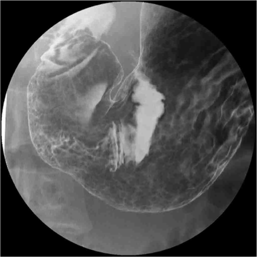
n The areae gastricae represent the normal reticular mucosal pattern in a well-distended stomach, with rugal folds becoming more prominent in a nondistended stomach (Fig. 3.1).
n It is bounded by the lesser omentum attached along the lesser curvature, and greater omentum attached to the greater curvature.
n The right and left gastric arteries supply blood to the lesser curvature, whereas the gastroepiploic and short gastric arteries supply the greater curvature and fundus, respectively.
n Venous drainage is provided by the gastric veins that drain into the portal vein, the short gastric and left gastroepiploic veins that drain into the splenic vein, and right gastroepiploic vein that drains into the superior mesenteric vein.
Techniques
Fluoroscopy
n Even in the modern age of fast and accessible crosssectional imaging, fluoroscopy has remained a prominent modality for assessing the stomach because of its superior spatial resolution and dynamic nature.
Tumor Staging/Classification Systems
Primary Tumor (T)
Nodal Status (N)
Metastases (M)
Key Elements of a Structured Report
n Double contrast technique: preferred method for evaluation of the gastric mucosa. The patient is given effervescent granules to distend the stomach followed by a barium suspension to coat the mucosa.
n Single contrast technique: less commonly performed, but useful for assessment of peristalsis, gastric outlet obstruction, postoperative patients, or suspected perforation.
Computed Tomography
n Better for depiction of extraluminal disease and associated complications.
n May use water or positive oral contrast.
Protocols
Fluoroscopy
n Upper gastrointestinal (GI) double contrast technique:
n To obtain adequate distension of the stomach, the patient should be given a dose of effervescent crystals at the start of the examination. Instruct the patient to resist belching.
n Most evaluations of the stomach are preceded by upright imaging of the esophagus with a thick barium solution. When completed, the patient should be lowered into the supine position, as to retain barium within the dependent fundus.
n The patient should then complete a 540 degree rotation to the left to coat all surfaces of the stomach, with final right anterior oblique (RAO) positioning.
n Image the contrast opacified duodenal bulb as barium exits the stomach.
n Rotate the patient into the right posterior oblique (RPO) position to obtain views of the anterior and posterior walls of the stomach.
n Rotate the patient into the supine position to obtain images of the lesser and greater curvatures (Fig. 3.2).
n Rotate the patient into the left posterior oblique (LPO) position to obtain air-contrast images of the antrum and duodenal bulb.
n Obtain a scout image of the abdomen to assess for free air. In postoperative patients, scout imaging also serves as a baseline comparison.
n Using thin barium, first obtain upright views of the esophagus in the LPO position. The patient should drink enough to fill the stomach.
n Obtain upright views of the gastric body and antrum in the LPO, anteroposterior (AP), and RPO positions. Use compression when possible.
n Bring the table to the horizontal position and take an image of the fundus in the supine position. As the fundus is often positioned beneath the ribcage, it cannot be compressed.
n Image the contrast-filled antrum and duodenal bulb in the RAO position. Compression may be performed from underneath.
n Finish by obtaining an overhead film of the stomach and small bowel.
n The aforementioned protocols are only to serve as a guide. Evaluation of gastric pathology requires prompt identification and real-time adjustment to obtain the best images possible while limiting radiation exposure.
Specific Disease Processes
BENIGN VERSUS MALIGNANT ULCERS
Fluoroscopy
n Benign ulcers (95%):
n Pooling of barium within a smooth, sharply defined mucosal defect that projects beyond the stomach contour.
n Radiating folds reach the edge of the ulcer (Fig. 3.3).
n More common on lesser curvature, antrum, and posterior wall.
n Giant ulcers over 3 cm are almost always benign.
n If further images of the esophagus are required in the prone position, these should be obtained at this time, followed by assessment for reflux, and a full field of view image of the opacified bowel.
n Upper GI single contrast technique:
n If there is question of GI leak, the examination should first be performed with water-soluble contrast before proceeding to barium.
Fig. 3.2 Anterior-posterior view of the stomach demonstrating the greater (arrow) and lesser (curved arrow) curvatures. The incisura angularis is seen along the lesser curvature (asterisk).
Fig. 3.1 Areae gastricae representing a normal mucosal pattern.
Fig. 3.3 Benign ulcer with smooth borders and radiating folds reaching the ulcer edge (arrows).
n Hampton line: Thin radiolucent line between the gastric lumen and the ulcer seen on profile view representing a layer of mucosa overhanging the ulcer edge.
n Malignant ulcers (5%):
n Irregular shape not extending beyond the stomach contour.
n Asymmetric radiating folds that do not reach the ulcer’s edge.
n More common on greater curvature.
n Carmen Meniscus sign: Radiolucent raised border convex toward the lumen surrounding the ulcer on profile view.
Computed Tomography
n Used to assess for complications, such as perforation, involvement of surrounding structures, and metastases.
n Depicts wall thickening, submucosal edema, and luminal narrowing.
FOCAL MASSES
Polyps
n Hyperplastic:
n Most common benign epithelial neoplasm of the stomach.
n Seen with chronic gastritis, no malignant potential.
n More likely in fundus or body.
n Smooth, sessile or pedunculated, smaller than 1 cm.
n Hamartomatous:
n Peutz-Jeghers and Cronkhite-Canada syndromes.
n No malignant potential.
n Clustered broad-based polyps.
n Adenomatous:
n Increased risk of malignancy, although higher risk of coexisting gastric cancer than of malignant transformation.
n Familial adenomatous polyposis syndrome.
n More likely in antrum and body.
n Single, lobulated or cauliflower-like, over 1 to 2 cm.
n Fluoroscopy:
n Seen as radiolucent filing defects if dependent.
n Rim of barium creating ringed shadows if nondependent (Fig. 3.4).
n May have central droplet.
n Primary malignancies:
n Gastric carcinoma:
n Accounts for more than 95% of gastric malignancies.
n Risk factors: Helicobacter pylori, smoked foods, nitrate/ nitrite heavy diet.
n Fairly even distribution throughout the stomach.
n May present as a malignant ulcer, polyp, or scirrhous type.
n Polypoid carcinomas present as lobulated or fungating masses that may contain irregular ulcerations.
n Calcifications suggest a mucinous subtype.
n Metastases:
n Virchow node: lymphatic spread to left supraclavicular node.
n Krukenberg tumor: ovarian metastasis.
n Sister Mary Joseph nodule: umbilical metastasis.
n Gastrointestinal stromal tumor:
n Submucosal tumor arising from the interstitial cells of Cajal.
n 70% of all gastrointestinal stromal tumors (GISTs) occur in the stomach.
n May be benign or malignant, which are not reliably distinguished by CT except for the presence of metastases.
n Associated with neurofibromatosis type 1, as well as part of the Carney triad (GIST, pulmonary chondroma, extraadrenal paraganglioma).
n Fluoroscopy:
n Submucosal round mass forming smooth obtuse angles with the gastric wall.
n May have central barium-filled ulcer when larger.
n CT:
n Well-defined, mostly exophytic mass, ulcerated when larger (Fig. 3.5).
n May contain air, fluid, or calcifications.
n Assess for metastases (liver, lungs, peritoneum).
n Metastases can appear cystic, especially following chemotherapy.
n Lymphadenopathy is not a typical feature.
n Lymphoma:
n 80% non-Hodgkin lymphoma, B-cell type.
n Chronic H. pylori infection may result in a more indolent lymphoma arising from mucosa-associated lymphoid tissue (MALT).
n Stomach is most common site of primary GI lymphoma.
n Fluoroscopy/CT:
n May present as a polypoid or ulcerative mass, multiple submucosal nodules, or diffuse thickening.
n Multiple lesions favor MALT lymphoma.
n Metastases:
n Most commonly hematogenous spread from breast or melanoma.