All rights reserved. No part of this publication may be reproduced, stored in a retrieval system, or transmitted, in any form or by any means, electronic, mechanical, photocopying, recording or otherwise, except as permitted by law. Advice on how to obtain permission to reuse material from this title is available at http://www.wiley.com/go/permissions.
The right of Robert H. Whitaker to be identified as the author of this work has been asserted in accordance with law.
Registered Office(s)
John Wiley & Sons, Inc., 111 River Street, Hoboken, NJ 07030, USA
John Wiley & Sons Ltd, The Atrium, Southern Gate, Chichester, West Sussex, PO19 8SQ, UK
Editorial Office
9600 Garsington Road, Oxford, OX4 2DQ, UK
For details of our global editorial offices, customer services, and more information about Wiley products visit us at www.wiley.com.
Wiley also publishes its books in a variety of electronic formats and by print‐on‐demand. Some content that appears in standard print versions of this book may not be available in other formats.
Limit of Liability/Disclaimer of Warranty
The contents of this work are intended to further general scientific research, understanding, and discussion only and are not intended and should not be relied upon as recommending or promoting scientific method, diagnosis, or treatment by physicians for any particular patient. In view of ongoing research, equipment modifications, changes in governmental regulations, and the constant flow of information relating to the use of medicines, equipment, and devices, the reader is urged to review and evaluate the information provided in the package insert or instructions for each medicine, equipment, or device for, among other things, any changes in the instructions or indication of usage and for added warnings and precautions. While the publisher and authors have used their best efforts in preparing this work, they make no representations or warranties with respect to the accuracy or completeness of the contents of this work and specifically disclaim all warranties, including without limitation any implied warranties of merchantability or fitness for a particular purpose. No warranty may be created or extended by sales representatives, written sales materials or promotional statements for this work. The fact that an organization, website, or product is referred to in this work as a citation and/or potential source of further information does not mean that the publisher and authors endorse the information or services the organization, website, or product may provide or recommendations it may make. This work is sold with the understanding that the publisher is not engaged in rendering professional services. The advice and strategies contained herein may not be suitable for your situation. You should consult with a specialist where appropriate. Further, readers should be aware that websites listed in this work may have changed or disappeared between when this work was written and when it is read. Neither the publisher nor authors shall be liable for any loss of profit or any other commercial damages, including but not limited to special, incidental, consequential, or other damages.
Library of Congress Cataloging‐in‐Publication Data
Names: Whitaker, R. H. (Robert H.), author, illustrator.
Title: A visual guide to clinical anatomy / Robert H. Whitaker.
Description: Hoboken, NJ : Wiley-Blackwell, 2021.
Identifiers: LCCN 2020027865 (print) | LCCN 2020027866 (ebook) | ISBN 9781119708100 (paperback) | ISBN 9781119708162 (adobe pdf) | ISBN 9781119708148 (epub)
Subjects: MESH: Body Regions–anatomy & histology | Clinical Medicine | Pictorial Work
5.10 Eye Light and Near Reactions, Eye Movements and Pupil Control, 398
5.11 Outer, Middle and Inner Ear, 406
5.12 Mouth and Mandible, 413
5.13 Nose and Face, 424
5.14 Neck Fascia, Vessels and General Anatomy, 430
5.15 Neck Triangles, Muscles, Thyroid and Parathyroid Glands, 440
5.16 Parotid and Submandibular Glands, 448
5.17 Larynx and Pharynx, 452
5.18 Autonomic Nervous System, in General, Sympathetic and Parasympathetic, 464
5.19 Temporal, Infratemporal and Pterygopalatine Fossae, 481
5.20 Cavernous Sinus and other Venous Sinuses, 489
Preface
Robert H. Whitaker
It is easier to say what this book is not, rather than what it is. It is definitely not a textbook or an atlas but simply a series of images that I have used for teaching over the last 30 years as an Anatomy Lecturer and Demonstrator in The University of Cambridge, UK. The images have also been used for numerous lectures, courses and meetings elsewhere.
Each image is designed to give a synopsis of anatomical and clinical information about an individual topic or clinical condition for both medical students and those in clinical practice. I have drawn each image myself using Adobe Illustrator, a graphics programme that I can recommend to all aspiring anatomical artists.
Anatomists have often been accused of teaching students too much detail that is not clinically relevant and I have been very much aware of this. I have strived only to teach anatomy that will be pertinent to their varied clinical careers and constantly endeavoured to find easy ways to learn and remember the vast amount of information that anatomy comprises.
I make no apology for a lack of neuroanatomy as it is outside my field. There is relatively little embryology amongst these images but six concise podcasts in embryology in the form of lectures with images can be found on www.instantanatomy.co.uk (a subscription website) which includes a panacea of descriptive anatomy. www.instantanatomy.net is a free, similar website with fewer lectures. I also make no apology that you will find duplication of both illustrations and text on two or more images throughout. This is intentional as often the same information is needed for any individual teaching slide.
I would be delighted if some of you wish to use the images in this book for your own lectures or teaching and you are very welcome to do so with an acknowledgement.
I am most grateful to Mr John Fergus FRCS who very kindly helped me to check every image for anatomical and clinical accuracy. I took advice from a team of experts for the sections on the heart (Professor John Wallwork, CBE, FRCS, FMedSci), the eye (Mr. Nick Sarkis, FRCS) and ear, nose and throat (Mr. Roger Gray, FRCS). To each of them I am eternally grateful.
Robert H. Whitaker
Foreword
By Professor Harold Ellis
Human Anatomy is intrinsically interesting - surely everyone likes to know how he or she is constructed. However, it is, of course, of immense practical importance to people in the healing professions. Just as the London taxi driver needs to learn “the knowledge” of the streets of the metropolis, so health workers need to know what lies below their palpating fingers. Of course, surgeons need to be especially competent anatomists.
The early anatomy textbooks had no or few illustrations, and these were mostly crude and grossly inaccurate. In 1543 came a revolution; the publication by the young anatomist Andreas Vesalius, of Padua, of “De Humani Corpora Fabrica”, (the structure of the human body). Its revolutionary contribution was not so much the text but the magnificent illustrations – good enough and often still used today. Interestingly, there is still debate about who some of these anonymous artists were.
Good teachers of Anatomy, without exception, can produce clear and accurate diagrams; those that are most appreciated are ones that the student can reproduce. Robert H. Whitaker, a retired urologist and now Anatomy teacher, is well known for his Anatomy texts and his lectures, which are illustrated by his own clearly reproducible illustrations. This new publication will prove useful to both undergraduate and graduate students in reinforcing their anatomy studies. I only wish a book like this had been available all those years ago, when I was an Anatomy student!
Professor Harold Ellis, CBE, DM, MCh, FRCS Emeritus Professor of Surgery, University of London; Clinical Anatomist, Guy’s Campus London SE1 1US, UK
Foreword
By Professor Sir Roy Calne
I have known Robert H. Whitaker for nearly 50 years as a surgical colleague, friend and fellow artist working together at Addenbrooke’s Hospital in Cambridge, UK. We have shared Surgical Grand Rounds, Committee Meetings and many other hospital and social activities. We have an underlying mutual respect. With the production of this unique anatomical book he has introduced us to a new approach to teaching in that it is neither an atlas nor is it a textbook. When you delve into this beautifully illustrated book you will be impressed with its fund of knowledge and bright, colourful images.
Robert has produced some 920 annotated anatomical images that provide a concise synopsis of what is important for students and others to know and retain in order to undertake safe and competent clinical practice in any branch of medicine. These clear and beautiful images are a feat indeed!
A knowledge of anatomy is as relevant today as it was a century ago and I remember a comment I made in the Christmas Edition of the British Medical Journal many years ago when endoscopic surgery was in its infancy, that there was a risk of endoscopic surgeons not being able to get out of trouble with an open operation if they did not have an accurate working knowledge of the anatomy of the region.
Each image tells a story of a particular aspect of anatomy and its relevance to clinical practice. This brings the subject to light and its beautiful presentation makes it interesting, enjoyable and memorable.
Professor Sir Roy Calne, MA, MS, FRS, FRCS, FRCP Emeritus Professor of Surgery, University of Cambridge and Addenbrooke’s Hospital, Cambridge CB2 0QQ, UK
About the Author
Robert H. Whitaker, MA, MD, MChir, FRCS, FMAA graduated from Selwyn College, Cambridge before undertaking his clinical training at University College Hospital, London. He spent a year at Johns Hopkins Hospital, Baltimore in the Urological Research Laboratories before returning to the St Peters Hospital Group in London to train as a urologist. He was a Senior Lecturer at the London Hospital Medical School before being appointed as a Consultant Urological Surgeon at Addenbrooke’s Teaching Hospital in Cambridge, UK in 1973.
He spent 20 years practising mostly paediatric urology during which time he co-founded the British Association of Paediatric Urologists. He was an examiner for the Primary FRCS and later the MRCS at all four Colleges of Surgeons in the UK and a Hunterian Professor at the Royal College of Surgeons of England in 1973. He retired in 1990 to teach anatomy in the Department of Anatomy in Cambridge for the next 30 years. He is an Honorary Fellow and Examiner for the Medical Artists’ Association of Great Britain and was awarded the Farquharson Teaching Award by the Royal College of Surgeons of Edinburgh in 2013. He was awarded the St Peter’s Medal of the British Association of Urological Surgeons in 1994 and was made an Honorary Member of the European Society of Paediatric Urology in 2019.
Robert H. Whitaker is author of the anatomy textbook Instant Anatomy, now in its fifth edition (co‐authored by Neil Borley), and has anatomy teaching websites – www.instantanatomy.net and www.instantanatomy.co.uk
Cambridge, UK 2020
Upper Limb
1.1 General Anatomy, 2
1.2 Shoulder and Arm, 13
1.3 Axilla, Brachial Plexus and Nerve Lesions, 31
1.4 Elbow and Forearm, 52
1.5 Wrist and Hand, 62
A Visual Guide to Clinical Anatomy, First Edition. Robert H. Whitaker.
1.1 General Anatomy
SURFACE ANATOMY
Function of any bone:
To give form
For muscle attachments
Movement
Protection of internal organs
Metabolic
Calcium, phosphorus Haemopoiesis
CLASSIFICATION OF JOINTS 1
FIBROUSCARTILAGINOUS
Skull sutures
Interosseous membranes
Inferior tibio bular
11th, 12th costotransverse
Primary
Costochondral
1st sternochondral
Spheno-occipital
Secondary
Midline symphyses
Intervertebral
Hyaline cartilage
SYNOVIAL
ATYPICAL SYNOVIALTYPICAL SYNOVIAL
Articular surface covered with brocartilage
Tempormandibular
Sternoclavicular
Acromioclavicular
2-7 Sternochondral
Articular surface covered with hyaline cartilage
All other synovial joints
PLANE
Tarsus and carpus
Gliding
CLASSIFICATION OF JOINTS 2
Flexion Extension HINGE Interphalangeal
SADDLE CONDYLOID 1st Carpometacarpal
Flexion Extension Adduction Abduction Circumduction “Controlled” rotation = opposition
BALL AND SOCKET Hip Shoulder (Sternoclavicular and Talocalcaneonavicular many features of one)
Flexion
Extension Adduction
Abduction
Circumduction “Free” rotation
Coracoid process of scapula
Clavicle
Pectoralis major
Palpable around the shoulder are:
The acromion
The head of the humerus
The corocoid process
The clavicle
Upper trunk over rst rib
Median over brachial artery
Ulnar behind medial epicondyle
Supraclavicular
Median between palmaris longus and exor carpi radialis
Ulnar lateral to pisiform
Extensor pollicis longus
Extensor pollicis brevis
Abductor pollicis longus
POSITION OF RELATIVELY SUPERFICIAL NERVES IN UPPER LIMB
Super cial branch of the radial nerve palpable over tendon of extensor pollicis longus
Cephalic vein
PALPABLE STRUCTURES IN THE UPPER LIMB
Both superior to deltopectoral groove
Coracoid process Lesser tuberosity
Acromioclavicular joint
Medial and lateral epicondyles
Olecranon
Head of radius
Anconeus (posterior to olecranon)
Radial and ulnar styloid processes
Dorsal (Lister’s) tubercle of radius
Hook of hamate
Biceps tendon and aponeurosis
Brachial, radial and ulnar pulses
VULNERABLE NERVES IN THE ARM
Acromion
Axillary nerve
Posterior axilla
Radial nerve
Lateral epicondyle
RADIAL NERVE
2/3 1/3
Passes from where the posterior axilla meets the arm to a point 2/3 down a line from acromion to the lateral epicondyle then it passes anterior to the lateral epicondyle
LEFT SCAPULA
T7
Rib 8
Covers half the ribs 2-7 8th rib is rst below Upper border at T2 Medial spine at T3 Lower border at T7
T2
T3
DERMATOMES IN THE OUTSTRETCHED UPPER LIMB (Anterior view)
UPPER LIMB DERMATOMES
ANTERIORPOSTERIOR
Note the axial lines that separate non-consecutive dermatomes
C4
C5
C6
T2
T1
C8
C7
C4
C5
C6
C7 C 8 T1
T2T3
C4
C5
C6
C7 C 8 T1 T2 T3
UPPER LIMB DERMATOMES
Supraclavicular nerves (cervical plexus)
Upper lateral cutaneous nerve of arm (axillary)
Lower lateral cutaneous nerve of arm (radial)
Lateral cutaneous nerve of forearm (musculocutaneous)
ANTERIOR
Intercostobrachial
Medial cutaneous nerve of arm (medial cord)
Medial cutaneous nerve of forearm (medial cord)
Note: The axial lines that separate non-consecutive dermatomes
CUTANEOUS NERVES OF THE UPPER LIMB
Upper lateral cutaneous nerve of arm (axillary C5,6)
Lower lateral cutaneous nerve of arm (radial C5,6)
Lateral cutaneous nerve of forearm (musculocutaneous C5,6)
Median (C6,7,8)
POSTERIORANTERIOR
Supraclavicular (C3,4)
Intercostobrachial (T2)
Medial cutaneous nerve of arm (C8,T1)
Post cutaneous nerve of arm and forearm (radial C5,6,7,8)
Medial cutaneous nerve of forearm (radial C5,6,7,8)
Ulnar (C8,T1)
Upper lateral cutaneous nerve of arm (axillary C5,6)
Lower lateral cutaneous nerve of arm (radial C5,6)
Lateral cutaneous nerve of forearm (musculocutaneous C5,6)
Radial (C6,7,8)
Lateral cordMedial cordPosterior cord
C4
Supraclavicular nerves (cervical plexus)
C5
Upper lateral cutaneous nerve of arm (axillary)
C5
Lower lateral cutaneous nerve of arm (radial) C6
Lateral cutaneous nerve of forearm (musculocutaneous)
C6,7 Radial nerve
C7
C8
T1
APPROXIMATE DERMATOMES OF UPPER LIMB
Medial cutaneous nerve of forearm T1 Medial cutaneous nerve of arm
Ulnar nerve and medial cutaneous nerve of forearm
Palmar and digital branches of median and ulnar nerves
Above the elbow the anterior dermatomes reach round posteriorly to meet an axial line. Supply posteriorly is by the posterior cutaneous nerve of arm. Below the elbow supply is by the posterior cutaneous nerve of forearm (C6,7)
Usual arrangment but in 15% the nerve passes behind artery
Mnemonic for anastomotic vessels:
Pus - Profunda
Spells - Superior ulnar collateral
Nasty - Nutrient
Infection - Inferior ulnar collateral
Deltoid
Cephalic vein
Pierces the clavipectoral fascia at upper end of the deltopectoral groove to enter axillary vein
Radial artery
Pulse. Lateral to the tendon of radialis
exor carpi
SUPERFICIAL VEINS AND PULSES IN UPPER LIMB
Pectoralis major
Brachial artery
Pulse. In the cubital fossa, lateral to the median nerve
Medial epicondyle
Basilic vein
Commencement of the basilic vein (medial side)
Pierces the fascia in the medial mid arm to join the venae commitantes which together, at the inferior border of teres major, become the axillary vein
Dorsal venous arch
Commencement of the cephalic vein (lateral side)
POSTERIORANTERIOR
lymphatics
LYMPHATIC DRAINAGE IN UPPER LIMB Super cial
lymphatics
Lymphatics from palm of hand and anterior forearm follow basilic vein to lymph nodes in the cubital fossa and then pass deep, to follow the deep veins to the lateral axillary and then central nodes
Lymphatics from dorsum of hand, posterior forearm and posterior arm follow cephalic vein to supraclavicular or infraclavicular lymph nodes
AXILLARY LYMPH NODES
Apical
Infraclavicular
Posterior
Supratrochlear
75% of lymphatics from the breast drain to axillary nodes. Others to internal thoracic, abdominal nodes or to other breast
Support: Rotator cu (subscapularis, supraspinatus, infraspinatus, teres minor), long head biceps, triceps in abduction, muscles from chest to arm
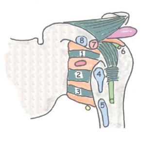
SHOULDER JOINT
(GLENOHUMERAL)
LIGAMENTS: Anterior
1,2,3: Glenohumeral
Anterior: superior, middle, inferior (weak thickenings of capsule)
4: Subscapularis
5: Teres major
6: Supraspinatus
7: Short head biceps
8: Pectoralis minor
Opening of subscapular bursa
Coraco-acromial (strong ++)
Subacromial bursa (large)
Coracohumeral (strong)
Transverse humeral (intertubercular)
Shallow glenoid fossa - deepened by glenoid labrum. Synovial, Ball and socket. Humeral head is 1/3 hemisphere
allow wide abduction, exion and extension.
Capsule: Strong and taut superiorly (anti-sag), inferiorly lax and inserted lower to Synovium: Envelops biceps tendon, communicates with bursae anteriorly and posteriorly
Long head of biceps
SHOULDER JOINT AND ROTATOR CUFF MUSCLES
Capsule
Glenoid labrum
Lax capsule inferiorly
Synovial sheath of biceps
Rotator Cu Muscles:
Subscapularis (anterior)
Infraspinatus (posterior)
Teres minor (posterior)
Supraspinatus (superior)
All blend with capsule of shoulder joint
Lax capsule inferiorly allows dislocation of head of humerus inferiorly and usually anteriorly
The tendon of the long head of biceps lies within the capsule but not within the synovial membrane. It attaches to the supraglenoid tubercle
SUBACROMIAL BURSA AND PAINFUL ARC SYNDROME
Subacromial bursa
Rotator cu (Tendon of supraspinatus)
During abduction the greater tuberosity impinges on the supraspinatus tendon at its point of least blood supply beneath the acromion
Abduction
Acromion
Greater tuberosity
Inferior capsule
Coracoacromial ligament (arch)
ACROMIOCLAVICULAR JOINT
Acromioclavicular ligament
Conoid
Coracoclavicular ligament
Trapezoid
Synovial
Atypical
Thick superior capsule (acromioclavicular ligament)
Incomplete fibrocartilaginous disc in upper joint
Strong coracoclavicular ligament
Nerve: Lateral supraclavicular (C4)
Movements: Gliding (passive) and 20° of rotation of scapula
ACROMIOCLAVICULAR JOINT AND CORACOCLAVICULAR LIGAMENTS
Coracoclavicular ligaments
ConoidTrapezoid
Acromioclavicular ligament
Costoclavicular ligament
ACROMIOCLAVICULAR JOINT
Synovial, Atypical, Plane (passive gliding)
Thick superior capsule (acromioclavicular ligament)
Incomplete fibrocartilaginous disc in upper joint
Strong coracoclavicular ligament
Nerve: Lateral supraclavicular (C4)
The acromioclavicular joint can dislocate but forces usually pass through coracoclavicular ligament to the clavicle then via the costoclavicular ligament to the manubrium and sternum. Excessive force will fracture the clavicle
Movements: gliding (passive) and 20o of rotation of scapula
Upper Limb: 1.2 Shoulder and Arm
STERNOCLAVICULAR JOINT
Capsule
Costoclavicular ligament (anterior and posterior bres)
Synovial
Interclavicular ligament
Atypical ( brocartilage on joint surfaces)
Fibrocartilaginous disc dividing it into 2 cavities
Manubrial surface is concave
All the features of a ball and socket joint
Disc attached to capsule, acts as shock absorber
Capsule thick above and posteriorly
Fulcrum at costoclavicular ligament
Clavicle rotates 40 degrees
Nerves: supraclavicular (C3,4)
Clavicle elevated At distal end, medial end depresses
At rest
Clavicle depressed at distal end, medial end elevates
Disc
1st costal cartilage with primary cartilaginous joint at either end
Ligaments:
Thickening of capsule (above and posteriorly) = anterior and posterior sternoclavicular ligaments
Interclavicular
Costoclavicular (strong)
MOVEMENTS AT THE STERNOCLAVICULAR JOINT
Distal clavicle is pushed forwards, medial end retracts
Distal clavicle pushed posteriorly, medial end protrudes
In view of all the movements described here and the ability to pivot on the costoclavicular ligament, the sternoclavicular joint can be regarded has having many the features of a ball and socket joint