A therapeutic non-self-reactive SARS-CoV-2 antibody protects from lung pathology in a COVID-19 hamster model
Jakob Kreye, S. Momsen Reincke, Hans-Christian Kornau, Elisa Sánchez-Sendin, Victor Max Corman, Hejun Liu, Meng Yuan, Nicholas C. Wu, Xueyong Zhu, ChangChun D. Lee, Jakob Trimpert, Markus Höltje, Kristina Dietert, Laura Stöffler, Niels von Wardenburg, Scott van Hoof, Marie A. Homeyer, Julius Hoffmann, Azza Abdelgawad, Achim D. Gruber, Luca D. Bertzbach, Daria Vladimirova, Lucie Y. Li, Paula Charlotte Barthel, Karl Skriner, Andreas C. Hocke, Stefan Hippenstiel, Martin Witzenrath, Norbert Suttorp, Florian Kurth, Christiana Franke, Matthias Endres, Dietmar Schmitz, Lara Maria Jeworowski, Anja Richter, Marie Luisa Schmidt, Tatjana Schwarz, Marcel Alexander Müller, Christian Drosten, Daniel Wendisch, Leif E. Sander, Nikolaus Osterrieder, Ian A. Wilson, Harald Prüss
PII: S0092-8674(20)31246-0
DOI: https://doi.org/10.1016/j.cell.2020.09.049
Reference: CELL 11649
To appear in: Cell
Received Date: 11 August 2020
Revised Date: 14 September 2020
Accepted Date: 18 September 2020
Please cite this article as: Kreye, J., Reincke, S.M., Kornau, H.-C., Sánchez-Sendin, E., Corman, V.M., Liu, H., Yuan, M., Wu, N.C., Zhu, X., Lee, C.-C.D., Trimpert, J., Höltje, M., Dietert, K., Stöffler, L., von Wardenburg, N., van Hoof, S., Homeyer, M.A., Hoffmann, J., Abdelgawad, A., Gruber, A.D., Bertzbach, L.D., Vladimirova, D., Li, L.Y., Barthel, P.C., Skriner, K., Hocke, A.C., Hippenstiel, S., Witzenrath, M., Suttorp, N., Kurth, F., Franke, C., Endres, M., Schmitz, D., Jeworowski, L.M., Richter, A., Schmidt, M.L., Schwarz, T., Müller, M.A., Drosten, C., Wendisch, D., Sander, L.E., Osterrieder, N., Wilson, I.A., Prüss, H., A therapeutic non-self-reactive SARS-CoV-2 antibody protects from lung pathology in a COVID-19 hamster model, Cell (2020), doi: https://doi.org/10.1016/j.cell.2020.09.049.
This is a PDF file of an article that has undergone enhancements after acceptance, such as the addition of a cover page and metadata, and formatting for readability, but it is not yet the definitive version of
record. This version will undergo additional copyediting, typesetting and review before it is published in its final form, but we are providing this version to give early visibility of the article. Please note that, during the production process, errors may be discovered which could affect the content, and all legal disclaimers that apply to the journal pertain.
A therapeutic non-self-reactive SARSCoV-2 antibody protects from lung pathology in a COVID-19 hamster model
Jakob Kreye1,2,3,4,20,21,*, S. Momsen Reincke1,2,3,5,20, Hans-Christian Kornau1,6, Elisa SánchezSendin1,2,3, Victor Max Corman7, Hejun Liu8, Meng Yuan8, Nicholas C. Wu8, Xueyong Zhu8 , Chang-Chun D. Lee8, Jakob Trimpert9, Markus Höltje10, Kristina Dietert11,12, Laura Stöffler1,3 , Niels von Wardenburg1,3, Scott van Hoof1,2,3, Marie A. Homeyer1,3,5, Julius Hoffmann1,3, Azza Abdelgawad9, Achim D. Gruber11, Luca D. Bertzbach9, Daria Vladimirova9, Lucie Y. Li2,10, Paula Charlotte Barthel10, Karl Skriner13, Andreas C. Hocke14, Stefan Hippenstiel14, Martin Witzenrath14, Norbert Suttorp14, Florian Kurth14,15, Christiana Franke3, Matthias Endres1,3,16,17,18 , Dietmar Schmitz1,6, Lara Maria Jeworowski7, Anja Richter7, Marie Luisa Schmidt7, Tatjana Schwarz7, Marcel Alexander Müller7, Christian Drosten7, Daniel Wendisch14, Leif E. Sander14 , Nikolaus Osterrieder9, Ian A. Wilson8,19, Harald Prüss1,2,3,*
1 German Center for Neurodegenerative Diseases (DZNE) Berlin, 10117 Berlin, Germany
3 Department of Neurology and Experimental Neurology, Charité-Universitätsmedizin Berlin, corporate member of Freie Universität Berlin, Humboldt-Universität Berlin, and Berlin Institute of Health, 10117 Berlin, Germany
4 Department of Pediatric Neurology, Charité-Universitätsmedizin Berlin, corporate member of Freie Universität Berlin, Humboldt-Universität Berlin, and Berlin Institute of Health, 10117 Berlin, Germany
5 Berlin Institute of Health (BIH), 10178 Berlin, Germany
JournalPre-proof
6 Neuroscience Research Center (NWFZ), Cluster NeuroCure, Charité-Universitätsmedizin Berlin, corporate member of Freie Universität Berlin, Humboldt-Universität Berlin, and Berlin Institute of Health, 10117 Berlin, Germany
7 Institute of Virology, Charité-Universitätsmedizin Berlin, corporate member of Freie Universität Berlin, Humboldt-Universität zu Berlin, and Berlin Institute of Health, 10117 Berlin, Germany, and German Centre for Infection Research (DZIF), 10117 Berlin, Germany
8 Department of Integrative Structural and Computational Biology, The Scripps Research Institute, La Jolla, CA 92037, USA
9 Institute of Virology, Freie Universität Berlin, 14163 Berlin, Germany
10 Institute of Integrative Neuroanatomy Berlin, Charité-Universitätsmedizin Berlin, corporate member of Freie Universität Berlin, Humboldt-Universität zu Berlin, and Berlin Institute of Health, 10117 Berlin, Germany
11 Institute of Veterinary Pathology, Freie Universität Berlin, 14163 Berlin, Germany
12 Veterinary Centre for Resistance Research, Freie Universität Berlin, 14163 Berlin, Germany
13 Department of Rheumatology and Clinical Immunology, Charité-Universitätsmedizin Berlin, corporate member of Freie Universität Berlin, Humboldt-Universität Berlin, and Berlin Institute of Health, 10117 Berlin, Germany
14 Department of Infectious Diseases and Respiratory Medicine, Charité-Universitätsmedizin Berlin, corporate member of Freie Universität Berlin, Humboldt-Universität Berlin, and Berlin Institute of Health, 10117 Berlin, Germany
15 Department of Tropical Medicine, Bernhard Nocht Institute for Tropical Medicine and I. Department of Medicine, University Medical Center Hamburg-Eppendorf, 20359 Hamburg, Germany
16 Center for Stroke Research Berlin, Charité-Universitätsmedizin Berlin, corporate member of Freie Universität Berlin, Humboldt-Universität Berlin, and Berlin Institute of Health, 10117 Berlin, Germany
17 Excellence Cluster NeuroCure Berlin, Charité-Universitätsmedizin Berlin, corporate member of Freie Universität Berlin, Humboldt-Universität Berlin, and Berlin Institute of Health, 10117 Berlin, Germany
18 German Centre for Cardiovascular Research (DZHK), partner site Berlin, Charité-Universitätsmedizin Berlin, corporate member of Freie Universität Berlin, Humboldt-Universität Berlin, and Berlin Institute of Health, 10785 Berlin, Germany.
19 The Skaggs Institute for Chemical Biology, The Scripps Research Institute, La Jolla, CA 92037, USA
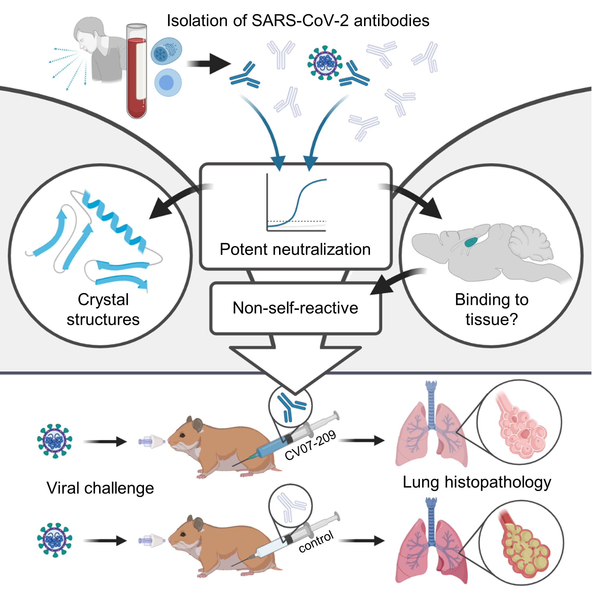
The emergence of SARS-CoV-2 led to pandemic spread of coronavirus disease 2019 (COVID-19), manifesting with respiratory symptoms and multi-organ dysfunction. Detailed characterization of virus-neutralizing antibodies and target epitopes is needed to understand COVID-19 pathophysiology and guide immunization strategies. Among 598 human monoclonal antibodies (mAbs) from ten COVID-19 patients, we identified 40 strongly neutralizing mAbs. The most potent mAb CV07-209 neutralized authentic SARS-CoV-2 with IC50 of 3.1 ng/ml. Crystal structures of two mAbs in complex with the SARS-CoV-2 receptor-binding domain at 2.55 and 2.70 Å revealed a direct block of ACE2 attachment. Interestingly, some of the near-germline SARS-CoV-2 neutralizing mAbs reacted with mammalian self-antigens. Prophylactic and therapeutic application of CV07209 protected hamsters from SARS-CoV-2 infection, weight loss and lung pathology. Our results show that non-self-reactive virus-neutralizing mAbs elicited during SARS-CoV-2infection areapromisingtherapeuticstrategy.
The severe acute respiratory syndrome coronavirus 2 (SARS-CoV-2) started emerging in humans in late 2019, and rapidly spread to a pandemic with millions of cases worldwide. SARS-CoV-2 infections cause coronavirus disease 2019 (COVID-19) with severe respiratory symptoms, but also pathological inflammation and multi-organ dysfunction, including acute respiratory distress syndrome, cardiovascular events, coagulopathies and neurological symptoms (Helms et al., 2020; Zhou et al., 2020; Zhu et al., 2020). Some aspects of the diverse clinical manifestations may result from a hyperinflammatory response, as suggested by reduced mortality in hospitalized COVID-19 patients under dexamethasone therapy (Horby et al., 2020).
JournalPre-proof
Understanding the immune response to SARS-CoV-2 therefore is of utmost importance. Multiple recombinant SARS-CoV-2 mAbs from convalescent patients have been reported (Brouwer et al., 2020; Cao et al., 2020; Ju et al., 2020; Kreer et al., 2020; Robbiani et al., 2020; Rogers et al., 2020; Wec et al., 2020). mAbs targeting the receptor-binding domain (RBD) of the viral spike protein S1 can compete with its binding to human angiotensin converting enzyme 2 (ACE2) and prevent viral entry and subsequent replication (Cao et al., 2020; Ju et al., 2020; Walls et al., 2020). Potent virus neutralizing mAbs that were isolated from diverse variable immunoglobulin (Ig) genes typically carry low levels of somatic hypermutations (SHM). Several of these neutralizing mAbs selected for in vitro efficacy showed prophylactic or therapeutic potential in animal models (Cao et al., 2020; Liu et al., 2020; Rogers et al., 2020; Zost et al., 2020). The low number of SHM suggests limited affinity-maturation in germinal centers compatible with an acute infection. Near-germline mAbs usually constitute the first line of defense to pathogens, but carry the risk of self-reactivity to autoantigens (Lerner, 2016; Liao et al., 2011; Zhou et al., 2007). Although critical for the therapeutic use in humans, potential tissue-reactivity of near-germline SARS-CoV-2 antibodies has not been examined so far.
Here, we systematically selected 18 strongly neutralizing mAbs out of 598 antibodies from 10 COVID-19 patients by characterization of their biophysical properties, authentic SARS-CoV-2 neutralization, and exclusion of off-target binding to murine tissue. Additionally, we solved two crystal structures of
neutralizing mAbs in complex with the RBD, showing antibody engagement with the ACE2 binding site from different approach angles. Finally, we selected mAb CV07-209 by its in vitro efficacy and the absence of tissue-reactivity for in vivo evaluation. Systemic application of CV07-209 in a hamster model of SARSCoV-2 infection led to profound reduction of clinical, paraclinical and histopathological COVID-19 pathology, thereby reflecting its potential for translational application in patients with COVID-19.
JournalPre-proof
RESULTS
Antibody repertoire analysis of COVID-19 patients
We first characterized the B cell response in COVID-19 using single-cell Ig gene sequencing of human mAbs (Figure 1A). From ten COVID-19 patients with serum antibodies to the S1 subunit of the SARSCoV-2 spike protein (Figure S1A, Table S1), we isolated two populations of single cells from peripheral blood mononuclear cells with fluorescence-activated cell sorting (FACS): CD19+CD27+CD38+ antibody secreting cells (ASC) reflecting the overall humoral immune response and SARS-CoV-2-S1-labeled CD19+CD27+ memory B cells (S1-MBC) for characterization of antigen-specific responses (Figure S1B and S1C). We obtained 598 functional paired heavy and light chain Ig sequences (Table S2). Of 432 recombinantly expressed mAbs, 122 were reactive to SARS-CoV-2-S1 (S1+), with a frequency of 0.018.2% (median 7.1%) within ASC and 16.7-84.1% (median 67.1%) within S1-MBC (Figure 1B and 1C).
Binding to S1 did not depend on affinity maturation as measured by the number of SHM (Figure 1D).
Compared to mAbs not reactive to SARS-CoV-2-S1, S1+ mAbs had less SHM, but equal lengths for both their light and heavy chain complementarity-determining region 3 (CDR3) (Figure S1D, S1E and S1F).
Within the ASC and S1-MBC population, 45.0% and 90.2% of S1+ mAbs bound the RBD, respectively (Figure S1G).
JournalPre-proof
S1+ mAbs were enriched in certain Ig genes including VH1-2, VH3-53, VH3-66, VK1-33 and VL2-14 (Figure S2). We identified both clonally related antibody clones within patients and public and shared S1+ clonotypes from multiple patients (Figure S3A and S3B). Some public or shared clonotypes had been previously reported, such as IGHV3-53 and IGHV3-66 (Figure S3D) (Cao et al., 2020; Yuan et al., 2020a), while others were newly identified, such as IGHV3-11 (Figure S3C).
Identification and characterization of potent SARS-CoV-2 neutralizing mAbs
We next determined the mAbs with the highest capacity to neutralize SARS-CoV-2 in plaque reduction neutralization tests (PRNT) using authentic virus (Munich isolate 984) (Wolfel et al., 2020). Of 87 mAbs strongly binding to RBD, 40 showed virus neutralization with a half-maximal inhibitory concentration (IC50)
≤250 ng/ml and were considered neutralizing antibodies (Figure 1A, Table S2), from which 18 (Top-18) were selected for further characterization (Table S3). The antibodies bound to RBD with a half-maximal effective concentration (EC50) of 3.8-14.2 ng/ml (Figure 1E) and an equilibrium dissociation constant (KD) of 6.0 pM to 1.1 nM (Figure S4, Table S3), thereby neutralizing SARS-CoV-2 with an IC50 of 3.1-172 ng/ml (Figure 1F, Table S3). The antibody with the highest apparent affinity, CV07-209, was also the strongest neutralizer (Figure 1G). We hypothesized that the differences in neutralizing capacity relate to different interactions with the ACE2 binding site. Indeed, the strongest neutralizing mAbs CV07-209 and CV07-250 reduced ACE2 binding to RBD to 12.4% and 58.3%, respectively. Other Top-18 mAbs including CV07-270 interfered only weakly with ACE2 binding (Figure S5A).
JournalPre-proof
The spike proteins of SARS-CoV-2 and SARS-CoV share more than 70% amino acid sequence identity, whereas sequence identity between SARS-CoV-2 and MERS-CoV and other endemic coronaviruses is significantly lower (Barnes et al., 2020). To analyze potential cross-reactivity of mAbs to other coronaviruses, we tested for binding of the Top-18 mAbs to the RBD of SARS-CoV, MERS-CoV, and the human endemic coronaviruses 229-E, NL63, HKU1 and OC43. CV38-142 detected the RBD of both SARS-CoV-2 and SARS-CoV, whereas no other mAb was cross-reactive to additional coronaviruses (Figure S5C and S5D). To further characterize the epitope of neutralizing mAbs, we performed ELISAbased epitope binning experiments using biotinylated antibodies. Co-applications of paired mAbs showed competition of most neutralizing antibodies for RBD binding (Figure S5B). As an exception, SARS-CoV cross-reactive CV38-142 bound RBD irrespective of the presence of other mAbs, suggesting an independent and conserved target epitope (Figure S5B).
Near-germline SARS-CoV-2 neutralizing antibodies can bind to murine tissue
Many SARS-CoV-2 neutralizing mAbs carry few SHM or are in germline configuration (Figure 1D) (Ju et al., 2020; Kreer et al., 2020). Such antibodies close to germline might be reactive to more than one target (Zhou et al., 2007). Prompted by the abundance of near-germline SARS-CoV-2 antibodies and to exclude
potential side effects of mAb treatment, we next analyzed whether SARS-CoV-2 antibodies can bind to self-antigens.
Therefore, we tested the binding of S1-mAbs to unfixed murine tissues. Surprisingly, four of the Top-18 potent SARS-CoV-2 neutralizing mAbs showed anatomically distinct tissue reactivities (Figure 2, Table S3). CV07-200 intensively stained brain sections in the hippocampal formation, olfactory bulb, cerebral cortex and basal ganglia (Figure 2A). CV07-222 also bound to brain tissue, as well as to smooth muscle (Figure 2B). CV07-255 and CV07-270 were reactive to smooth muscle from sections of lung, heart, kidney and colon, but not liver (Figure 2C and 2D, Table S3). None of the Top-18 mAbs bound to HEp-2 cells, cardiolipin or beta-2 microglobulin as established polyreactivity-related antigens (Jardine et al., 2016) (Figure S5E).
JournalPre-proof
Crystal structures of two mAbs approaching the ACE2 binding site from different angles
Diffraction-quality crystals were obtained for SARS-CoV-2 RBD complexed with two individual neutralizing mAbs, CV07-250 and CV07-270, which have notable differences in the number of SHM, extent of ACE2 competition and binding to murine tissue. CV07-250 (IC50=3.5 ng/ml) had 33 SHM (17/16 on heavy and light chain, respectively), strongly reduced ACE2 binding and showed no binding to murine tissue. In contrast, CV07-270 (IC50= 82.3 ng/ml) had only 2 SHM (2/0), did not reduce ACE2 binding in our assay, and showed binding to smooth muscle tissue. Using X-ray crystallography, we determined structures of CV07-250 and CV07-270 in complex with SARS-CoV-2 RBD to resolutions of 2.55 and 2.70 Å, respectively (Figure 3, Table S4, Table S5).
The binding mode of CV07-250 to RBD is unusual in that it is dominated by the light chain (Figure 3A and 3D), whereas in CV07-270, the heavy chain dominates as frequently found in other antibodies (Figure 3B and 3E). Upon interaction with the RBD, CV07-250 has a buried surface area (BSA) of 399 Å2 and 559 Å2 on the heavy and light chains, respectively, compared to 714 Å2 and 111 Å2 in CV07-270. CV07-250 uses CDR H1, H3, L1, L3, and framework region 3 (LFR3) for RBD interaction (Figure 3D and Figure 4A, 4B
and 4C), whereas CV07-270 interacts with CDR H1, H3, L1, and L2 (Figure 3E and Figure 4D, 4E and 4F).
The epitope of CV07-250 completely overlaps with the ACE2 binding site with a similar angle of approach as ACE2 (Figure 3A, 3C, 4G and 4I). In contrast, the CV07-270 epitope only partially overlaps with the ACE2 binding site and the antibody approaches the RBD from a different angle compared to CV07-250 and ACE2 (Figure 3B, 3C, 4H, 4I), explaining differences in ACE2 competition. Although CV07-250 and CV07-270 both contact 25 epitope residues, only seven residues are shared (G446/G447/E484/G485/Q493/S494/Q498). Furthermore, CV07-270 binds to a similar epitope as SARSCoV-2 neutralizing antibody P2B-2F6 (Ju et al., 2020) with a similar angle of approach (Figure S5F). In fact, 18 out of 20 residues in the P2B-2F6 epitope overlap with the CV07-270 epitope, although CV07-270 and P2B-2F6 are encoded by different germline genes for both heavy and light chains. Thus, these two mAbs represent antibodies encoded by different germline genes that bind to the same epitope in the RBD with near-identical binding modes and approach angles. This structural convergence is also encouraging for targeting this highly immunogenic epitope for vaccine development.
Interestingly, CV07-250 was isolated 19 days after symptom onset, but already acquired 33 SHM, the highest number among all S1+ MBCs (Figure S1D). Some non-germline amino acids are not directly involved in RBD binding, including all five SHMs on CDR H2 (Figure S6). This observation suggests that CV07-250 could have been initially affinity-matured against a different antigen.
JournalPre-proof
Prophylactic and therapeutic mAbs in a COVID-19 animal model
Next, we selected mAb CV07-209 for evaluation of in vivo efficacy based on its high capacity to neutralize SARS-CoV-2 and the absence of reactivity to mammalian tissue. We used the hamster model of COVID19, as it is characterized by rapid weight loss and severe lung pathology (Osterrieder et al., 2020). In this experimental set-up, hamsters were intranasally infected with authentic SARS-CoV-2. Nine hamsters per group received either a prophylactic application of CV07-209 24 hours before viral challenge, or a
therapeutic application of CV07-209 or control antibody mGO53 two hours after viral challenge (Figure 5A).
Hamsters under control mAb treatment lost 5.5±4.4% (mean±SD) of body weight, whereas those that received mAb CV07-209 as a therapeutic or prophylactic single dose gained 2.2±3.4% or 4.8±3.4% weight after 5 days post-infection (dpi), respectively. Mean body weights gradually converged in the animals followed up until 13 dpi, reflecting the recovery of control-treated hamsters from SARS-CoV-2 infection (Figure 5B).
To investigate the presence of SARS-CoV-2 in the lungs, we measured functional SARS-CoV-2 particles from lung tissue homogenates. Plaque forming units were below the detection threshold for all animals in the prophylactic and in 2 of 3 in the treatment group at 3 and 5 dpi (Figure 5C and 5D). qPCR measurements of lung viral genomic RNA copies revealed a 4-5 and 3-4 log reduction at both time points in the prophylactic and therapeutic group, indicating a drastic decrease of SARS-CoV-2 particles in lungs after CV07-209 application. Reduced virus replication and cell infection was confirmed by lowered detection of subgenomic viral RNA (Figure 5C and 5D). However, genomic and subgenomic RNA levels from nasal washes and laryngeal swaps were similar between all groups, indicating virus replication in the upper airways (Figure 5C and 5D).
JournalPre-proof
Additionally, we performed histopathological analyses of infected hamsters. As expected, all lungs from control-treated animals sacrificed at 3 dpi revealed typical histopathological signs of necro-suppurative pneumonia with suppurative bronchitis, necrosis of bronchial epithelial cells and endothelialitis (Figure 6A). At 5 dpi, control-treated animals showed marked bronchial hyperplasia, severe interstitial pneumonia with marked type II alveolar epithelial cell hyperplasia and endothelialitis (Figure 6D). In contrast, animals receiving prophylactic treatment with CV07-209 showed no signs of pneumonia, bronchitis, necrosis of bronchial epithelial cells, or endothelialitis at 3 dpi. A mild interstitial pneumonia with mild type II alveolar epithelial cell hyperplasia became apparent 5 dpi. Animals receiving therapeutic treatment with CV07-209 also showed a marked reduction of histopathological signs of COVID-19 pathology, although at both time points one out of three animals showed mild bronchopulmonary pathology with signs of interstitial
pneumonia and endothelialitis. These qualitative findings were mirrored in the reduction of the bronchitis and edema scores (Figure 6B, 6E and Table S6).
To confirm the absence of viral particles under CV07-209 treatment, we performed in-situ hybridization of viral RNA at 3 dpi. No viral RNA was detectable in the prophylactic group, whereas all animals in the control group and one in the therapeutic group revealed intensive staining of viral RNA in proximity of bronchial epithelial cells (Figure 6C). Taken together, these findings show that systemic application of SARS-CoV-2 neutralizing mAb CV07-209 protects hamsters from COVID-19 lung pathology and weight loss in both the prophylactic and the therapeutic setting.
DISCUSSION
JournalPre-proof
Driven by the pandemic spread of COVID-19 in early 2020, numerous groups have reported the isolation, characterization, structural analysis and animal model application of SARS-CoV-2 neutralizing mAbs (Barnes et al., 2020; Brouwer et al., 2020; Cao et al., 2020; Ju et al., 2020; Kreer et al., 2020; Robbiani et al., 2020; Rogers et al., 2020; Wec et al., 2020). In many places, our work confirms previous results, including the observation of a shared antibody response against the SARS-CoV-2 spike protein, the identification of ACE2 blocking as an important mechanism of virus neutralization, the isolation of highaffinity near-germline antibodies, and the in vivo efficacy of prophylactic mAb applications. Additionally, our results add several findings to the growing knowledge on the humoral immune response in SARS-CoV-2 infections.
First, we provide two structures of neutralizing mAbs identified in this study binding to the RBD of SARSCoV-2 at resolutions of 2.55 and 2.70 Å, allowing detailed characterization of the target epitopes and the SARS-CoV-2 neutralization mechanism of these two mAbs. SARS-CoV-2 mAbs can compete with ACE2 binding and exert neutralizing activity by inhibiting viral particle binding to host cells (Barnes et al., 2020; Brouwer et al., 2020; Cao et al., 2020; Ju et al., 2020; Kreer et al., 2020; Robbiani et al., 2020; Rogers et al., 2020; Wec et al., 2020), a key mechanism previously identified in SARS-CoV neutralizing antibodies (Prabakaran et al., 2006; ter Meulen et al., 2006). Steric hindrance of mAbs blocking ACE2 binding to the
RBD provides one mechanistic explanation of virus neutralization (Barnes et al., 2020; Cao et al., 2020; Wu et al., 2020). CV07-250 clearly belongs to this category of antibodies, as its epitope lies within the ACE2 binding site and it approaches the RBD from a similar angle as ACE2. In contrast, the epitope of CV07-270 only partially overlaps with the ACE2 binding site and approaches the RBD ridge from a different angle. In line with these findings, competition of CV07-270 with ACE2 binding as detected by ELISA was very weak. Its mechanism of virus neutralization therefore remains elusive. Of note, there have been reports of neutralizing antibodies targeting epitopes distant to the ACE2 binding site (Chi et al., 2020). Future research will need to clarify if additional mechanisms like triggering conformational changes in the spike protein upon antibody binding contribute to virus neutralization, as reported for SARS-CoV (Walls et al., 2019).
JournalPre-proof
Secondly, the majority of our SARS-CoV-2 mAbs are close to germline configuration, supporting previous studies (Kreer et al., 2020; Robbiani et al., 2020). Binding of some antibodies to HEp-2 cells was reported before (Kreer et al., 2020), a finding we could confirm in our cohort. Given the increased probability of auto-reactivity of near-germline antibodies, we additionally examined for reactivity of SARS-CoV-2 mAbs with unfixed murine tissue, allowing the detection of reactivity to potential self-antigens in their natural conformation. Indeed, we found that a fraction of SARS-CoV-2 neutralizing antibodies also bound to brain, lung, heart, kidney or gut expressed epitopes. Such reactivity with host antigens should ideally be prevented by immunological tolerance mechanisms, but complete exclusion of such antibodies would generate “holes” in the antibody repertoire. In fact, HIV utilizes epitopes shared by its envelope and mammalian self-antigens, thus harnessing immunological tolerance to impair anti-HIV antibody responses (Yang et al., 2013) and impeding successful vaccination (Jardine et al., 2016). To defy viral escape in HIV, but similarly COVID-19, anergic strongly self-reactive B cells likely enter germinal centers and undergo clonal redemption to mutate away from self-reactivity, while retaining HIV or SARS-CoV-2 binding (Reed et al., 2016). Interestingly, longitudinal analysis of mAbs in COVID-19 showed that the number of SHM in SARS-CoV-2-neutralizing antibodies only marginally increased over time (Kreer et al., 2020). This finding suggests that the self-reactivity observed in this study may not be limited to mAbs of the early humoral immune response in SARS-CoV-2 infections. Whether self-reactive antibodies could contribute to extra-
pulmonary symptoms in COVID-19 awaits further studies and should be closely monitored also in vaccination trials.
Finally, we evaluated in detail the in vivo efficacy of the most potent neutralizing antibody CV07-209 in a Syrian hamster model of SARS-CoV-2 infection. This model is characterized by a severe phenotype including weight loss and distinct lung pathology. Our results demonstrated that prophylaxis and treatment with a single dose of CV07-209 not only led to clinical improvement as shown by the absence of weight loss, but also to markedly reduced lung pathology. While the findings confirm the efficacy of prophylactic mAb administration as described by other groups in mice, hamsters and rhesus macaques (Cao et al., 2020; Liu et al., 2020; Rogers et al., 2020; Zost et al., 2020), our work also demonstrates the efficacy of post-exposure treatment in hamsters leading to viral clearance, clinical remission and prevention of lung injury. We provide detailed insights into the lung pathology of SARS-CoV-2 infected hamster at multiple times of the disease course including the regeneration phase. It complements two very recent demonstrations of a therapeutic effect of mAbs in a hamster model of COVID-19 (Baum et al., 2020; Li et al., 2020). These data expand the growing knowledge on post-exposure treatment from transgenic hACE2 mice (Cao et al., 2020) and a mouse model using adenovector delivery of human ACE2 before viral challenge (Liu et al., 2020). Collectively, our results indicate that mAb treatment can be fine-tuned for exclusion of self-reactivity with mammalian tissues and that mAb administration can also be efficacious after the infection, which will be the prevailing setting in COVID-19 patients.
JournalPre-proof
LIMITATIONS OF STUDY
While our study confirms the potential of therapeutic mAb applications for the treatment of COVID-19, the interpretation of the data is limited to a first exploration of a short window between viral infection and antibody administration. Although our paradigm mimics the relevant scenario of immediate post-exposure treatment, we cannot conclude whether the therapeutic benefit can also be translated into the more common clinical setting of treatment at heterogenous timepoints after symptoms have occurred. For this, follow-up studies will have to focus on delayed mAb application after symptom onset.
Also, we here describe the reactivity of SARS-CoV-2 mAbs to self-antigens from different tissues. These findings obligate attention, but simultaneously careful interpretation and require thorough investigations to provide better understanding of their functional relevance beyond the observed binding. Amongst others, this includes the identification of non-viral target antigens, functional in vitro studies and in vivo models. The self-reactive mAbs identified in this study derived from patients without severe extra-pulmonary symptoms. To address a possible connection between self-reactive antibodies and the diverse clinical manifestations in COVID-19, the expression and characterization of mAbs from patients with such disease courses will be needed.
ACKNOWLEDGMENTS
JournalPre-proof
We thank Stefanie Bandura, Matthias Sillmann and Doreen Brandl for excellent technical assistance, Christian Meisel for performing a cardiolipin ELISA and Martin Barner for assistance in generating the circos plot in Figure S4B. We acknowledge BIAFFIN GmbH & Co KG (Kassel, Germany) for performance of SPR measurements and Dr. Désirée Kunkel from Flow & Mass Cytometry Core Facility at Charité - Universitätsmedizin Berlin for support with single cell sorting. SMR is participant in the BIHCharité Junior Clinician Scientist Program funded by the Charité – Universitätsmedizin Berlin and the Berlin Institute of Health. Work at Scripps was supported by NIH K99 AI139445 (NCW) and the Bill and Melinda Gates Foundation OPP1170236 (IAW). Use of the SSRL, SLAC National Accelerator Laboratory, is supported by the U.S. Department of Energy, Office of Science, Office of Basic Energy Sciences under Contract No. DE-AC02–76SF00515. The SSRL Structural Molecular Biology Program is supported by the DOE Office of Biological and Environmental Research, and by the National Institutes of Health, National Institute of General Medical Sciences (including P41GM103393). This work was supported by COVID-19 grants from Freie Universität Berlin and Berlin University Alliance to NO and LES; and by the German Research Foundation (DFG) to ADG, ACH, SH, NS, MW, CD and LES (grant SFB-TR84), to ME and DS (EXC2049) and to HP (grant numbers PR 1274/2-1, PR 1274/3-1, PR 1274/5-1); and by the Helmholtz Association to HCK (grant ExNet0009) and to HP (grant HIL-A03); and by the Federal Ministry of Education and Research to HP (Connect-Generate, 01GM1908D) and MW, ACH, SH, NS and LES (PROVID, 01KI20160A and SYMPATH, 01ZX1906A).
Related to this work the German Center for Neurodegenerative Diseases (DZNE) and the Charité–Universitätsmedizin Berlin have filed a patent application on which JK, SMR, HCK, ESS, VMC, MAM, DW, LES and HP are named as inventors.
MAIN FIGURE TITLES AND LEGENDS
Figure 1. Identification and characterization of potent SARS-CoV-2 neutralizing mAbs
(A) Diagram depicting the strategy for isolation of 18 potently neutralizing mAbs (Top-18).
(B) Normalized binding to S1 of SARS-CoV-2 for mAbs isolated from antibody secreting cells (▼; blue = S1-binding, grey = not S1-binding). (OD=optical density in ELISA)
(C) Normalized binding to S1 of SARS-CoV-2 for mAbs isolated from S1-stained memory B cells (▲; colors like in (B))
(D) S1-binding plotted against the number of somatic hypermutations (SHM) for all S1-reactive mAbs.
(E) Concentration-dependent binding of Top-18 SARS-CoV-2 mAbs to the RBD of S1 (mean±SD from two wells of one experiment).
(F) Concentration-dependent neutralization of authentic SARS-CoV-2 plaque formation by Top-18 mAbs (mean±SD from two independent measurements).
(G) Apparent affinities of mAbs to RBDs (KD determined by surface plasmon resonance) plotted against IC50 of authentic SARS-CoV-2 neutralization.
See also Figures S1, S2, S3, S4, S5 and Tables S1, S2, S3.
Figure 2. SARS-CoV-2 neutralizing antibodies can bind to murine tissue
Immunofluorescence staining of SARS-CoV-2 mAbs (green) on murine organ sections showed specific binding to distinct anatomical structures, including
(A) staining of hippocampal neuropil with CV07-200 (cell nuclei depicted in blue),
(B) staining of bronchial walls with CV07-222,
(C) staining of vascular walls with CV07-255, and
(D) staining of intestinal walls with CV07-270.
Smooth muscle tissue in (B-D) was co-stained with a commercial smooth muscle actin antibody (red). Scale bars: 100 µm.
See also Table S3.
Figure 3. Crystal structures of mAbs in complex with SARS-CoV-2 RBD
(A) CV07-250 (cyan) in complex with RBD (white).
(B) CV07-270 (pink) in complex with RBD (white).
(C) Human ACE2 with SARS-CoV-2 RBD (PDB 6M0J) (Lan et al., 2020).
(D-E) Epitopes of (D) CV07-250 and (E) CV07-270. Epitope residues contacting the heavy chain are in orange and the light chain in yellow. CDR loops and framework region that contact the RBD are labeled.
(F) ACE2-binding residues on the RBD (blue) in the same view as (D) and (E). The ACE2 interacting region is shown in green within a semi-transparent cartoon representation.
See also Figures S5, S6 and Tables S4, S5.
Figure 4. Interactions and angle of approach at the RBD-antibody interface
(A-C) Key interactions between CV07-250 (cyan) and RBD (white) are highlighted.
(A) CDR H3 of CV07-250 forms a hydrogen-bond network with RBD Y489 and N487.
(B) VH Y100b (CDR H3), VL F32 (CDR L1), and VL Y91 (CDR L3) of CV07-250 form a hydrophobic aromatic patch for interaction with RBD L455 and F456.
(C) The side chain of VL S67 and backbone amide of VL G68 from FR3 is engaged in a hydrogen-bond network with RBD G446 and Y449.
(D-F) Interactions between CV07-270 (cyan) and RBD (white) are illustrated.
(D) Residues in CDR H1 of CV07-270 participate in an electrostatic and hydrogen-bond network with RBD R346 and K444.
JournalPre-proof
(E) VH W100h and VH W100k on CDR H3 of CV07-270 make π-π stacking interactions with Y449. VH W100k is also stabilized by a π-π stacking interaction with VL Y49.
(F) VH R100g on CDR H3 of CV07-270 forms an electrostatic interaction with RBD E484 as well as a π-cation interaction with RBD F490. Oxygen atoms are in red, and nitrogen atoms in blue. Hydrogen bonds are represented by dashed lines
(G-I) Zoomed-in views of the different RBD ridge interactions with (G) CV07-250, (H) CV07-270, and (I) ACE2 (PDB 6M0J) (Lan et al., 2020). The ACE2-binding ridge in the RBD is represented by a backbone ribbon trace in red.
See also Figures S5, S6 and Tables S4, S5.
Figure 5. Prophylactic and therapeutic application of mAb CV07-209 in a COVID-19 hamster model
(A) Schematic overview of the animal experiment.
(B) Body weight of hamsters after virus challenge and prophylactic (pink) or therapeutic (blue) application of SARS-CoV-2
neutralizing mAb CV07-209 or control antibody (mean±SEM from n = 9 animals per group from day -1 to 3, n = 6 from days 4 to 5; n = 3 from days 6 to 13; mixed-effects model with posthoc Dunnett’s multiple tests in comparison to control group; significance levels shown as * (p<0.05), ** (p<0.01), *** (p<0.001), **** (p<0.0001), or not shown when not significant).
(C-D) Left: Quantification of plaque forming units (PFU) from lung homogenates. Right: Quantification of genomic SARS-CoV-2
RNA (gRNA) as copies per 105 cellular transcripts (left y-axis, filled circles) and cycle threshold (ct) of subgenomic SARS-CoV-2
RNA (sgRNA) detection (right y-axis, unfilled circles) from samples and timepoints as indicated. Values for PFU were set to 5 when not detected, gRNA copies below 1 were set to 1 and ct of sgRNA to 46 when not detected. Bars indicate mean. Dotted lines represent detection threshold.
See also Figure 6 and Table S6.
Figure 6. Histopathological analysis of hamsters after SARS-CoV-2 infection
(A) Histopathology of representative haematoxylin and eosin stained, paraffin-embedded bronchi with inserted epithelium (upper row) and lung parenchyma with inserted blood vessels (lower row) at 3 dpi. Severe suppurative bronchitis with immune cell infiltration (hash) is apparent only in the control-treated animals with necrosis of bronchial epithelial cells (diagonal arrows). Necrosuppurative interstitial pneumonia (upward arrows) with endothelialitis (downward arrows) is prominent in control-treated animals. Scale bars: 200 µm in bronchus overview, 50 µm in all others.
(B) Bronchitis and edema score at 3 dpi. Bars indicate mean.
(C) Detection of viral RNA (red) using in situ hybridization of representative bronchial epithelium present only in the control group.
Scale bars: 50 µm.
JournalPre-proof
(D) Histopathology of representative lung sections from comparable areas as in (A) at 5 dpi. Staining of bronchi of control-treated animals showed a marked bronchial hyperplasia with hyperplasia of epithelial cells (diagonal arrow) and still existing bronchitis (hash), absent in all prophylactically treated and in 2/3 therapeutically treated animals (upper row). Lung parenchyma staining of control-treated animals showed severe interstitial pneumonia with marked type II alveolar epithelial cell hyperplasia and endothelialitis (insets, downward arrows). Compared to control-treated animals, prophylactically treated animals showed only mild signs of interstitial pneumonia with mild type II alveolar epithelial cell hyperplasia (upward arrow), whereas therapeutically treated animals showed a more heterogeneous picture with 1/3 showing no signs of lung pathology, 1/3 animal showing only mild signs of interstitial pneumonia, and 1/3 animal showing a moderate multifocal interstitial pneumonia.
Scale bars: 200 µm in bronchus overview, 50 µm in all others.
(E) Bronchitis and edema score at 5 dpi. Bars indicate mean.
See also Figure 5 and Table S6.
SUPPLEMENTAL FIGURE TITLES AND LEGENDS
Figure S1. SARS-CoV-2-S1 serum IgG response from COVID-19 patients, flow cytometry gating and characteristics of immunoglobulin sequences. Related to Figure 1 and Tables S1 and S2.
(A) Serum IgG response determined as the normalized optical density (OD) in a SARS-CoV-2-S1 ELISA in relation to the time point of diagnosis defined by the first positive qPCR test. Upward arrowhead denotes the appearance of first symptoms. Downward arrowhead denotes the PBMC isolation. From patient CV01, PBMC samples were isolated at two time points as indicated by the second downward arrow with an asterisk (*).
(B-C) A representative flow cytometry plot from patient CV38 indicating gating on (B) CD19+CD27+antibody-secreting cells (ASC) and (C) SARS-CoV-2-S1-stained memory B cells (S1-MBC). Cells were pre-gated on live CD19+ B cells.
(D) Comparison of somatic hypermutation (SHM) count within immunoglobulin V genes combined from heavy and light chains of S1-reactive (S1+, blue) and non-S1-reactive (S1-, grey) mAbs. Statistical significance was determined using a Kruskal-Wallis test with Dunn’s multiple comparison test. (ASC: n = 20 S1+, n = 260 S1-; S1-MBC: n = 102 S1+, n = 50 S1-, n-values represent number of mAbs). All expressed mAbs are displayed. Each triangle represents one mAb, isolated from an ASC (▼) or a S1-MBC (▲). Bars indicate mean.
(E-F) Length comparison of complementarity-determining region (CDR) 3 amino acid sequences between S1+ and S1- mAbs within (E) heavy and (F) light chains. Bars indicate mean. Symbols and colors have the same meaning as in (D).
(G) Frequency of RBD-binder (S1+RBD+) and non-RBD-binder (S1+RBD-) relative to all expressed mAbs (upper lanes) and relative to S1+ mAbs (lower lanes).
Figure S2. Comparison of variable gene usage. Related to Figure 1 and Table S2.
JournalPre-proof
Comparison of gene usage between SARS-CoV-2-S1-reactive (S1+) and non-reactive (S1-) mAbs is shown for immunoglobulin (A) variable heavy (IGHV), (B) variable kappa (IGKV) and (C) variable lambda (IGLV) genes. Bars depict percentage of gene usage of all expressed mAbs within each group.
Figure S3. Clonal expansion and public or common clonotypes. Related to Figure 1 and Table S2.
(A) Pie charts represent clonal relationship of all expressed mAbs from each donor separately for antibody secreting cells (ASC) and S1-stained memory B cells (S1-MBC). mAbs were considered S1-reactive (S1+) or non-S1-reactive (S1-) based on SARSCoV-2-S1 ELISA measurements. Antibodies were considered to be clonally expanded when they were isolated from multiple cells.
(B) Circos plot displays all isolated mAbs from ten donors. Interconnecting lines indicate relationship between mAbs that share the same V and J gene on both Ig heavy and light chain. Such public or shared clonotypes in which more than 50% of mAbs are S1reactive are represented as colored lines. Small black angles at the outer circle border indicate expanded clones within the respective donor.
(C) Properties of public clonotypes from S1+ mAbs according to the colors used in (B) with sequence similarities between mAbs isolated from different donors, also within CDR3.
(D) Public or common antibody response using VH3-53 and VH3-66 genes.
IGHV, IGHJ IGKV, IGKJ, IGLV, IGLJ = V (variable) and J (joining) genes of immunoglobulin heavy, kappa, lambda chains; CDR = complementarity-determining region; n.exp. = not expressed.
Figure S4. Binding kinetic measurements of mAbs to RBD. Related to Figure 1 and Table S3.
Binding kinetics of mAbs to RBD were modeled (black) from multi-cycle surface plasmon resonance (SPR) measurements (blue, purple, orange). Fitted monovalent analyte model is shown. For CV07-200, neither a bivalent nor a monovalent analyte model described the data accurately (no model is shown). Three out of the 18 selected mAbs for detailed characterization (Top-18) were not analyzed using multi-cycle-kinetics: CV07-270 was excluded as it interacted with the anti-mouse IgG reference surface on initial qualitative measurements. CV07-255 and CV-X2-106 were not analyzed since they showed biphasic binding kinetics and relatively fast dissociation rates in initial qualitative measurements. Non-neutralizing CV03-191, a mAb not included in the Top-18 mAbs, was included in the multicycle experiments as it has the same clonotype as strongly neutralizing CV07-209 (Figure S4C). All measurements are performed by using a serial 2-fold dilution of mAbs on reversibly immobilized SARS-CoV-2-S1 RBD-mFc.
Figure S5. Bind epitope Characterization of selected mAbs. Related to Figures 1, 3, 4 and Table S3.
(A) Competition for RBD binding between Top-18 mAbs and ACE2. ELISA-based measurements of human ACE2 binding to SARSCoV-2 RBD after pre-incubation with the indicated neutralizing mAbs. Values are shown relative to antibody-free condition as mean+SD from three independent measurements.
JournalPre-proof
(B) Competition for RBD binding between combinations of potent neutralizing mAbs is illustrated as a heat map. Shades of green indicate the degree of competition for RBD binding of detection mAb in presence of 100-fold excess of competing mAb relative to non-competition conditions. Green squares indicate no competition. Values are shown as mean of two independent experiments.
(C) Representative immunofluorescence staining on VeroB4 cells overexpressing spike protein of indicated coronavirus with SARSCoV-2 mAb CV07-209 at 5 µg/ml. For all other 17 of the selected 18 mAbs (Top-18, Table S3), similar results were obtained.
(D) Binding of indicated mAbs to fusion proteins containing the RBD of indicated coronaviruses and the constant region of rabbit IgG revealed by ELISA. For all other Top-18 mAbs, similar results were obtained as for CV07-209. Values indicate mean+SD from two wells of one experiment.
(E) Representative HEp-2 cell staining with a commercial anti-nuclear antibody as positive control revealed nuclear binding (top). S1-reactive non-neutralizing mAb CV38-148 exhibited cytoplasmatic binding (middle). Neutralizing mAb CV07-209 showed no binding (bottom). All mAbs selected for detailed characterization (Top-18, Table S3) revealed similar results like CV07-209 when used at 50 µg/ml. Representative scale bar: 25 µm.
(F) Structural comparison of CV07-270/RBD and P2B-2F6/RBD complexes. Structure of CV07-270 (pink, left) and structure of P2B2F6 (PDB 7BWJ) (Ju et al., 2020) (blue, middle) in complex with RBD (white), as well as superimposition of the structures of CV07270/RBD and P2B-2F6/RBD based on the RBD (right).
Figure S6. Comparison of sequences of CV07-250 and CV07-270 to their putative germline sequences.
Related to Figures 3, 4.
(A) Alignment of CV07-250 with the germline IGHV1-18 sequence (nucleotide SHM rate 5.8%) and IGLV2-8 (nucleotide SHM rate 5.4%).
(B) Somatic mutations VH S31H, VL G29A, VL N31H, VL Y32F, VL S34T, and VL L46V are located in the CV07-250 paratope with other somatic mutations in all of the CDRs that may affect overall CDR conformation and interactions. Hydrogen bonds are represented by dashed lines. Distances between atoms are shown in solid lines. CV07-250 heavy chain is in dark cyan and light chain is in light cyan. SARS-CoV-2 RBD is in light grey.
(C) Alignment of CV07-270 with the germline IGHV3-11 sequence (nucleotide SHM rate 0.7%) and IGLV2-14 (nucleotide SHM rate 0%). The regions that correspond to CDR H1, H2, H3, L1, L2, and L3 are indicated. Residues that differ from the germline are highlighted in red. Residues that interact with the RBD are highlighted in yellow. Residue positions in the CDRs are labeled according to the Kabat numbering scheme.
STAR*METHODS
Detailed methods are provided in the online version of this paper and include the following:
• KEY RESOURCES TABLE
• RESOURCE AVAILABILITY
o Lead Contact
o Materials Availability
o Data and Code Availability
• EXPERIMENTAL MODEL AND SUBJECT DETAILS
o SARS-CoV-2-infected individuals and sample collection
o Animal experiment approval and animal care
• METHOD DETAILS
o PBMC collection and FACS staining
o Generation of recombinant human monoclonal antibodies
o SARS-CoV-2-S1 ELISA
o RBD ELISA
o Circos plot of public clonotypes
o Identification of 18 strongly neutralizing antibodies
o Surface plasmon resonance measurements
JournalPre-proof
o Plaque reduction neutralization test
o Immunocytochemistry
o Crystal structure determination of Fab-RBD complexes
o Murine tissue reactivity screening
o HEp2 cell assay
o Polyreactivity screening ELISA
o Hamster model of SARS-CoV-2 infection
o Histopathology and in situ hybridization
o Virus titrations, RNA extractions and RT-qPCR
• QUANTIFICATION AND STATISTICAL ANALYSIS
KEY RESOURCES TABLE
RESOURCE AVAILABILITY
Lead Contact
Further information and requests for resources and reagents should be directed to and will be fulfilled by the Lead Contact, Jakob Kreye (jakob.kreye@dzne.de).
Materials Availability
All requests for materials including antibodies, viruses, plasmids and proteins generated in this study should be directed to the Lead Contact author. Materials will be made available under a Material Transfer Agreement (MTA) for non-commercial usage.
Data and Code Availability
JournalPre-proof
X-ray coordinates and structure factors are deposited at the RCSB Protein Data Bank under accession codes 6XKQ and 6XKP. The nucleotide sequences of the Top-18 antibodies have been deposited to GenBank (accession numbers MW002770 - MW002805). The raw sequencing data associated with this manuscript together with the analysis using custom BASE software have been deposited to Code Ocean (https://codeocean.com/capsule/7823731/tree/v1, DOI: 10.24433/CO.1724316.v1). The software used for Ig sequence analysis is available on https://github.com/automatedSequencing/BASE. The published article includes all data generated or analyzed during this study and are available from the corresponding author on request. Additional Supplemental Items are available from Mendeley Data at http://dx.doi.org/ 10.17632/f6tb3csgjt.1.
EXPERIMENTAL MODELS AND SUBJECT DETAILS
SARS-CoV-2-infected individuals and sample collection
The patients have given written informed consent and analyses were approved by the Institutional Review Board of Charité - Universitätsmedizin Berlin, corporate member of Freie Universität Berlin, HumboldtUniversität Berlin, and Berlin Institute of Health, Berlin. All patients in this study were tested positive for SARS-CoV-2 infection by RT-PCR. Most patients belong to a prospective COVID-19 cohort (Kurth et al., 2020). Patient characteristics are described in Table S1.
Animal experiment approval and animal care
The animal experiment was approved by the Landesamt für Gesundheit und Soziales in Berlin, Germany (approval number 0086/20) and performed in compliance with relevant national and international guidelines for care and humane use of animals. In vitro and animal work was conducted under appropriate biosafety precautions in a BSL-3 facility at the Institute of Virology, Freie Universität Berlin, Germany. Twenty-seven six-week old female and male golden Syrian hamsters (Mesocricetus auratus; outbred hamster strain RjHan:AURA, Janvier Labs) were kept in groups of 3 animals in enriched, individually ventilated cages. The animals had ad libitum access to food and water and were allowed to acclimate to these conditions for seven days prior to prophylactic treatment and infection. Cage temperatures and relative humidity were recorded daily and ranged from 22-24°C and 40-55%, respectively.
METHOD DETAILS
JournalPre-proof
PBMC collection and FACS staining
Recombinant SARS-CoV-2-S1 protein produced in HEK cells (Creative Diagnostics, DAGC091) was covalently labeled using CruzFluor647 (Santa Cruz Biotechnology, sc-362620) according to the manufacturer’s instructions.
Using fluorescence-activated cell sorting we sorted viable single cells from freshly isolated peripheral blood mononuclear cells (PBMCs as 7AAD-CD19+CD27+CD38+ antibody-secreting cells (ASCs) or SARSCoV2-S1-enriched 7AAD-CD19+CD27+ memory B cells (MBCs) into 96-well PCR plates. Staining was performed on ice for 25 minutes in PBS with 2 % FCS using the following antibodies: 7-AAD 1:400