Another random document with no related content on Scribd:
METHODS IN PLASTIC OPERATIONS
THERE ARE FIVE DISTINCTIVE METHODS EMPLOYED IN PERFORMING PLASTIC OPERATIONS:
I. Stretching of the margins of the skin.
II. Sliding flaps of adjacent skin.
III. Twisting pedunculated flaps.
IV. Implantation of pedunculated flaps by bridging.
V. Transplantation of nonpedunculated flaps or skin grafting.
This classification differs from that heretofore generally given in the meager literature on the subject, but the author believes his arrangement to be more scientifically exact as well as simpler for recording and history purposes.
I. STRETCHING METHOD
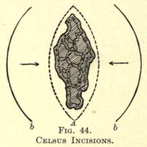
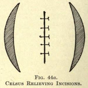
In the stretching method the defect is neatly excised, so as to freshen the margins to be brought together. It may be necessary, if the defect is too large for free apposition, to dissect the skin away from the underlying tissue to render it more movable and to overcome tension. The shape of the incision depends largely upon the nature of the defect and must be made with a view of leaving as little scar as possible. Where the defect is somewhat linear, or elongated, an elliptical incision (A) is made, as in Fig. 44, and, if necessary because of too great tension, the skin is undermined sufficiently to allow the parts to come together; if this cannot be done readily, two semilunar incisions (b, b) must be added. This will allow of ready coaptation. The wound is then brought together with an interrupted suture, appearing as in Fig. 44a, the semilunar spaces being allowed to heal by granulation.
FIG. 44.
CELSUS INCISIONS.
FIG. 44a.
CELSUS RELIEVING INCISIONS.
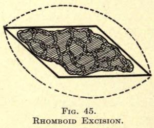
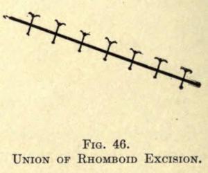
In excisions in small rhomboidal form, the skin is merely dissected up and around the wound, the same as in Fig. 45, and the wound is sutured in linear form, as shown in Fig. 46.
.
FIG. 45.
RHOMBOID EXCISION
FIG. 46.
UNION OF RHOMBOID EXCISION.
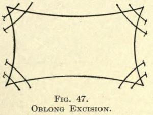
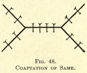
If the defect is oblong in form, the angles are brought together wholly, leaving a small surface to granulate, as in Fig. 47, or they are drawn toward the center, leaving the remainder of the parallel lines to be sutured, as shown in Fig. 48.
FIG. 47.
OBLONG EXCISION.
FIG. 48.
COAPTATION OF SAME.
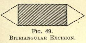
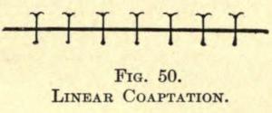
Another method of overcoming a smaller defect of similar form is to excise a small triangular portion of skin at either side of the oblong, as in Fig. 49, and then with or without dissection bringing the margins together in linear form, as in Fig. 50.
FIG. 49.
BITRIANGULAR EXCISION.
FIG. 50.
LINEAR COAPTATION.
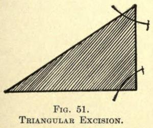
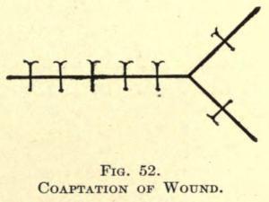
Likewise can a triangular fault be brought together by sewing in the greater angles and making a linear wound of the remaining part, as in Figs. 51 and 52.
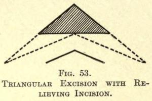
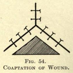
Again, a triangular defect may be remedied by adding a smaller triangle at each end involving healthy skin, utilizing, if need be, the relieving incisions, as in Figs. 53 and 54.
FIG. 51.
TRIANGULAR EXCISION.
FIG. 52.
COAPTATION OF WOUND.
FIG. 53.
TRIANGULAR EXCISION WITH RELIEVING INCISION.
IG. 54.
COAPTATION OF W
.
II. SLIDING METHOD
Following the principle of Celsus, as mentioned on page 8, defects can be overcome in various ways. The incisions may be straight or curved, and one or more flaps of skin are raised, slid, and sutured
F
OUND
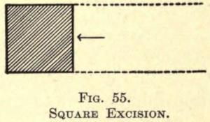
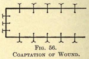
over the part to be covered. The simplest form is the covering of a square, as shown in Figs. 55 and 56.
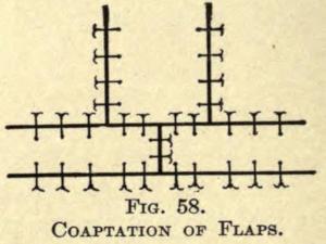
If the square be too large for the above method, the incisions can be carried to the other side and above or below the defect, as shown in Figs. 57 and 58.
FIG. 55.
SQUARE EXCISION.
FIG. 56.
COAPTATION OF WOUND.
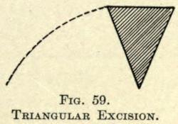
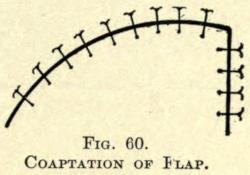
For triangular areas the curved incisions can be made, as in Fig. 59, rotating the flap into place, as shown in Fig. 60.
FIG. 57.
SQUARE EXCISION.
FIG. 58.
COAPTATION OF FLAPS.
COAPTATION OF FLAP.
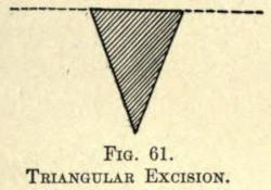
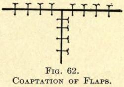
Or bilateral flaps may be utilized by straight incisions, stretched and sewn, as in Figs. 61 and 62.
FIG. 59.
TRIANGULAR EXCISION.
FIG. 60.
COAPTATION OF FLAPS.
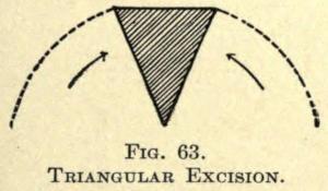
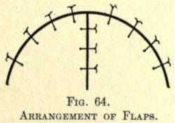
Again, two curved incisions are made to obtain rotating flaps, Fig. 63, and sewn, as shown in Fig. 64.
FIG. 61.
TRIANGULAR EXCISION.
FIG. 62.
IG. 63. T
EXCISION.
IG. 64.
ARRANGEMENT OF FLAPS.
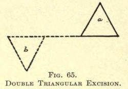
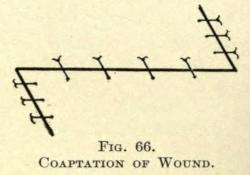
Bürow introduced a method for closing over defects by sliding flaps in which he utilized the mobility of skin obtained by the excision of triangles of healthy skin. The results are exceedingly good, but, unfortunately, the sacrifice of skin affects its general use, inasmuch as patients can afford but little loss of healthy skin; besides, there is the objection of added scarring. The closing of a triangular defect by this method is shown in Figs. 65 and 66, in which a is the triangular defect and b the triangle of healthy skin excised. The skin about the incisions is dissected up and the flaps are sutured into position, as shown in Fig. 66.
F
F
FIG. 65.
FIG. 66.
COAPTATION OF WOUND.
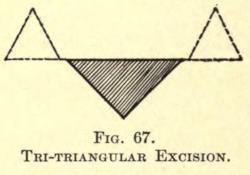
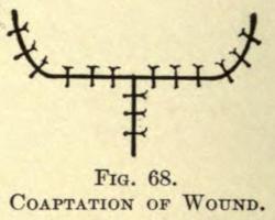
Where the triangular defect has a wide base, bilateral triangular sections of skin are removed (Fig. 67), and the flaps are coapted, as in Fig. 68.
DOUBLE TRIANGULAR EXCISION.
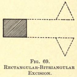
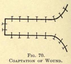
Through the sacrifice of two triangles a large oblong or square defect may be covered, the excisions being shown in Fig. 69 and the suturing in Fig. 70.
FIG. 67.
TRI-TRIANGULAR EXCISION.
FIG. 68.
COAPTATION OF WOUND.
RECTANGULAR-BITRIANGULAR EXCISION.
Although in several of the above methods the flaps are rotated and slightly twisted, the following are only classified with those under this division.
FIG. 69.
FIG. 70.
COAPTATION OF WOUND.
III. TWISTING METHOD
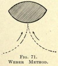
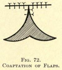
Where an elliptical defect is to be obliterated the curved incision shown in Fig. 71 can be satisfactorily employed, leaving but a small area to granulate over. The suturing is depicted in Fig. 72.
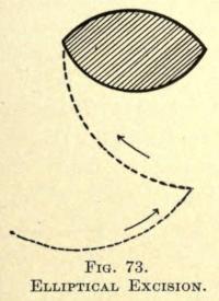
In this the twisting of the flaps is but little, while in the following, in which the defect is of similar shape, the twisting is more apparent; so much so, that a fold at the root of the flap may be
FIG. 71.
WEBER METHOD.
FIG. 72.
COAPTATION OF FLAPS.
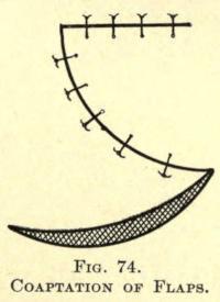
induced to some extent. The excision and incisions are shown in Fig. 73, and the method of bringing the parts together in Fig. 74, leaving a small area for granulation.
FIG. 73.
ELLIPTICAL EXCISION.
FIG. 74.
COAPTATION OF FLAPS.
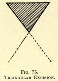
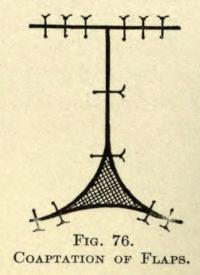
Considerable twisting of flaps is shown in covering triangular parts in Figs. 75 and 76.
FIG. 75.
TRIANGULAR EXCISION.
FIG. 76.
COAPTATION OF FLAPS.
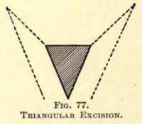
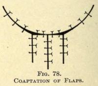
In this only a small surface is left to granulate over, while in the following the parts are entirely covered. The excision and incisions are shown in Fig. 77, and the method of approximation and suturing in Fig. 78.
TRIANGULAR EXCISION.
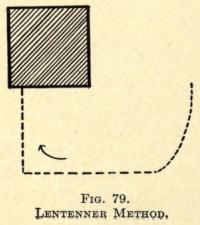
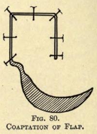
In covering a square area considerable twisting must be resorted to, as shown in Figs. 79 and 80, leaving a portion to granulate.
FIG. 77.
FIG. 78.
COAPTATION OF FLAPS.
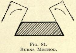
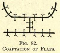
Where the area is irregular and formed somewhat as in Fig. 81, bilateral incisions are made and the flaps twisted into place and sewn, as in Fig. 82.
FIG. 79.
LENTENNER METHOD.
FIG. 80.
COAPTATION OF FLAP.
FIG. 81.
BURNS METHOD.
FIG. 82.
COAPTATION OF FLAPS.
IV. IMPLANTATION OF PEDUNCULATED FLAPS BY BRIDGING
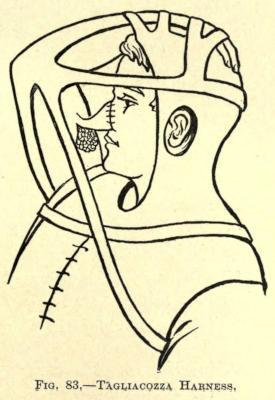
In this method the flap to be utilized in covering a defect is taken from a distant part of the body, as, for instance, from the arm. The flap thus taken at first remains attached at its distal end to the tissue of the arm by a pedicle, which is not severed until a circulation has been established between the flap and the part of the human economy to which its free end has been attached by suture, the arm being held in position in the meantime by a suitable contrivance, as shown in Fig. 83.
IG. 83. TAGLIACOZZA HARNESS.
These pedunculated flaps, bridging over space, may likewise be taken from the forearm, the hand, or the thoracic region.
When thoracic flaps are used they may be directly sewn at their free ends to the part to be covered, as, for instance, in the forearm or arm, or they are stitched to the forearm to be later transferred to another part of the body after their circulation had become established.
The various methods of the employment of these bridging flaps will be taken up individually in their respective places farther on.
V. TRANSPLANTATION OF NONPEDUNCULATED FLAPS OR SKIN-GRAFTING
Where there is loss of skin due to injury or operative procedure the parts may heal by granulation, but as this requires much time, and the consequent cicatrice causes considerable deformity, the granulating or freshly made wounds are covered with so-called
detached skin flaps or grafts, when the former methods of plastic surgery cannot be followed.
The methods employed in skin-grafting may be classified as: 1, autodermic; 2, heterodermic; 3, zoödermic.
1. Autodermic, when the grafts are taken from the tissue of the patient.
2. Heterodermic, when the grafts for the patient are taken from other persons.
3. Zoödermic, when the grafts for the patient are taken from the lower species.
The former two classes may for convenience be again subdivided into
1. (a) Auto-epidermic.
(b) Autodermic.
2. (c) Hetero-epidermic.
(d) Heterodermic.
The third class will permit of a great many subdivisions, too numerous to mention, each taking its name from the source of the graft.
1. Autodermic Skin-grafting
a. Auto-epidermic Skin-grafting. The method of covering granulation areas with small circular pieces of detached skin, pin grafts, was first advocated by J. Reverdin in 1870. The Reverdin method is applicable to healthy granulating surfaces only. The small lentil-form skin grafts are obtained from the arm or other suitable part of the body by raising the superficial layer of the skin with a
tenaculum hook and cutting the conelike elevation off with delicate scissors. The grafts thus obtained contain the epiderm and corium and a slight base of the Malpighian layer. They are immediately transferred, without handling, to the granulating surface and fixed by the gentle pressure of the hook point.
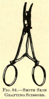
The skin may be transfixed with an ordinary sewing needle and the graft cut away with a delicate flat knife or razor blade, or scissors especially designed for the purpose may be used. (See Fig. 84.)
FIG. 84.—SMITH SKIN GRAFTING
SCISSORS.
A number of these grafts are often needed to cover a defect, in which case they are placed side by side upon the surface with a little space between their borders. Several such operations may be necessary, as many of the grafts are liable to die from malnutrition, pressure, or defective cutting.
The granulating surface to be covered in this manner must first be cleansed with a weak sublimate solution, followed by a sterilized normal salt solution. When an ulcerated or denuded surface requires skin-grafting, the best time to begin is as soon as there is evidence of the formation of new skin at the edges of the wound; in other words, when reparative action is becoming established. This does not apply to surfaces just denuded over healthy areas for plastic purposes, which should be grafted immediately.
The grafts, having been placed, are covered with a layer of very thin protective silk, or gutta percha, over which a soft gauze or
cotton dressing may be applied, borated absorbent cotton being most suitable.
Thiersch recommends the use of gauze compresses saturated in the normal salt solution, which are changed each day.
Another method of covering the grafts is to use perforated silk or small strips of the same material, which permit the dressings to absorb the excretions from the wound and also allow of the free use of either weak antiseptic or sterile salt solutions.
The use of silk or rubber prevents the adhesion of the grafts, which would otherwise be torn away by the removal of dressings, although iodoform gauze answers the purpose very well. It can be safely lifted by first thoroughly wetting it with the normal salt solution.
Strips of tinfoil, first rendered aseptic by immersion in a 1-1,000 sublimate solution and then dipped into sterilized oil or two-per-cent salicylized oil, have been recommended by Socin. Goldbeaters’ skin has also been advocated.
A method that has proved of great value in America is that of skingrafting in blood. In this method the grafted site is covered with perforated protective silk or rubber tissue, covered with a thin layer of absorbent cotton, or, better, several folds of sterilized gauze, which is kept wet constantly with bovinine. The latter undoubtedly is the means of keeping life in the grafts by supplying the necessary nutrition until the grafts have formed vascular connection, have become firmly adherent, and begin to spread or grow out at their edges.
The living grafts remain as pale islets of skin, which throw out thin epidermal films that meet and grow thicker, until finally the interjoined grafts assume all the functions of normal skin.
It is often necessary to reduce or scarify the edges of the healthy skin that has become thickened where the grafts meet it. This is permissible only when the grafts have become firm and thrive, and may be accomplished by the careful and intelligent use of pure
carbolic acid applied with a wooden pick, or by the employment of a stick of fused nitrate of silver, care being taken not to come in contact or to allow the cauterant to touch directly or in solution the new skin.
b. Autodermic Skin-grafting.—Larger pieces of skin may be excised from selected parts of the body, preferably the outer side of the arm, and utilized to cover the entire defect. The piece of skin is cut about one third larger than the size and shape of the area to be covered. This method was first introduced by R.Wolfe in 1876, and gives splendid results. He advises removing all subcutaneous adipose tissue from the graft by gently cutting it away with fine scissors or the razor, and then loosely suturing the flap to the skin surrounding the denuded defect.
Granulating surfaces must first be freed of their loose superficial layers with a sharp curette and the bleeding controlled by spongepressure before the flaps are placed. The edges of the wound made by the excision of the flap are simply sewn together, or one of the plastic methods may be used to accomplish the same. Unfortunately these flaps, if they thrive, contract, leaving uncovered spaces, which must be treated separately or allowed to granulate. The dressing in this case is the same as in the Reverdin process.
F . Krause, of Altoona (1896), advocates the use of freed flaps from which the subcutaneous adipose tissue has not been removed, holding that in the healing of such there is less contraction to follow. The success in both of the above methods depends upon an early vascular connection, as considerable nutrition is necessary to supply their want. The blood dressing has aided much in bringing about a happy result. The latter is continued in the manner described for about ten or twelve days, when the grafts may be allowed to depend upon their own circulatory supply. The parts must, in the meantime, be kept at rest and all undue pressure is to be avoided.
These grafts, while becoming organized, change in color more or less from a light gray to a bluish gray and shed off their epitheliar
layers, while the cutis vera remains, rebuilding its squamous covering eventually and leaving the surface quite normal.
At times small points of the flap, where subjected to undue pressure or interference, will turn dark and break down, sloughing away and leaving the granulating surface exposed. These areas are, however, soon recovered by skin cells being thrown out from the infral edges of the graft. Often the use of the nitrate-of-silver stick, applied gently at various tardy points, will hasten the process of repair.
The most satisfactory results in skin-grafting are those obtained by the method introduced by Ollier, of Lyons, in 1872, and perfected by Thiersch, of Leipzig, 1874. His method is now almost entirely used for covering large defects. The grafts can be applied over connective tissue, periosteum, bone, and even adipose tissue. The grafts consist of very thin strips of skin taken from the extensor surface of the arm or the anterior region of the thigh, after thorough antiseptic preparation. They should be taken from the patient in preference to those of other individuals or the new-dead or freshly amputated parts.
Granulating surfaces are scraped clean of their superficial or loose layer, while fresh wounds may be covered at once or a few days after having been made, antiseptic compresses being used in the meantime. Hemorrhage is controlled at the time of grafting by sponge-pressure or torsion of the small vessels.
In this, as in the former method, it is desirable that the surface to be covered be free from loose tissue and dry (Garre).
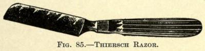
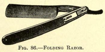
For the removal of the strips the Thiersch razor is to be used. It is concave on its upper side and plane below, the blade being bent at an angle to the handle (Fig. 85). Folding razors of the same type can be procured; their advantage lies in having a protecting case when not in use.
Slide fixation locks are a valuable addition to the latter, as they hold the blade in place when open. See Fig. 86.
FIG. 86. FOLDING RAZOR.
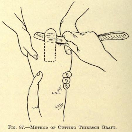
The site from which the graft is to be taken is first thoroughly scrubbed and washed, then cleansed with an antiseptic solution. The skin of the anterior surface of the arm or upper thigh is usually chosen. The skin of the part is made tense with the left hand, while the point of beginning is slightly raised by the assistant with the aid of a tenaculum hook. The razor, dipped into sterile salt solution, is now taken in the right hand and by quick sawing movements, the plane side being placed next to the limb, a strip of skin is detached (Fig. 87), which, as it is cut, glides in folds upon the concave side of the razor.
FIG. 85. THIERSCH RAZOR.
The uppermost layer of the skin is removed, including epidermis, the Malpighian and papillary layers, as well as a small portion of the stroma. Hübscherincludes only the epidermis and the upper portion of the papillary layer, with equal success.
The length and width of the strips so removed must be made according to the defect to be covered. Their width may be made as much as two inches and their length not to exceed four inches.
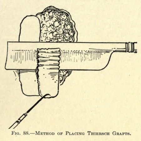
The collected strip of skin, still on the razor, is now brought to the place of grafting and, with the point of a needle placed at its farther end, is slid off upon the part to be covered and allowed to fall in place by the gentle backward withdrawal of the razor blade, as shown in Fig. 88.
FIG. 87.—METHOD OF CUTTING THIERSCH GRAFT.
The graft may be smoothed out with the needle held flatwise or be stroked down gently, so that its fresh surface makes contact with every portion of the part covered, a precaution the author considers important to obtain the best results.
If the defect is large, and where several grafts are needed, the second flap thus obtained is made to slightly overlap the one already placed, and so on. The free, or distal, ends of the flaps are made to slightly overlap the skin or that of a graft placed endwise to it. Every part of the wound should be covered.
As soon as this has been accomplished the strips are powdered over with iodol or aristol or protected with some antiseptic gauze (boric or iodoform), or covered with strips of lint smeared with borated petrolatum, over which light, teased-out pieces of sterilized