Contributors
Mohamed E. Abdelsalam, MD, MSc
Interventional Radiology
The University of Texas MD Anderson Cancer Center Houston, Texas
Andreas Adam, PhD, FRCP, FRCS, FRCR
Professor Department of Interventional Radiology
King’s College London Consultant Radiologist St. Thomas’ Hospital London United Kingdom
Vijay Agarwal, MD
Assistant Professor
The Leo M. Davidoff Department of Neurological Surgery
Albert Einstein College of Medicine
New York, New York
Kamran Ahrar, MD
Professor Departments of Interventional Radiology and Thoracic and Cardiovascular Surgery
Divisions of Diagnostic Imaging and Surgery
The University of Texas MD Anderson Cancer Center
Professor Department of Diagnostic and Interventional Imaging
The University of Texas Medical School at Houston Houston, Texas
Andrew Akman, MD
Interventional Radiologist
Suburban Hospital Bethesda, Maryland
Ziyad Al-Otaibi, MD
Critical Care and Emergency Medicine Attending Director
Emergency Medicine Department
Prince Sultan Medical Military City
Riyadh Saudi Arabia
Morvarid Alaghmand, MD
Internal Medicine
Doctors Community Health System Lanham, Maryland
Ali Albayati, MD, FRCR, MMED, MBChB
Vascular and Interventional Radiologist Progressive Radiology
Assistant Clinical Professor of Vascular and Interventional Radiology
George Washington University Hospital Washington, DC
Ajlan Alzaki, PhD Medical Student SMHS
The George Washington University School of Medicine and Health Sciences Washington, DC
Seyed Ameli-Renani, MBBS, FRCR, EBIR Radiology
St George’s University Hospitals NHS Foundation
London
United Kingdom
Sandeep Amin, MD Anesthesiology
Rush Oak Park Hospital
Rush University Medical Center Oak Park, Illinois
John F. Angle, MD Professor
Department Radiology and Medical Imaging University of Virginia School of Medicine Division of Vascular and Interventional Radiology Director
Division of Angiography and Interventional Radiology
University of Virginia Health System Charlottesville, Virginia
Gary M. Ansel, MD, FACC Director, Center for Critical Limb Care
Riverside Methodist Hospital Columbus, Ohio
Assistant Clinical Professor of Medicine Medical University of Ohio Toledo, Ohio
Chad Baarson, MD
Vascular and Interventional Radiologist
Walter Reed National Military Medical Center Bethesda, Maryland
Arya Bagherpour, DO Assistant Professor
Vascular and Interventional Radiology Department of Radiology
University of Texas Medical Branch Galveston, Texas
Alex M. Barnacle, BM, MRCP, FRCR Consultant Interventional Radiologist Department of Radiology
Great Ormond Street Hospital for Children
London
United Kingdom
Bradley P. Barnett, MD, PhD Research Fellow
Department of Radiology
Johns Hopkins Medical Institutions Baltimore, Maryland
Richard A. Baum, MD, MPA, MBA Interventional Radiologist Radiology
Brigham and Women’s Hospital
Associate Professor Department of Radiology
Harvard Medical School Boston, Massachusetts
Charles F. Batti, Jr., MD
MidOhio Cardiology and Vascular Consultants
Riverside Methodist Hospital Columbus, Ohio
Jennifer Berkeley, MD, PhD Director of Neurocritical Care
Department of Neurology Sinai Hospital of Baltimore Baltimore, Maryland
Anuj Bhatia, MBBS, MD, FRCA, FFPMRCA, FIPP, FRCPC (Anesthesia and Pain Medicine), EDRA, CIPS, ASRAPMUC
Associate Professor
University of Toronto Director
Anesthesia Chronic Pain Clinical Services Staff
Department of Anesthesia and Pain Medicine
University Health Network
Mount Sinai Hospital and Women’s College Hospital
Clinical Investigator
Krembil Research Institute
PhD Candidate in Clinical Epidemiology at IHPME
University of Toronto
Altum Medical Director
Anesthesia and Pain Medicine
Affiliated Faculty
Techna Institute for the Advancement of Technology for Health Toronto, Ontario
Canada
Jose I. Bilbao, MD, PhD
Professor of Radiology
Universidad de Navarra
Consultant Radiologist
Clínica Universitaria
Pamplona Spain
Tiago Bilhim, MD, PhD
Centro Hospitalar de Lisboa Central Lisbon Portugal
James H. Black III, MD, FACS
Chief
The Johns Hopkins Hospital Vascular Surgery and Endovascular Therapy
The David Goldfarb, MD, Associate Professor of Surgery
The Johns Hopkins University School of Medicine
Baltimore, Maryland
Brian M. Block, MD
Anesthesiologist Towson, Maryland
Franz Edward Boas, MD, PhD
Assistant Attending Interventional Radiology Service Department of Radiology Memorial Sloan Kettering Cancer Center
New York, New York
Steven R. Bonebrake, DO Associate Interventional Radiology
Medical Student Clerkship Director Department of Radiology
Geisinger Medical Center
Danville, Pennsylvania
Maryam Boumezrag, MD Department of Radiology
The George Washington University Washington, DC
Louis Boyer, MD, PhD Department of Radiology University Hospital Gabriel Montpied Clermont Ferrand
France
Zoe Brady, BSc (Hons), PhD (verify degrees)
Chief Medical Physicist Department of Radiology
Alfred Health
Melbourne, Victoria Australia
Peter R. Bream, Jr., MD, FSIR
Professor of Radiology
Vascular and Interventional Radiology
Program Director
Diagnostic Radiology
University of North Carolina at Chapel Hill
Chapter Hill, North Carolina
Rachel F. Brem, MD, FACR, FSBI
Professor and Vice Chair
Breast Imaging and Interventional Center
Department of Radiology
Breast Cancer Program Leader
The George Washington University Cancer Center
The George Washington University Washington, DC
Allan Brook, MD, FACR, FSIR
Director of Interventional Neuroradiology
Montefiore Medical Center
Professor of Radiology and Neurosurgery
Albert Einstein College of Medicine
New York, New York
Benjamin S. Brook, MD
Chief
Vascular Surgery
University of Utah
Salt Lake City, Utah
Mark Duncan Brooks, MBBS, FRANZCR Consultant Radiologist
Department of Radiology
Austin Hospital
University of Melbourne
Melbourne, Victoria Australia
Elias Brountzos, MD, EBIR
Professor of Interventional Radiology
Second Department of Radiology
Medical School
National and Kapodistrian University of Athens
Athens
Greece
Brendan Buckley, BSc, MB, BCh BAO, MRCS, FRCR, FRANZCR, EBIR
Consultant Interventional Radiologist
Auckland City Hospital Auckland
New Zealand
Charles T. Burke, MD
Professor of Radiology
University of North Carolina School of Medicine
Chapel Hill, North Carolina
Patricia E. Burrows, MD Professor
Department of Diagnostic and Interventional Imaging
University of Texas Medical School at Houston
Director of Vascular Anomalies Program
Department of Diagnostic and Interventional Imaging
Memorial Hermann and Children’s Memorial Hermann Hospital
Houston, Texas
Justin J. Campbell, MD
Department of Radiology
Southshore Hospital
Weymouth, Massachusetts
Colin P. Cantwell, MB, BCh, BAO, MSc, MRCS, FRCR, FFR(RCSI)
Consultant Radiologist
Department of Radiology
St. Vincent’s University Hospital
Dublin Ireland
Thierry Carreres, MD
Cardio-vasculaire Radiologie
Hopital Européen Georges-Pompidou
Paris France
John A. Carrino, MD, MPH
Vice Chairman of Radiology
Department of Radiology and Imaging
Hospital for Special Surgery
New York, New York
David Casper, BS
University of Arizona College of Medicine
Phoenix, Arizona
Lucie Cassagnes, MD, PhD
Department of Radiology
University Hospital Gabriel Montpied
Clermont Ferrand
France
Pascal Chabrot, MD, PhD
Department of Radiology
University Hospital Gabriel Montpied
Clermont Ferrand
France
Ajay Chavan, MD, DMRD, MBBS
Prof. Dr.
Institute of Diagnostic and Interventional Radiology
Klinikum Neustadt am Ruberberge Oldenburg Germany
Lakhmir S. Chawla, MD
Professor of Medicine
Department of Anesthesiology and Critical Care Medicine
The George Washington University Medical Center Washington, DC
Yung-Hsn Chen, MD
Musculoskeletal Imaging
The Johns Hopkins University School of Medicine Baltimore, Maryland
Wayne Cheng, MD
New York Hospital – Weill Cornell Medical Center
New York, New York
Rush Chewning, MD
Instructor in Radiology
Radiology Department
Section of Pediatric Interventional Radiology
Boston Children’s Hospital/Harvard Medical School
Boston, Massachusetts
Albert K. Chun, MD, MBA
Clinical Assistant Professor of Radiology Department of Radiology
The George Washington University Washington, DC
Warren Clements, BBiomedSc(Hons), MBBS, FRANZCR, EBIR
Consultant Interventional Radiologist Department of Radiology
Alfred Health
Adjunct Lecturer Department of Surgery Monash University Melbourne, Victoria Australia
Wendy A. Cohen, MD Professor of Radiology University of Washington School of Medicine
Chief of Service Department of Radiology Harborview Medical Center Seattle, Washington
Sarah E. Connolly, MD
Assistant Program Director
Diagnostic Radiology Residency
Assistant Professor of Radiology and Surgery Vascular and Interventional Radiology Section
Mallinckrodt Institute of Radiology
Washington University School of Medicine in St. Louis Saint Louis, Missouri
Anne M. Covey, MD
Attending Interventional Radiologist Diagnostic Radiology
Memorial Sloan-Kettering Cancer Center Professor of Radiology Diagnostic Radiology
Weill Medical College of Cornell University New York, New York
Laura Crocetti, MD, PhD
Prof. Dr. Division of Interventional Radiology University of Pisa Pisa
Italy
Michael D. Dake, MD
Senior Vice President for Health Sciences Professor of Medical Imaging, Medicine and Surgery
University of Arizona Tucson, Arizona
Michael J. Darcy, MD
Chief of Interventional Radiology Professor of Radiology and Surgery
Mallinckrodt Institute of Radiology
Washington University School of Medicine in St Louis Saint Louis, Missouri
Raj Das, MBBS, BMedSci, MSc, MRCS, FRCR, EBIR
Consultant Interventional Radiologist Department of Radiology
St George’s University Hospitals NHS Foundation Trust
London
United Kingdom
Thierry de Baere, MD Oncology
Interventional Radiology Image Guided Therapy Gustave Roussy Cancer Center Villejuif
France
Professor of Medicine
University of Paris Sud
Paris
France
Diederick De Boo, MD, PhD, FRANZCR, EBIR
Interventional Radiologist Radiology
John Hunter Hospital
Newcastle New South Wales
Australia
Khanant M. Desai, MD Brigham and Women’s Hospital Department of Radiology
Harvard Medical School Boston, Massachusetts
Sudhen B. Desai, MD Department of Radiology
Section of Interventional Radiololgy Texas Children’s Hospital Houston, Texas
Massimiliano di Primio, MD Division of Interventional Radiology Hopital Européen Georges-Pompidou
Paris
France
Robert G. Dixon, MD, FSIR Professor of Radiology
Interventional Radiology Program Director Department of Radiology
University of North Carolina Chapel Hill, North Carolina
Pablo D. Dominguez, MD Department of Radiology
Clínica Universitaria de Navarra
Pamplona Spain
Damian E. Dupuy, MD
Cape Cod Hospital
Department of Radiology
Director of Tumor Ablation
Hyannis, Massachusetts
Professor of Diagnostic Imaging
Brown University
Providence, Rhode Island
Yasser El-Abd, MD
Department of Radiology and Medical Imaging
Division of Vascular and Interventional Radiology
University of Virginia Health System
Charlottesville, Virginia
Alireza Esfandiari-Namini
Department of Radiology
The George Washington University
Washington, DC
Clifford J. Eskey, MD, PhD
Professor of Radiology and Surgery
Department of Radiology
Dartmouth-Hitchcock Medical Center
Lebanon, New Hampshire
Gian Marco Falcone
Vascular and Interventional Radiology Department
Careggi University Hospital
Florence Italy
Chieh-Min Fan, MD
Associate Director
Division of Angiography and Interventional Radiology
Brigham and Women’s Hospital
Assistant Professor
Department of Radiology
Harvard Medical School
Boston, Massachusetts
Fabrizio Fanelli
Director
Vascular and Interventional Radiology Department
Careggi University Hospital
Florence Italy
Laura M. Fayad, MD
Professor of Radiology
Orthopaedic Surgery and Oncology
Chief of Musculoskeletal Imaging Director of Translational Research for Advanced Imaging
The Russell H. Morgan Department of Radiology and Radiological Science
The Johns Hopkins University School of Medicine
Baltimore, Maryland
Rukshan Fernando, MBBS, MRes, FRCR
Consultant Interventional Radiologist
Auckland City Hospital
Auckland
New Zealand
Contributors
Dimitrios Filippiadis, MD, PhD, MSc, EBIR
Associate Professor of Radiology
National and Kapodistrian University of Athens
Second Department of Radiology Attikon University Hospital
Athens Greece
Kathleen R. Fink, MD
Assistant Professor of Neuroradiology University of Washington School of Medicine
Seattle, Washington
Karen Flood, BMed Sci, BM BS, MMedSciClinEd, MRCS, FRCR
Consultant Vascular and Interventional Radiologist
Leeds Teaching Hospitals NHS Trust Leeds
United Kingdom
Matthew D. Forrester, MD Cardiothoracic Surgery Providence Medical Group Spokane, Washington
Bradley Fox, MD
Diagnostic Radiology Resident Jackson Memorial Hospital/University of Miami Miami, Florida
Philippe Gailloud, MD Director Division of Interventional Neuroradiology
The Johns Hopkins Hospital Baltimore, Maryland
Debra A. Gervais, MD Department of Radiology Section of Interventional Radiology Massachusetts General Hospital Boston, Massachusetts
Basavaraj V. Ghodke, MD
Associate Professor Departments of Neuroradiology and Neurological Surgery University of Washington School of Medicine Director Neuro-Interventional Radiology Childrens Hospital and Research Center Seattle, Washington
Gunvir Gill, MD
Assistant Professor and Division Chief Vascular and Interventional Radiology Department of Radiology
University of Texas Medical Branch Galveston, Texas
Mark F. Given, FFR, FRANZCR Beaumont Private Clinic
Beaumont, Dublin
Ireland
Michael Gonsalves, BSc MBBS FRCR
EBIR
Dr Radiology
St.George’s University Hospitals NHS Foundation Trust
London
United Kingdom
Theodore S. Grabow, MD
Baltimore Spine Center and Maryland
Pain Specialists
Adjunct Assistant Professor
Department of Anesthesiology and Critical Care Medicine
The Johns Hopkins University School of Medicine
Baltimore, Maryland
Brian Grieme, MD Vascular and Interventional Radiology
Oklahoma City, Oklahoma
Jonathan S. Gross, MD
Associate Clinical Professor of Medicine Department of Radiology
New York University School of Medicine
New York, New York
Gianluigi Guarnieri, MD
Neuroradiology Service
Cardarelli Hospital
Naples
Italy
Jeffrey P. Guenette, M.D.
Neuroradiology Fellow Department of Radiology
Brigham and Women’s Hospital
Harvard Medical School
Boston, Massachusetts
Wasim Hakim, BSc, MBBS, MRCS, FRCR Doctor of Radiology
St. Mary’s Hospital
London
United Kingdom
Danial K. Hallam, MD, MSc Associate Professor
Departments of Radiology and Neurological Surgery University of Washington School of Medicine
Seattle, Washington
Mohamad Hamady, MBChB, FRCR, FSIR, EBIR
Consultant and Interventional Radiologist
Senior Lecturer (Hons) Imperial College
Training Program Director for Interventional Radiology
Incoming Chair of Education Committee—British Society of Interventional Radiology
Incoming Chair of Scientific Committee—IDEAS/CIRSE
London
United Kingdom
Christopher Harnain, MD
Interventional Radiology
Weill Cornell Medicine
New York, New York
Hasnain Hasham
Fellow
Interventional Radiology
Dotter Institute of Radiology
Oregon Health & Science University
Portland, Oregon
Philip John Haslam, MBBS MRCP FRCR
Doctor of Radiology
Freeman Hospital
Newcastle upon Tyne
Tyne and Wear
United Kingdom
Stéphan Haulon, MD, PhD
Professor
Department of Surgery
Université de Lille 2
Chief of Vascular Surgery
Hôpital Cardiologique - CHRU Lille
Lille
France
Klaus A. Hausegger, MD
Professor and Head Department of Radiology
Institute of Interventional and Diagnostic Radiology
Klagenfurt Austria
Michael C. Hill, MD Diagnostic Radiologist
Washington, DC
Joshua A. Hirsch, MD
Vice Chair
Radiology
Harvard Medical School Department of Radiology
Massachusetts General Hospital
Boston, Massachusetts
Tyler Hoelscher, BS
University of Arizona College of Medicine
Phoenix, Arizona
Andrew Hugh Holden, MBChB, FRANZCR, EBIR, ONZM
Associate Professor
Consultant Interventional Radiologist
Auckland City Hospital
Auckland
New Zealand
Michael Hoy
Medical Student
SMHS
The George Washington University School of Medicine and Health Sciences
Washington, DC
Joseph A. Hughes III, MD
Assistant Professor of Radiology
Section Chief
Interventional Radiology
Virginia Tech Carilion School of Medicine Roanoke, Virginia
Ashley I. Huppe, MD
Assistant Professor
Department of Radiology
The University of Kansas Health System Kansas City, Kansas
Rabia Idrees, MD
Department of Radiology
Interventional Radiology Division
The George Washington University School of Medicine Washington, DC
Uzoma Igboagi, MD Department of Radiology
Interventional Radiology Division
The George Washington University Washington, DC
Elizabeth Ann Ignacio, MD, FSIR
Associate Professor
Interventional Radiology
The George Washington University Medical Center Washington, DC
James E. Jackson, MD
Department of Radiology
Imperial Healthcare NHS Trust London
United Kingdom
Tim Joseph, BSc, MBBS, FRANZCR Consultant Interventional Radiologist
Radiology
Alfred Health Melbourne, Victoria Australia
Irem Kapucu, MD Research Fellow
Rush University Medical Center Chicago, Illinois
Claire S. Kaufman, MD Assistant Professor
Radiology University of Utah Salt Lake City, Utah
John A. Kaufman, MD, MS Director
Dotter Interventional Institute
Oregon Health & Science University Portland, Oregon
Pavan Kavali, MD
Assistant Professor
Radiology and Surgery
Mallinckrodt Institute of Radiology
Washington University School of Medicine in St. Louis Saint Louis, Missouri
Jacob Kazmi-Bowdoin
Medical Student
Albert Einstein College of Medicine New York, New York
Alexis D. Kelekis, MD, PhD, EBIR
Assistant Professor of Interventional Radiology
National and Kapodistrian University of Athens
Second Radiology Department Attikon University Hospital
Athens
Greece
Frederick S. Keller, MD
Cook Professor
Charles T. Dotter Department of Interventional Radiology
Oregon Health Sciences University
Portland, Oregon
Nadia J. Khati, MD, FACR
Associate Professor of Radiology Abdominal Imaging Division
The George Washington University Medical Center
Washington, DC
Neil M. Khilnani, MD Department of Radiology Division of Interventional Radiology
Weill Cornell Medicine
New York, New York
Iman Khodarahmi, MD, PhD
Assistant Professor of Radiology
Musculoskeletal Imaging
New York University School of Medicine
New York, New York
Darren D. Kies, MD
Assistant Professor of Radiology Department of Radiology and Imaging Sciences
Emory School of Medicine Atlanta, Georgia
Hyun S. Kim, MD
Diagnostic Radiology
Vascular/Interventional Radiology
Yale New Haven Hospital New Haven, Connecticut
Tammy Kim, MD Internal Medicine
Washington, DC
Kun Yung Kim, Dr. Department of Radiology
Asan Medical Center
University of Ulsan College of Medicine
Seoul
Republic of Korea
Jennifer L. Kissane, MD
Assistant Professor of Radiology, Medicine and Surgery
Cardiovascular and Interventional Radiology Department of Radiology
Pennsylvania State University College of Medicine
Penn State Hershey Medical Center
Hershey, Pennsylvania
Hiro Kiyosue, MD
Associate Professor
Department of Radiology
Oita Medical University
Yufu, Oita
Japan
Jim Koukounaras, BMBS, FRANZCR, EBIR
Interventional Radiologist
Department of Radiology
The Alfred Hospital
Adjunct Lecturer, Department of Surgery
Monash University
Honorary Adjunct, Department of Radiology
University of Melbourne
Melbourne, Victoria
Australia
Andres Krauthamer, MD
Assistant Professor of Vascular and Interventional Radiology
University of Miami Miller School of Medicine
Miami, Florida
Venkatesh P. Krishnasamy, MD
Staff Clinician
Interventional Radiology
National Institutes of Health Bethesda, Maryland
William T. Kuo, MD, FSIR, FCCP, FSVM, FCIRSE
Professor of Radiology—Interventional Radiology
Division of Vascular and Interventional Radiology
Stanford University School of Medicine
Stanford, California
Maxim Kupershmidt, MBBS, FRANZCR
Barwon Medical Imaging
Geelong, Victoria
Australia
Pierre-Yves Laffy, MD
Cardio-vasculaire Radiologie
Hopital Européen Georges-Pompidou
Paris France
Nicole Lamparello, MD
Assistant Professor of Radiology
Division of Interventional Radiology
New York Hospital – Weill Cornell Medical Center
New York, New York
Leo Lawler, MB, BCh, BAO, FRCR, FFRRCSI, Hon Professor
Consultant Interventional Radiologist
Department of Radiology
Mater Misericordiae University Hospital
Dublin Ireland
Christopher Lawrence, MD
Department of Radiology
Interventional Radiology Division
The George Washington University Hospital
Washington, DC
Judy M. Lee, MD
Assistant Professor Department of Obstetrics and Gynecology
The Johns Hopkins University School of Medicine Baltimore, Maryland
Michael J. Lee, EBIR Professor of Radiology
Royal College of Surgeons in Ireland Beaumont Hospital
Consultant Interventional Radiologist and Clinical Director Department of Radiology Beaumont Hospital
Dublin Ireland
Riccardo Lencioni, MD, FSIR, EBIR
Hon. Res. Prof of Interventional Oncology
Miami Cancer Institute Miami, Florida
Professor of Radiology School of Medicine
University of Pisa
Pisa
Italy
Robert J. Lewandowski, MD Department of Radiology
Section of Interventional Radiology Northwestern Memorial Hospital Chicago, Illinois
Robert P. Liddell, MD
Assistant Professor Radiology and Radiological Sciences
The Johns Hopkins School of Medicine Baltimore, Maryland
Rafael H. Llinas, MD
Associate Professor Department of Neurology
The Johns Hopkins University School of Medicine
Clinical Vice Chair of Neurology
The Johns Hopkins Hospital Baltimore, Maryland
Romaric Loffroy MD, PhD Professor, Head Department of Vascular and Interventional Radiology François-Mitterrand University Hospital
Dijon
France
Brendan Logiurato, MD
New York Hospital – Weill Cornell Medical Center
New York, New York
Rahul Lohan, MBBS, M.Med, FRCR, MCI
Radiology Department
Khoo Teck Puat Hospital
Singapore
Stuart M. Lyon, MBBS, FRANZCR, EBIR
Interventional Radiologist
Monash Health
Melbourne, Victoria Australia
Anoop Madan, MBBS, FRANZCR
Senior Staff Specialist
Radiology
The Alfred Hospital
Melbourne, Victoria Australia
Jeevan Kumar Mahaveer, MBBS, MRCS, FRCR
Consultant Vascular and Interventional Radiologist
Leeds Teaching Hospitals NHS Trust
Leeds
United Kingdom
Julian Maingard, Bachelor of Medicine/ Bachelor of Surgery
Neurointerventional Radiology Monash Health
Melbourne, Victoria Australia
Patrick C. Malloy, MD, FSIR
Executive Chief of Staff
VA New York Harbor Healthcare System
Assistant Clinical Professor of Radiology
Department of Radiology
New York University School of Medicine
New York, New York
Mark D. Mamlouk, MD
Department of Radiology
Chief Fellow
University of California School of Medicine San Francisco
San Francisco, California
Michael J. Manzano, MD Radiology
USC Verdugo Hills Hospital
Los Angeles, California
Irfan Masood, MD
Diagnostic and Interventional Radiology University of Texas Medical Branch Galveston, Texas
Surena F. Matin, MD
Urology
The University of Texas MD Anderson Cancer Center Houston, Texas
Jean-Baptist Martin, MD
Interventional Neuroradiologist
Pain Management Centre Jean-Viloette Geneva Switzerland
Antonio Martinez-Cuesta, MD, MSc, FRCR
Department of Radiology
Hospital de Navarra
Pamplona
Spain
Alan H. Matsumoto, MD, FSIR, FACR, FAHA
Professor and Chair
Department of Radiology and Medical
Imaging
University of Virginia School of Medicine
University of Virginia Health System
Charlottesville, Virginia
David M. Mauro, MD
Assistant Professor of Radiology and Surgery
University of North Carolina at Chapel Hill
Chapel Hill, North Carolina
Matthew A. Mauro, MD, FSIR, FACR, FAHA
Ernest H. Wood Distinguished Professor
Department of Radiology
Professor of Surgery
UNC Medical Center
President
UNC Faculty Physicians
Chapel Hill, North Carolina
Ian McCafferty, Bsc MB BS MRCP FRCR
Consultant Diagnostic and Interventional Radiologist
University Hospital Birmingham
Birmingham Women’s and Children’s Hospital Birmingham United Kingdom
Timothy McClure, MD
Assistant Professor of Urology, Division of Urology
New York Hospital – Weill Cornell Medical Center
New York, New York
Ellen C. McCormick, MD
Department of Radiology
Interventional Radiology Division
The George Washington University Hospital
Washington, DC
Simon John McPherson, BSc, MRCP, FRCS, EBIR
Doctor of Vascular Radiology
Leeds General Infirmary
Leeds, West Yorkshire
United Kingdom
Anita K. Mehta, MD
Assistant Professor
Department of Radiology
The George Washington University
Washington, DC
Stephen Merrilees, MBChB, FRANZCR, EBIR
Consultant Interventional Radiologist
Auckland City Hospital
Auckland New Zealand
Olivier Meyrignac, MD, PhD
Department of Radiology
CHU Rangueil Toulouse France
Robert J. Min, MD, MBA
Chairman of Radiology
Division of Interventional Radiology
Weill Cornell Medicine
New York, New York
Fatima Mokrane, MD, PhD Department of Radiology CHU Rangueil Toulouse France
Robert A. Morgan, FRCR, FSIR, FCIRSE, EBIR
Consultant Interventional Radiologist Reader in Interventional Radiology St. George’s University of London London United Kingdom
Hiromu Mori, MD, PhD Chairperson Department of Radiology Faculty of Medicine Oita University Yufu, Oita Japan
Paul R. Morrison, MS Medical Physicist
Brigham and Women’s Hospital Harvard Medical School Boston, Massachusetts
Connor A. Morton, MD Interventional Radiology McGraw Medical Center Northwestern Memorial Hospital Chicago, Illinois
Giovanna Moscato, MD Radiology Service Vanvitelli University Naples Italy
Kieran P. Murphy, MB, FRCPC, FSIR Professor of Interventional Neuroradiology University of Toronto Toronto, Ontario Canada
Timothy E. Murray, MD Vascular and Interventional Radiology Fellow University of British Columbia Vancouver British Columbia
Gianluca Muto, MD Radiology Service CHU Geneva Switzerland
Mario Muto, MD Neuroradiology Service Cardarelli Hospital Naples Italy
Andrew Nelson, MD Department of Radiology Interventional Radiology Division The George Washington University Hospital Washington, DC
Albert A. Nemcek, Jr., MD Department of Radiology
Section of Interventional Radiology Northwestern Memorial Hospital Chicago, Illinois Illinois
Patrick Nicholson, MB, BCh, BAO, FFRRCSI
Clinical Fellow in Interventional Neuroradiology
Toronto Western Hospital Toronto, Ontario Canada
Rory L. O’Donohoe, MB, BCh, BAO, MRCPI, FFRRSCI Department of Radiolog St. Vincent’s University Hospital Dublin Ireland
Murat Osman, BS. Medical Student SMHS
The George Washington University School of Medicine and Health Sciences Washington, DC
Philippe Otal, MD Department of Radiology CHU Rangueil Toulouse France
Auh Whan Park, MD
Associate Professor
Department of Radiology and Medical Imaging
University of Virginia School of Medicine Division of Vascular and Interventional Radiology
University of Virginia Health System Charlottesville, Virginia
David A. Pastel, MD Director
Division of Neuroradiology Dartmouth-Hitchcock Medical Center Lebanon, New Hampshire
Aalpen A. Patel, MD, FSIR Chair
Department of Radiology Vice Chair of Informatics Medical Director 3D Laboratory Department of Radiology
Geisinger Medical Center Danville, Pennsylvania
Jai V. Patel, MB ChB, MRCP, FRCR Consultant Vascular and Interventional Radiologist
Leeds Teaching Hospitals NHS Trust Leeds United Kingdom
Sandhya Patel, MD
US Naval Medical Center
Department of Internal Medicine San Diego, California
Olivier Pellerin, MD, MSc
Faculté de Médecine
Université Paris Descartes
Sorbonne Paris-Cité Paris France
Philip Peng, MBBS, FRCPC Director Professor
Department of Anesthesia
Toronto Western Hospital and University Health Network Toronto, Ontario Canada
Chiara Perazzini, MD
Department of Radiology
University Hospital Gabriel Montpied Clermont Ferrand France
João M. Pisco, MD, PhD† Hospital St. Louis Lisbon Portugal
Jeffrey S. Pollak, MD
The Robert I. White, Jr. Professor of Interventional Radiology Department of Radiology and Biomedical Imaging
Yale University School of Medicine New Haven, Connecticut
Rupert H. Portugaller, MD
Associate Professor Department of Vascular and Interventional Radiology
University Clinic of Radiology
Medical University Graz Graz Austria
Stephen J. Power, MB, BCh, BAO, BPharm, MRCPI, FRCR, FFRRCSI Vascular and Interventional Radiology Fellow
Department of Medical Imaging St. Michael’s Hospital Toronto, Ontario Canada
Hemkumar Pushparaj, MBBS, MD Department of Anesthesia University of Toronto Toronto, Ontario Canada
Zain Naeem Qazi, MD Department of Radiology
George Washington University Hospital Washington, DC
Martin G. Radvany, MD
Professor of Radiology and Neurosurgery Division of Interventional Neuroradiology
University of Arkansas for Medical Sciences Little Rock, Arkansas
†Deceased
Batya R. Radzik, MSN, CRNP, BC
Nurse Practitioner
Neurocritical Care
Department of Anesthesia and Critical Care Medicine
The Johns Hopkins Hospital Baltimore, Maryland
Dheeraj Rajan, MD, FRCPC, FSIR
Professor Division of Vascular and Interventional Radiology University of Toronto Toronto, Ontario
Canada
Anne Ravel, MD
Department of Radiology University Hospital Gabriel-Montpied Clermont Ferrand
France
Faezeh Razjouyan, MD, MS
Radiology Resident Department of Radiology
The George Washington University School of Medicine and Health Sciences Washington, DC
Paul Revel, MD
Department of Radiology
CHU Rangueil
Toulouse
France
Anne Roberts, MD Professor Department of Radiology
University of California
Chief
Vascular and Interventional Radiology
Thornton Hospital-UCSD Medical Center San Diego, California
Alain Roche, MD
Radiology
Ultrasonography Unit
Imaging Department Institut Gustave Roussy Villejuif
France
Hervé Rousseau, MD, PhD
Professeur et Chef de Service Service de Radiologie L’Hôpital Rangueil Universitaire de Toulouse
Toulouse
France
Noé Roussel, MD
Department of Radiology
CHU Rangueil
Toulouse
France
Diego San Millan Ruiz, MD
Neuroradiologist
Department of Diagnostic and Interventional Radiology
Hospital of Sion
Sion
Switzerland
Wael E.A. Saad, MD, FSIR
Associate Professor of Radiology
Division of Angiography and Interventional Radiology
University of Virginia School of Medicine
Charlottesville, Virginia
Tarun Sabharwal, FRCR
Consultant
Interventional Radiologist
Guy’s and St.Thomas’ Hospitals
NHS Foundation Trust
London
United Kingdom
Arsalan Saleem, MD
Clinical Instructor and Chief Resident
Vascular and Interventional Radiology
Department of Radiology
University of Texas Medical Branch
Galveston, Texas
Anam Salman, MD
Department of Radiology
Diagnostic Radiology
The George Washington University Hospital Washington, DC
Marc Sapoval, MD, PhD
Head Division of Interventional Radiology
Hopital Européen Georges-Pompidou
Paris
France
Shawn Sarin, MBA, MD
Director
Department of Vascular and Interventional Radiology
The George Washington University Hospital Washington, DC
Paola Scalise, MD
Department of Diagnostic Imaging
Pisa University Hospital
Pisa Italy
Matthew P. Schenker, MD
Associate Radiologist
Division of Angiography and Interventional Radiology
Brigham and Women’s Hospital Boston, Massachusetts
Ryan Carl Schenning, MD
Assistant Professor, Vascular & Interventional Radiology
Charles T. Dotter Department of Interventional Radiology
Oregon Health & Science University
Portland, Oregon
Daniel Scher, MD
Department of Vascular and Interventional Radiology
George Washington University Hospital Washington, DC
Marc H. Schiffman, MD
Assistant Professor of Radiology
Division of Interventional Radiology
New York Hospital—Weill Cornell Medical Center
New York, New York
Liqi Shu, MD
Department of Radiology
Interventional Radiology Division
The George Washington University Hospital Washington, DC
Sameer Singhal
Medical Student
SMHS
The George Washington University School of Medicine and Health Sciences
Washington, DC
Ji Hoon Shin, MD, PhD
Professor
Department of Radiology
University of Ulsan College of Medicine
Asian Medical Center
Seoul Republic of Korea
Stuart G. Silverman, MD
Professor of Radiology
Chief of Abdominal Imaging and Intervention
Brigham and Women’s Hospital
Harvard Medical School
Boston, Massachusetts
Constantinos T. Sofocleous, MD, PhD, FSIR
Attending Interventional Radiology Service
Department of Radiology
Memorial Sloan Kettering Cancer Center
New York, New York
Ho-Young Song, MD, PhD
Department of Radiology
Asan Medical Center
University of Ulsan College of Medicine
Seoul
Republic of Korea
Thomas A. Sos, MD
Professor
Department of Radiology
Weill Medical College of Cornell University
Division of Interventional Radiology
New York—Presbyterian Hospital
New York, New York
Vlasios Sotrichos, MD
Department of Radiology
University of Pennsylvania
Philadelphia, Pennsylvania
Michael C. Soulen, MD, FSIR, FCIRSE
Professor of Radiology
Department of Interventional Radiology
University of Pennsylvania School of Medicine
Philadelphia, Pennsylvania
James Spies, MD, MPH Professor and Chair Department of Radiology
MedStar Georgetown University Hospital
Senior Associate Executive Director MedStar Medical Group Radiology Washington, DC
Stavros Spiliopoulos, MD, PhD, EBIR
Assistant Professor 2nd Radiology Department, Division of Interventional Radiology University of Athens Athens Greece
Brian Stedman, BSc MB BS FRCS FRCR
Consultant Radiologist Department of Radiology University Hospital Southampton Southampton United Kingdom
Jessica K. Stewart, MD Assistant Professor Department of Radiology University of North Carolina at Chapel Hill Chapel Hill, North Carolina
Paul V. Suhocki, MD Associate Professor Department of Radiology Duke University Medical Center Durham, North Carolina
M. Reza Taheri, MD, PhD Assistant Professor Department of Radiology
George Washington University School of Medicine and Health Sciences Washington, DC
Denise M. Thigpen, MD Breast Imaging and Intervention Assistant Professor Department of Radiology Medical Faculty Associates The George Washington University Hospital Washington, DC
Kenneth Robert Thomson, MD, FRANZCR, EBIR
Honorary Consultant Radiology Alfred Hospital Melbourne, Victoria Australia
Raymond H. Thornton, MD
Vice Chair for Quality, Safety, and Performance Improvement Department of Radiology Division of Interventional Radiology Memorial Sloan-Kettering Cancer Center New York, New York
Constantinos Tingerides, BSc(Hons), MBBS (London), MRCS, FRCR
Consultant Vascular and Interventional Radiologist
Radiology
Leeds Teaching Hospitals, NHS Trust Leeds
United Kingdom
Scott O. Trerotola, MD, FSIR Professor of Radiology in Surgery Associate Chair and Chief Interventional Radiology Vice Chair for Quality Perelman School of Medicine at the University of Pennsylvania Philadelphia, Pennsylvania
David W. Trost, MD
Associate Professor of Clinical Radiology Department of Interventional Radiology Weill Cornell Medicine
New York Presbyterian Hospital—Weill Cornell New York, New York
Maria Tsitskari, MD
St. George Hospital Pafos Cyprus
Kemal Tuncali, MD
Assistant Professor of Radiology
Brigham and Women’s Hospital Harvard Medical School Boston, Massachusetts
Ulku Cenk Turba, MD Professor Interventional Radiology Rush University Medical Center Chicago, Illinois
Mark R. Tyrrell, PhD, FRCS Consultant
Department of Vascular Surgery St. Thomas’ Hospital Tunbridge Wells Kent United Kingdom
Otto M. van Delden, prof Interventional Radiology
Academic Medical Center of the University of Amsterdam Amsterdam Netherlands
Eric vanSonnenberg, MD
Professor of Radiology
University of Arizona College of Medicine
Professor
Arizona State University Phoenix, Arizona
Visiting Professor of Medicine
Professor of Radiology
David Geffen School of Medicine at UCLA Los Angeles, California
Pasquale Vassallo, MD Neuroradiology Service
Cardarelli Hospital Naples Italy
Anthony C. Venbrux, MD
Professor of Radiology and Surgery
Division of Interventional Radiology
The George Washington University School of Medicine
Washington, DC
Isabel Vivas, MD
Professor of Radiology
Universidad de Navarra Consultant Radiologist
Clínica Universitaria de Navarra Pamplona Spain
Dierk Vorwerk, MD
Professor and Director
Department of Radiology
Klinikum Ingolstadt Ingolstadt Germany
Eric M. Walser, MD
Professor and Chairman Department of Radiology University of Texas Medical Branch Galveston, Texas
Anthony S. Walton, MBBS, FRACP, FCSANZ
Interventional Cardiologist Alfred Hospital Prahran, Victoria Australia
Sophie Wang Vascular Resident Beth-Israel Hospital Boston, Massachusetts
Peter N. Waybill, MD, FSIR Professor of Radiology, Medicine, and Surgery Chief
Division of Cardiovascular and Interventional Radiology Director
Interventional Oncology
Department of Radiology
Pennsylvania State University College of Medicine
Penn State Hershey Medical Center Hershey, Pennsylvania
Chapman Wei, MD
Department of Radiology
Interventional Radiology Division
The George Washington University Hospital
Washington, DC
C. Jason Wilkins, BMBCh, MRCP, FRCR
Consultant Interventional Radiologist
Department of Radiology
King’s College Hospital
London
United Kingdom
Robert J. Wityk, MD
Associate Professor of Neurology Neurology
The Johns Hopkins University School of Medicine
Baltimore, Maryland
Gerald M. Wyse, MB, BCh, BAO, FFRRSCI, MRCPI
Consultant Interventional Neuroradiologist
Department of Radiology
Cork University Hospital
Cork Ireland
Hyeon Yu, MD
Associate Professor Department of Radiology
University of North Carolina School of Medicine
Chapel Hill, North Carolina
Charline Zadro, MD Department of Radiology
CHU Rangueil
Toulouse France
Yuanlong Zhao, MD Department of Radiology
Interventional Radiology Division
The George Washington University Hospital Washington, DC
Christoph L. Zollikofer, MD Professor Department of Radiology
Kantonsspital Baden
Baden, Aargau Switzerland
Another random document with no related content on Scribd:
For the correction of this defect various methods are given, and all of these must be modified more or less, to meet the requirements or extent of lost tissue. In some cases the entire subseptum is absent, while in others there is more or less of a stump remaining. Again in some, the subseptum required is unusually wide and in others quite narrow.
While a number of surgeons prefer making the flap to restore it from part or the whole thickness of the upper lip, as will be shown, the author believes the best results are to be obtained with the Italian flap method, if there be great loss of tissue, or to attempt to restore smaller defects with cartilage-supported nonpedunculated flaps taken from back of the ear, as heretofore described, or the cartilage to be used as a support may be taken from the nasal septum itself, having its pedicle posteriorly.
This strip of cartilage is brought downward, freed at either side from its mucosal attachment, and the skin flap to be used is then made wide enough to be sutured to the inferior mucosa margins as well as to the skin of the lobule.
The method of taking a sliding flap from the healthy skin of the nose is not advisable, because of the resultant disfigurement.
The tissue of the lip, on the other hand, can be used, since the secondary wound can be readily drawn together, leaving only a linear scar. In men, this may be hidden by the mustache.
When the Italian method is used, the method referred to in restoration of the lobule is to be followed.
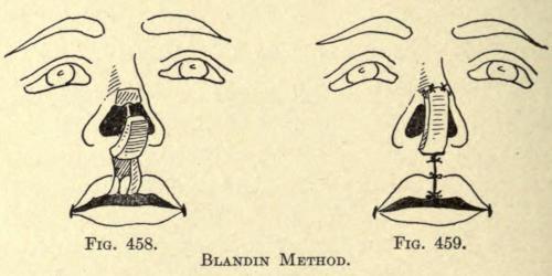
Blandin Method.—The flap is taken vertically from the entire thickness of the upper lip, as shown in Fig. 458, having its pedicle at the base of the nose.
This strip of tissue is turned upward, mucosa outward, and its freshened free end is sutured to the raw surface of the lobule.
The secondary wound of the lip is sutured as in ordinary harelip, as shown in Fig. 459.
The mucosa soon takes on the appearance of skin, but in most cases remains pink in color.
The flap taken in this way should not be made too wide.
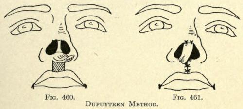
Dupuytren Method.—The flap is taken vertically from the skin of the upper lip, reaching down at its free end to the vermilion border, as shown in Fig. 460.
The flap is twisted upon its pedicle and sutured to the skin of the lobule; to facilitate this the left incision is made higher than that on the right.
The pedicle may be cut as with all such flaps, and it may be allowed to remain, if not too disfiguring.
The secondary wound of the upper lip is drawn together by suture, as shown in Fig. 461.
The mucosa of the nose is to be sutured to the raw edge of the flap when that is possible.
FIG. 458. FIG. 459.
BLANDIN METHOD.
FIG. 460. FIG. 461.
DUPUYTREN METHOD.
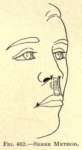
Serre Method.—This author advises dissecting up a flap from the upper lip, including the skin only, leaving it attached just above the vermilion border, as in Fig. 462.
The free and upper end is sutured to the lobule. When union has taken place, the pedicle is divided and is brought upward and sutured into place. The secondary wound repaired by suturing finally. There is some difficulty in dressing the wound during the time required to have it unite to the skin of the lobule, because of the danger of pressure and consequent gangrene.
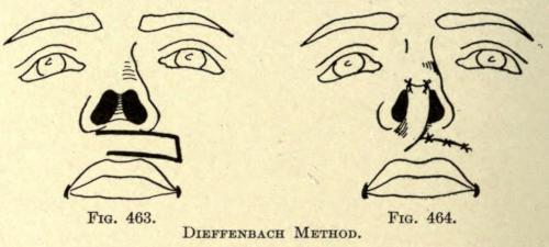
Dieffenbach Method.—This author took up the skin flap transversely or obliquely, as shown in Fig. 463, and twisted it into position, as shown in Fig. 464.
The objection to the direction of making the flap in this manner is that the consequent cicatrization has a tendency to draw the mouth out of its normal position on the wounded side.
FIG. 462. SERRE METHOD.
FIG. 463. FIG. 464.
IEFFENBACH METHOD.
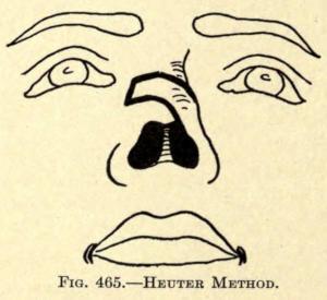
The following methods show the taking of the flap from the skin of the nose itself. Unless the defect be very small such methods are objectionable.
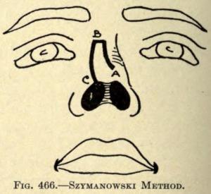
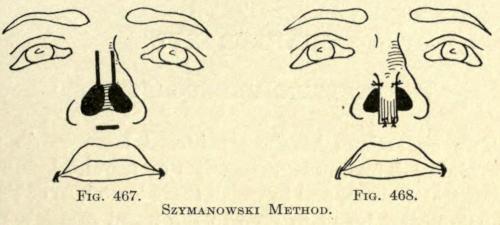
Szymanowski Method. In the latter method of Szymanowski the flap must be stretched considerably, to close over a lengthy deformity, encouraging gangrene. The deformity is not so great, however, as with the two preceding methods.
FIG. 465. HEUTER METHOD.
FIG. 466. SZYMANOWSKI METHOD.
FIG. 467. FIG. 468.
SZYMANOWSKI METHOD.
The author believes a nonpedunculated flap with or without a cartilaginous support should be tried before other methods are resorted to, in all cases, with the hope of healing the graft in place. The fact that the mucosa can in some cases be sutured to the margins of the flap adds much to the possibility of its subsequent life by adding its nutriment to the graft.
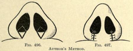
ANGULAR EXCISION TO CORRECT LOBULE
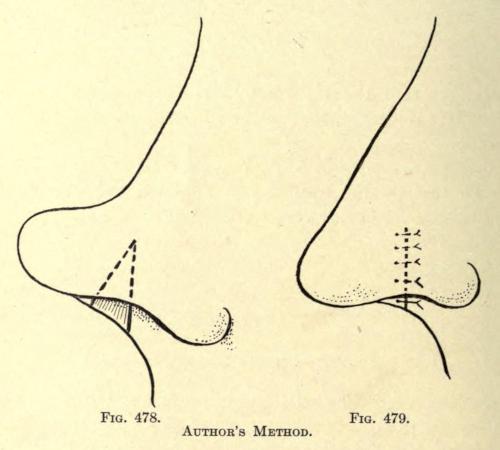
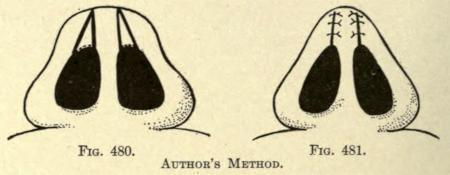
When the lobule is unduly broad at its base and is more or less concave above the rim of the alæ, it can be reduced by removing a diamond-shaped piece of tissue at either side of the subseptum.
The bases of the two triangles making up the diamond at its widest area meet at the anterior rim of the nostrils, extending with their apices upward and backward, as shown in Fig. 480.
If there be a prominence of the cartilaginous structure of the lobule, this may be removed subcutaneously after the two ablations have been made.
F
IG. 480. FIG. 481.
AUTHOR’S METHOD.
Before suturing the wounds, it is advisable to free the skin of the inferior lobule to overcome tension.
The sutures are applied as in Fig. 481. None are used to unite the mucosa unless the interior wounds are large enough to permit of their use.
CORRECTION OF MALFORMATIONS ABOUT THE NASAL LOBULE
The operations herein described apply particularly to the correction or reduction of an overprominent nasal tip due to an excessive growth of congenital malformation of that part of the nose, giving the organ undue prominence and a hooklike appearance, usually associated with a narrow, sharply upward inclined upper lip.
Pozzi Method.—The same operation, on a larger scale, can be readily employed for the correction of hyperplasia nasi and rhinophyma.
In the operation of Pozzi (Bulletin et mémoire de Société de chirurgie, 1897, p. 729) an elliptical section of skin and cartilage are removed from the lobule with its widest part corresponding to the point of the nose; the cicatrices occasioned thereby are practically as bad, if not worse, than the unscarred overprominent nose, while the submucous procedure of Roe (MedicalRecord, July 18, 1891) is not only insufficient in these cases, but, according to my experience, practically useless.
Roe Method.—Roe’s method requires a submucous extirpation of the redundant cartilage at the tip through a necessarily small opening within the nasal orifice, also the division in several places of the anterior fold of the lower lateral cartilage with the object of reducing the undue convexity of the alæ. The latter is, we might say, impossible, since the cartilages will be reduced by such a method, even under pressure dressings, which are likely to cause gangrene of the skin of the wings; or if this be avoided, the cicatrix resulting from such division usually restores the very fault that it is expected to overcome, while the mucous lining of the alæ becomes thickened and more firmly tied down than previous to the operation.
One is tempted to exsect the major curvature of the lower lateral cartilage, but this leads to a flattening of the wings of the nose, partial atresia of the nasal orifice, and a decided lack in its symmetry.
Secondly, in Roe’s operation there is always a lack of knowing how much or how little to remove of the cartilage of the tip, a second cosmetic operation being made necessary after the parts have contracted and healed, a common fault with most cosmetic plastic operations performed under local anesthesia, owing to the immediate edematous enlargement following its hypodermic use.
Operation as Commonly Practiced.—The operation heretofore most commonly practiced is one in which an elliptical piece of skin is cut from the tip of the nose, followed by the extirpation of the anterior prominences of the lateral cartilages, and amputation of the septal cartilages. Unfortunately, the result, at first quite satisfactory to the eye, culminates in the pulling apart of the cicatrix formed by bringing the sides of the wound together along the median line with a later depression of the tip in this median line, occasioned by the outward traction of the lower lateral cartilages. Even a second or third operation does not overcome this result entirely, and at best leaves an ugly irregular gash in the median line of the tip and the columna.
In one of the cases here cited this same operation had been unsuccessfully tried twice by another surgeon, with very unsatisfactory and unsightly result. (Case II.)
The ideal operation for all of this type of cases from the view of the surgeon is to leave as little disfigurement as possible, and the method to be here considered, when properly followed, leaves no scar whatever, except for a slight white line across the columna of the nose, where it is out of view, and when contracted offers no objection on the part of the hypercritical patient.
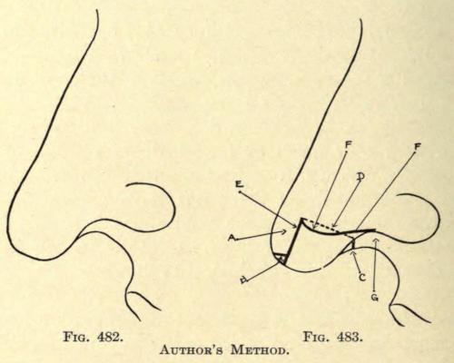
Author’s Method.—The method of the author is as follows: Given a nose, typified by the illustration in Fig. 482, the skin above the site of the operation is thoroughly cleansed with soap and hot water, then rinsed with alcohol, ninety-five per cent, and vigorously scrubbed with gauze sponges, dipped into hot bichlorid solution, 1 to 2,000, followed with a thorough lavage with sterilized water. Both nostrils are now cleansed with warm boric-acid solution by the aid of small tufts of absorbent cotton wound over a dressing forceps. The patient is then instructed to breathe through the mouth during the operation. A number of small round gauze sponges dipped into sterilized water and squeezed dry are placed within reach of the assistant. About one drachm of two-per-cent Beta Eucain solution is
now injected about the tip of the nose, the columna, and the alæ, as far back as their posterior fold.
FIG. 482. FIG. 483. AUTHOR’S METHOD.
A thin bistoury is then thrust into the nose from right to left, entering at the point E(Fig. 483), and brought down parallel to the anterior line of the nose, and emerging below the tip in a line with the anterior border of the nasal orifices. This procedure leaves a strip (A) about one quarter inch wide, laterally, rounded at its inferior extremity, and attached superiorly to the nose. Next the round inferior tip (B) is cut away obliquely, sloping inward toward the nose by the aid of a small angular scissors. Each blade of the angular scissors is now placed into each nostril, the tips of the blades inclined forward, and the columna or subseptum is divided at C, also the septum along the line D up to a point a little above the first incision made externally at E. The two arterioles of the columna are controlled by the use of mosquito-bill forceps. The two projecting
folds of the lower lateral cartilage in the columna are next severed as deeply as possible to give mobility to the stump, a step necessary to overcome the changed position, otherwise resulting in a droop, which would have to be corrected at a later sitting.
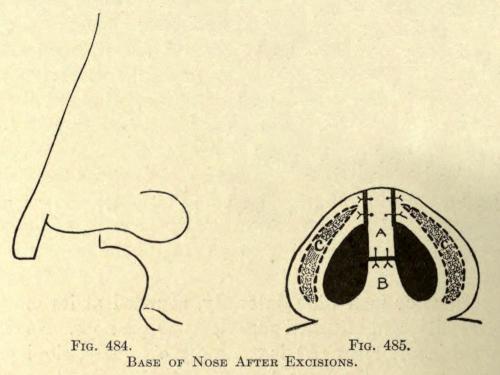
The next step is to give the needed shape to both wings. This is accomplished with a specially designed scissors, so curved on the flat that its convexity facing upward corresponds to the normal curvature of the orificial rim. A clean cut with these scissors, beginning at Gand ending at the point E, is made, leaving the base of the nose, as shown in Fig. 484. The anterior flap A is now bent backward to meet the stump of the columna at C. If it does not fall readily into place a little more of the septal cartilage is removed along the line Duntil this is accomplished.
It may be necessary to shorten the flap A in cases where a very prominent hook is to be corrected.
BASE OF NOSE AFTER EXCISIONS.
FIG. 484. FIG. 485.
The free end of the flap A is now sutured with No. 4 sterilized twisted silk to the stump of the columna at B. Two stitches usually suffice (see Fig. 485). One or two sutures may also be taken across the angles of union of the alæ and the flap A. The inferior raw surface of each wing may be found to be too wide, owing to the presence of the thickened cartilage at this point of the wing. The skin and the mucous membrane are then carefully peeled away from the cartilage, and the latter cut away as high as possible, or a gutterlike incision is made along its edges as shown in C(Fig. 485), excising the elongated elliptical piece of tissue which includes the cartilage. The raw mucocutaneous edges of the wings are now brought together with a No. 1 twisted silk continuous suture, completing the operation.
An antiseptic powder is dusted over the parts operated on, and small gauze dressings are applied with the aid of strips of silk isinglass plaster. A small tampon of cotton, well dusted over with an antiseptic powder, is placed into each nostril.
The dressings are changed the second day, when the resultant swelling will have practically subsided. The sutures in the columna are removed the fourth day preferably, and those of the wings about the sixth day. Complete cicatrization follows in about ten days, when the patient can be discharged.
The following cases are given to show the types of cases thus far operated upon and to illustrate the results obtained:
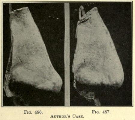
FIG. 486. FIG. 487.
AUTHOR’S CASE.
Case I.—Mr. R., aged thirty-two; foreman mechanic. Had been operated upon for angular nose, also at point of nose by Dr. S. Presented himself for operation October 19, 1904, when cast was made (see Fig. 486). Bromides given during recovery. Patient had been subject to fits of depression on account of his nose for over a year. Wounds healed in ten days, when second cast was made (Fig. 487). Complete recovery.
Case II.—Miss B. P., aged twenty-two; actress. Patient presented herself for operation March 22, 1905. A long, irregular depressed cicatrix showing at point of nose, the result of an attempt to reduce tip of nose by an elliptical extirpation of the lobule (Dr. N.). No cast was made of the case at the time, so that a second cast showing the result would be of no use. Recovery complete in twelve days. Patient returned to her profession three weeks later much pleased with the result.
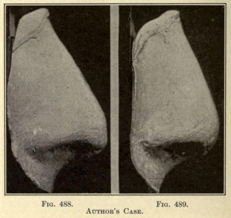
FIG. 488. FIG. 489. AUTHOR’S CASE.
Case III.—Mr. L. L., aged twenty-eight; broker. Presented himself, at the advice of Dr. T., for operation May 2, 1905. Cast of cast made and shown in Fig. 488. Uneventful recovery in twelve days, when case Fig. 489 was made.
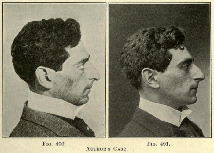
Case IV.—Mr. M. B., aged twenty-eight; operatic baritone. Presented himself for operation June 4, 1906. Photograph shown in Fig. 490. Uneventful recovery in fifteen days, when photograph in Fig. 491 was made; angular nose operated upon (at this time discharged; recovery complete).
FIG. 490. FIG. 491.
AUTHOR’S CASE.
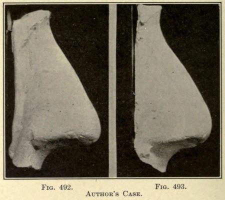
Case V.—Miss L. W., aged twenty-seven. Presented herself for operation and cast (Fig. 492) made August 4, 1906. Uneventful recovery in ten days. Cast of result made August 18, 1906 (see Fig. 493).
In each of these cases the patient was discharged highly satisfied and well pleased with the result of the operation, although in Case V the patient was requested to return in about one month for an operation to reduce the width of the wings of the nose, which was not attempted at the first sitting, but could have been with little difficulty by beginning the primary incision at E, Fig. 483, higher up, and cutting out a triangular section on either side of the flap A, the apex of each triangle being at point E, and the base along the line D. The wounds are sutured along the dorsum of the nose with No. 1 twisted silk, after exsecting much of the lower lateral cartilages of
the wings, as can easily be reached in the triangular point formed by the raw dorsal border and the inferior edge (F). The latter method, however, would be likely to leave a slight cicatricial line on either side of the nose. This could be much overcome by making the incision from point Eto Bobliquely to the plane of the skin, likewise the posterior sides of the triangles mentioned, just as the incisions at B, and across the columna at C, are made. Recovery should be complete in five days.
DEFICIENCY OF NASAL LOBULE
Where there is a lack of lobular prominence it may be enlarged and brought forward by a subcutaneous prothesis if the skin is flexible enough to permit of injection, as has heretofore been described. If this cannot be done, the following operation may be
FIG. 492. FIG. 493.
employed to advance the point of the nose, and reduce the width at its base so commonly observed with these cases.
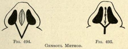
Gensoul Method.—A deep incision is made from the floor of each nostril downward and backward, meeting at a point just below the union of the subseptum with the upper lip, as in Fig. 494.
The deeper tissues are loosened from their attachments to the bone until the subseptum at its base, including the triangular appendage thus made, is freely movable.
The lobule is now drawn forward to its required prominence and the parts are sutured Y fashion, as in Fig. 495.
If the subseptum be too wide, an elliptical section is removed, including the cartilage, sufficient to give it the desired thickness when brought together, as illustrated. The lips of the wound are brought together as shown.
CORRECTION OF WIDENED BASE OF NOSE
When the base of the nose at its juncture with the lip is too broad, the reversed procedure mentioned under correction of a broad lobule is to be employed.
The diamond-shaped section is removed from the posterior rim of the nares as shown in Fig. 496.
FIG. 494. FIG. 495.
GENSOUL METHOD.
The tissues at either side are freed from their subcutaneous attachments so as to render them mobile.
The mucosa and skin wounds are sutured as in Fig. 497.
A retention splint or suture is to be employed to retain the parts as with the anterior lobule operation just described until healing has taken place.
FIG. 496. FIG. 497.
AUTHOR’S METHOD.
REDUCTION OF THICKNESS OF ALÆ
When the alæ are thickened they add to the width of the nasal bone and cause more or less atresia of the nostrils. The cause may be due to superabundant connective tissue or a congenital enlargement of the lower lateral cartilage.
To overcome this deformity the following operations may be followed:
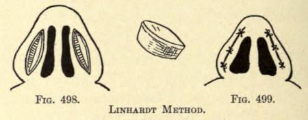
Linhardt Method. This author excises an elliptical section of tissue from the inferior base of both nasal wings, as shown in Fig. 498.
A similar procedure has heretofore been described in Fig. 485 in connection with correction of the lobule.
The section removed includes as much of the cartilage as is necessary to thin out the wing of the nose and to overcome the
atresia.
The parts are sutured as shown in Fig. 499.
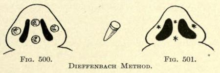
Dieffenbach Method.—In this method cone-shaped section of skin and cartilage are removed from the wings of the nose, as shown in Fig. 500.
If the septum is too wide, two or three of the same shaped sections are removed from it.
The skin wounds are drawn together by suture, as shown in Fig. 501.
CORRECTION OF NASAL DEVIATION
In this deformity the nose is bent or twisted to one side. The cause is usually traumatism, but may be congenital.
FIG. 498. FIG. 499.
LINHARDT METHOD.
FIG. 500. FIG. 501.
DIEFFENBACH METHOD.
The interior cartilaginous septum is usually found malformed on one or both sides.
To correct the deviation, the redundant cartilaginous septum is cut or sawed away to clear both nares and the anterior nasal vestibule. After this has been done the nasal attachments are freed subcutaneously, until the nasal organ is freely movable from its attachment to the superior maxillary bones.
The nose is now placed in the position desired, somewhat overdoing the correction, and is held in place by gauze packs in the nares or by Roberts’ spear-pointed pins thrust through the lateral skin of the nose at either side and through the septum, as shown in Fig. 339, p. 365.
The use of the pins placed as shown allows of free drainage to the nares and gives little inconvenience to the patient.
Plugs of gauze contract and harden, thus overcoming the object of their use and cause a disturbance of the wounds and pain when reapplied.
The pins should not be withdrawn until the nose has healed into its new position, or begin to cause irritation of the parts punctured.
Where the deviation is unilateral it should be corrected by subcutaneous injection, as previously described.
UNDUE PROMINENCE OF NASAL PROCESS OF THE SUPERIOR MAXILLARY
The protuberance of bone lies external to the middle meatus, involving an abnormal convexity of the nasal process of the superior maxillary. Its external removal or reduction involves considerable tissue and would leave a conspicuous linear scar, therefore the surgeon must attempt its reduction from the inner nose.
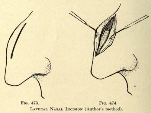
The author prefers to make a horizontal incision below the inferior border of the process, beginning anteriorly just before the