1 I: CELLS AND TISSUES THE CELL
1.1 Overview
1.2 Microscopes and Techniques
1.3 Different Appearances of Cells According to Technique
1.4 Ultrastructure and Function of Cell Membranes
1.5 Intercellular Junctions: Ultrastructure and Function of Tight Junctions
1.6 Intercellular Junctions: Ultrastructure and Function of Anchoring Junctions
1.7 Intercellular Junctions: Ultrastructure and Function of Gap Junctions
1.8 Ultrastructure and Function of the Nucleus and Nucleolus
1.9 Ultrastructure and Function of the Nucleus: Chromatin and Matrix
1.10 Ultrastructure and Function of the Nuclear Envelope
1.11 Ultrastructure and Function of Mitochondria
1.12 Ultrastructure and Function of Mitochondrial Cristae and Matrix
1.13 Ultrastructure and Function of Smooth Endoplasmic Reticulum
1.14 Ultrastructure and Function of Rough Endoplasmic Reticulum
1.15 Ultrastructure and Function of Ribosomes
1.16 Ultrastructure of the Golgi Complex
1.17 Functions of the Golgi Complex
1.18 Ultrastructure and Function of Lysosomes
1.19 Ultrastructure and Function of Peroxisomes
1.20 Ultrastructure and Function of Inclusions: Glycogen
1.21 Ultrastructure and Function of Inclusions: Lipid Droplets
1.22 Ultrastructure and Function of Cytoplasmic Vesicles: Endocytosis, Transcytosis, and Exocytosis
1.23 Ultrastructure and Function of Microtubules
1.24 Ultrastructure and Function of Cytoplasmic Filaments
1.25 Ultrastructure and Function of the Centrosome and Centrioles
1.26 The Cell Cycle, Mitosis, and Other Cellular Processes
1.27 Specializations of the Cell Surface: Cilia and Basal Bodies
1.28 Histopathology and Disease
1.29 Pathology of the Cell
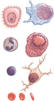
Schematic showing wide variation in shapes, sizes and tinctorial properties of different cells as seen via light microscopy. Names of cells often reflect structural or functional characteristics: Keratinocyte (or prickle cell) in epidermis (A); Macrophage (or phagocyte) in connective tissue (B); Polymorphonuclear leukocyte (or neutrophil) in peripheral blood (C); Plasma cell in connective tissue (D); Lymphocyte (a type of agranular leukocyte) in blood (E); Nerve cell (or neuron) in nervous tissue (F); Erythrocyte (red blood cell) in circulation (G). cell in connective tissue ( ); Lymphocyte (a type of agranular
1.1 OVERVIEW
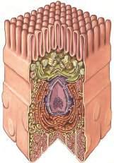
Microvilli
Centriole
Rough endoplasmic reticulum
Smooth endoplasmic reticulum
Plasma membrane
Golgi
Nucleus
Nucleolus
Mitochondria
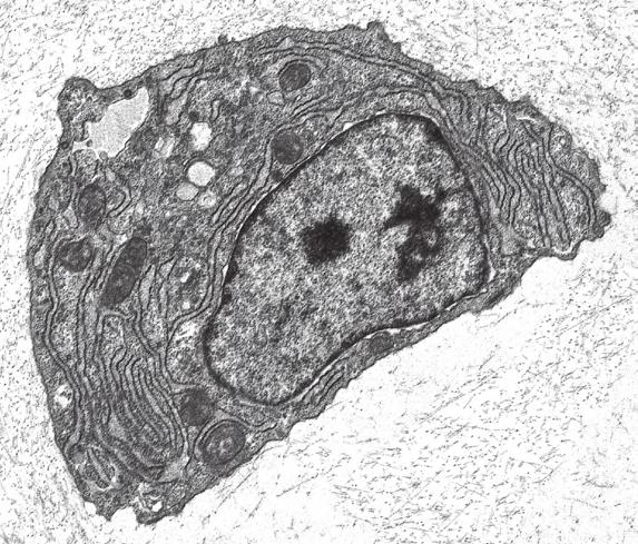
A composite cell cut open to show organization of its main components, as seen via electron microscopy. A plasma membrane surrounds the cell, which is polarized, with basal, lateral, and apical domains. Its cytoplasm contains various organelles and inclusions, which surround a nucleus. Some organelles are membrane bound, but some are not. The apical cell border has many finger-like projections called microvilli. Lateral cell borders are areas with intercellular junctions.
NerveCellMegakaryocytes
Light micrograph (LM) of part of the dorsal root ganglion. A large nerve cell contrasts with smaller cells that surround it. 235×. H&E.
The human body is organized into four basic tissues (epithelial, muscle, nervous, and connective) that consist of cells and associated extracellular matrix. The cell is the fundamental structural and functional unit of all living organisms. The body contains about 60 × 1012 cells—some 200 different types whose size and shape vary widely—but all have a common structural plan. The eukaryotic cell is a mass of protoplasm surrounded by an external plasma (limiting) membrane. The two components of the protoplasm are the nucleus, which holds the genome consisting of chromosomes, and the cytoplasm, a complex aqueous gel made of water (about 70%), proteins, lipids, carbohydrates, and organic and inorganic molecules. Organelles (specialized structures with functional capability) and inclusions (relatively inert, transitory structures) are in the cytoplasm. Except for mature erythrocytes, without a nucleus, most cells have one nucleus that conforms to the cell’s shape. A few cells, such as osteoclasts and skeletal muscle cells, may be multinucleated. A nuclear envelope invests the nucleus, whose substance, called chromatin, contains one
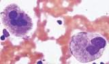
LM of megakaryocytes in a bone marrow smear. Each cell has one large multilobulated nucleus that is polyploid and intensely basophilic. 350×. Wright’s.
or more nucleoli. Internal cell structure is modified to reflect function: Muscle cells, for example, are modified for contraction; nerve cells (or neurons), for conduction; connective tissue cells such as fibroblasts, for support; and glandular epithelial cells, for secretion.
HISTORICAL POINT
German scientists biologist Theodor Schwann (1810-1882) and botanist Matthias Schleiden (1804-1881) proposed the cell theory, which states that all living organisms are composed of similar units of organization called cells. For his observations on normal animal cells, Schwann is recognized as the father of modern histology. Later, renowned German pathologist Rudolph Virchow (1821-1902) proposed that disease originates in cells, not in tissues or organs. Because he was the first to use microscopes and histologic specimens as a basis for the study of pathology, he is credited as the founder of modern cytopathology. With advances in medical science more than a century later, knowing the light and electron microscopic appearance of cells has become fundamental to diagnosis, treatment, and clinical management of many common and rare diseases.
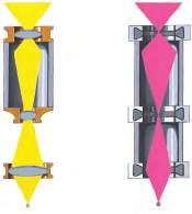
Optical parts of a conventional light (or bright-field) microscope. This compound microscope transmits light through three glass lenses. Light, first focused on a stained specimen by a substage condenser lens, passes through the specimen and then an objective lens, which magnifies and projects the illuminated image to the ocular lens. The ocular lens further magnifies the image and projects it to the eye of the viewer or a photographic plate. Most tissues are colorless, so color dyes serve as stains that differentially absorb light so that structures in specimens may be distinguished.
Optical parts of a conventional lenses. on ocular to photographic may
1.2 MICROSCOPES AND TECHNIQUES
Projector
Intermediate
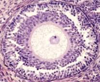
Comparative views of the ovary as seen with light (Left) and electron (Right) microscopes. Images show a large oocyte surrounded by smaller follicular cells (FC). The LM is a paraffin section stained with hematoxylin and eosin (H&E). Hematoxylin, a blue cationic stain, binds to anionic (negatively charged) basophilic sites in tissue sections. Eosin, a pink anionic stain, binds to acidophilic (positively charged) tissue components. The EM is a thin plastic section stained with heavy metals (lead citrate and uranyl acetate). Left: 200×; Right: 1800×. microscopes. Images show a with hematoxylin and eosin (H&E). Hematoxylin, a blue
Histology is the study of body tissues and cells, their constituents. Cells cannot be seen with the naked eye, so the primary tool used to study them is the microscope. It produces enlarged images of cells and enhances contrast for resolving details. Of several kinds of microscopes, two major ones are light and electron microscopes They have different lenses and sources of illumination and provide complementary information at different levels of resolution and magnification. The ability to discriminate two points that are close together is the resolving power of a microscope. It is related to the light wavelength. A conventional light microscope uses bright-field illumination, with a resolving power of about 0.2 µm. Study specimens absorb visible light; glass lenses focus and magnify specimens. Most cells absorb very little light, so staining is needed to increase light absorption. Cells and tissues first undergo sequential
Optical parts of a transmission electron microscope (TEM). A TEM transmits a beam of electrons through an ultrathin section of tissue that has been cut via an ultramicrotome. Several coiled electromagnetic lenses deflect electrons and use the same principle as that of light microscope lenses to condense, focus, and magnify images. Electrons from a heated tungsten filament (or cathode) are drawn toward an anode within a vacuum column. Electrons are not visible to the naked eye, so a fluorescent screen or photographic plate records the image as a black and white electron micrograph (EM). The advantage of the TEM is great resolving power. electron microscope (TEM). TEM electromagnetic electrons and use the same principle
filament (or cathode) are drawn toward naked

processing steps. Fixation in aldehydes and dehydration in alcohols are followed by embedding in paraffin or plastic. Specimen sections (or slices) are made with a microtome, followed by staining with color dyes. The illumination source of the transmission electron microscope (TEM) is a beam of electrons, which has a smaller wavelength. The resolving power of the TEM, 0.2-0.5 nm, is about 103 greater than that of the light microscope. For the TEM, ultrathin sections are cut after specimens have been fixed and embedded in plastic. Sections are then stained with heavy metals to enhance contrast, and black-and-white, not color, images result. A scanning electron microscope (SEM) is used for thick specimens or whole cells that have been fixed, dried, and coated with a thin metal film. It provides three-dimensional surface views. A high-resolution SEM (HRSEM) allows internal morphology of cells and organelles to be discerned with great depth of focus.
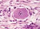
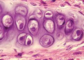
LM of chondrocytes in hyaline cartilage. The main function of these principal cells of cartilage is to synthesize and secrete surrounding extracellular matrix (ECM). Each cell has one round to ellipsoid nucleus and pale-stained cytoplasm. The ECM, which is also stained, contains proteins and carbohydrates secreted by the cells. 400×. H&E.
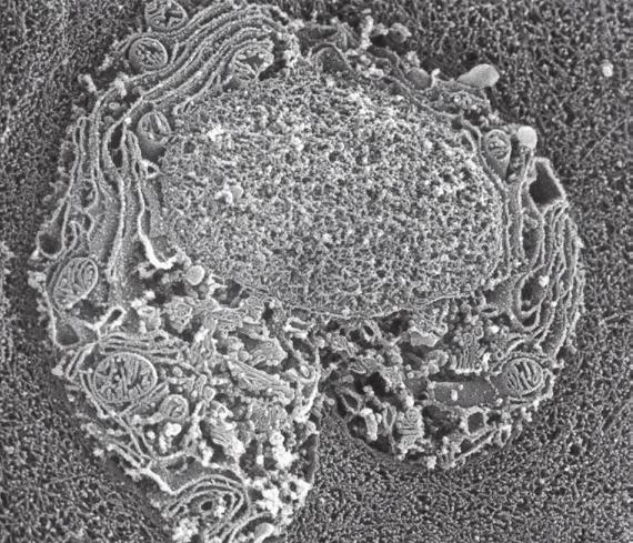
High-resolution scanning EM (HRSEM) of a chondrocyte. This image shows internal surface contours of a cell in three dimensions. Cells are frozen, fractured open, and coated with a thin metal film, and then surfaces are scanned. The resolving power of the SEM is not as great as that of the TEM, but tissue sections need not be cut for an SEM Complementary information is obtained from the two microscopes. 2000×. (Courtesy of Dr M. J. Hollenberg)
1.3 DIFFERENT APPEARANCES OF CELLS ACCORDING TO TECHNIQUE
EM of a chondrocyte with its nucleus and cytoplasm. Heavy metal stains combine with different parts of the cell to render them dark or light. Areas that appear dark, such as cell membranes and organelles, are electron dense they scatter electrons that have passed through the section. Conversely, areas that do not scatter electrons are lighter (electron lucent). Note that assorted organelles pack the cytoplasm. 2000×. (Courtesy of Dr. B. J. Crawford)
Histologic techniques provide different but complementary views of cells and thus a useful morphologic base, which can aid understanding of cell function in health and disease. Paraffin sections are routinely stained with hematoxylin and eosin (H&E) and examined with a light microscope. Cell nuclei (which are rich in nucleic acids such as DNA and RNA) have an affinity for hematoxylin (a basic dye), stain blue, and are termed basophilic. In contrast, the cytoplasm of cells and extracellular matrix typically
have an affinity for eosin (an anionic dye), stain pink, and are eosinophilic (or acidophilic). With superior resolving power, a TEM provides better elucidation of cell details, such as membranes and organelles, than a light microscope. Different parts of cells have distinct affinities for metal stains used on thin sections, so resulting two-dimensional images show variations in electron density, recorded in black and white. HRSEM images of freezefractured cells show three-dimensional spatial relationships of organelles and inclusions.
Another random document with no related content on Scribd:
and men felt the disbandment keenly.
By the 8th of February only a few headquarter details were left. Lieut.-Colonel Morgan Owen, commanding the 10th R.B., returned on the 12th from a conference to find that his battalion no longer existed. About the middle of the month the last few details went to the Divisional Wing of the Corps Reinforcement Camp.
The 7th K.O.Y.L.I. remained with the Division until the 15th, when they came under the O.C. Reinforcements XXIInd Corps; on the 20th they became the 14th Entrenching Battalion.
The 2nd Scottish Rifles, a very fine battalion over 1000 strong, under Lieut.-Col. H. C. H. Smith, D.S.O., came from the 8th Division to the 20th on the 3rd of February, and from that time formed part of the 59th Brigade.
In the Trench Mortar Batteries the personnel of V/20 Battery was transferred to the XXIInd Corps (H) T.M.B.; X, Y, and Z/20 were then reorganised to form the new X and Y/20 Batteries. At the same time, twelve mortars were handed over in exchange for ten six-inch Newtons.
After this the 20th Division was relieved by the 37th. All units were out of the line by the 19th, and on the following day began to entrain for the area of the Fifth Army south of the Somme.
CHAPTER XI
THE GERMAN OFFENSIVE ON THE SOMME
21st February to 25thMarch1918
Occupation of the rear zone defences Retreat to the Somme Defence of the Libermont Canal Actions of the 61st Infantry Brigade and 91st Field Artillery Brigade under the 36th Division. (Vide Map IV.)
On the 23rd of February 1918 Divisional Headquarters was opened at Ercheu in the area of the Fifth Army. The 60th Brigade was billeted in the Ham area; the 59th near Beaulieu, three miles south of Ercheu; the 61st near Freniches, three miles south-east of Ercheu; the 91st Field Artillery Brigade at EsmeryHallon; the 92nd Field Artillery Brigade at Rouy-le-Grand, two and a half miles north-east of Nesle. The Division was in G.H.Q. reserve but was to be placed at the disposal of the XVIIIth Corps, commanded by Lieut.-Gen. Sir F. I. Maxse, in the event of a German attack on the Corps front. During the rest of February and the first three weeks of March all units worked from time to time on the defences behind the battle zone and reconnoitred the positions which they would have to occupy in accordance with various schemes which had been prepared to meet a German offensive. All these schemes entailed considerable work for the 20th Divisional Signal Company. The Divisional Artillery, under Brig.-General Christie, took full advantage of the unique experience of being out of the line for a whole month to train batteries in open warfare, the ground lending itself admirably to this kind of work.
At the end of February the four machine gun companies of the Division were formed into the 20th Battalion, Machine Gun Corps, under Lieut.-Colonel H. L. Riley, D.S.O. The 59th, 60th, 61st and 217th Companies became “A,” “B,” “C,” and “D” Companies of the battalion respectively. In the middle of March Brig.-General Banbury,
commanding the 61st Infantry Brigade, went home for six months’ rest. He was succeeded by Brig.-General J. K. Cochrane, C.M.G.
It soon became clear that the expected German offensive would not be long delayed. On arrival in this area troops had been ordered to be ready to move at twenty-four hours’ notice; this was reduced to twelve hours on the 10th of March, and on the 20th to one hour. Just after 5 A.M. on the 21st, XVIIIth Corps issued the order “Man Battle Stations.”
The Fifth Army, under General Sir Hubert Gough, consisting of the IIIrd, XVIIIth, XIXth and VIIth Corps, held an extended front of some forty-two miles from the Oise to Gouzeaucourt, where the Third Army, under General the Hon. Sir Julian Byng, continued the line to the Scarpe. The XVIIIth Corps held a line facing St Quentin from Urvillers to Gricourt, with the 36th Division on the right, the 30th opposite St Quentin, and the 61st on the left. On the right of the 36th Division was the 14th Division of the IIIrd Corps. On the left of the 61st Division was the XIXth Corps. The 20th Division, if called upon to support this line, was to move forward ready to man the rear zone defences between the Somme and the Omignon river from St Simon to Trefcon, sending a brigade of artillery to each of the 36th and 30th Divisions.
At 4.40 A.M. on the 21st of March the enemy opened an intense bombardment along practically the whole front of the Fifth and Third Armies, using a great quantity of gas shells. The infantry attack, launched in a thick fog after several hours of this bombardment, developed on the XVIIIth Corps front about 10.30 A.M.
By noon the line had been penetrated at various points, although on all parts of the front isolated detachments held out most gallantly. The infantry of the 20th Division was ordered at 1 P.M. to concentrate behind the rear zone defences, and the artillery brigades moved forward to their allotted divisions, the 91st (Lieut.-Colonel Erskine) to the 36th Division and the 92nd (Lieut.-Colonel Balston) to the 30th.
In the course of the day the enemy pushed forward, particularly on the right of the Corps line, where by 3 P.M. the situation had become critical. The 61st Brigade (Brig.-General Cochrane) was ordered therefore to man the bridge-heads at St Simon and Tugny immediately on arrival in that area. Pressed on the front and outflanked on the right, the 36th Division retired during the night south of the Somme. The 61st Infantry Brigade and the 91st Field Artillery Brigade covered this retirement, and from that time came under the orders of the 36th Division. Here we will leave them for the moment in order to trace the movements of the rest of the 20th Division during the following four days.
On the left the Germans had penetrated the line at the junction of the XVIIIth and XIXth Corps and at Roupy, and had gained a footing in Grand Seraucourt. The 60th Brigade (Brig.-General Duncan) therefore pushed out covering troops on the defensive line from the Somme to Vaux, and as troops of the 30th Division were retiring on the left, the 59th Brigade (Brig.-General Hyslop) manned the defences between Vaux and the Corps left boundary at Trefcon. The 92nd Field Artillery Brigade under the 30th Division was in action south of Fluquières, covering a line between Roupy and Savy.
All details were formed into a Divisional Reinforcement Battalion under Major Storr, second in command of the 12th King’s, and moved to a position between Matigny and Douilly. The 11th D.L.I. concentrated at Golancourt and sent “D” Company to reinforce the 61st Brigade. Divisional Headquarters came forward to Ham.
This was the situation on the night of the 21st, when the enemy had advanced west and south-west of St Quentin for a distance of about three miles. Although he had made an important advance along the whole front, he had not yet broken through the battle zone.
Taking advantage of the thick mist which still hung over the country, the enemy worked down the St Quentin Canal on the 22nd towards Happencourt. This threatened the right flank of the 60th Brigade. The 59th was therefore withdrawn from the rear zone
defences between Vaux and Trefcon, leaving there only a skeleton force, and assembled in the Germaine—Foreste area, ready to launch a counter attack, if necessary, towards the south-east. But at 5.50 P.M. the enemy turned the left flank of the Corps north of Holnon Wood, driving back the 61st Division, which retired towards the Somme. To meet this new situation the 59th Brigade occupied a previously sited rearguard position between Douilly and Lanchy.
The 60th Brigade was attacked after heavy shelling at 3.50 P.M. Although their front line was driven back for a short distance, all battalions withstood the attack until they were ordered to withdraw. The 6th K.S.L.I. (Lieut.-Colonel Welch) on the right made a fine stand west of Happencourt with the enemy on both flanks; two companies of the 12th R.B. (Lieut.-Colonel MacLachlan) in the centre were surrounded, but fought their way back; and the 12th K.R.R.C. (Lieut.-Colonel Moore) on the left were heavily engaged with the enemy, who penetrated through Vaux and Fluquières. “A” Company of the 12th K.R.R.C. was practically cut off, but gallantly held on. In front of this company there was a gap in the wire through which the Germans tried to force their way four times, but two Lewis guns so successfully covered the gap that all efforts failed. Eventually, when all officers and many N.C.O.’s had been wounded or killed, the company was forced to fall back.
The pressure on the Corps front continued, and in view of the general situation the forward divisions were ordered to retire across the Somme during the night through the 59th and 60th Brigades, which after covering the withdrawal were to move back, fighting rearguard actions, to a position between Canizy and Béthencourt, and blow up all bridges on their front. At 8 P.M., before this movement was timed to take place, the enemy was reported to have broken through Vaux with a force of all arms, and the 20th Division was ordered to prolong the rearguard position held by the 59th Brigade to the right. The 60th Brigade accordingly occupied the line Bray—Douilly. Just before the troops moved back the Germans were heard advancing down a sunken road from Happencourt, singing for all they were worth. They had probably seen men of other divisions
retiring and thought that the whole line had gone, and they were very much surprised and completely scattered when they suddenly came under Lewis gun fire from the 6th K.S.L.I.
During the night the 20th covered the withdrawal of the 30th and 61st Divisions, and then fell back in close contact with the enemy across the Somme Canal. Most of the fighting occurred on the front of the 60th Brigade. Brig.-General Duncan received the orders for this retirement at 10.45 P.M., and made repeated efforts to send them on to battalions, but, owing to the heavy fighting which was going on, none of the orderlies got through. All units, however, had been told what the plans were, and they successfully carried them out. At 11 P.M. the Germans, who had crept up close to the position under cover of a dense mist, drove a wedge between two companies of the 12th K.R.R.C. and got into the line of the 6th K.S.L.I., cutting off practically the whole of one company. Lieut.-Colonel Welch, commanding the 6th K.S.L.I., led the battalion headquarters forward to counter attack, but the enemy was found to be too strong. The companies of the K.S.L.I. were driven back in various directions, and the headquarters and most of the men became separated from the brigade until the afternoon of the 24th. The 12th R.B. particularly distinguished themselves; they inflicted very heavy casualties on the enemy and caused a stampede among his transport, which was moving down the main St Quentin—Ham road, but they lost their commanding officer, Lieut.-Colonel MacLachlan, who was killed while gallantly leading his men.
“D” Company of the 11th D.L.I. (Lieut.-Colonel Hayes) had been sent to reinforce the 61st Brigade. The rest of the battalion had come under the orders of the 60th during the afternoon, and by 8 P.M. had taken up a position between Tugny and Bray to fill a gap between the 61st and 60th Brigades. Soon afterwards the 12th King’s, on the left of the 61st Brigade, were forced back, leaving the right flank of the D.L.I. exposed. The enemy could be heard in Tugny shouting in English and making a lot of noise, and patrols sent out by the battalion found that he had worked round the right flank in the fog. About midnight “B” Company on the left was rushed from
the right rear. A strong party of “A” Company on the right, under Capt. Endean, however, held out until the battalion was ordered to retire through Ham and hold the bridge-head at Offoy. By this time the enemy had broken through the line and the casualties were heavy; but leaving twenty men under 2nd Lieut. English as a rearguard with two Vickers guns on the main road, the remains of the battalion withdrew. Capt. Endean, two other officers, and forty men of “A” Company, although surrounded, fought their way through, and a small party of “B” Company, under C.S.M. Craggs, who was afterwards awarded the D.C.M., got back with the 12th R.B. and took part in the rearguard actions fought by that battalion. In the early hours of the 23rd, 97 men of “A” and “B” Companies were formed into one company under Lieut. Bushell and occupied the Offoy bridge-head.
The bridges over the Somme Canal were reported to have been prepared for demolition. The 83rd Field Company R.E. (Major Massie) and the 96th Field Company R.E. (Major Story), on arrival at the canal found that this work had been most inadequately done. In many cases the charges were far too small, the main girder at one bridge had been ignored, and some bridges had not been prepared at all. This, of course, threw a great deal of extra work on the field companies, as many new charges had to be laid. Where the existing charges were fired they failed to make complete gaps in the bridges; extra charges had to be laid afterwards, and by this means the work was satisfactorily completed.
By dawn on the 23rd the Division had successfully crossed the canal, the 83rd and 96th Field Companies R.E. had blown up the bridges, and the 60th Brigade on the right and the 59th on the left had occupied the line from Canizy to Béthencourt. At the same time the 92nd Field Artillery Brigade, which had helped to beat off heavy attacks on Roupy on the 22nd, rejoined the Division temporarily and came into action about one and a half miles south-east of Nesle, covering the crossings of the Somme opposite the front of the 60th Infantry Brigade. Divisional Headquarters was opened at Nesle, and the 20th Divisional Signal Company (Major Brace), whose signal lorry
had been the last vehicle to cross at Ham, quickly established communications with brigades.
The 12th K.R.R.C. took over the defences opposite Offoy from the D.L.I., who then prolonged the line to the right as far as Canizy, where they were joined by 34 more men of “A” Company, under Capt. Endean. About 26 men of the K.S.L.I. and parties from various divisions joined the 60th Brigade on this flank. On the left were the 12th R.B. Brig.-General Duncan found a large number of men of the 30th Division holding the canal, and these he took under his command and placed in support on the railway. On the 59th Brigade front the 11th K.R.R.C. (Lieut.-Colonel Priaulx) held the line about Voyennes, with the Scottish Rifles (Lieut.-Colonel H. C. H. Smith) in the centre and the 11th R.B. (Lieut.-Colonel Cotton) on the left at Béthencourt.
The work of the R.E. was made particularly difficult at this time, as someone had set fire to the R.E. park at Chaulnes long before the enemy got anywhere near it. In consequence, for two or three days hardly any shovels or R.E. stores could be obtained. Parties went to the park with lorries but were unable to do very much good, owing to the great heat and to the impossibility of getting at much of the most necessary material. The 83rd and 96th Field Companies now joined their respective brigades, to which they had already sent two sections each, the 83rd going to the 60th Brigade and the 96th to the 59th Brigade.
The 60th, 61st and 62nd Field Ambulances had been attached respectively to the 59th, 60th and 61st Brigades, but during the successive retirements they had to deal with the casualties of many different units. Their work was splendid. Many times when communication was difficult and the situation uncertain, advanced dressing stations were established close to the front line and the wounded carried back, often under heavy fire, with great perseverance and courage.
On the 22nd, between 2000 and 3000 cases passed through a temporary aid post of the 60th Field Ambulance established at
Matigny by Major C. A. Boyd before the post was abandoned at the last possible moment that night when the enemy was entering the village. Major Boyd and his party crossed at Voyennes just before the bridges were destroyed.
At 8 A.M. on the 23rd the enemy was reported to have broken through at Ham and to be advancing on Esmery Hallon, through which the 30th Division was retiring towards the Libermont Canal. Brig.-General Duncan was ordered to counter attack, and elements of the 182nd Brigade (61st Division) were placed at his disposal to assist this operation.
Lieut.-Colonel Bilton, of the 61st Division, was placed in command of the counter attack, and was assisted by Major A. E. Sanderson, the Brigade Major of the 60th Brigade. Lieut.-Colonel Bilton moved off with about 800 men and at 4 P.M. launched the attack from the main Ham—Nesle road, about 1500 yards west of Eppeville. The attack, delivered with great dash, drove back the enemy, captured Verlaines, and restored the situation.
The Division held the canal crossings throughout the day in spite of many attempts by the enemy to force them. Fighting was heavy at Offoy on the front of the 12th K.R.R.C.; at Béthencourt the 11th R.B. drove back with considerable loss several parties which tried to cross the canal.
In the evening the 182nd Brigade (61st Division), which had been under the orders of Major-General Douglas Smith since the morning, was transferred to the 30th Division, and the 183rd and 184th Brigades (61st Division) came under the 20th Division for counterattack purposes and moved to Nesle. The Division was further reinforced by two batteries of a Canadian Motor Machine Gun Battalion under Capt. Merling. “In all subsequent operations,” the official report states, “up to the 31st March, these batteries performed yeoman service, and on account of their mobility were of inestimable service in holding the extended fronts allotted to the Division.”
As soon as night came on, a great noise of traffic and shouting was heard in Offoy, where “C” Company of the 12th K.R.R.C. guarded the crossing. Under the fire of Vickers and Lewis guns the Germans became quieter, but at various points along the canal they could be heard driving in stakes, moving planks, and making evident preparations to throw out bridges. They planked what remained of the Offoy bridge during the night and made several attempts to cross it, but the 12th K.R.R.C. held on with great courage and complete success.
All through the 24th the Division was repeatedly attacked, particularly on the flanks, and the whole line was severely shelled. At 5 A.M. it was reported that the enemy had crossed the canal at Pargny, forcing a gap between the 20th and 8th Divisions, and was moving south along the west bank. Dispositions to meet this were made by the 11th R.B. and by battalions of the East Lancashire Regiment and the Royal Berkshire Regiment, who prolonged the flank to the left. Under cover of a thick mist the Germans shortly afterwards worked their way into Béthencourt, where their machine guns caused many casualties among the headquarters of the 11th R.B. At 8 A.M. an attempt to cross the canal on the front of the 11th R.B. was frustrated. “C” Company counter attacked the enemy in Béthencourt about 10 A.M., but was hopelessly outnumbered, and only a few men returned.
In spite of the flanks being in the air, the line held until 11.30 A.M., when a heavy trench mortar bombardment, directed by low-flying aeroplanes, forced the left flank to withdraw to a ridge 200 yards in rear. In making a further counter attack another company of the 11th R.B. was almost wiped out by artillery and machine-gun fire.
Shortly after mid-day the 183rd Brigade, reinforced by eight motor machine guns, counter attacked, and temporarily relieved the pressure. Later, as the enemy was getting further and further round the left flank, these three battalions retired to a better position in rear. Here Lieut.-Colonel Cotton was wounded, and was succeeded by Major the Hon. A. M. Bertie, D.S.O., M.C. By 4 P.M. “A” Company
consisted of 1 officer and 25 men; “D” Company had 1 officer, and “B” and “C” Companies none at all.
About 2 P.M. the 11th K.R.R.C. at Voyennes lost their commanding officer, Lieut.-Colonel Prialux, who was killed by a shell, with most of the battalion headquarters. He was succeeded by Major M. S. Ormrod. Soon afterwards the enemy crossed the canal and turned the line, forcing the battalion back to the high ground west of the village. The Scottish Rifles were then attacked from the rear. “B” Company, under Capt. Steward, made a fine stand, and eventually withdrew from a difficult position only 24 men strong.
Meanwhile a critical situation had developed on the right. At 8 A.M. the enemy, advancing from Ham, forced our troops out of Canizy and back to the railway. Lieut.-Colonel Moore, commanding the 12th K.R.R.C., immediately led “B” and “D” Companies of his battalion forward to counter attack. It was a great charge, and the bayonet was used with wonderful effect, but when it was over, Lieut.-Colonel Moore was missing. The Germans were driven back into the village, but later they came on again in greater numbers than before, and under very strong pressure the right of the 12th K.R.R.C. swung back to the railway again. In the course of this fighting our aeroplanes gave invaluable help. At one time, when “C” Company of the 12th K.R.R.C. was holding on to its position with great difficulty under heavy trench mortar fire, they nose-dived to a height of 200 feet and dropped bomb after bomb on to the German trench mortars until they put them completely out of action.
At 9.30 A.M. Major Massie, commanding the 83rd Field Company R.E., with about thirty of his men, reported to Brig.-General Duncan, who ordered him to take up a position south of Canizy on the main Ham road. Moving forward behind cyclist scouts and an advanced guard Major Massie took up the allotted position, keeping one section in reserve. He collected a considerable number of stragglers of various units and took them under his command. He was in touch with the 12th K.R.R.C. on his left, but had no troops on his right.
During the morning, repeated attempts of the enemy to advance were held up by the rifle fire of this party, with the help of one Lewis gun of the 30th Division. At 1.15 P.M. the pressure on the right increased, and half an hour later the reserve section was sent up to reinforce this flank with about thirty men of other divisions and some two or three hundred rounds of ammunition which had been collected. By 3 P.M. the right flank was being very heavily pressed and ammunition was running out, and at 3.30 the enemy made a heavy and determined attack against the whole company front. The losses from enfilade machine-gun fire were severe, but although the right was forced back the company held on until the troops on the left had retired. Then, having made a very gallant stand, Major Massie gradually withdrew his party while heavily engaged with the enemy, and eventually reported to the 60th Brigade at 7 o’clock that evening.
By 1.20 P.M. the 30th Division had been forced back to the line of the Libermont Canal, leaving the right flank of the 60th Brigade exposed. At Pargny the enemy had increased the gap between the 20th and the 8th Divisions, while he maintained great pressure along the whole Divisional front. There was no course, then, but to fall back to the line Buverchy—Mesnil-St Nicaise.
Major Boyd, 60th Field Ambulance, made two attempts on the 23rd to establish an A.D.S. at Béthencourt, but in the afternoon the shelling was so heavy that the party could not get through, and at night the danger of the enemy turning the flank was too great to make this a suitable place. An A.D.S. was therefore formed between Béthencourt and Mesnil-St Nicaise, where many casualties were brought in. Severe shelling forced Major Boyd and his party to evacuate this dressing station on the 24th and to take shelter wherever possible. The stretcher cases were collected in a cowshed, but as the shelling showed no signs of abating and the enemy was pressing forward, this A.D.S. was definitely abandoned and the wounded carried back by repeated journeys through the barrage to a place on the side of the road. Here, fortunately, a motor lorry came up, steering an erratic course between the shell-holes. All
cases were crowded into it and got away, while the party moved off to Nesle, picking up and carrying back many more wounded men on the way.
Lieut.-General Sir F. I. Maxse, the Corps Commander, states in his report on these operations: “Throughout the 23rd March and until the afternoon of the 24th the 20th Division not only held their own new line, but also counter attacked with the 60th Brigade to restore the situation south of Ham. The 12th Battalion K.R.R.C. particularly distinguished themselves on this date, as did also the 11th Battalion of the Rifle Brigade at Béthencourt in the area of an adjoining Corps.”
The following extract from Sir Douglas Haig’s despatches[13] deals with the situation on the 24th:
“At nightfall the line of the river north of Epenancourt was still held by us, but the gap opposite Pargny had been enlarged, and the enemy had reached Morchain. South of that point the 20th Division, with its left flank in the air and having exhausted all reserves in a series of gallant and successful counter attacks, fell back during the afternoon to the line of the Libermont Canal, to which position the great weight of the enemy’s attacks from Ham had already pressed back the troops on its right.”
On the Libermont Canal, on the afternoon of the 24th, the right of the 60th Brigade rested about Buverchy; the 83rd Field Company R.E., the 11th D.L.I., and the 6th K.S.L.I., who had rejoined the brigade, held this flank; the 12th K.R.R.C. were opposite Bacquencourt, and the 12th R.B. continued the line to Quicquery. The troops of the 59th Brigade had become rather mixed, but generally the Scottish Rifles held the right from Quicquery to the railway, and the 11th R.B., with elements of the 61st and 8th Divisions, the left as far as Mesnil-St Nicaise.
The 11th K.R.R.C. had not received the orders to retire, but having been forced out of their line at Voyennes, had fallen back to Quicquery and had taken up a position on the high ground west of
the village under the 184th Brigade. The 183rd Brigade formed a defensive flank to the north, as touch with the 8th Division had not been gained. Divisional Headquarters moved to Réthonvillers.
At this time French troops began to arrive on the British front. Four companies from the 22nd French Division and a mitrailleuse company were sent to reinforce the Divisional right flank, and were placed under the 60th Brigade.
By the evening of the 24th the situation on the left flank had become serious. The gap between the 20th and the 8th Divisions had still further increased, as the 8th had retired due west. The enemy was in Morchain. It soon became evident that the 59th Brigade, already weakened by heavy attacks during the day, would be called upon for further resistance during the night. The brigade was therefore reinforced by four motor machine guns, the 25th Entrenching Battalion, which had been placed under the orders of Major-General Douglas Smith, and the Divisional Reinforcement Battalion. The 183rd Brigade extended the left flank as far as Potte, but could not obtain touch with the 8th Division.
At night, on the left of the 59th Brigade, the 11th R.B., the Divisional Reinforcement Battalion and the 25th Entrenching Battalion were forced back by strong attacks to positions about halfway between Mesnil and Nesle, facing north-east. This new position was then held by a most mixed force composed of the above three battalions and elements of the 61st and 8th Divisions.
During the morning of the 25th, while the right of the line between Buverchy and Quicquery stood firm, the left was continually outflanked and was gradually pressed back.
Soon after dawn on the 25th the Germans, attacking down the Mesnil—Nesle road, drove in the front line at this point. Later they got round the flank, and these troops were forced to fall back to a line south of Nesle, where the Lewis gunners of the 11th R.B. did excellent work and captured three German machine guns.
The Scottish Rifles on the right of the 59th Brigade found the enemy working round their left early in the morning, and formed a defensive flank with “A” Company, under 2nd Lieut. H. Grant. About 11 A.M. the French mitrailleuse company came up, and firing over the heads of the infantry on to the Nesle road, gave invaluable help. By mid-day “A” Company, after a very gallant fight, was driven back to a sunken road which ran at a distance of 150 yards behind the front line, and the whole battalion came under intense machine-gun fire from the left flank. The Nesle road was swarming with Germans, who by 2 P.M. were firing straight down the sunken road, making the position untenable. The only way of escape lay along a marshy stream with thickly wooded banks, which ran through the line towards Nesle. By this way small parties were sent back, and by 2.30 P.M. the last man was out of the sunken road. Many were caught by shell and machine-gun fire on their way, and most of those who survived appear to have gone into Nesle, where they must have fallen into the hands of the enemy. After making a fine stand, only 7 officers and 55 men got back to the brigade.
The line of the 59th Brigade then ran from Quicquery back to the high ground north of Billancourt.
At Quicquery the 12th R.B. beat off a determined attack, and on the whole front south of this the troops of the 60th Brigade held their line with very little artillery support and under fire from German field guns which had been brought up close to the front line. Once more, however, they were left with their right flank in the air, for by 6 o’clock the 30th Division was back at Moyencourt. At the same time the outflanking movement on the left of the 59th Brigade became more dangerous, and the two brigades were therefore ordered to retire to the line Cressy (two miles north-west of Ercheu) —Billancourt—Réthonvillers.
Just as the 12th K.R.R.C. were moving out of their position the enemy broke across the canal, led by an officer dressed in British uniform, who called on the K.R.R.C. to halt. The officer was bayoneted at once, but his men pushed on and captured the
battalion headquarters. On the whole of the 60th Brigade front, in spite of a good deal of fighting, the retirement was successfully carried out. Divisional Headquarters moved at 9.45 P.M. to Roye.
The 92nd Field Artillery Brigade had been fighting meanwhile with the 30th Division, coming under the orders of the 20th only for a short time on the 23rd of March. At 11 A.M. on that day the brigade marched south to positions of observation to cover the crossings at Libermont and at Ramecourt, a mile and a half further north. Between 3 and 4 P.M. it was decided that guns were wanted just north of Esmery Hallon, so four 18-pdrs. and four howitzers, from A/92 and D/92 Batteries respectively, were pushed forward. By the time these had come into action it was getting dark, and, beyond the fact that the enemy was shelling Esmery Hallon, the situation was obscure. It was a bad bit of country, low-lying, marshy, and full of irrigation cuts, and observation was made difficult by small scattered osier beds.
The early morning of the 24th was misty. Under cover of darkness the enemy had worked his way almost up to the rising ground immediately east of Esmery Hallon and on the flank of the guns. With the enemy established on this vantage point, the guns would be cut off. Fortunately Lieut. Patten, M.C., who had been sent to warn the batteries of the situation, fell in with the limbers already on their way up, and pushing on at the gallop succeeded in withdrawing the guns under the nose of the enemy.
The guns then crossed the canal and took up their former positions. By this time Esmery Hallon was partially enveloped by the enemy, whose infantry, just north of the village, was engaged by C/92. The French, who had come up during the night, were making a gallant stand opposite the western exits of the village. In order to help them the 92nd Field Artillery Brigade maintained constant fire throughout the day, inflicting heavy casualties on the enemy. Further south the situation was somewhat quieter; Libermont had been occupied by the French and liaison had been established between them and B/92 and D/92 Batteries. At the request of the battalion
commander in Libermont these two batteries searched and swept the Bois de l’Hôpital. This wood, lying east and south of Libermont and extending right up to the canal, hid all the movements of the enemy at this point. By dawn on the 25th the enemy had drawn up to the bridge-heads, but all attacks that morning were beaten off with heavy loss. The French had no guns east of Ognolles (one mile south-west of Ercheu), so the 92nd Brigade found itself responsible for the immediate support of the French infantry. B/92 and D/92 were in and near Ercheu, covering Libermont and the crossings in its neighbourhood; A/92 and C/92 were further north, covering the bridge at Ramecourt and the roads from Esmery Hallon. These two latter batteries accounted for a large number of the enemy, who in this area was forced to attack down an exposed slope. Opposite Libermont the problem was very different. It had become increasingly evident that there was no defensive line on the right, where officers’ patrols had failed to gain touch with anybody except scattered parties of French infantry. It was also reported that the enemy was working his way through the Bois de l’Hôpital. At 12.30 P.M. the enemy was known to be across the canal south-west of Libermont.
At 1 P.M. French infantry began to take up an outpost line southwest of Ercheu, and to dig in along the railway further to the rear, in front of Ognolles.
The position of B/92 and D/92 was becoming critical, as the ground on their right flank not only commanded their positions, but with its scattered copses, orchards and straggling hedgerows gave excellent cover to small parties of the enemy and machine guns which might now be momentarily expected. Libermont, too, had been turned, and French infantry, after putting up a most gallant defence, began to retire.
Both batteries were then ordered to withdraw to Ognolles, leaving one gun of B/92 and three howitzers of D/92 in close support to oppose any machine guns which might try to establish themselves on the slight ridge which commanded Ercheu from the south.
These guns dispersed the first machine-gun detachment which appeared and, firing at close range, most successfully held the enemy at bay until the remainder of the brigade had withdrawn and the enemy, working through the enclosed country further south, had completely turned the position. They then rejoined the rest of the brigade, which at dusk moved to Roiglise.
We must now go back to the 61st Brigade and the 91st Field Artillery Brigade, which we left on the 21st of March near St Simon and Tugny under the 36th Division, and follow the actions of these troops until on the 25th the 61st Brigade returned to the Division and the 91st Field Artillery Brigade came under the French. By dark on the 21st of March the 61st Brigade was east of the St Quentin Canal, covering the perimeter of the St Simon and Tugny bridgeheads, the 7th D.C.L.I. (Lieut.-Colonel Burges-Short) on the right, east of Avesne (one mile east of St Simon), the 12th King’s (Lieut.Colonel Vince) on the left as far as Pont de Tugny. The 7th Somerset L.I. (Lieut.-Colonel Troyte-Bullock) were in reserve on a line northeast of St Simon. The German advance on this day forced the 36th Division to retire during the night to the line Tugny—Happencourt— Fontaine. The 61st Brigade covered this retirement, and at 11.30 P.M. received orders from the 36th Division to withdraw to the west bank of the canal. By the morning of the 22nd the Somersets had taken up a position on the south-west bank of the St Quentin Canal from a point about one and a half miles north-west of Jussy to the canal junction west of St Simon. On their right the only troops of the 14th Division whom they met were odd patrols. The 12th King’s continued the line northwards to a point half a mile north-east of Tugny, where they joined the right of the 36th Division and gained touch also with the 60th Brigade. These two battalions thus held a front of 6000 yards.
From mid-day on the 22nd the 7th D.C.L.I. (less one company) held the south bank of the Somme Canal from the left of the Somersets at the canal junction to half a mile west of the Dury Ollezy road. They were in touch on their left with a brigade of the 36th Division. By 11 P.M. this division had another brigade at Cugny
and another at Brouchy. One company of the D.C.L.I. had been sent shortly before mid-day to fill the gap between the right of the 7th Somerset L.I. and the 14th Division. This company got into touch with the 6th Somerset L.I. (14th Division) and helped to stem the heavy attacks on Jussy before rejoining the battalion on the morning of the 23rd.
“C” Company, 20th Machine Gun Battalion, was distributed so as to cover the front of the 7th Somerset L.I. and the 7th D.C.L.I., and “D” Company 11th D.L.I., under the 12th King’s, dug trenches near Dury and Tugny. The 61st T.M.B. and Brigade Headquarters were at Ollezy.
The retirement on the 21st necessitated a reorganisation of the artillery covering the 36th Division. Three groups were formed. The Right Group under Lieut.-Colonel Erskine, consisting of A/91 and C/91 Batteries and the 232nd Heavy Battery, was detailed to cover the 61st Brigade. B/91 was attached to the Centre Group, under Lieut.-Colonel Potter, and D/91 to the Left Group, under Lieut.Colonel Eley, both covering the 36th Division. On the night 21st/22nd Lieut.-Colonel Erskine established his headquarters at Ollezy, and placed his batteries west and north-west of the village to cover the crossings near St Simon.
During the 22nd the 61st Brigade maintained its position along the canal. Enemy patrols crossed near Artemps in the morning and entered St Simon at 1.40 P.M., when the Somersets came under gun and trench mortar fire. During the evening the 91st Field Artillery Brigade, now reinforced by D/173 (Howitzer) Battery, was withdrawn to positions near Eaucourt, where the crossings of the canal could be as easily covered while the guns were less exposed.
That night a company of the 12th King’s was driven out of Tugny, and soon afterwards, owing to the retirement of the 36th Division, the battalion, with “D” Company 11th D.L.I., fell back through the 7th D.C.L.I. to the railway, leaving one company to reinforce the D.C.L.I. at the Ollezy—Dury crossing.
At 7.45 A.M. on the 23rd the 12th King’s were sent back to positions guarding Cugny. At 11.30 A.M. came the serious news that the Germans were advancing on both flanks. On the right they were reported to be approaching Cugny from the east, on the left they had broken through at Ham and were advancing south on Brouchy. Here they were held up by the 36th Division, which formed a defensive flank on the line Brouchy—Golancourt, and also sent up troops to fill the gap between that line and the left of the 61st Brigade on the Somme Canal. Near Cugny a company of the 12th King’s, supported by three platoons of the 11th D.L.I., beat off an attack and drove the enemy back into the woods east of the village. The remaining platoon of the 11th D.L.I. was last seen advancing to support another company of the 12th King’s. During this fighting, Lieut.-Colonel Vince, commanding the 12th King’s, was killed.
During the morning the enemy forced the passage of the canal at Jussy, on the front of the 14th Division. This completely turned the flank of the three companies of the 7th Somerset L.I. on the canal bank. “A” Company was in reserve and was then the only company in touch with battalion headquarters, which had moved back, by order of the Brigade Commander, to the railway west of Annois, so as to be in more direct communication with Brigade Headquarters at Eaucourt. “A” Company endeavoured to form a defensive flank between battalion headquarters and the companies on the canal, but owing to the fog and marshy ground this was found to be impossible.
The three companies on the canal were eventually surrounded, but they fought on until all ammunition and bombs were spent before the survivors fell into the hands of the enemy. Battalion headquarters and “A” Company fell back fighting, with both flanks turned, and joined the headquarters and one company of the 7th D.C.L.I., who by this time were on the railway embankment south of Ollezy. During this fighting, Lieut.-Colonel Troyte-Bullock, their commanding officer, had been badly wounded.
At the time when the enemy broke through at Jussy the 7th D.C.L.I. were disposed as follows: one company, with the attached company of the 12th King’s, was holding the Somme Canal north of Ollezy; one company was east of Ollezy, supporting the left company of the Somersets; one company and battalion headquarters were on the railway embankment south of Ollezy; the company which had returned from fighting with the 14th Division was posted south-east of Ollezy, south of the railway.
Shortly after mid-day Lieut.-Colonel Troyte-Bullock arrived at the headquarters of the 7th D.C.L.I. Although he was very exhausted as the result of his severe wound, he managed to give Lieut.-Colonel Burges-Short most valuable information. The attack on the 7th D.C.L.I. developed in great strength against the company east of Ollezy, but was repulsed about 6 P.M., largely by enfilade fire from the company south of the railway. On the evening of the 23rd, owing to the enemy’s advance at Annois and to the retirement of the 36th Division from the canal to a position north of Eaucourt, Lieut.-Colonel Burges-Short withdrew the forward companies of the 7th D.C.L.I. and the attached company of the 12th King’s to the railway embankment.
The front then held extended from the stream running north from Cugny to the Eaucourt—Ollezy road. The remnants of the 7th Somerset L.I., about 120 men, were disposed between Eaucourt and the railway facing north-west.
All batteries of the 91st Brigade barraged the canal bank during the morning, and later, when the attack on Brouchy developed, were ordered back, A/91 to a position one mile south-east of Brouchy, C/91 to north of Beaumont, and D/173 to Villeselve, where the headquarters of the 61st Brigade and of the 91st Field Artillery Brigade were then established. The 232nd (Heavy) Battery was transferred at this period to another command.
Soon after 6 P.M. the enemy took Brouchy, forcing back the troops of the 36th Division on to the gun position of A/91 Battery south-
east of the village, where the gunners helped the worn-out infantry to dig trenches alongside the guns.
At the same time the position about Eaucourt was obscure. It was decided that Brouchy should be retaken by the 36th Division, and that the 61st Brigade should clear up the situation at Eaucourt. The attack on Brouchy did not take place, but about 11 P.M. 100 men of the 284th A.T. Company R.E., under Lieut. Jones (7th Somerset L.I.), the intelligence officer of the 61st Brigade, and 2nd Lieut. Arnold (12th King’s), the brigade gas officer, advanced to Eaucourt. The party consolidated a position on the north side of the village, joining up with the D.C.L.I. on the railway.
The 62nd Field Ambulance had been established at Dury on the 21st. On the morning of the 22nd, leaving squads of bearers in Dury and Ollezy, and sending a party to Eaucourt to form an A.D.S., the unit moved back to Brouchy. From the A.D.S. at Eaucourt the only way to get the wounded to Brouchy was by horse ambulance over high ground and across open country. Time after time this journey was made under shell-fire in full view of the enemy.
The bearers who had been left at Dury were cut off on the morning of the 23rd on the far side of the canal. By that time the bridges had been blown up and all crossing points were being shelled by our guns, so that with no way of escape left open the party fell into the enemy’s hands. The field ambulance moved back from Brouchy to Guiscard and in the evening opened an A.D.S. at Villeselve.
In the early hours of the 24th the 7th D.C.L.I. on the railway became almost isolated; they nevertheless held on until they were ordered to withdraw at 11 P.M. to Villeselve. By that time the enemy was in Golancourt and Brouchy on the left, and on the right was advancing on Cugny, out of which he drove our troops at 1.30 P.M. No attack on the D.C.L.I. developed from Ollezy, but the company on the right had to fight a hard rearguard action to gain time for the remainder to withdraw. This was successfully accomplished, and the battalion reached Villeselve at about 12.30 P.M.
The 91st Field Artillery Brigade, to which B/91 Battery had returned, took up positions of readiness in the morning in a thick fog south-west of Villeselve. At 1.10 P.M. 61st Brigade Headquarters left Villeselve for Beines.
When the troops at Cugny were driven back at 1.30 P.M. they also retired on Villeselve. By the time the remnants of the 7th D.C.L.I. and 12th King’s reached that place a number of French troops with various details of other divisions were occupying a line east and north-east of the village. To cover the right flank of these troops three companies of the D.C.L.I. took up a position on a ridge about 500 yards south-west of Beaumont; the remaining company of this battalion was in Villeselve, and a company of the 12th King’s supported the French.
The 91st Field Artillery Brigade had meanwhile been ordered to take up a position at Beines to cover the approaches to Villeselve. The troops of the 14th Division were falling back through Beines at 2.30 P.M. French troops also were coming back, and there was considerable confusion. What was left of the 61st Brigade was almost isolated, and the enemy was threatening Beaumont and was advancing from Brouchy and Golancourt.
Realising that immediate action was necessary to save the brigade from being surrounded, and being unable to communicate with the headquarters of the 36th Division, Brig.-General Cochrane explained the situation to Brig.-General Harman, 3rd Cavalry Division, who also was most anxious to do anything he could to check the retreat. Under the orders of Brig.-General Harman a composite squadron of cavalry, commanded by Major E. H. Watkins Williams, 10th Hussars, made a most gallant charge on the Germans north-west of Villeselve, killing a great many and taking 107 prisoners. This action of the cavalry considerably eased the situation on that flank.
From their position south-west of Beaumont the three companies of the D.C.L.I were able to prevent the Germans from entering Beaumont until 4.30 P.M. when, under cover of an intense trench
mortar barrage, parties of the enemy began to filter through the village. At the same time the enemy was seen moving southwestwards across the Brouchy-Villeselve road. At 5 P.M. Lieut-Colonel Burges-Short was severely wounded, and shortly afterwards was taken prisoner as the enemy advanced.
“D” Company of the 11th D.L.I. had become separated from the 12th King’s in the fighting north of Cugny on the 23rd and retired on the village, carrying back two wounded officers of the King’s with them. No troops could be found except a company belonging to the 36th Division, whose line the D.L.I. prolonged until at 6.45 P.M. the enemy was reported to be working round their right. It was then decided to withdraw. Second-Lieut. Banks went to the assistance of the company of the 36th Division. Touch with this company was lost in the darkness, and 2nd Lieut. Banks was not seen again. “D” Company of the D.L.I. retired to Villeselve, meeting no organised body of troops before coming within three-quarters of a mile of the village. Having reported to Brigade Headquarters and procured three boxes of ammunition and a few badly-needed rations, the company commander led his company forward at 6 A.M. on the 24th, to try and get into touch with the rest of the brigade. In the thick mist he was unable to do so and dug in about a mile north-east of Villeselve, facing north, with a section of the machine gun company and a company of the 36th Division on his left, and some troops of the 14th Division facing east on his right. At noon the mist cleared, and the company, coming under heavy shell-fire from the east and south, had to swing round to the east. Although about this time the remnants of the 61st Brigade fell back, covering Villeselve further to the right, “D” Company was not in touch with them.
The troops on the left retired at 3 P.M., and after holding on for another half hour “D” Company fell back to a sunken road on the eastern outskirts of Villeselve. There a little later it gained touch with the reserve company of the 7th D.C.L.I. The Germans gradually worked their machine guns forward and swept the bank of the sunken road with fire; their field guns came into action about 6 P.M. at a range of 1500 yards, firing shrapnel which burst most effectively
right on to the line. Half an hour later the troops were shelled out of the position, and retired along the road to Villeselve, which for the first 200 yards was swept by the enemy’s machine guns.
The batteries of the 91st Field Artillery Brigade held on in their position near Beines until 5.30 P.M., when orders were received to retire the brigade to Guiscard with a view to joining the 9th French Division. All batteries were accordingly withdrawn to Guiscard except C/91, which remained in position until dark, and was able to give most valuable support to the French on the right and to the cavalry on the north of the Villeselve—Guiscard road. C/91 managed to get back by the last available road after the enemy had captured Berlancourt and was close to Buchoire, about a mile south-east of Guiscard.
By 7 P.M. the remnants of the 61st Brigade, now very much mixed and scattered, had retired to a line covering Guiscard and Buchoire; Brigade Headquarters was 2000 yards south-west of Guiscard on the main road to Noyon. Large numbers of troops and stragglers retiring from Guiscard were stopped by a party of 100 men of various units of the brigade, who were formed up at 9 P.M. on a defensive line across the main road 400 yards in front of Brigade Headquarters. At 10 P.M. the right of the 61st Brigade line was found to be in the hands of the enemy.
The A.D.S. at Villeselve was abandoned shortly before the enemy entered the village. In consequence of the rapid advance of the Germans, all transport and surplus personnel of the 62nd Field Ambulance in Guiscard were sent back. A small party under Lieut.Colonel Stack, the commanding officer, remained in the village collecting, dressing and evacuating crowds of wounded, British and French. Shells fell into the garden of the house which served as a dressing station, but Lieut.-Colonel Stack refused to go until all cases had been cleared.
The 91st Field Artillery Brigade came in the evening under the G.O.C. 9th French Division, from whom orders were received at 10 P.M. to move to Crisolles. Guiscard was being heavily shelled at this
time, and soon afterwards was captured by the enemy. Having lost a certain number of men during the shelling of the town the brigade successfully withdrew at 11 P.M., although the enemy had advanced by then to within a few hundred yards of the guns.
What remained of the 61st Brigade was withdrawn to Neuvilly, where it was reorganised on the morning of the 25th into a composite battalion of four companies, one from each battalion in the brigade, with a headquarters company in addition. The total strength was then nine junior officers and 440 other ranks. At 1.45 P.M. the brigade marched to Avricourt and then moved by ’bus to Gruny to rejoin the 20th Division, under which in the course of two days’ intense fighting the three battalions together were destined to be reduced to under 100 men.
CHAPTER XII
THE GERMAN OFFENSIVE ON THE SOMME
(Continued)
26thMarchto 28thApril1918
The March to Le Quesnel—Defence of Le Quesnoy—Actions at Arvillers and Mezières—Retirement to the line of the Luce—Relief of the Division—Actions of the 91st Field Artillery Brigade under the French—Divisional Artillery at the Battle of Villers-Bretonneux.
We left the 20th Division on the evening of the 25th of March holding the line Cressy—Billancourt—Réthonvillers, with the 60th Brigade on the right, the 59th on the left, and Divisional Headquarters at Roye.
By that time all local reserves on this part of the battle-front had been thrown into the fighting, but French forces were coming up in increasing numbers to reinforce the British line. On the 25th the French took over the whole of the front south of the Somme,[1] the 20th Division coming under the orders of the G.O.C. 133rd French Division. “The situation still remained critical, however, as every mile of the German advance added to the length of the front to be held, and while the exhaustion” of the British divisions “was hourly growing more acute, some days had yet to pass before the French could bring up troops in sufficient strength to arrest the enemy’s progress.”[14][**2nd anchor “1”]
The Division was under orders from XVIIIth Corps to conform with the retirement of the French towards Roye, should such retirement become necessary. On arrival at Roye the Division was to move north-west to Le Quesnel, covered by the 61st Brigade as a left flank guard. The 133rd French Division, which was to have relieved the 20th, had orders to retire during the night, and French
and British troops were so mixed that a relief in the ordinary sense of the word would have been impossible. At an interview between Lieut.-Colonel Haskard, G.S.O.I. of the 20th Division, and the G.O.C. 133rd French Division, it was decided that the 20th should withdraw at midnight, at the same time as the French. The French Divisional Commander, however, said that he could give no orders as to the withdrawal of the 20th Division, as his line of retreat was towards the south. He had no objection to the 20th moving on Le Quesnel, although as the enemy was already in Liancourt he doubted if this could be done.
It was obviously a difficult task, for it meant a flank march in the face of an advancing enemy who at the beginning was not more than four miles away, and who before the march was completed penetrated to within half a mile of the road on which the troops were moving. It is due to the exceptionally gallant action of the 61st Brigade that this operation was brought to a successful close.
On arrival at Gruny late in the evening of the 25th, the 61st Brigade moved into its allotted position. The battalion headquarters company was on the eastern outskirts of Gruny, the Somerset L.I. company on the eastern outskirts of Cremery, and the D.C.L.I. and King’s companies at Liancourt. The headquarters and Somerset L.I. companies took up their positions without incident. The D.C.L.I. and King’s, on the other hand, had some difficulty at Liancourt, as although a line was established in touch with the French on the east side of the village, German machine guns and patrols were in occupation of the south-west outskirts. No touch was obtained with the 24th Division on the left, and during the night the position in the village itself was not quite clear, as British, French and German patrols were continually challenging each other in different languages.
The 60th and 59th Brigades withdrew to Roye at midnight, and before dawn this difficult manœuvre was successfully accomplished and outposts were thrown out east, north and west of the town.