Part 1 Pharmacology Basics
1 The Nursing Process and Drug Therapy
Overview of the Nursing Process
Assessment
Identification of Human Need Statements
Planning: Outcome Identification
Implementation
Evaluation
Key Points
Critical Thinking Exercises
Review Questions
References
2 Pharmacologic Principles
Overview
Pharmaceutics
Pharmacokinetics
Pharmacodynamics
Pharmacotherapeutics
Pharmacognosy
Pharmacoeconomics
Summary
Key Points
Critical Thinking Exercises
Review Questions
References
3 Lifespan Considerations
Overview
Drug Therapy During Pregnancy
Drug Therapy During Breastfeeding
Considerations for Neonatal and Pediatric Patients
Considerations for Older Adult Patients
Nursing Process
Key Points
Critical Thinking Exercises
Review Questions References
4 Cultural, Legal, and Ethical Considerations
Cultural Considerations
Legal Considerations
Ethical Considerations as Related to Drug Therapy and Nursing Practice
Nursing Process
Key Points
Critical Thinking Exercises
Review Questions
References
5 Medication Errors
Medication Errors
Issues Contributing to Errors
Preventing, Responding to, Reporting, and Documenting Medication Errors: a Nursing Perspective
Errors Related to the Transition of Care
Summary
Key Points
Critical Thinking Exercises
Review Questions
References
6 Patient Education and Drug Therapy
Overview
Assessment of Learning Needs Related to Drug Therapy
Human Need Statements Related to Learning Needs and Drug Therapy
Planning: Outcome Identification as Related to Learning Needs and Drug Therapy
Implementation Related to Patient Education and Drug Therapy
Evaluation of Patient Learning Related to Drug Therapy
Summary
Key Points
Critical Thinking Exercises
Review Questions
References
7 Over-the-Counter Drugs and Herbal and Dietary Supplements
Over-the-Counter Drugs
Herbals and Dietary Supplements
Nursing Process
Key Points
Critical Thinking Exercises
Review Questions
References
8 Gene Therapy and Pharmacogenomics
Overview
Basic Principles of Genetic Inheritance
Discovery, Structure, and Function of DNA
Gene Therapy
Pharmacogenetics and Pharmacogenomics
Application of the Nursing Process as Related to Genetic Principles
Summary
Key Points
Critical Thinking Exercises
Review Questions
References
9 Photo Atlas of Drug Administration
Preparing for Drug Administration
Enteral Drugs
Parenteral Drugs
Topical Drugs
References
Part 2 Drugs Affecting the Central Nervous System
10 Analgesic Drugs
Overview
Treatment of Pain in Special Situations
Pharmacology Overview
Opioid Drugs
Drug Profiles
Nonopioid and Miscellaneous Analgesics
Drug Profiles
Nursing Process
Assessment
Key Points
Critical Thinking Exercises
Review Questions
References
11 General and Local Anesthetics
Overview
General Anesthetics
Drug Profiles
Drugs for Moderate Sedation
Local Anesthetics
Drug Profiles
Neuromuscular Blocking Drugs
Drug Profiles
Nursing Process
Key Points
Critical Thinking Exercises
Review Questions
References
12 Central Nervous System Depressants and Muscle Relaxants
Overview
Physiology of Sleep
Benzodiazepines and Miscellaneous Hypnotic Drugs
Drug Profiles
Barbiturates
Drug Profiles
Over-the-Counter Hypnotics
Muscle Relaxants
Drug Profiles
Nursing Process
Key Points
Critical Thinking Exercises
Review Questions
References
13 Central Nervous System Stimulants and Related Drugs
Overview
Attention-Deficit/Hyperactivity Disorder
Narcolepsy
Obesity
Migraine
Analeptic-Responsive Respiratory Depression Syndromes
Drugs for Attention-Deficit/Hyperactivity Disorder and Narcolepsy
Drug Profiles
Anorexiants
Drug Profiles
Antimigraine Drugs
Drug Profiles
Drugs for Specific Respiratory Depression Syndromes: Analeptics
Drug Profiles
Nursing Process
Key Points
Critical Thinking Exercises
Review Questions
References
14 Antiepileptic Drugs
Epilepsy
Antiepileptic Drugs
Drug Profiles
Nursing Process
Key Points
Critical Thinking Exercises
Review Questions
References
15 Antiparkinson Drugs
Indirect-Acting Dopaminergic Drugs
Direct-Acting Dopamine Receptor Agonists
Critical Thinking Exercises
Review Questions References
16 Psychotherapeutic Drugs
Anxiety Disorders
Affective Disorders
Psychotic Disorders
Critical Thinking Exercises
Another random document with no related content on Scribd:
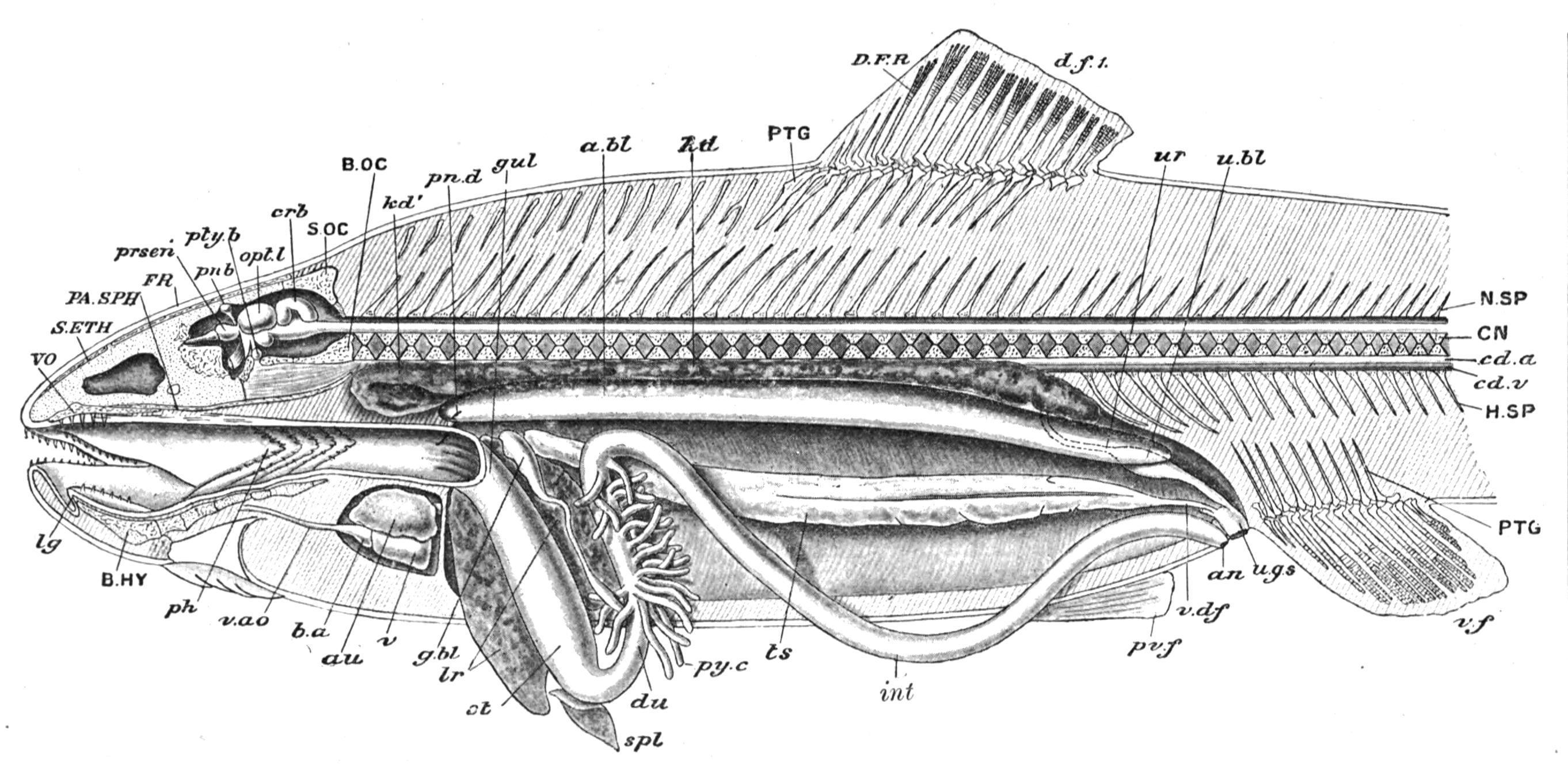
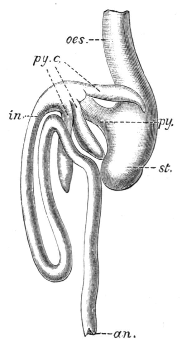
FIG. 154. Dissection of a male Teleost (Salmo fario) from the left side. a.bl, Air-bladder opened; an, anus; au, auricle; b.a, bulbus aortae; B.HY, basi-hyal; B.OC, basioccipital; cd.a, caudal artery; cd.v, caudal vein; CN, centrum; crb, cerebellum; d.f.1, first dorsal fin; D.F.R, dermal fin-rays; du, duodenum or anterior segment of the intestine; FR, frontal; g.bl, gall-bladder; gul, oesophagus or gullet; H.SP, haemal spine; int, intestine; kd, kidney; kd′, "headkidney"; lg, tongue; lr, liver; N.SP, neural spine; opt.l, optic lobes; PA.SPH, parasphenoid; ph, pharynx; pn.b, pineal body; pn.d, bristle passed into ductus pneumaticus; prsen, prosencephalon; pty.b, pituitary body; PTG, pterygiophores, or radial elements of dorsal and ventral fins; pv.f, pelvic fin; py.c, pyloric caeca; S.ETH, supra-ethmoid; S.OC, supra-occipital; spl, spleen; st, stomach; ts, testis; u.bl, urinary bladder; u.g.s, urino-genital sinus and its external aperture; ur, ureter or kidney-duct; v, ventricle; v.ao, ventral aorta; v.df, vas deferens; v.f, ventral fin; VO, vomer. (From Parker and Haswell.)
In the remaining Fishes the degree of convolution varies within rather wide limits. The oesophagus is usually straight and wide, but in Lutodeira, among Teleosts, it is long and even convoluted, and in the Plectognath Teleosts it gives off a large sac-like outgrowth ("airsac"), which extends anteriorly as far as the head, and posteriorly to the beginning of the tail, and communicates with the oesophagus by two apertures. The stomach may be U-shaped with the concavity directed forwards, and consisting of a right limb passing backwards from the oesophagus, and a left limb curving forwards to its junction with the intestine (Fig. 153). In such instances as these the stomach and the adjacent section of the intestine describe a characteristic
siphonal curve. In certain other Fishes (Fig. 160), the oesophageal portion of the stomach terminates behind in a tubular or sac-like dilatation at some distance posterior to the laterally situated pylorus, which indicates the origin of the intestine. The intestine is straight, or nearly so, in Elasmobranchs, Crossopterygii, and Dipnoi, and also in a few Teleosts; but sometimes, and very generally in Teleosts, it is more or less convoluted, notably in some of the Mugilidae, and in the Loricariidae, where, as in Plecostomus, it is disposed in numerous spiral coils like a watch-spring. The terminal portion of the intestine or rectum either opens into a cloaca, which also receives the urinary and genital ducts, as in Elasmobranchs (Fig. 153), and Dipnoi (Fig. 155, A), or opens externally by an anus, situated in front of the separate or united urinogenital ducts, as is the case with all the remaining groups of Fishes (Fig. 154). The cloacal aperture is invariably situated near the junction of the caudal and trunk regions, and as a rule is median in position, rarely, as in the Dipnoi, displaced to the right or left of the middle line; but the anus differs greatly in position, sometimes retaining its primitive position at the hinder end of the trunk, as in the Holocephali, Chondrostei, Crossopterygii, Holostei, and many Teleosts, or occupying almost any position between that point and, as in the "Electric Eels" (Gymnotidae), the ventral surface of the throat (Fig. 351.)

FIG. 155. A, alimentary canal and liver of a female Protopterus, from the left side. Part of the left wall of the stomach and intestine, and the peritoneal investment of the spleen have been removed. a.p, Abdominal pore; b.d, bile-duct; b.ent, Bursa Entiana; cl, cloaca; cl.ap, cloacal aperture; cl.c, caecum cloacae; c.m.a, coeliaco-mesenteric artery; cy.d, bile duct; k.d, kidney duct; m.a, mesenteric arteries; od, oviduct; pt.c, post-caval vein or inferior vena cava; p.v, portal vein; the other reference letters as in B. (From Newton Parker.) B, viscera of an adult female Lepidosteus, ventral view. The oesophagus, the commencement of the intestine and the rectum have been laid open. ab, air-bladder; an, anus; b.d, intestinal aperture of the bile-duct; g.b, gall-bladder; gl, oesophageal aperture of the air-bladder; h.d, hepatic duct; l, liver; oes, oesophagus; py, pylorus; py.c; pyloric caeca; py.c′, the four intestinal orifices of the pyloric caeca; r, rectum; s, spleen; sp.v, spiral valve; st, stomach. (From Balfour and Newton Parker.)
FIG. 156. Transverse section of a Fish, diagrammatic. cn, Centrum; coel, coelome; d.a, dorsal aorta; d.f, dorsal fin; d.m, dorsal muscles; d.ms, dorsal mesentery; f.r, fin ray; gon, gonad; int, intestine; l.v, lateral vein; msn, mesonephros; msn.d, mesonephric duct; n.a, neural arch; p, parietal layer of the peritoneum; p′, visceral layer; p.c.v, posterior cardinal vein; pn.d, Müllerian duct; r, ventral rib; r′, dorsal rib; sp.c, spinal cord; t.p, transverse process; v.m, ventral muscles; v.ms, ventral mesentery. (Modified, after Parker and Haswell.)
The whole length of the alimentary canal from the oesophagus to the rectum is invested externally by the visceral layer of the peritoneum (Fig. 156), which histologically consists of a stratum of connective tissue, supporting on its free surface an epithelial stratum (coelomic epithelium). Primarily, the investing peritoneum is continued both dorsally and ventrally into bilaminar suspensory folds, the dorsal and ventral mesenteries (d.ms, v.ms), which extend to the mid-dorsal or mid-ventral line of the abdominal cavity. The two layers then separate and become continuous with the parietal layer of the peritoneum lining the whole of the inner surface of the body-wall. Embryologically, the two mesenteries owe their formation to the fusion above and below the mesenteron of the contiguous walls of two laterally situated and primitively distinct coelomic cavities. The dorsal mesentery in the adult is occasionally complete, as in the Myxinoid Cyclostomata and in the Elasmobranch Hypnos subnigrum,[230] and also in some Dipnoi and in a few Teleosts, but much more frequently it is reduced by absorption to anterior and posterior remnants, or to a series of isolated bands, or even, as in the Lamprey (Petromyzon), to a few filaments accompanying the intestinal blood-vessels. The ventral mesentery, on the contrary, is rarely present, and if present is never complete. In Lepidosteus[231]
a ventral mesentery is said to be present in connexion with that part of the intestine which contains the spiral valve. In Protopterus,[232] and also in Neoceratodus,[233] there is a well-developed ventral mesentery in relation with the greater part of the length of the intestine, although in the former Dipnoid its continuity is interrupted by one or two vacuities, and in the latter the mesentery is incomplete posteriorly. A ventral mesentery is also present in the intestinal region of some of the Muraenidae among Teleosts.[234]

FIG. 157. Transverse section through a portion of the wall of the intestine, combined from the condition seen in both the higher and the lower Vertebrata. Semi-diagrammatic. a.c, Epithelial cells in the amoeboid state; b.v, blood-vessels; c.m, circular muscular layer; g, one of Lieberkühn's glands in the higher Vertebrates; i.ep, intestinal epithelium; l, leucocytes; l′, leucocytes in the intestinal epithelium; l.f, lymph follicles; l.m, longitudinal muscular layer; lym, lymphatic vessels; p, visceral layer of the peritoneum; sm, the submucosa; v, villi of the higher Vertebrates. (From Wiedersheim.)
Internal to its peritoneal investment the wall of the alimentary canal consists in succession from without inwards of (1), a muscular coat, (2) the submucosa, and (3) an epithelial stratum or mucous membrane, the first two of these layers, with the addition of the peritoneum, being derivatives of the inner or splanchnic portion of the embryonic mesoblast.[235]
Excluding the oesophagus, where the muscular coat is mainly composed of striated fibres, the musculature of the alimentary canal usually consists solely of non-striated, spindle-shaped fibres disposed in two layers, an external stratum of longitudinally arranged fibres, and an inner stratum of circularly disposed fibres (Fig. 157), with the addition, in the stomach, of an oblique layer between the two. In the oesophagus the reverse arrangement may exist, the circular layer being external and the longitudinal internal. The muscular coat varies considerably in thickness in different regions and in different Fishes, and in the Cyclostomata, the Holocephali, some Teleosts, and the Dipnoi may be very feebly developed, or even entirely absent, as in the intestine of the HagFish (Myxine). In the Gillaroo Trout (Salmostomachicus),[236] on the contrary, the distal section of the siphonal stomach has its musculature unusually thickened, so as to form an incipient gizzard for the crushing of the shells of the freshwater Molluscs on which the Fish feeds. In some of the Mullets (Mugilidae),[237] a true gizzard is developed by the enormous thickening of the muscular coat of the caecal stomach, the cavity of which, in consequence, is reduced to a mere vertical fissure, and is lined by an exceptionally thick, horny epithelium.
There are a few exceptions to the rule that the muscular fibres are of the non-striated variety. Thus in some Teleosts, as in the Tench (Tincavulgaris), striated fibres are continued from the oesophagus into the walls of the stomach and intestine, and there form an outer longitudinal and an inner circular layer, situated externally to the corresponding layers of the non-striated stratum. In the Siluroid, Amiurus, the striated fibres of the outer circular layer of the oesophagus are continued, although but sparsely, into the inner circular layer of the stomach.
The submucosa (Fig. 157) lies between the muscular layer externally and the epithelial lining internally, and is characteristically developed
in the stomach, and even more so in the intestine. Histologically, it consists of a framework of connective tissue, enclosing in its meshes masses of leucocytes (lymphoid tissue), some of which are amoeboid and migratory, and may even be found between the cells of the intestinal epithelium (including in some instances the cloacal epithelium), probably actively participating in the transmission of food material from the alimentary canal to the lymphatics and bloodvessels; while other and somewhat similar, but larger, leucocytes (phagocytes), are concerned with the elimination of waste substances or noxious micro-organisms. In addition to the diffused lymphoid tissue of the submucosa, special rounded or oval, and sometimes encapsuled, masses of this tissue (lymph follicles) are common in the intestinal wall (Fig. 157) of Acipenser, the Dipnoi and some Elasmobranchs, and are perhaps the only representatives in Fishes of the solitary follicles or "Peyer's patches" of the higher Vertebrates. A mass of lymphoid tissue exists in the axis of the spiral valve of Acipenser, which has been compared with a similarly situated structure in Lepidosiren.[238] In some Elasmobranchs a large lymphoid organ is imbedded in the submucosa of the oesophageal wall, while a local thickening of the tissue is met with in the pyloric sphincter. Protopterus is remarkable among Vertebrates for the extraordinary development of lymphoid tissue,[239] which, apart from its distribution in the submucosa, is abundantly present between the longitudinal and circular muscle layers, and the peritoneal and muscular coats of the intestine.
In addition to the lymphoid tissue the submucosa contains nonstriated muscle cells and plexuses of capillary blood-vessels, which in certain Loaches (e.g. Misgurnus), where intestinal respiration occurs, extend between the cells of the intestinal epithelium. A network of lymphatic spaces or vessels surrounds the blood-vessels. In some Elasmobranchs the small arteries of the submucosa of the stomach are provided with singular sphincter muscles, which occasionally encircle both the artery and the corresponding vein.[240]
The lining epithelium differs considerably in character in different portions of the alimentary canal. The epithelium of the mouth, pharynx, and anterior section of the oesophagus is often squamous and is succeeded in the hinder part of the oesophagus, and in the stomach and intestine, by a columnar epithelium. As a rule the epithelium of the rectum is also columnar, but in Elasmobranchs it may become squamous. Goblet cells are of very frequent occurrence throughout the whole length of the alimentary canal, from the mouth to the rectum inclusive, interspersed between the superficial epithelial cells; in the same position in the intestine migratory leucocytes have been found. The primitive ciliation of the Vertebrate alimentary canal is retained to a greater or less extent in many Fishes, and is sometimes, but not always, associated with a feeble development of the musculature. In the larval form of Petromyzon (Ammocoetes), the whole canal is ciliated except the pharynx and rectum; but in the adult ciliation is retained only in places which gradually become fewer as the rectum is approached. In the Myxinoids, however, cilia are said to be absent.
In the Dipnoi (e.g. Protopterus) the epithelium of the stomach and intestine is largely ciliated, but in Elasmobranchs, ciliation is usually restricted to the posterior portion of the oesophagus and the edge of the spiral valve. Among the more generalised Teleostomi (e.g. Acipenser, Lepidosteus, Amia), the oesophagus, stomach, and intestine may be ciliated, but to an extent which varies in different genera. The pyloric appendages, when present, are also more or less extensively ciliated. In Teleosts, however, the recorded instances of ciliation are relatively rare. Nevertheless, ciliated epithelium has been found in the intestine of a few species (e.g. Rhombus aculeatus and Syngnathus acus), and also in the pyloric appendages; in the stomach (e.g. Perca and Esox), and even in the oesophagus (e.g. Perca).
The mucous membrane, including the submucosa, is frequently developed into variously arranged ingrowths projecting into the
lumen of the alimentary canal; these are generally of the nature of longitudinal or transverse ridges, or a combination of the two, giving rise to retiform structures. The simple longitudinal folds, which are sometimes found in the oesophagus, stomach, and rectum, often disappear on distension, and probably merely provide for the enlargement of these cavities during the deglutition of relatively large prey, or for the accumulation of faeces. On the other hand, the permanent and often complicated folds of the intestinal mucous membrane are probably related to an increase in the secretive or absorptive area of this portion of the alimentary canal. In the stomach the mucous membrane is usually smooth, rarely, as in the "Electric Eel" (Gymnotus), reticulate. In the intestine the folds assume a highly characteristic and often complicated disposition. [241] In the Cyclostomata the folds are simple and longitudinally arranged. In Elasmobranchs (Fig. 158, A), obliquely transverse folds are present in addition, and, uniting with the longitudinal ridges, bound linear depressions.
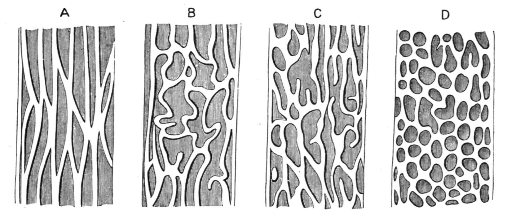
FIG. 158. The intestinal mucous membrane of different Fishes, to show the transition from simple longitudinal and transverse folds to crypts. A, Of an Elasmobranch; B, C, and D, of various Teleosts. (After Wiedersheim.)
In various Teleostomi (Fig. 158, B, C, D), the union of the two series of folds becomes more or less retiform, and the network of intersecting ridges bounds a series of deep tubular crypts which appear to penetrate to a considerable distance into the intestinal wall, and possibly foreshadow the characteristic Lieberkühn's glands of Mammalia. Crypts may also be found in the stomach, where they receive the apertures of the gastric glands, as in Amiurus, but more
usually they are restricted to the intestine. In the Dipnoi (e.g. Protopterus) the mucous membrane of the stomach, and—excluding the Bursa Entiana where a number of oblique folds are present—of the intestine also, is, on the contrary, perfectly smooth.
In addition to transverse and longitudinal folds the mucous membrane of the various sections of the alimentary canal is often developed into outgrowths which are more or less linear.[242] In the oesophagus these may be papilliform, as in Box and Caesio; obtuse in Acipenser, hard and almost spine-like in species of Rhombus; or in the form of pyramidal retroverted processes with jagged or fringed edges, as in the Spiny Dog-Fish (Acanthiasvulgaris). In the Basking Shark (Selache) similar processes are present, which, near the stomach, become unusually long and branched, so that the entrance to that cavity is surrounded by a series of backwardly-directed arborescent tufts. Peculiar papillose or tag-like processes of the mucous membrane are frequently present on the spiral valve of Elasmobranchs, in the intestine of such Teleosts as Balistes, Mugil and some Pleuronectidae, and also in the rectum of Rhombus maximus.
Of all the outgrowths from the mucous membrane of the alimentary canal the so-called "spiral valve" of the Cyclostomata, Elasmobranchs, Holocephali, Chondrostei, Crossopterygii, Amiidae, Lepidosteidae and Dipnoi is the most characteristic. The first appearance of this structure was probably in the form of a straight longitudinal fold or ridge projecting into the cavity of the intestine, similar, perhaps, to the typhlosole of many Invertebrata. This primitive condition is not retained in any existing Fishes, although it may be closely approached in the larval Cyclostome (Ammocoetes), and is perhaps also indicated in the straight anterior portion of the spiral valve of Polypterus. Absent altogether in the Myxinoids, the valve is represented in its simplest condition, as in certain other Cyclostomata (e.g. Petromyzon), by a ridge of mucous membrane which commences anteriorly on the dorsal side, and, after describing
a partial spiral as it passes backwards, terminates posteriorly on the ventral side, the width of the valve not exceeding half the diameter of the intestine. This simple type of valve is repeated in embryo Elasmobranchs, but in the adults of these Fishes the valve becomes much more complicated, and exhibits a wide range of structural variation. The increased complexity of the valve seems to depend on several factors, the effect of which, in different Elasmobranchs, is best studied in a series of valves of progressively higher differentiation.[243]
In a hypothetical simple type of valve, easily derivable from the more primitive type of Petromyzon, it may be conceived that, while not exceeding in width the semi-diameter of the intestine, the valve becomes disposed in several complete and more or less closely approximated spiral turns, the free edge of the valve being on the same level as its attached margin, and leaving an open axial canal along the centre of the gut. The nearest approach to this hypothetical type, which has been compared, not inaptly, to un escalier tournant sans noyau, is perhaps to be found in the Thresher-Shark (Alopeciasvulpes).
The structure of the more complicated spiral valves of other Elasmobranchs are well illustrated within the limits of the single genus Raia.
In one specimen of Raia sp. (Fig. 159, A) the last four coils of the valve are similar to those of the hypothetical type, but the more anterior ones, owing to the greater width of the valve, which here exceeds the semi-diameter of the intestine, have their free margins deflected downwards, while that portion of the valve which forms the first half turn is coiled inwards upon itself, so as to form a hollow cone, open dorsally, and having its apex directed forwards. In other examples a further modification is introduced by the increasing width of the valve, which now, throughout its whole length, equals the
semi-diameter of the intestine; and by the formation of an axial columella by the thickened free edge of the valve, which is traversed by a central band of unstriped muscle, as well as by the intraintestinal artery and vein, and takes the place of the central canal of the preceding types. The valve is, however, still regular, and its free margin remains on the same level as the corresponding portion of the attached edge. In other specimens, again, additional complications are introduced by a still further increase in the width of the valve, which now exceeds, often considerably, the semidiameter of the intestine, and the consequent deflection of the free edge of the valve either forwards or backwards (C and D). As shown in C the valve, in consequence of the backward deflection of its free margin, presents the appearance of a nest of imperfect truncated cones with their apices directed backwards, the successive cones adhering so closely to one another that they combine to form a central conical chamber with a spirally disposed cavity winding round it. In D, on the contrary, the free edge of the valve is deflected forwards, so that, as in C, a nest of cones is formed, but the apices of the successive cones are directed forwards instead of backwards. Notwithstanding these variations in the structure of the valve as a whole, the first coil or half coil nearly always resembles that described in A.

FIG. 159. Examples of various types of the spiral valve in Elasmobranchs. A, B, C, and D in specimens of Raia spp.; E, in Sphyrna malleus. A, B, and D represents longitudinal sections of
the intestine, the ventral portion of the valve being removed. In C successive portions of the ventral wall of the intestine have been cut out. In E the intestine has been opened along the mid-ventral line and its wall reflected to the right and left; the ventral portion of each coil of the "scroll" valve has been removed. In most of the figures the pylorus is shown in the upper part, and the "rectal" gland in the lower. (From T. Jeffery Parker.)
It is obvious that the structure of the valve varies considerably within the limits of the genus, and it may be added that various intermediate types of structure occur between A and B, A and C, and A and D. The individual variations are perhaps even more remarkable, and appear to be quite independent of age and sex. By way of example it may be mentioned that valves approximating to one or other of those represented by C and D occur in different individuals of Raiamaculataof the same sex and similar in size, even in young specimens not more than three inches in length.
As regards other Elasmobranchs, the common Dog-Fish (Scyllium canicula)[244] has a well-developed spiral valve disposed in twelve coils, which structurally represents a more highly developed example of the type D. The existence of considerable individual variation is nevertheless indicated by the fact that in one specimen examined the valve was intermediate between C and D, five of the eight cones projecting forwards and three backwards. In a specimen of Notidanus sp.[245] there were as many as twenty coils, which in disposition were intermediate between B and C, approximating, however, more nearly to B. In a specimen of the Port Jackson Shark (Heterodontus)[246] the valve had eight coils, and in structure was also intermediate between B and C, but approached more nearly to C. Some of the Hammer-headed Sharks (e.g. Sphyrna malleus)[247] possess a type of spiral valve which differs considerably from any of those hitherto described, and is termed a "scroll" valve (Fig. 159, E). The attached edge of the valve pursues a straight longitudinal course, or at any rate only describes a half turn and back again in
passing from the pyloric to the cloacal extremity of the gut. In the middle of its course the width of the valve is about equal to twothirds of its length, but towards either extremity it gradually diminishes until the free and attached margins meet. The valve thus constituted is rolled upon itself from left to right, the successive coils being comparable to a series of cylinders placed one inside the other, and becoming gradually larger both in length and diameter from within outwards. A similar valve is present in some of the Carchariidae.
In the Holocephali (e.g. Chimaera monstrosa)[248] the valve describes only three and a half coils, and is further remarkable in that the attached margin, for a considerable portion of its extent, does not form a regular spiral but describes only a slightly sinuous course. Posteriorly, the valve is more normal, and consists of about two cones with their apices directed forwards.
In the Dipnoi the spiral valve is well developed, and in Neoceratodus[249] describes nine coils, and in Protopterus[250] six or seven. The structure of the valve in the latter Dipnoid resembles that of Scylliumcanicula, except for the smaller number of cones.
In the more generalised Teleostomi the valve is best developed in the Sturgeon (Acipenser) and in Polypterus. In the former[251] the valve is restricted to the posterior half of the total length of the intestine, often extending to within an inch of the anal aperture, and describing in its backward course about seven or eight coils. The width of the valve is about equal to the semi-diameter of the intestine, and the thickened free margin forms a well-marked axial columella, round which the cavity of the gut winds, as in the type B, except that the spiral is a more open one. In Polypterus the valve begins close to the solitary pyloric caecum, and for some distance pursues a straight longitudinal course, but eventually forms a few spiral coils, ceasing, however, at a considerable distance from the
anus. The evidence afforded by petrified faeces or "coprolites" proves that certain extinct Crossopterygii (e.g. Macropoma, Megalichthys), like their living representative, Polypterus, possessed a spiral valve.[252] In Amiaand Lepidosteus[253] the valve is almost vestigial, being restricted to the terminal portion of the intestine, and is somewhat variable as to the precise number of its coils. In Amia there are nearly four coils, extending over 3 cm., that is less than a tenth of the total length of the intestine, but in some specimens the coils do not exceed two and a half or three in number.
Lepidosteus[254] has a still shorter valve which, in specimens of 7-10 cm. in length, may not consist of more than three and a half coils, and in much larger specimens may be reduced to less than two coils, a variation which suggests that a reduction takes place in the number of coils as the fish increases in age and size. The structure of the valve in the three last-mentioned genera resembles that described in Acipenser, and in none of them does the width of the valve so far exceed the semi-diameter of the intestine as, by forward or backward deflection, to give rise to the highly characteristic cones of Elasmobranchs and Dipnoi.
In the more specialised Teleostomi (Teleostei) the spiral valve is wholly wanting, except perhaps as a vestigial structure in certain Clupeoids, as, for example, Chirocentrus,[255] and possibly also in some Salmonidae.[256]
From what has been said as to the structure of the spiral valve in the different groups of Fishes, it may be concluded that the valve most nearly retains its primitive condition in the Cyclostomata; attains its maximum development in the Elasmobranchs, especially in the Notidanidae, and shows no indication of degeneration in the Dipnoi. In the Holocephali and the lower Teleostomi, on the other hand, the valve exhibits various stages of retrogressive modification, and in the Teleosts is either absent altogether or persists only as a vestigial structure in a very few species.
From a physiological point of view the object of the spiral valve is to increase the absorptive inner surface of the intestine,[257] but, from what has been said as to the structural variability of the valve, it is obvious that its efficacy from a functional standpoint must be equally variable. The value of the valve as an absorptive mechanism necessarily depends on the area of absorption-surface which it provides, as well as on the degree of resistance which it offers to the passage of food material along the cavity of the intestine. These factors will in turn depend on the number of coils, on the width of the valve, and on the extent to which its free margin is deflected in forming the series of cones, but these again are precisely the structural features which are most liable to variation. The total absorption area in the four types of valve characteristic of the genus Raia has been calculated, and may be expressed in square centimetres as follows:—A, 136.64; B, 143.82; C, 254.3; and D, 276.7.[258] Hence as regards mere absorption area a spiral valve of the type D has twice the extent of a valve of the type A, and if, in addition, account be taken of the retardation of the food due to the increased obstruction offered by the columella and cones in D, it is clear that the difference in physiological value between the two types must be far more considerable than is indicated by a comparison of their relative superficial areas alone.
The evolution of the spiral valve was probably due to the necessity of increasing the absorptive area of an almost straight unconvoluted intestine, a result which in other animals is often obtained by an increase in the length and concurrent convolution of the intestine itself. Any attempt to correlate the variations in the degree of perfection or imperfection of the valve considered as an absorptive mechanism with any special variations in the nature or quality of the food is, however, a very difficult problem, and a satisfactory explanation has yet to be found. The difficulty, moreover, is increased by the fact that the majority of Fishes with a spiral valve are mainly carnivorous; the Elasmobranchs, in which this structure is at the same time most highly developed and most variable,
exclusively so. On the other hand, the term "carnivorous" covers a multiplicity of minor differences in the nature and relative digestibility of different forms of animal food, and it is quite possible that it is with differences of this kind that the specific or individual variations in the development of the spiral valve are associated. The absence of the valve in the variously nourished Teleosts, save perhaps as a vestige in one or two, is also difficult to account for, although it is not improbable that compensating structural modifications exist in this group. As a rule, the intestine is much more convoluted in these Fishes, but to an extent which varies greatly in different species, while the characteristic pyloric caeca and the spiral valve appear to a certain extent to be developed in inverse proportion to one another.
The Glands.
The glands associated with the alimentary canal in different Fishes are (1) the gastric glands, (2) the liver, (3) the pancreas, (4) the pyloric appendages, and (5) the "rectal" gland.
Oral salivary glands are wanting in all Fishes, the only secretory structures in the mouth being numerous mucus-secreting goblet cells, which here, as elsewhere throughout the alimentary canal, are intermixed with the ordinary epithelial cells.
The
Gastric
Glands.
—The Cyclostomata and Dipnoi do not possess any specially differentiated gastric glands, and it is probable that in these Fishes the secretion of the digestive fluids is effected by the ordinary lining epithelium of the stomach or intestine, or both. In the remaining groups gastric glands are generally present in the form of simple caecal structures embedded in the submucosa and opening on the surface of the mucous membrane into the cavity of the stomach. The glands differ in different Fishes in the character of
their lining epithelium and in the extent to which their component cells are differentiated from the epithelium of the stomach. There does not appear, however, to be any distinction into "central" (pepsin-forming) and "parietal" (acid-secreting) cells, as is the case in the higher Vertebrata. Towards the pyloric end of the stomach the true gastric glands are often replaced by mucous glands. There are, nevertheless, not a few Teleosts in which special gastric glands are absent, as, for example, Syngnathus acus, and several species of Cyprinidae, Labridae, and Blenniidae, etc. In at least two genera (Gastrosteus and Cobitis), belonging to widely different families, gastric glands are present in certain species but absent in others. As suggested by Edinger,[259] the absence of these glands may possibly be due to degeneration.
It may be remarked that the formation of such digestive ferments as pepsin and trypsin, which are associated with the stomach and pancreas respectively, in the higher Vertebrates, is not nearly so strictly localised in Cyclostomes and Fishes. So far from peptic digestion being limited to the stomach, it may take place in the pharynx, stomach, and intestine of Ammocoetes, and in some Elasmobranchs (e.g. Scyllium), and in such Teleosts as the Pike, Eel, and Carp, the peptic region extends from the stomach for some distance along the intestine, while trypsin has been obtained from the mucous membrane of the stomach, intestine and pyloric caeca, as well as from the pancreas.[260]
Intestinal glands analogous to the glands of Lieberkühn in the higher Vertebrates seem to be entirely wanting in Fishes, unless represented by the sac-like or tubular crypts which are so generally present in the Teleostomi.
The Liver.—Phylogenetically the oldest gland in connexion with the Vertebrate alimentary canal, and in size by far the largest, the liver arises as a caecal outgrowth from the embryonic mesenteron, and in
this primitive stage recapitulates a condition which is retained throughout life in Amphioxus. By the subsequent division and branching of this outgrowth the massive compound tubular gland of the adult Fish is eventually formed.
The liver of Fishes (Figs. 153, 154) is very variable in size, shape, colour, and degree of lobulation. Anteriorly, it is usually moulded to the posterior face of the transverse septum between the pericardial and abdominal portions of the coelom, and from thence extends backwards in the abdominal cavity to a varying distance, in some Sharks as far as the cloaca. Externally, the gland is invested by the peritoneum, which extends on to it from the pericardial septum and forms a suspensory fold, and also from the oesophagus and stomach. The shape of the liver usually bears some relation to that of the body, being, for example, longest in the Eels and broadest in the Rays. In the great majority of Fishes the liver is bilobed, consisting of two sub-equal lateral lobes, disposed longitudinally and confluent anteriorly for a portion of their extent. From this normal type there are a few minor variations.[261] In Petromyzon, Lepidosteus (Fig. 155, B), and a few Teleosts (e.g. the Gymnodontes, Lophobranchii, and some Salmonidae) the liver is unilobed. In the Myxinoids and in the Dipnoi (e.g. Protopterus), the organ is bilobed, but the small anterior lobe lies immediately in front of the much larger posterior lobe, with the gall-bladder between the two (Fig. 155, A). In some Teleosts (e.g. Scomber), the liver is trilobed. A gall-bladder is invariably present in either the larval or adult Cyclostomata, in the Chrondrostei, Holostei, Crossopterygii and Dipnoi, and generally also in Elasmobranchs and Teleosts. In the Elasmobranchs it is rarely entirely wanting, as in Sphyrnaand Pristis, and in the Teleosts in some of the Gurnards (Trigla). The gallbladder and bile-duct of Petromyzon fluviatilis atrophy after the metamorphosis which follows the larval Ammocoetes stage, but in Petromyzonmarinusthe duct, although usually absent, is sometimes retained. In the Ammocoetes the epithelium lining the gall-bladder is ciliated. In some Fishes, as, for example, in many Elasmobranchs,
the gall-bladder is more or less completely embedded in the substance of the liver; in others, as in most Teleostomi, the organ is quite distinct from the gland (Fig. 154).
A simple arrangement of the ducts from the liver and gall-bladder is that found in the common Dog-Fish (Scyllium canicula). In this Elasmobranch a cystic duct leaves the gall-bladder, and, after receiving several hepatic ducts from the lobes of the liver, becomes the bile-duct and opens into the commencement of the intestine. In the Myxinoids and in the Dipnoi (e.g. Protopterus), there are but two hepatic ducts, one from each lobe of the liver; these unite and then meet the cystic duct to form the bile-duct (Fig. 155, A). The number of hepatic ducts may, however, be considerably increased, as, for example, in the Siluroid Amiurus,[262] where 8-10 separate ducts join the cystic duct. In a few instances one of the hepatic ducts opens directly into the intestine, independently of that which unites with the cystic duct in forming the bile-duct. In the Dipnoi (e.g. Protopterus),[263] and in some Teleostomi (e.g. Lepidosteus),[264] the bile-duct receives the duct from the pancreas before opening into the intestine.
The Pancreas.—In the Cyclostomes (e.g. Petromyzon, Bdellostoma, Myxine) a rudimentary pancreas is apparently present, but the evidence as to its identity is not wholly conclusive. A well-developed pancreas occurs in Elasmobranchs, in at least one of the Dipnoi, and probably in most Teleostomi.[265]
In Elasmobranchs the pancreas is a compact structure, uni- or bilobed, and entirely distinct from the liver. In Scyllium canicula (Fig. 153), the bilobed gland lies in the angle between the distal limb of the stomach and the adjacent portion of the intestine, and from the smaller of its two lobes the duct issues to pass to its intestinal aperture near the commencement of the spiral valve. In most of the Teleostomi in which its existence has hitherto been recorded, the
pancreas is a singularly diffuse gland; and usually a considerable portion, or even the whole of it, is embedded in the substance of the liver, its lobules accompanying the ramifications of the hepatic artery and duct, and the portal vein. The pancreatic duct usually opens into the intestine near the aperture of the bile duct (e.g. Amiurus); sometimes the two ducts open on the apex of a common papilla (e.g. Acipenser and Amia), or by their union form a common duct (e.g. Lepidosteus). Among the Dipnoi a well-developed pancreas is present in Protopterus,[266] embedded in the wall of the stomach and intestine, internal to the peritoneal investment of these organs, and extending even into the first fold of the spiral valve. The gland is traversed by fine ductules which unite together and open into the bile-duct just before the latter enters the intestine. In the remaining Dipnoi the existence of a pancreas has yet to be ascertained. Developmentally, the pancreas resembles the liver, and, histologically, is very similar to that of the higher Vertebrates, consisting of terminal glandular alveoli continuous with intermediary tubular portions, and eventually with the finer ductules, which, by their union, form the main efferent duct.
The Pyloric Caeca.—These structures are caecal outgrowths from the intestine, and are situated close to the pyloric extremity of the stomach and the intestinal apertures of the bile and pancreatic ducts. Wholly wanting in the Cyclostomata and Dipnoi, and, unless represented by a pair of caeca opening into the long, tubular, nonvalvate anterior portion of the intestine in the Greenland Shark (Laemargus borealis),[267] in the Elasmobranchs also, they are very generally present in the Teleostomi, although extremely variable both in number and arrangement in different families. In Amiathere is no trace of pyloric caeca. Polypterus has a single short caecum with a thick muscular wall. In Acipenser, Polyodon, and Lepidosteus, on the contrary, pyloric caeca are unusually well developed. In Acipenser the caeca are not only numerous, but are so connected together by connective tissue and blood-vessels, and so invested externally by the peritoneum, as to form a large, compact, gland-like
mass, communicating with the intestine by a single wide duct. In Polyodon the organ is essentially similar, but is lobed externally. In Lepidosteus (Fig. 155, B, py.c), the caeca are also very numerous, but relatively short, and, although united into a compact mass, open by four pit-like orifices into the intestinal cavity. In Teleosts the caeca are subject to extraordinary variations in number, size, and arrangement.[268] In some families, and even in groups of higher taxonomic value, they are entirely absent, as is the case with the Siluridae, Esocidae, Cyprinodontidae, Labridae, Plectognathi, and Lophobranchii. The "Sand-eel" (Ammodytes) has but a single caecum; the Turbot (Rhombus maximus) two, and other Pleuronectidae three to five; and the Perch (Perca), three (Fig. 160, py.c).
In other Teleosts, on the contrary, these structures are much more numerous. In Labrus labrax there are about 60, in the Whiting (Gadusmerlangus) 120, while in the Mackerel (Scomber scombrus) there are no fewer than 191. If few in number the caeca open separately into the intestine, but when numerous, more or fewer of them may unite to form a smaller number of efferent ducts, as in the Whiting, where four such ducts are formed. In some instances, as in the Tunny (Thunnus), the union of the caeca by connective tissue leads to the formation of a compact mass. As regards their arrangement, the caeca may either be disposed in a whorl round the intestine, as in the Whiting, or in a linear series, as in the Salmon (Salmo) and in some of the Clupeidae.
The mucous membrane lining the anterior pyloric caeca is often developed into a network of ridges, limiting crypt-like or tubular depressions; and not infrequently the epithelium is ciliated.
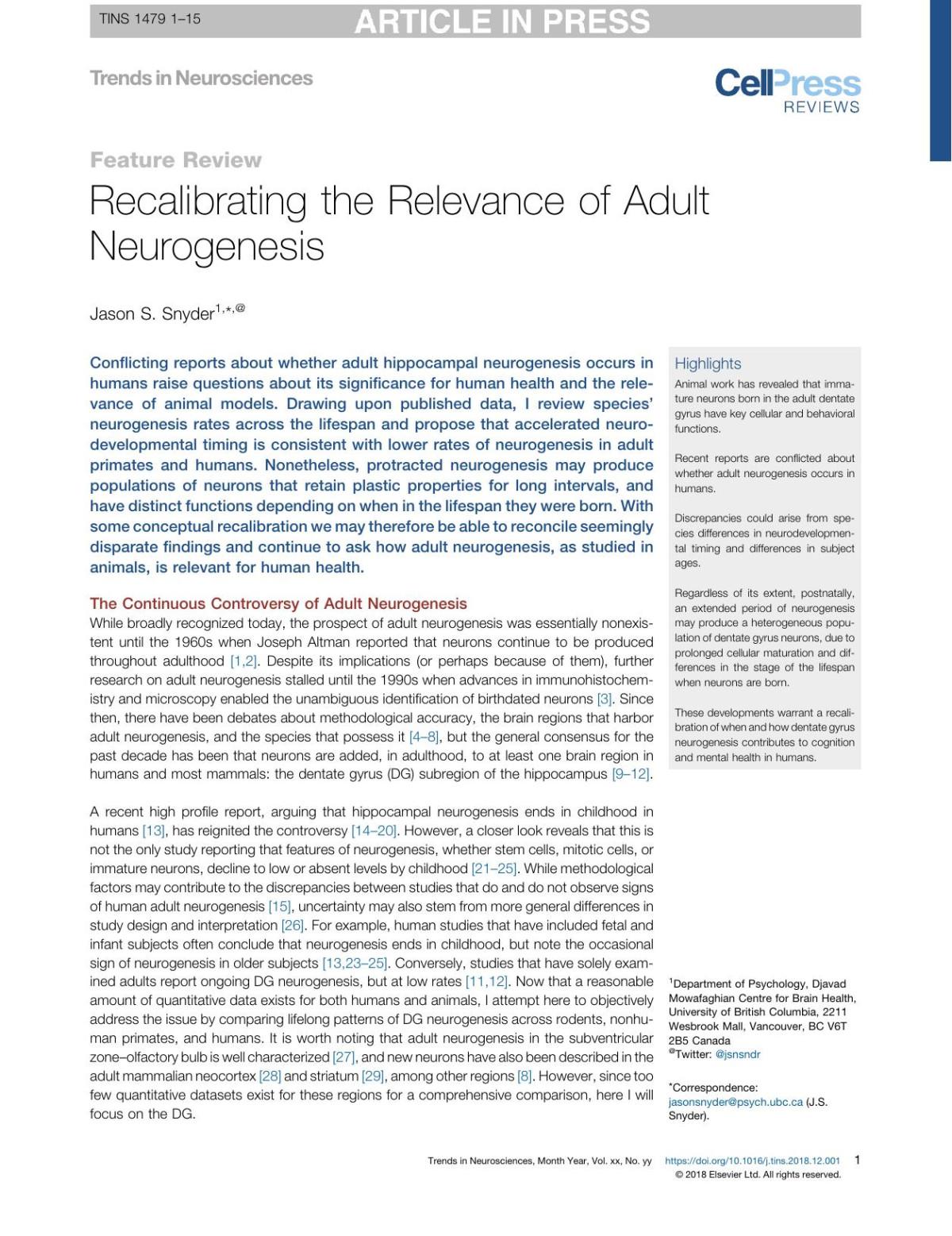
FIG. 160. The alimentary canal of a Perch (Perca). an, Anus; in, intestine; oes, oesophagus; py, pylorus; py.c, pyloric caeca; st, stomach. (After Wiedersheim.)
The precise function of these organs, whether digestive or absorptive, is still uncertain.[269] That they may be digestive is suggested by the presence of certain amylolytic and proteolytic enzymes, but this obvious conclusion is to some extent vitiated by the close proximity of these organs to the stomach, and more especially to the intestinal orifice of the pancreatic duct. It is by no means improbable, however, that the caeca are both digestive and absorptive organs. An attempt has been made to show that the pyloric caeca and the spiral valve vary inversely as regards the extent of their development in different groups of Fishes.[270] To some extent the reciprocal variation of these structures supports this view, but it is also evident that there are obvious objections to its unqualified acceptance. Thus, in some Teleostomi (e.g. Acipenser, Polyodon), exceptionally well-developed and numerous caeca and a spiral valve are both present. Amia with an almost vestigial spiral valve has no trace of pyloric caeca, and in Teleosts the absence of a spiral valve is associated with the complete suppression of the caeca in many large and important groups.
The Rectal Gland.—The "rectal" gland, or appendix digitiformis, is a small organ of unknown function with complex glandular walls, and a central duct opening dorsally into the terminal portion of the intestine.[271] The organ is generally present in Elasmobranchs (Fig. 153, rct.gl), in which group the intestinal orifice of its duct may
either be close to the termination of the spiral valve, or, as in Chlamydoselachus,[272] near the cloacal outlet of the gut. An apparent representative of the gland, the "caecum cloacae," is also present in the Dipnoi,[273] but communicates directly with the cloaca (Fig. 155, A, cl.c). The "rectal" gland is perhaps homologous with the intestinal caecum which is to be found in some Teleosts (e.g. Box vulgaris), and possibly also with the "caecum" (caecum coli), and its vermiform appendix in the higher Vertebrata.[274] The caecum cloacae, on the contrary, is morphologically a urogenital sinus, formed as a dilatation of the fused hinder portions of the mesonephric ducts, and probably comparable with the sperm sacs of male Elasmobranchs, and also with the urinary bladder of Teleostomes.[275]
CHAPTER X
THE RESPIRATORY ORGANS
The principal respiratory organs consist of a series of pairs of branchial clefts in the form of perforations in the side walls of the throat, which place the pharynx in free communication with the exterior. The first and most anterior of these clefts, the mandibulohyoid cleft or "spiracle," is situated between the mandibular and hyoid arches; the second, the hyo-branchial or hyoidean cleft, between the hyoid arch and the first branchial arch; and the remaining clefts between the succeeding branchial arches. On the anterior and posterior walls of more or fewer of the clefts highly vascular plate-like, or variously shaped filamentous outgrowths of their lining membrane are developed, which subserve the purpose of exposing the blood to the influence of the oxygen-containing water, and are termed branchial lamellae or "gills." In addition to their
usual respiratory organs, the gills, a few Fishes utilise the air-bladder either as a functional lung or as an oxygen reservoir, and in others accessory breathing organs of various kinds are developed.
The arrangement of the branchial clefts and the gills may be conveniently studied first in the Elasmobranchs. Excluding the spiracles, there are usually in this group (Fig. 161, A), five pairs of branchial clefts, but in certain primitive members of the group the number may be larger. Thus, in Notidanusgriseus (Hexanchus) and in Chlamydoselachus there are six, and in Notidanus cinereus (Heptanchus), seven clefts. The pharyngeal apertures of the clefts are relatively wide, but their external openings, which are freely exposed on the lateral surface of the head between the eye and the pectoral fin, are usually narrow and slit-like.

FIG. 161. A, Horizontal section through the head of an Elasmobranch; B, similar section of a Teleost (diagrammatic). b.c, Branchial cavity; b.l, branchial lamellae; c, coelom; e.b.a, external branchial aperture; hy.a, hyoid arch; hy.c, hyo-branchial cleft; l.s, interbranchial septum; n, nasal organ; oes, oesophagus; op, operculum; p.q, palato-quadrate cartilage; Ph, pharynx; sp, spiracle; s.ps, spiracular pseudobranch; 1-5, 1st to 5th branchial arches. (From Boas, slightly altered.)
The successive clefts are separated from one another by a series of inter-branchial septa, each of which consists of the lining membrane of two contiguous clefts and a median fibrous sheet; it is further strengthened on its pharyngeal margin by a branchial arch, and more externally by the fringe of cartilaginous rods (branchial rays)
with which the outer convex edge of each arch is provided. The anterior and posterior walls of each septum are produced into a number of outwardly-radiating vascular plates or folds (branchial lamellae or "gills"), which by their free edges project into the cavity of the cleft (Fig. 161, A). Although slightly free at their outer extremities, the lamellae do not extend so far as the external margin of the septum to which they are attached (Fig. 164, B). Each series of lamellae is termed a "hemibranch," and, from what has been said, it is obvious that each inter-branchial septum and its supporting branchial arch carry two hemibranchs, an anterior and a posterior, the two forming a complete biserial gill or "holobranch." The hyoid arch, however, has only a single hemibranch, viz. that pertaining to the anterior wall of the hyo-branchial cleft, and as the fifth or last cleft has a hemibranch only on its anterior wall, the fifth arch is gillless.[276] The spiracle is a vestigial cleft. At an early stage of embryonic growth it differs but little from its fellows, but subsequently degenerating it is represented in the adult by a tubular passage between the oral cavity and the exterior, which, however, is often complicated by the development of caecal outgrowths.[277] The anterior wall of the spiracle often retains a rudiment of a hemibranch in the shape of more or fewer vascular lamellae, which, as they are supplied with arterial blood, and not with venous blood like the ordinary gills, are said to form a mandibular or spiracular "pseudobranch." The spiracle varies greatly in size in different families, being largest in the Trygons and Torpedos, and very small, or even absent in the Lamnidae. Its pseudobranch is best developed in the Notidanidae, where it has the essential structure of a true hemibranch, and, as in other Elasmobranchs, but to a greater extent, probably aids in the additional aeration of the blood which is distributed to the eye and brain. The characteristic opercular covering of the external apertures of the gill-clefts in the Teleostomi and Dipnoi is wanting in Elasmobranchs. It is interesting to note, however, that in Chlamydoselachus[278] curious frilled cutaneous folds are developed as extensions of the outer edges of the interbranchial septa, as well as of the hyoid region, and, like a series of
incipient opercula, project backwards over the successive branchial clefts (Fig. 252).
While in many respects more primitive than in Elasmobranchs the branchial system of the Cyclostomata presents certain special and peculiar features. The branchial clefts assume the form of oval, antero-posteriorly flattened pouches or sacs, varying, however, in number, and in their mode of communicating with the exterior, in different genera. In the Lamprey (Petromyzon) there are seven pairs of obliquely-disposed gill-sacs opening externally by small rounded orifices, and by similar apertures, not directly into the pharynx, but into a branchial canal (Fig. 162, r.t), which underlies the oesophagus, and, while ending blindly behind the last pair of sacs, communicating in front with the oral cavity.[279] The first of the series of gill-sacs corresponds to the hyo-branchial or hyoidean cleft of Elasmobranchs and other Fishes. Spiracles are absent in the adult, but in the embryo are represented by pouch-like outgrowths of the hypoblast of the oral cavity, which subsequently undergo singular changes.[280] Thus, the outgrowths become converted into the lateral halves of a complete ciliated circum-oral groove, which is retained even in the Ammocoetes stage, and recalls the ciliated peripharyngeal ring of Ascidians. Another archaic feature is also to be noted in the continuity of the groove with a ciliated mid-dorsal pharyngeal ridge, which has been compared to the "dorsal lamina" of Ascidians, and to the equally characteristic hyperbranchial groove of Amphioxus.[281] Ventrally also, the lateral halves of the groove unite to form a single groove, which, after receiving the median aperture of the thyroid rudiment,[282] is continued backwards in the mid-ventral line of the pharyngeal wall as far as the last branchial arch. No trace of these ciliated structures is, however, to be met with in the adult.
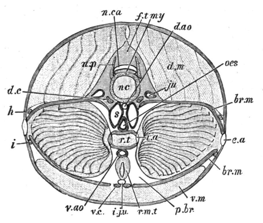
FIG. 162.—Petromyzon marinus. Transverse section through the branchial region (semi-diagrammatic). br.m, Branchial membrane; d.ao, dorsal aorta; d.c, dorsal cartilage of the branchial basket; d.m, dorsal muscles; e.a, external aperture of a gill-sac; f.t, fibrous tissue enclosing neural canal; h, i, lateral longitudinal cartilages of the branchial basket; i.a, internal aperture of a gillsac; i.ju, inferior jugular vein; ju, jugular vein (anterior cardinal); my, spinal cord; nc, notochord; n.ca, neural canal; n.p, neural process; oes, oesophagus; p.br, peri-branchial lymph sinus; r.m.t, retractor muscle of the tongue; r.t, respiratory tube or branchial canal; s, circum-oesophageal lymph sinus; v.ao, ventral aorta; v.c, ventral cartilage of branchial basket; v.m, ventral muscles. (From T. J. Parker.)
The branchial lamellae are represented by a series of vascular horizontal and parallel ridges radiating outwards along the roof, floor, and lateral walls of each gill-sac, and invested by an epithelium which is partially ciliated. The inter-branchial septa are much thicker than in Elasmobranchs, and include not only the walls of adjacent sacs and the branchial muscles, but also contain cavernous peribranchial lymph-sinuses. The cartilaginous branchial skeleton is situated wholly external to the gill-sacs, the so-called branchial arches lying between the external apertures of the sacs, and directly beneath the superficial skin, or, in other words, on the outer margins of the inter-branchial septa, and not on the inner, as is invariably the case with the branchial arches of Fishes.
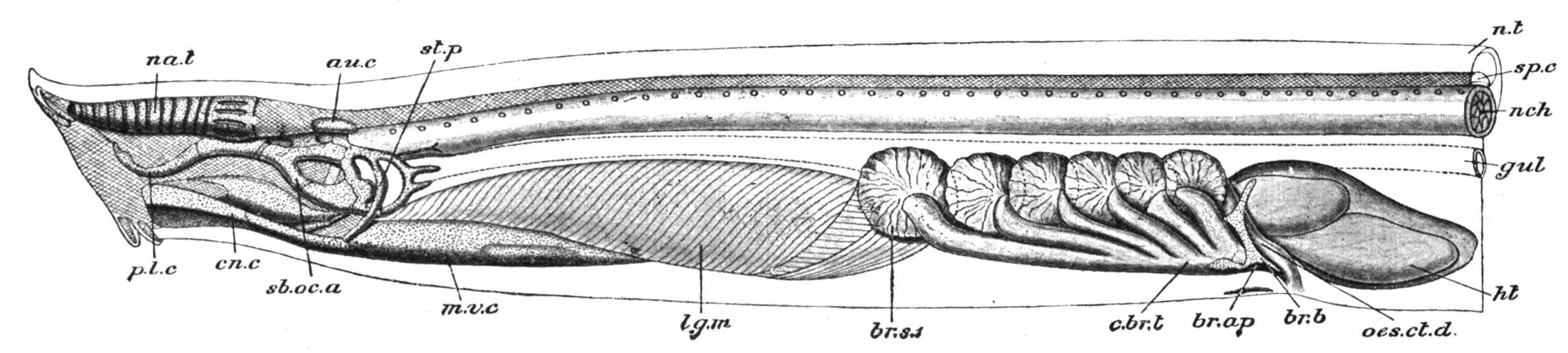
FIG. 163 Dissection of Myxine glutinosa from the left side. au.c, Auditory capsule; br.ap, left branchial aperture; br.b, rudiment of branchial basket; br.s.1, first gill-sac; c.br.t, common branchial tube; cn.c, cornual cartilage; gul, gullet; ht, heart; lg.m, lingual muscles; m.v.c, median ventral cartilage; na.t, nasal tube; nch, notochord; n.t, neural tube; oes.ct.d, oesophageo-cutaneous duct; p.l.c, posterior lateral cartilage; sb.oc.a, subocular arch; sp.c, spinal cord; st.p, styloid process. (After W. K. Parker, from Parker and Haswell's Zoology.)
In the Hag-Fish (Myxine) (Fig. 163), there are usually six, very rarely seven, pairs of gill-sacs, all of which open directly into the pharynx, and not into a branchial canal as in the Lampreys. On the other hand, Myxineis unique in having the outer extremities of its gill-sacs produced into a corresponding number of tubular canals which, after a longer or shorter course obliquely backwards and outwards, unite to form on each side a ventrally-situated external aperture (Fig. 163). In the same genus a short canal, or oesophageo-cutaneous duct, passes from the pharynx behind the last gill-sac of the left side, and opens externally with the common external branchial aperture of that side.
In Bdellostoma there are usually six or seven pairs of gill-sacs, but some species have ten or even fourteen pairs.[283] They agree with those of the Lamprey in having independent external apertures, but resemble the corresponding organs in Myxinein opening directly into the pharynx. An oesophageo-cutaneous duct is also present.[284]
In the Holocephali there are but four branchial clefts, the fifth cleft being closed. Spiracles are absent in the adult, although present in the young of Chimaera. The branchial lamellae resemble those of Elasmobranchs, but the inter-branchial septa are somewhat shorter, so that the lamellae project slightly beyond their outer margins (Fig. 164, B). A hyoidean hemibranch is present. A noteworthy feature is the development of a cutaneous fold from the outer surface of the hyoid arch, which grows backwards over the gill-clefts, and, uniting
above and below with the body-wall, terminates in a free posterior margin, just behind the last gill-cleft. By the growth of this opercular fold the gills become enclosed in a spacious branchial cavity, and the clefts communicate with the exterior through a slit-like opening between the free margin of the fold and the body-wall.
The reduction in the extent of the inter-branchial septa which is initiated in the Holocephali is carried to a still further extent in the Teleostomi. Commencing with the Chondrostei, and passing thence to the more specialised Teleostei, the septa become gradually reduced in length, and the branchial lamellae project freely beyond their outer margins to an increasing extent.
This modification, least marked in Acipenser (Fig. 164, C) and Polyodon, attains its maximum in the Teleosts (Fig. 164, D and E), where the branchial lamellae take the form of a double series of free filaments disposed along the convex outer margin of each branchial arch, and attached by their bases only to the reduced and inconspicuous septa. As a general rule each of the first four arches supports two hemibranchs,[285] forming a biserial gill or holobranch. In shape the branchial filaments are usually somewhat triangular, and consist of an axial supporting cartilage or bone, invested superficially by a highly vascular mucous membrane. As in most of the preceding groups the fifth branchial arch is gill-less. All Teleostomi possess a well-developed movable operculum, supported by a more or less complete series of opercular bones, with or without the addition of branchiostegal rays (Fig. 161, B). The size of the external branchial aperture varies considerably. Usually the hinder and lower margins of the operculum are free, and then the aperture is spacious. Not infrequently, however, the more or less extensive fusion of the ventral and hinder edges of the operculum with the body-wall reduces the aperture to a narrow slit, as in the Eels and some Siluridae, or to a small upwardly directed pore, as in the "Sea-Horse" (Hippocampus). In the Symbranchidae the branchial