
Unconventional magnification behaviour in microsphere-assisted microscopy Stephane Perrin & Hongyu Li & Sylvain Lecler & Paul Montgomery
https://ebookmass.com/product/unconventionalmagnification-behaviour-in-microsphere-assistedmicroscopy-stephane-perrin-hongyu-li-sylvainlecler-paul-montgomery/

Download more ebook from https://ebookmass.com
More products digital (pdf, epub, mobi) instant download maybe you interests ...

Elsevier Weekblad - Week 26 - 2022 Gebruiker
https://ebookmass.com/product/elsevier-weekbladweek-26-2022-gebruiker/

Jock Seeks Geek: The Holidates Series Book #26 Jill Brashear
https://ebookmass.com/product/jock-seeks-geek-the-holidatesseries-book-26-jill-brashear/
The New York Review of Books – N. 09, May 26 2022
Various Authors
https://ebookmass.com/product/the-new-york-review-ofbooks-n-09-may-26-2022-various-authors/

Calculate with Confidence, 8e (Oct 26, 2021)_(0323696953)_(Elsevier) 8th Edition Morris Rn Bsn Ma Lnc
https://ebookmass.com/product/calculate-withconfidence-8e-oct-26-2021_0323696953_elsevier-8th-edition-morrisrn-bsn-ma-lnc/

1 st International Congress and Exhibition on Sustainability in Music, Art, Textile and Fashion (ICESMATF 2023) January, 26-27 Madrid, Spain Exhibition
Book 1st Edition Tatiana Lissa
https://ebookmass.com/product/1-st-international-congress-andexhibition-on-sustainability-in-music-art-textile-and-fashionicesmatf-2023-january-26-27-madrid-spain-exhibition-book-1stedition-tatiana-lissa/

Elephants under human care : the behaviour, ecology, and welfare of elephants in captivity Paul A. Rees
https://ebookmass.com/product/elephants-under-human-care-thebehaviour-ecology-and-welfare-of-elephants-in-captivity-paul-arees/

Eco-design of
Maritime Infrastructures Sylvain Pioch
https://ebookmass.com/product/eco-design-of-maritimeinfrastructures-sylvain-pioch/

Scanning Nonlinear Dielectric Microscopy Yasuo Cho
https://ebookmass.com/product/scanning-nonlinear-dielectricmicroscopy-yasuo-cho/

Vaccine sentiments and under-vaccination: Attitudes and behaviour around Measles, Mumps, and Rubella vaccine (MMR) in an Australian cohort Mathew Toll & Ang Li
https://ebookmass.com/product/vaccine-sentiments-and-undervaccination-attitudes-and-behaviour-around-measles-mumps-andrubella-vaccine-mmr-in-an-australian-cohort-mathew-toll-ang-li/

ResearchNote
OpticsandLaserTechnology
journalhomepage: www.elsevier.com/locate/optlastec
Unconventionalmagnificationbehaviourinmicrosphere-assisted microscopy
StephanePerrin⁎,HongyuLi,SylvainLecler,PaulMontgomery ICubeLaboratory,UniversityofStrasbourg – CNRS,67412Illkirch,France
HIGHLIGHTS
• Magnificationinmicrosphere-basedmicroscopybehavesdifferentlythaninopticalmicroscopy.
• Lateralmagnificationincreasesalongthemicrosphereimagingdepth.
• Magnificationfactordependsonthemicrospheresizeandilluminationconditions.
• Reducingthespectralbandwidthofthelightsourcenarrowsthemagnificationrange.
ARTICLEINFO
Keywords:
Microscopy
Super-resolution
Microsphere
Magnification
Imageformation


ABSTRACT
Microsphere-assistedmicroscopyisanoriginalsub-diffraction-limitimagingtechniqueallowingtoreachafew hundrednanometresoflateralresolutioninaironlybyplacingamicrobeadinaclassicalopticalmicroscope. Thisworkaimstohighlightthemagnificationprocessinmicrosphere-assistedmicroscopywhichbehavesdifferentlyfromthecaseinopticalmicroscopy.Asamatteroffact,thelateralmagnificationofanopticalmicroscopedoesnotchangeaccordingtofocusplanepositions.Experimentsonthesuper-resolutionimaging technique,performedinairthroughsoda-lime-glassmicrospheresandattestedbysimulations,demonstratea significantincreaseinthemagnificationfactoralongthemicrosphereimagingdepth, i.e. atdifferentobjectfocal-planepositionoftheobjective.Moreover,itisshownthatthemagnificationrange,aswellasitsslope, dependonthesizeofthemicrosphere.Additionally,theinfluenceofthespectralwidthoftheilluminationlight sourceonthemagnificationrangeishighlighted.
1.Introduction
Resolvingpowerinclassicalopticalmicroscopyislimitedbydiffractionoflight,resultinginaminimaldistancebetweentwodistinct objectdetailsofhalfofthewavelengthinairusingalow-coherentillumination [1].Theideaofsub-diffraction-limitimagingtechniques appearedwiththeneedtovisualizeincreasinglysmallerelements [2], andwiththenotionof ultra-microscopy [3].Severalsuper-resolution imagingtechniqueshavethenbeendevelopedinthelastcenturysuch asconfocalmicroscopy [4,5] (enhancedfurtherusingadouble-pass configuration [6] andthephotonicjetphenomenon [7]),scanning near-fieldopticalmicroscopy [8,9],structuredilluminationmicroscopy [10,11] andmetamaterials-basedsuperlenses [12,13].Theycontributedtobringingopticalnanoscopytotheforefront,reinforcedby theNobelPrizeforChemistryin2014withsuper-resolved fluorescence microscopy [14] andsinglemoleculelocalizationmicroscopy [15].
⁎ Correspondingauthor.
E-mailaddress: stephane.perrin@unistra.fr (S.Perrin).
https://doi.org/10.1016/j.optlastec.2019.01.030
Nevertheless,thesesuper-resolutionimagingtechniquesrequirehigh stabilitysystems,complexalignmentorlongacquisitiontime.
In2011,Wangetal.experimentallydemonstratedfull-fieldlabelfreesuper-resolutionmicroscopyusinglow-refractive-indexmicrospheres(silicamicrosphereswithn 1.46) [16].Relativelyeasy-toimplement,thisapproachconsistsinintroducingatransparentmicrosphereinaclassicalwhite-lightmicroscope, i.e. betweenthesample andthemicroscopeobjective.Amagnifiedimage,providingsub-diffraction-limitinformation,isthengeneratedandcollectedbythemicroscope.Inthepastyears,severalpapershavefocusedontheperformanceofthesuper-resolutionimagingtechnique.In2012, Darafshehetal.suggestedplacinghigh-refractive-indexmicrospheres (barium-titanate-glasswithn ∼ 2.0)inanimmersionliquid [17] and, morerecently,embeddinginanelastomerlayer [18].Studiesofthe immersionmediuminfluenceontheimagecontrast [19,20] andonthe imagenature [21,22],aswellasoftheroleofthecoherenceoflighton
Received7August2018;Receivedinrevisedform7December2018;Accepted15January2019
Availableonline25January2019
0030-3992/©2019ElsevierLtd.Allrightsreserved.
thelateralresolution [23,24],leadtoabetterunderstandingofthe imagingtechnique.Inaddition,themanipulationofthemicrospheres hasbeenstudied,makingitpossibletoperformcontact-lesssuper-resolutionmeasurements [25–28];methodswhichcould,forexample, avoiddamagingbiologicalsamplesduringtheacquisitions [29–31]. Thesecontributionsarepromisingasregardsthefabricationofnovel opticaldevices(e.g.,microsphere-embeddedmicroscopeaccessories), enablingtoimproveopticalmicroscopy.
Experimentally,microsphere-assistedmicroscopyisabletoreacha lateralresolutionhigherthanconfocalmicroscopyandsolidimmersion lens [32],andsimilartostructuredilluminationmicroscopyandnegative-indexsuperlenses [33].However,beingarecentimagingtechnique,somephenomenaarestillnotfullyexplainedsuchastheorigin ofthesuper-resolutionresolvingpower.Duetothemicron-scalesizeof thespheres,theirimagingpropertiesdonotexactlyfollowgeometrical optics [34,35] andtheoreticalanalysesthusremaincomplex.Apossible collectionoftheevanescentwavesbythemicrospherefollowedbytheir conversioninthefar fieldyetappearsrelevanttoexplainthesuperresolvingpower [36,37].Inmicrosphere-basednanoscopy,itisnotonly thesuper-resolutionphenomenonisstillnotfullyexplained,butalso theimageprocessinvestigatedinthiswork.Unlikeopticalmicroscopy, microsphere-assistedmicroscopyprovideslongaxialfocusingranges, thereforetheimagingfactorwithintheimagingdepthbehavesinan unconventionalway.Asamatteroffact,thelateralmagnificationdiffersaccordingtotheaxialpositionofthemicroscopeobjective.Asan example, SupplementaryMovie illustratesthechangeinthemagnificationalongtheopticalaxisusingasoda-lime-glassmicrospherewitha diameterof29 μm.Thiseffectcouldleadtodivergentinterpretationsof results(e.g.,themagnificationfroma4.7 μm-diametermicrosphere equals × 4.1andeven × 8inRef. [16],and × 2.8,inRef. [28]).Therefore, arangeofmagnificationfactorissometimespreferredinordertoavoid anyposition-dependent-magni ficationconfusions [29,38]
Thisworkexposesafundamentalpropertyinmicrosphere-assisted microscopybyhighlightingthenon-classicalevolutionofthelateral magnificationalongtheimagingdepth.Throughexperimentalmeasurementsandnumericalanalyses,theinfluenceofthemicrosphere diameter,aswellasthebandwidthofthelightsource,onthemagnificationbehaviourisshowninair.
2.Methods
Aclassicalopticalmicroscopeinreflectingmodehasthusbeen enhancedbyintroducingglassmicrospheres(CosphericLLC, California)havingdifferentdiameters.Thesoda-lime-glassmicrosphereshavearefractiveindexof1.52.Theilluminationpartconsistsof awhite-lightsource( λ 0 =650nm, λΔ =400nm)wheretheemitting beampassesthroughaKöhlerarrangementtoprovideamorehomogeneousintensitydistribution.The fielddiaphragmwasclosedtoimprovetheimagingcontrast [39].Inaddition,wavelength filters(acyan filterwith λ 0 =567nmand λΔ =90nm,andablue-line filterwith λ 0 =445nmand λΔ =11nm)canbeintroducedinordertolimitthe spectralwidthofthelightsource.Theobjectbeamisthendirected towardstheimagingpartandpassesthroughthetransparentmicrosphereandthemicroscopeobjective(Zeiss, × 50,NA=0.55),asshown in Fig.1(a).Here,theobjectisacontrastgratingtargethavingaperiod of400nm(fabricatedattheMIMENTOTechnologyCentre,FEMTO-ST Institute,France).Itcanbenotedthattheopticalmicroscopealoneis notabletovisualizethe200-nm-groove-widthfeatures(Fig.1(b)).Indeed,consideringthetransferfunctionofthewholewhite-lightmicroscope,theresolvingpowerequalsonly840nm.Themicrosphereis thusplacedinaironthesurfaceoftheobject,allowingthesuper-resolutionphenomenontooccur.Acamera finallyrecordsthesuper-resolutionimage(Fig.1(c))fromtheobjectivelensandarelaylens.In ordertocapturethevirtualimagesatdifferentpositionsalongthe opticalaxis,apiezoelectricdeviceisverticallydisplaced.Thedepthof fieldofthemicroscopeobjective( 1 μ m)definestheaxialsamplingof

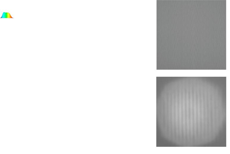
Fig.1. (a)Layoutofopticalheadofthemicrosphere-basednanoscope.Amicroscopeobjective(MO)collectsthevirtualimage(VI)oftheobject(OB)which wasmagnifiedbeforebya25-μ m-diameterglassmicrosphere(MS).Apiezoelectricdevice(PZT)allowstheMOtofocusondifferentaxialVIplanesalong theimagingdepth(ID)bydisplacingOBandMS.Comparisonofperformance between(b)theMOaloneand(c)theMS-basednanoscope,byimaginga200nm-line-widthgrating.

Fig.2. (a)Experimental(redsolidline)andnumerical(bluedottedline)evolutionsofthelateralmagnificationfroma25-μ m-diametermicrosphereplaced inairusingawhite-lightsource,alongthemicrobeadimagingdepth(Zaxial position).(b.i),(b.ii)and(b.iii)arethevirtualimagesofthe400nmperiodic objectatZpositionsof30 μ m,55 μ mand90 μ m,respectively.Scalebarsrepresent1 μ m.(c)Simulationofthevirtualimageformation.Theobjectistwo pointsourcesplacedagainstthemicrosphere.Thewhitedottedlineillustrates theinitialmicrospherelocation.(Forinterpretationofthereferencestocolour inthis figurelegend,thereaderisreferredtothewebversionofthisarticle.)
themeasurements.Duringtheexperiments,themeasurementswere repeatedusingtensimilarmicrospheres.Forexample,experimental curverepresentedin Fig.2(a)resultsthusofanaveragingofthemagnificationfactorsthroughten25-μ m-diametermicrospheres.Moreover, ateachaxialposition,themagnificationmeasurementswereperformed tentimes.
3.Resultsanddiscussion
Thelateralmagnificationgeneratedbythemicrosphere,definedas theratiobetweenthegratingperiodsintheimageplaneandtheobject plane,was firstmeasuredaccordingtotheaxialpositionwherethe objectivefocuseson(Fig.2(a)).Asoda-lime-glassmicrospherehavinga
diameterof25 μ mwasusedtodemonstratetheinfluenceoftheaxial imagepositiononthemagnificationfactor.Alinearevolutionalongthe microsphereimagingdepth(∼60 μ m)isshown,changingfrom × 4to × 8whilemaintainingthesuper-resolutionphenomenon.Thiseffectis significantlydifferenttoconventionalfull-fieldopticalsystem.Indeed, themagnificationofaconventionalopticalmicroscoperemainsconstantovertheclearestimagingdepth.Obviously,thevirtualimagesat positionsdeeperthan60 μ mhaveacontrastdecreased(Fig.2(b.iii)).In ordertoconfirmtheexperimentalresults,arigorous2Delectromagneticmodelwasalsoimplementedbyusinga finiteelement method(ComsolMultiphysics).Thesimulationofsuper-resolution virtualimagingconsistsintwosteps [23,40,41]: firstly,theelectric fieldsfromtheobject, i.e. twopointsources,interactswithamicrosphere.Then,thetransmittedelectric fieldistime-reversepropagated infree-space, i.e. propagatedintheoppositeaxialdirectionwithoutthe microsphere,inordertoretrievethetwoimages.Thebroadbandilluminationofthelightsourceisconsideredbyrepeatingthetwosteps overthevisiblespectralrange( λΔ =400nm,step=50nm)andthen thewavelength-dependantirradiancedistributionsaresummed (Fig.2(c)).Thesimulatedmagnificationfactor, i.e. thetransversedistanceratiobetweenthetworesultingimagesandthetwopointsources, isrepresentedbythebluedottedlinein Fig.2(a)accordingtothe longitudinalpositionoftheimageplane,showingmagnificationvalues ingoodagreementwiththeexperiments.
Afterwards,theinfluenceofthemicrospherediameterontheposition-dependentmagnificationwerestudied.Eightglassmicrospheres havingdifferentdiameterswerehenceplacedonthe400-nm-period grating.Inordertobereadable, Fig.3(a)representsthelinearevolution ofthemagnificationfactorforonlythreemicrospherediameters, i.e. 7 μ m,46 μ mand125 μ m.Thecurvesallowtoaffirmthat,atagiven axialimageposition,thelargemicrospheresprovideamagnification factorlowerthanwhenusingsmallmicrospheres.Moreover,themicrosphere-diameterdependenceonthemagnificationcurveslope α has
Fig.3. (a)Evolutionofthelateralmagnificationinairfrommicrospheres havingdiametersof7 μ m(inred),46 μ m(inorange)and125 μ m(inblue),asa functionofaxialpositionZ.Themicrospheresareilluminatedbyawhite-light source.(b)Evolutionofthemagnificationinclination α (inred)andtheinitial imageposition(inblue)fordifferentdiameters D.Acurve fitting1/D function issuperimposedtothecalculatedvalues α .(Forinterpretationofthereferences tocolourinthis figurelegend,thereaderisreferredtothewebversionofthis article.)
Whitelight Cyan-filteredwhitelight Blue-filteredwhitelight
Axialposition ( m) 7 6 5 4
Fig.4. Evolutionofthelateralmagnificationfroma25-μ m-diametermicrosphereplacedinairasafunctionofaxialpositionunderthreetypesoflight sourcehaving400nm(inredline),90nm(incyanline)and11nm(inblue line)ofbandwidth.(Forinterpretationofthereferencestocolourinthis figure legend,thereaderisreferredtothewebversionofthisarticle.)
beenestimatedandisrepresentedin Fig.3(b)(red-colorcurve).A fittingcurveisjuxtaposed,highlightingtheinverseproportionalityofthe slope α accordingtothediameter D ofthesoda-lime-glassmicrospheres.Inairandusingsoda-lime-glassmicrospheres,thisrelationis expressedas:
= α D [inμm] 1.83 [inμm] 1
Inaddition, Fig.3(a)showsthatthesmallerthediameter,thenarrower theimagingdepth.Indeed,notonlythemagnificationfactorbutalso theimagingdeptharehighlyaffectedbythesizeofthemicrosphere. Largemicrospheresareabletoformvirtualimagesatdeeperpositions, requiringfocuswiththemicroscopeobjectiveatfurtherpositions.The linearproportionalityoftheinitialpositionoftheimagingdepthasa functionofthediameterofsoda-lime-glassmicrospheresisshownin Fig.3(b)(blue-colorcurve).
Finally,theroleofthespectralbandwidthofthelightsourceonthe magnificationwasinvestigated.Forthispurpose,theperformanceof the25-μ m-diametermicrosphereusingthewhitelightsource(representedbytheredlinein Fig.4)iscomparedwithtwospectrallygatedilluminations, i.e. usingthecyan filter(cyanline)andtheblueline-filter(blueline).Themagnificationfactorremainslinearlyproportionalaccordingtotheaxialpositionandthemagnificationvalues donotdiffer,regardlessofthespectralbandwidthofthelightsource. However,onlythemagnificationrangeappearslimited.Asamatterof fact,thesmallerthespectralbandwidthis,thenarrowerthemicrosphereimagingdepthis.Thisspectralwidthdependenceshowsthatthe unconventionalmagnificationbehaviourismainlyduetoasignificant contributionofthechromaticaberrations.Wavelength-dependent beamsarefocusedindifferentimageplanes,eachhavingadifferent magnification.Thisleadstodifficultiestodefineauniquemagnification.Nevertheless,thisyieldsadegreeoffreedomduringthemeasurement, i.e. thepossibilitytoadjusttherequiredmagnification.This assumptioncanberetrievedthroughglassmicrosphereshavinganother diameter.
Inthiswork,theunconventionalbehaviourofthelateralmagnificationinmicrosphere-assistedmicroscopyhasbeenachievedinair usingsoda-lime-glassmicrospheres.Itshouldhoweverbementioned thatthiseffectoccursalsousingmicrosphereshavingahigherrefractiveindexandinadifferentimmersionconfiguration [35,42].
4.Conclusions
Thisworkexposesafundamentalpropertyinmicrosphere-assisted microscopy:thenon-classicalbehaviourofthelateralimagemagnification.Contrarilytoclassicalopticalmicroscopy,themagnification factorissubjecttoalinearevolutionalongthemicrosphereimaging depth,andbothgeometricalandopticalparametershaveaninfluence ontheposition-dependentmagnificationslopeandrange.Indeed, throughexperimentsandnumericalsimulations,itwasshownthatthe
microspheresizeisinverselyproportionaltothemagnificationgrowth andislinearlyproportionaltotheinitialpositionoftheaxial fieldof view.Furthermore,thereductionofthespectralbandwidthoftheilluminationlightsourceindeedlimitsthemagnificationrange.
Acknowledgements
TheauthorsthankA.Leong-Hoiforherfruitfulcontribution.This workreceivedfundingfromSATTConectusAlsaceandwaspartly supportedbytheFrenchRENATECHnetwork(FEMTO-STInstitute, Besançon)andtheUniversityofStrasbourg.
AppendixA.Supplementarymaterial
Supplementarydataassociatedwiththisarticlecanbefound,inthe onlineversion,at https://doi.org/10.1016/j.optlastec.2019.01.030
References
[1]H.vonHelmholtz,H.E.Fripp,Onthelimitsoftheopticalcapacityofthemicroscope,MonthlyMicroscop.J.16(1876)15–39, https://doi.org/10.1111/j.13652818.1876.tb05606.x
[2]Editorial,Beyondthediffractionlimit,NaturePhoton.3(2009)361.doi:https:// doi.org/10.1038/nphoton.2009.100
[3]E.H.Synge,Asuggestedmethodforextendingmicroscopicresolutionintotheultramicroscopicregion,Phil.Mag.7(6)(1928)356–362, https://doi.org/10.1080/ 14786440808564615
[4]H.Goldmann,Spaltlampenphotographieundphotometric,Ophthalmologica98 (1939)257–270, https://doi.org/10.1159/000299716 .
[5]H.Naora,Microspectrophotometryandcytochemicalanalysisofnucleicacids, Science114(1951)279, https://doi.org/10.1126/science.114.2959.279
[6] C.J.R.Sheppard,Y.Gong,Improvementinaxialresolutionbyinterferenceconfocal microscopy,Optik87(1991)129–132
[7]Z.Chen,A.Taflove,V.Backman,Photonicnanojetenhancementofbackscattering oflightbynanoparticles:apotentialnovelvisible-lightultramicroscopytechnique, Opt.Exp.12(2004)1214–1220, https://doi.org/10.1364/OPEX.12.001214
[8]E.A.Ash,G.Nicholls,Super-resolutionaperturescanningmicroscope,Nature237 (1972)510–512, https://doi.org/10.1038/237510a0
[9] D.Courjon,Near-FieldMicroscopyandNear-FieldOptics,ImperialCollegePress, London,2003
[10]W.Lukosz,M.Marchand,OptischenAbbildungUnterUberschreitungder BeugungsbedingtenAuflösungsgrenze,Opt.Acta10(1963)241–255, https://doi. org/10.1080/713817795
[11]M.Saxena,G.Eluru,S.S.Gorthi,Structuredilluminationmicroscopy,Adv.Opt. Photon.7(2015)241–275, https://doi.org/10.1364/AOP.7.000241
[12]V.G.Veselago,Theelectrodynamicsofsubstanceswithsimultaneouslynegative valuesof ∊ and μ ,Sov.Phys.Usp.10(1968)509–514, https://doi.org/10.1070/ PU1968v010n04ABEH003699
[13]X.Zhang,Z.Liu,Superlensestoovercomethediffractionlimit,Nat.Mater.7(2008) 435–441, https://doi.org/10.1038/nmat2141
[14]S.W.Hell,Far-fieldopticalnanoscopy,Science316(2007)1153–1158, https://doi. org/10.1126/science.1137395
[15]R.M.Dickson,A.B.Cubitt,R.Y.Tsien,W.E.Moerner,On/off blinkingandswitching behaviourofsinglemoleculesofgreen fluorescentprotein,Nature388(1997) 355–358, https://doi.org/10.1038/41048
[16]Z.Wang,W.Guo,L.Li,B.Luk’yanchuk,A.Khan,Z.Liu,Z.Chen,M.Hong,Optical virtualimagingat50nmlateralresolutionwithawhite-lightnanoscope,Nat. Commun.2(2011)218, https://doi.org/10.1038/ncomms1211
[17]A.Darafsheh,G.F.Walsh,L.DalNegro,V.N.Astratov,Opticalsuper-resolutionby high-indexliquid-immersedmicrospheres,Appl.Phys.Lett.101(2012)141128, https://doi.org/10.1063/1.4757600
[18]A.Darafsheh,C.Guardiola,A.Palovcak,J.C.Finlay,A.Carabe,Opticalsuper-resolutionimagingbyhigh-indexmicrospheresembeddedinelastomers,Opt.Lett.40 (2015)5–8, https://doi.org/10.1364/OL.40.000005
[19]A.Darafsheh,Influenceofthebackgroundmediumonimagingperformanceof
microsphere-assistedsuper-resolutionmicroscopy,Opt.Lett.42(2017)735–738, https://doi.org/10.1364/OL.42.000735
[20]Y.Zhou,Y.Tang,Y.He,X.Liu,S.Hu,Effectsofimmersiondepthonsuper-resolutionpropertiesofindex-differentmicrosphere-assistednanoimaging,Appl. Phys.Exp.11(2018)032501, https://doi.org/10.7567/APEX.11.032501
[21]L.Yao,Y.-H.Ye,H.FengMa,L.Cao,J.Hou,Roleoftheimmersionmediuminthe microscalesphericallensimaging,Opt.Commun.335(2015)23–27, https://doi. org/10.1016/j.optcom.2014.08.051
[22]H.S.S.Lai,F.Wang,Y.Li,B.Jia,L.Liu,W.J.Li,Super-resolutionrealimagingin microsphere-assistedmicroscopy,PLoSONE11(2016)e0165194, https://doi.org/ 10.1371/journal.pone.0165194
[23]A.V.Maslov,V.N.Astratov,Imagingofsub-wavelengthstructuresradiatingcoherentlynearmicrospheres,App.Phys.Lett.108(2016), https://doi.org/10.1063/ 1.4941030
[24]S.Perrin,S.Lecler,A.Leong-Hoi,P.C.Montgomery,Roleofcoherenceinmicrosphere-assistednanoscopy,Proc.SPIE10330(2017)103300V, https://doi.org/10. 1117/12.2270246
[25]L.A.Krivitsky,J.J.Wang,Z.Wang,B.Luk’yanchuk,Locomotionofmicrospheresfor super-resolutionimaging,Sci.Rep.3(2013)3501, https://doi.org/10.1038/ srep03501.
[26]J.Li,W.Liu,T.Li,I.Rozen,J.Zhao,B.Bahari,B.Kante,J.Wang,Swimming microrobotopticalnanoscopy,NanoLett.16(2016)6604–6609, https://doi.org/ 10.1021/acs.nanolett.6b03303
[27] F.Wang,L.Liu,H.Yu,Y.Wen,P.Yu,Z.Liu,Y.Wang,W.J.Li,Scanningsuperlens microscopyfornon-invasivelarge field-of-viewvisiblelightnanoscaleimaging, Nat.Commun.7(2016)13748, https://doi.org/10.1038/ncomms13748
[28]M.Duocastella,F.Tantussi,A.Haddadpour,R.P.Zaccaria,A.Jacassi,G.Veronis, A.Diaspro,F.DeAngelis,Combinationofscanningprobetechnologywithphotonic nanojets,Sci.Rep.7(2017)3474, https://doi.org/10.1038/s41598-017-03726-5
[29]L.Li,W.Guo,Y.Yan,S.Lee,T.Wang,Label-freesuper-resolutionimagingof adenovirusesbysubmergedmicrosphereopticalnanoscopy,LightSci.Appl.2 (2013)e104, https://doi.org/10.1038/lsa.2013.60
[30]H.Yang,N.Moullan,J.Auwerx,M.A.M.Gijs,Super-resolutionbiologicalmicroscopyusingvirtualimagingbyamicrospherenanoscope,Small10(2014) 1712–1718, https://doi.org/10.1002/smll.201302942
[31]S.Perrin,H.Li,K.Badu,T.Comparon,G.Quaranta,N.Messaddeq,N.Lemercier, P.Montgomery,J.-L.Vonesch,S.Lecler,Transmissionmicrosphere-assisteddarkfieldmicroscopy,Phys.StatusSolidiRRL(2018)1800445, https://doi.org/10. 1002/pssr.201800445
[32]A.Darafsheh,N.I.Limberopoulos,J.S.Derov,D.E.WalkerJr.,V.N.Astratov, Advantagesofmicrosphere-assistedsuper-resolutionimagingtechniqueoversolid immersionlensandconfocalmicroscopies,Appl.Phys.Lett.104(2014)061117, https://doi.org/10.1063/1.4864760
[33]X.Hao,C.Kuang,X.Liu,H.Zhang,Y.Li,Microspherebasedmicroscopewithopticalsuper-resolutioncapability,Appl.Phys.Lett.99(2011)203102, https://doi. org/10.1063/1.3662010
[34]A.Darafsheh,Opticalsuper-resolutionandperiodicalfocusingeffectsbydielectric microspheres,Ph.D.dissertation,UniversityofNorthCarolinaatCharlotte,2013.
[35] S.Lecler,S.Perrin,A.Leong-Hoi,P.Montgomery,Photonicjetlens,Sci.Rep. (2019)acceptedforpublication
[36]Y.Duan,G.Barbastathis,B.Zhang,Classicalimagingtheoryofamicrolenswith super-resolution,Opt.Lett.38(2013)2988–2990, https://doi.org/10.1364/OL.38. 002988
[37]Y.Ben-Aryeh,Increaseofresolutionbyuseofmicrospheresrelatedtocomplex Snell’slaw,J.Opt.Soc.Am.A33(2013)2284–2288, https://doi.org/10.1364/ JOSAA.33.002284
[38]S.Yang,F.Wang,Y.-H.Ye,Y.Xia,Y.Deng,J.Wang,Y.Cao,Influenceofthe photonicnanojetofmicrospheresonmicrosphereimaging,Opt.Exp.25(2017) 27551–27558, https://doi.org/10.1364/OE.25.027551
[39] S.Perrin,H.Li,A.Leong-Hoi,S.Lecler,P.Montgomery,Illuminationconditionsin microsphere-assistedmicroscopy,J.Microsc.(2018)submittedforpublication
[40]S.Perrin,A.Leong-Hoï,S.Lecler,P.Pfeiffer,I.Kassamakov,A.Nolvi, E.Haeggström,P.Montgomery,Microsphere-assistedphase-shiftingprofilometry, Appl.Opt.56(2017)7249–7255, https://doi.org/10.1364/AO.56.007249
[41]I.Kassamakov,S.Lecler,A.Nolvi,A.Leong-Hoi,P.Montgomery,E.Haeggstrom, 3Dsuper-resolutionopticalprofilingusingmicrosphereenhancedmirauinterferometry,Sci.Rep.7(2017)3683, https://doi.org/10.1038/s41598-017-03830-6
[42] H.C.VandeHulst,LightScatteringbySmallParticles,JohnWiley&Sons,New York,1957.
Another random document with no related content on Scribd:

The Project Gutenberg eBook of Kyllikki ja Lemminkäinen
This ebook is for the use of anyone anywhere in the United States and most other parts of the world at no cost and with almost no restrictions whatsoever. You may copy it, give it away or re-use it under the terms of the Project Gutenberg License included with this ebook or online at www.gutenberg.org. If you are not located in the United States, you will have to check the laws of the country where you are located before using this eBook.
Title: Kyllikki ja Lemminkäinen Laulurunoja
Author: Hilja Liinamaa-Pärssinen
Release date: April 1, 2024 [eBook #73309]
Language: Finnish
Original publication: Helsinki: E. E. Sundvall, 1902
Credits: Tapio Riikonen *** START OF THE PROJECT GUTENBERG EBOOK KYLLIKKI JA LEMMINKÄINEN ***