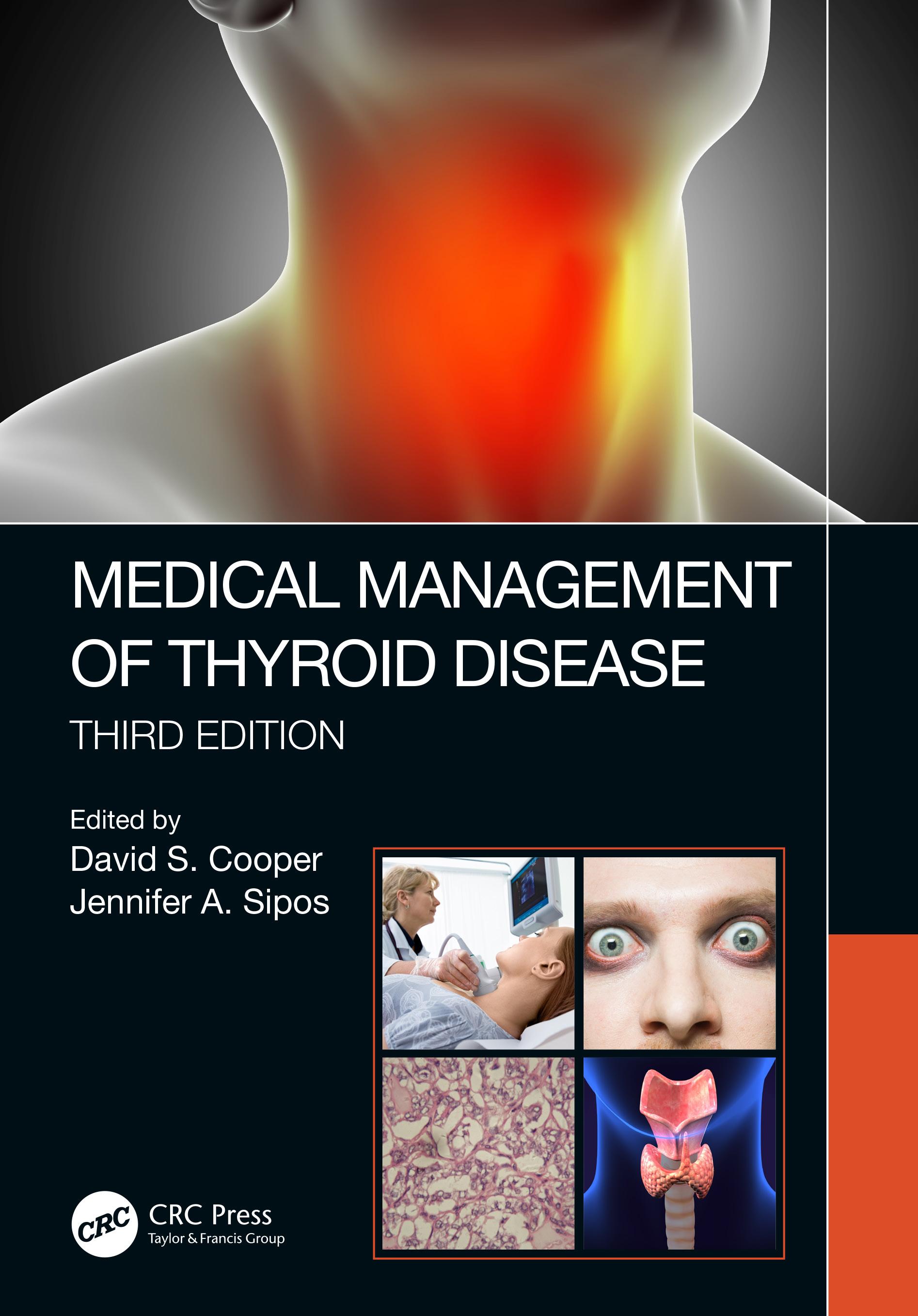
Medical Management of Thyroid Disease
Third Edition
Edited by David S. Cooper and Jennifer A. Sipos
CRC Press
Taylor & Francis Group
6000 Broken Sound Parkway NW, Suite 300
Boca Raton, FL 33487-2742
© 2019 by Taylor & Francis Group, LLC
CRC Press is an imprint of Taylor & Francis Group, an Informa business
No claim to original U.S. Government works
Printed on acid-free paper
International Standard Book Number-13: 978-1-138-57723-7 (Hardback)
This book contains information obtained from authentic and highly regarded sources. While all reasonable efforts have been made to publish reliable data and information, neither the author[s] nor the publisher can accept any legal responsibility or liability for any errors or omissions that may be made. The publishers wish to make clear that any views or opinions expressed in this book by individual editors, authors or contributors are personal to them and do not necessarily reflect the views/opinions of the publishers. The information or guidance contained in this book is intended for use by medical, scientific or health-care professionals and is provided strictly as a supplement to the medical or other professional’ s own judgement, their knowledge of the patient’ s medical history, relevant manufacturer’ s instructions and the appropriate best practice guidelines. Because of the rapid advances in medical science, any information or advice on dosages, procedures or diagnoses should be independently verified. The reader is strongly urged to consult the relevant national drug formulary and the drug companies’ and device or material manufacturers’ printed instructions, and their websites, before administering or utilizing any of the drugs, devices or materials mentioned in this book. This book does not indicate whether a particular treatment is appropriate or suitable for a particular individual. Ultimately it is the sole responsibility of the medical professional to make his or her own professional judgements, so as to advise and treat patients appropriately. The authors and publishers have also attempted to trace the copyright holders of all material reproduced in this publication and apologize to copyright holders if permission to publish in this form has not been obtained. If any copyright material has not been acknowledged please write and let us know so we may rectify in any future reprint.
Except as permitted under U.S. Copyright Law, no part of this book may be reprinted, reproduced, transmitted, or utilized in any form by any electronic, mechanical, or other means, now known or hereafter invented, including photocopying, microfilming, and recording, or in any information storage or retrieval system, without written permission from the publishers.
For permission to photocopy or use material electronically from this work, please access www.copyright.com (http://www.copyright. com/) or contact the Copyright Clearance Center, Inc. (CCC), 222 Rosewood Drive, Danvers, MA 01923, 978-750-8400. CCC is a not-forprofit organization that provides licenses and registration for a variety of users. For organizations that have been granted a photocopy license by the CCC, a separate system of payment has been arranged.
Trademark Notice: Product or corporate names may be trademarks or registered trademarks, and are used only for identification and explanation without intent to infringe.
Library of Congress Cataloging in Publication Data
Names: Cooper, David S. (Physician), editor. | Sipos, Jennifer, editor.
Title: Medical management of thyroid disease / [edited by] David S.Cooper and Jennifer Sipos.
Description: Third edition. | Boca Raton : Taylor & Francis, 2019. | Includes bibliographical references and index.
Identifiers: LCCN 2018030183| ISBN 9781138577237 (hardback : alk. paper) | ISBN 9781351267489 (ebook)
Subjects: | MESH: Thyroid Diseases--therapy | Thyroid Diseases--diagnosis
Classification: LCC RC655 | NLM WK 267 | DDC 616.4/4--dc23
LC record available at https://lccn.loc.gov/2018030183
Visit the Taylor & Francis Web site at http://www.taylorandfrancis.com
and the CRC Press Web site at http://www.crcpress.com
Preface
It has been more than 10 years since the second edition of Medical Management of Thyroid Disease was published. When I was asked by the publisher to edit this third edition of the text, I invited Dr. Jennifer Sipos from The Ohio State University to be my coeditor. Together, we have continued the tradition of this book, which was initially developed to be a practical guide on the management of both common and uncommon thyroid problems. We have tried, as much as possible, to limit the discussion to the clinical manifestations, diagnostic procedures, and treatment of the gamut of thyroid disorders in adults. As before, to the greatest degree possible, all of the recommendations in the text are “ evidence-based” or recapitulate evidence-based clinical practice guidelines. We have invited a number of new authors to provide a fresh approach to some of the topics.
Since the last edition of this text was published in 2008, there have been remarkable strides in our ability to care for thyroid patients. In the realm of benign thyroid disease, we now recognize that drug-induced thyroid dysfunction includes a large array of new drugs that inhibit tyrosine kinases, have effects on the immune system as “ checkpoint inhibitors,” or have other more ill-defined effects.
An entire chapter is devoted to this topic, in recognition of its importance. In the treatment of hypothyroidism, clinicians are now feeling more justified in using T4/T3 combination therapy in some patients, reflecting a better understanding that T4 monotherapy may not recapitulate the serum hormonal profile of the thyroid gland itself. There has been a revolution in the management of thyroid nodules, including a new classification for cytopathology (the Bethesda system), as
well as the development of molecular testing for improved diagnosis of indeterminate thyroid nodules. There has also been a sea change in the way low-risk thyroid cancer is managed, based on the 2015 American Thyroid Association clinical practice guidelines. Instead of a “ one-size-fits-all” approach, we now have a more personalized set of management strategies, based on the recognition that more aggressive treatment (i.e., total thyroidectomy, radioiodine ablation, and full suppression of serum TSH) is not necessary for the vast majority of thyroid cancer patients. Furthermore, there are now a number of randomized clinical trials which have helped to define the best management for advanced thyroid cancers.
Dr. Sipos and I want to thank the contributors to this text for their time and expertise. We also want to express our gratitude to two of our mentors, Dr. E. Chester Ridgway and Dr. Ernest Mazzaferri. Both were giants in the field of thyroidology, both contributed to the first and second editions of this text, and both have sadly passed away in the last several years. We wish to recognize them for their guidance, and for being inspiring role models and colleagues. Finally, we hope that practitioners will benefit from reading this textbook, but we understand that the ultimate beneficiaries of the knowledge gained will be the millions of patients suffering from thyroid disease around the world.
David S. Cooper, MD
The Johns Hopkins University School of Medicine
Jennifer A. Sipos, MD
The Ohio State University Wexner Medical Center
Editors
David S. Cooper, MD, MACP, received his medical degree from Tufts University School of Medicine and completed his endocrinology fellowship training at the Massachusetts General Hospital/Harvard Medical School. He is Professor of Medicine and Radiology at The Johns Hopkins University School of Medicine and Director of The Johns Hopkins Thyroid Clinic. He serves as editor-in-chief for endocrinology at Up-to-Date . He is a former contributing editor at JAMA and former deputy editor of the Journal of Clinical Endocrinology and Metabolism . He is the past chair of the Subspecialty Board for Endocrinology, Diabetes, and Metabolism of the American Board of Internal Medicine. Dr. Cooper is the past president of the American Thyroid Association and the recipient of the American Thyroid Association’ s Distinguished Service Award and its Paul Starr Award. He is also the recipient of the Distinction in Clinical Endocrinology Award from the American College of Endocrinology and the Endocrine Society’ s 2016 Outstanding Scholarly Physician Award.
Jennifer A. Sipos, MD, is a Professor of Medicine and Director of the Benign Thyroid Disorders Program at The Ohio State University. She obtained her medical degree and received her internal medicine residency training at Wake Forest University. She completed her endocrinology and metabolism fellowship at the University of North Carolina in Chapel Hill. Dr. Sipos has developed an interest in the use of ultrasonography for the diagnosis and management of thyroid cancer and has taught and served as a course director for numerous ultrasound courses nationally and internationally, including meetings for the Endocrine Society, American Thyroid Association, European Thyroid Association, American Association for Clinical Endocrinologists, Asia and Oceania Thyroid Association, Indian Endocrine Society, and International Congress for Endocrinology. Additionally, she is actively involved in several clinical research projects with a particular interest in factors implicated in the development of salivary damage after radioiodine therapy. She also participates in clinical trials for the evaluation of multikinase inhibitor therapies in refractory thyroid cancer and the diagnostic use of molecular markers in thyroid nodules.
Contributors
Victor Bernet
Mayo Clinic
Jacksonville, Florida
Keith C. Bible
Mayo Clinic
Rochester, Minnesota
Henry B. Burch
National Institutes of Health
Bethesda, Maryland
Ashish V. Chintakuntlawar
Mayo Clinic
Rochester, Minnesota
David S. Cooper
The Johns Hopkins University School of Medicine
Baltimore, Maryland
Vaninder K. Dhillon
The Johns Hopkins University Baltimore, Maryland
Poorani N. Goundan
Boston Medical Center Boston, Massachusetts
Elizabeth G. Grubbs
The University of Texas MD Anderson Cancer Center
Houston, Texas
Mimi I. Hu
The University of Texas MD Anderson Cancer Center
Houston, Texas
Jacqueline Jonklaas
Georgetown University School of Medicine
Washington, DC
Stephanie L. Lee
Boston Medical Center
Boston, Massachusetts
Susan J. Mandel
University of Pennsylvania Philadelphia, Pennsylvania
Carolyn Maxwell
Stony Brook University Hospital Stony Brook, New York
Michael T. McDermott
University of Colorado Denver School of Medicine
Denver, Colorado
Jennifer A. Sipos
The Ohio State University Columbus, Ohio
Robert C. Smallridge Mayo Clinic
Jacksonville, Florida
Julie Ann Sosa
University of California at San Francisco San Francisco, California
Ralph P. Tufano
The Johns Hopkins University Baltimore, Maryland
Nicole O. Vietor
Walter Reed National Military Medical Center
Bethesda, Maryland
Alisha N. Wade
University of the Witwatersrand Johannesburg, South Africa
The laboratory and imaging approaches to thyroid disorders
JACQUELINE JONKLAAS AND DAVID S. COOPER
INTRODUCTION
The central role of the thyroid gland in controlling metabolism was recognized in the 19th century, but evaluation of the function of the thyroid remains an evolving science. Initial approaches to the assessment of thyroid function centered on measuring end-organ responses as biological markers of thyroid hormone actions. Development of in vitro competitive binding assay methods allowed the direct quantification of hormone
levels in serum, and sensitive immunoassays have demonstrated the subtleties of pituitary and hypothalamic control of the thyroid. Abnormalities of hormone binding by serum proteins necessitated sensitive estimation of free hormone levels. With the detection of serum markers of autoimmune and malignant diseases of the thyroid gland, earlier diagnosis and improved monitoring of these conditions have been achieved, often with greater sensitivity than may be clinically relevant. Limitations to the measurement methods utilized
exist, however, particularly when underlying assumptions about the comparability of patient and control specimens are invalid. Nonetheless, the clinician can now effectively confirm suspected diagnoses of thyroid dysfunction, cost-effectively screen asymptomatic populations for common diseases, and appropriately monitor the treatment of patients with disorders of the thyroid.
PHYSIOLOGY OF THE HYPOTHALAMIC-PITUITARYTHYROID AXIS
Excellent reviews and books provide detailed explorations of the physiology of the hypothalamic-pituitary-thyroid axis, and the reader is invited to delve into those worthwhile sources (1). For the purposes of this chapter, a brief review of the biosynthesis and transport of thyroid hormones and the regulation of thyroid function by the hypothalamic-pituitary complex will suffice (Figure 1.1).
The synthesis of thyroxine (T4 ) and triiodothyronine (T3 ) begins with the active transport of iodide into the cell via a sodium-iodine symporter
located in the basal membrane. Following oxidation by thyroid peroxidase, the iodide moiety is covalently attached to tyrosyl residues of thyroglobulin, and the resulting iodotyrosines are coupled and cleaved from thyroglobulin to form T4 and T3 , normally in a 10:1 ratio. Thyroid hormone secretion requires endocytosis and degradation of iodinated thyroglobulin, followed by the release of T4 and T3 into the circulation. This process results in the total daily output of 80 to 100 µ g of T4 . In contrast, only 20% of the circulating T3 is produced by the thyroid, the remaining 80% is derived from the enzymatic outer-ring or 5¢ -monodeiodination of T4 in extrathyroidal tissues such as the liver, kidney, brain, muscle, and skin. Removal of the inner-ring or 5-iodine of T4 forms the inactive metabolite reverse T3 (rT3 ). Other inactivating pathways for T4 and T3 include glucuronidation, sulfation, deamination, and cleavage. The normal daily fractional turnover rates for T4 and T3 are 10% and 75%, respectively.
In serum, at least 99.95% of T4 and 99.5% of T3 molecules are bound by the transport proteins thyroxine-binding globulin (TBG), transthyretin (thyroxine-binding prealbumin [TBPA]), and
Figure 1.1 The hypothalamic pituitary thyroid axis. (From Refetoff S, Dumitrescu A. Best Pract Res Clin Endocrinol Metab. 2007;21:277– 305. Used with permission.)
albumin. Although TBG is present in lower concentrations than either transthyretin or albumin, its greater affinity for thyroid hormones makes it the predominant serum carrier of T4 and T3
Variations in binding characteristics among normal and abnormal thyroid hormone-binding proteins are responsible for much of the methodologic limitations in assays that attempt to measure concentrations of free T4 and T3 . This large pool of protein-bound hormone provides a stable reservoir that maintains the supply of free, unbound hormone available for transport into the cells. Once within target cells, T4 is further deiodinated to T3 , which in the nucleus binds to the thyroid hormone receptor, modulating the transcription of thyroid hormone-responsive genes and producing most of the clinical effects recognized as the metabolic effects of thyroid hormones.
The primary regulatory influence on thyroid gland function is the circulating level of thyrotropin (thyroid stimulating hormone, or TSH).
Produced by thyrotroph cells of the anterior pituitary, TSH is a two-subunit glycoprotein, the specificity of which is conferred by its β -subunit; the α -subunit is structurally similar to that of follicle-stimulating hormone, luteinizing hormone, and human chorionic gonadotropin. Negative feedback by T4 and T3 influences TSH synthesis and release, as evidenced by a complex inverse relationship between the concentrations of TSH and free iodothyronine (2, 3). It is likely that each individual has a genetically determined set-point for this TSH/free T4 relationship, based on twin studies (4, 5). TSH levels peak just before nocturnal sleep, and the nadir occurs in the late afternoon; this nocturnal surge is lost early in the course of nonthyroidal illness. TSH levels in various populations conform best to a logGaussian rather than Gaussian distribution (6). The hypothalamic tripeptide thyrotropin-releasing hormone (TRH) stimulates TSH secretion and modulates thyrotroph response to altered thyroid hormone levels. In conjunction with the suppressive effects of dopamine, corticosteroids, somatostatin, androgens, and endogenous opioids, TRH may be responsible for modulating the setpoint for the negative feedback loop that controls thyroid hormone levels. Hypothalamic production of TRH itself is regulated by circulating thyroid hormones, as well as by multiple central nervous system factors.
LABORATORY EVALUATION OF THYROID FUNCTION
Assays of thyroid hormones
TOTAL SERUM IODOTHYRONINE CONCENTRATIONS
When concentrations and binding affinities of thyroid hormone -binding proteins are normal, there exists at physiologic equilibrium a direct relationship between levels of total hormone and free hormone (7). Thus, measurement of total iodothyronine concentration can provide a reasonable surrogate for estimating the amount of free iodothyronine present. Either serum or plasma can be used to assay hormone concentrations, although serum is generally preferred. The most commonly employed technique for the determination of total T4 (TT4 ) and T3 (TT3 ) concentrations is competitive immunoassay, using either polyclonal or monoclonal “ capture” antibodies directed against the specific iodothyronine. To ensure measurement of bound as well as free hormones, inhibitors of iodothyronine binding are added— e.g., 8- anilino-1-naphthalene sulfonic or salicylic acids for TBG and barbital for TBPA. These agents successfully dissociate the hormone from binding proteins without interfering with hormone binding to immunoglobulin.
Radioimmunoassay (RIA) depends upon measurement of the distribution of a tracer quantity of radiolabeled hormone that competes with the endogenous hormone in the patient’ s specimen for binding to a capture antibody. The higher the serum hormone concentration, the lower the amount of radiolabel that binds to the antibody. Following the addition of a limited amount of capture antibody and the radiolabeled iodothyronine to be measured, the antibody-antigen complexes are separated from the serum. Separation techniques vary, including ammonium sulfate or second antibody precipitation. Newer methods that facilitate automated separation include attachment of the anti-T4 antibody to a solid phase, such as the wall of the assay tube or magnetizable particles. The concentration of either TT4 or TT3 is then determined by comparison of the amount of antibody-bound radiolabel with a simultaneously derived standard curve. A fundamental assumption, therefore, is that there is no difference in the
assay conditions (including protein binding and other constituents found in the serum) between the patient’ s sample and the control standards, an assumption that is often invalid.
Nonisotopic methods avoid reliance upon radioactive reagents and are now the most commonly used assays. The heterogeneous enzymelinked immunosorbent assay (ELISA) incorporates enzymes, fluorescent, or chemiluminescent molecules that create a quantitative signal when interacting with a specific enzyme bound to the tracer hormone— e.g., alkaline phosphatase, horseradish peroxidase, or glucose-6-phosphate dehydrogenase. As in RIA, numerous physical and chemical approaches exist for separating signal bound to the anti-iodothyronine antibody from unbound signal. In contrast, homogeneous enzyme immunoassays do not require a separation step. Instead, the binding of the antibody to a tracer hormone directly affects the activity of the signal-generating enzyme bound to the tracer. Other technologies, such as liquid chromatograph-tandem mass spectroscopy (LC-MS/MS) have also been applied to provide greater specificity and less analytical interference (8).
Due to common alterations in serum TBG levels, TT4 and TT3 are generally not used as standalone tests in clinical practice, but are combined with direct measurements of TBG or TBG-binding capacity, which can then be used to calculate a Free Thyroxine Index (see below).
Reference ranges vary to some degree, but commonly cited ranges are 4.5– 12.6 mcg/dL (58– 160 nmol/L) for TT4 and 80– 180 ng/dL (1.2– 2.7 nmol/L) for TT3 (9). As developed by their manufacturers, these assay techniques have similar performance characteristics, although each may be affected by different sources of interference. TT4 assays tend to be more reliable than TT3 assays. For example, in a recent study, in which 11 TT4 and 12 TT3 assays were compared, with LC-MS/MS values as the reference serum concentrations, only 4/10 TT4 assays and 4/11 TT3 assays failed to agree to within 10% of the reference concentrations, with greater deviation seen with the TT3 assay (10). Contributing factors to measurement error include qualitative differences between the protein constituents of sample diluents used for calibration and those found in patient sera, leading to differential dissociation of hormone from binding proteins.
DETERMINATION OF FREE T4 AND T3 CONCENTRATIONS
Because T4 and T3 are highly bound to serum proteins, alterations in either the levels of these proteins or their binding characteristics can significantly alter the concentration of total hormone. As it is the free hormone that is biologically active, however, techniques are required to permit either direct measurement or estimation of the serum free hormone levels. All methods that have been developed face the identical problem: distinguishing between the 3– 4 orders of magnitude difference in the concentrations of the free and the proteinbound hormones. In all free hormone assays, the central assumption is that the effectiveness of separating the free from the bound hormone is identical in both the patient samples and the standards used to calibrate the assay, an assumption that is difficult to validate in all potential clinical situations. As a result, in a study comparing the results of 15 FT4 and 13 FT3 immunoassays to values obtained by the reference method equilibrium dialysis-LC-MS/ MS, all the FT4 immunoassays and 9 of the FT3 immunoassays produced results that were outside the 10% agreement with the reference method (11). Direct methods for measuring FT4 and FT3 include equilibrium dialysis, ultrafiltration, and gel filtration to separate the free hormone from its binding proteins. In the case of equilibrium dialysis, undiluted patient serum is dialyzed overnight across a membrane with pores that allow free but not protein-bound hormone to partition, allowing equilibration of the free hormone concentration across the membrane. A highly sensitive RIA, capable of detecting nanogram (or picomole) quantities of hormones, is then used to measure the hormone content of the protein-free dialysate, comparing to a standard curve generated with gravimetrically determined amounts of hormone (12). Faster turnaround can be achieved by using ultrafiltration rather than equilibrium dialysis, but greater variability can result from minimal amounts of serum proteins that leak through the filtration device as well as a hormone that is adsorbed to either the membrane or container surface. Such direct measurements are generally expensive, time consuming, and not widely used commercially. Expected adult values for these direct methods are about 0.8 to 2.3 ng/dL for free T4 and 210 to 440 pg/dL for free T3 . As mentioned above, the LC-MS/MS assay
used above as the reference method for assessing FT4 and FT3 assays employed separation by equilibrium dialysis (11). Separation by ultrafiltration has also been combined with LC-MS/MS (13). The LC-MS/MS technique to measure free T4 levels provides high specificity; hence its use as a reference assay (11). LC-MS/MS can also offer simultaneous measurement of other thyroid analytes (13).
Immunoassay methods for estimation of free hormone concentration are now widely used. In the “ analogue” or “ one-step” free T4 method, a labeled T4 analogue that does not bind to serumbinding proteins is added to serum and the mixture is either incubated with an anti-T4 antibody or allowed to bind to antibody attached to a solid phase. At equilibrium, the amount of analogue complexed to the antibody is inversely proportional to the amount of free T4 that is available. One-step methods require structurally modified analogues that do not displace hormone from protein-binding sites, but a complete lack of displacement is rarely achieved. Therefore, these methods depend on the assumption that there is no difference in hormone-binding affinity for proteins between the sample to be measured and the assay controls or calibrators, both for the actual analyte as well as the analogue. This assumption is particularly at risk when there are circulating inhibitors of hormone binding in serum, such as occurs in renal failure or other nonthyroidal illnesses, or major alterations in hormone-binding protein concentrations (14). Because the analogues used generally bind to albumin, although not with the same kinetics as T4 or T3 , this method may not correct for abnormalities in albumin binding.
In “ two-step” assays, serum is exposed to a solid phase containing an anti-T4 antibody, binding a certain amount of free hormone to the solid phase. By diluting the specimen and limiting the duration of incubation, there should be minimal disruption of endogenous hormone binding to serum proteins (12). After removal of the serum and its proteins, a tracer quantity of radiolabeled T4 is incubated with the solid phase, equilibrating with the remaining unoccupied antibody molecules. The amount of radiolabeled T4 complexed to the solid phase is thus inversely proportional to the free T4 concentration of the serum. Because the label is unable to interact with serum-binding proteins or endogenous inhibitors of hormone binding to protein (due to the physical separation step), the
“ two-step” method has a good correlation with the free T4 determined by direct equilibrium dialysis. Nonradioactive assays have also been developed, and automated two-step procedures are in common use.
For free T3 measurements, methods that rely upon physical separation of bound from free hormones, such as dialysis or ultrafiltration, are not generally commercially available. The same technology for “ one-step” assays of free T4 is used to measure free T3 . Interference from serum proteins and difficulty avoiding stripping T3 from its binding proteins is a greater problem than in free T4 assays (15). New methods that utilize tandem mass spectrometry following equilibrium dialysis or ultrafiltration may allow faster and more reliable assays (16).
The thyroid hormone-binding ratio (THBR), another calculated value proportional to the fraction of hormone that is free in circulation, derives from measurement of the availability of protein-binding sites in the patient’ s serum. In the traditional uptake method, a tracer quantity of radiolabeled iodothyronine is added to the serum and allowed to partition between unoccupied specific protein-binding sites and a nonsaturable adsorbent— e.g., talc, charcoal, resin, or anti-iodothyronine antibodies. T3 is generally preferred as the labeled ligand, as it has a lower affinity for TBG and therefore does not displace T4 from its binding sites. There is an inverse relationship between the amounts of radiolabel adsorbed by the inert solid phase and unoccupied serum protein-binding sites. The percent uptake derives from the ratio of tracer bound by the adsorbent to the tracer bound by serum proteins; an alternative but less reliable formula expresses the ratio as the amount of tracer attached to adsorbent to the amount initially added. The THBR is then calculated as the percent uptake in the patient’ s serum and normalized to that of a control or reference serum; the expected normal range is centered around unity. The THBR is increased when there are few endogenous binding sites, which can occur with an increased amount of T4 available to bind (thyrotoxicosis), the presence of competing ligands (certain drugs and nonthyroidal illness), or a decreased amount of binding protein (TBG deficiency). Conversely, hypothyroidism and TBG excess will produce an increased number of available binding sites, producing a decreased THBR. As a general rule, true
thyroid function abnormalities produce concordant increases or decreases in the total serum T4 and THBR, whereas discordant changes in the two tests typically result from protein-binding abnormalities. Alternate methods use nonisotopic labels, such as enzyme-linked tracers and light emitters. These all rely on the similar principle of estimating the partitioning of the labeled hormone between serum-binding proteins and a solid phase. A free hormone index is estimated by multiplying the total serum hormone concentration by the THBR. In most conditions of endogenous thyroid function abnormalities or protein-binding alterations, the index corrects for effects of protein binding on total T4 levels, and correlates well with free T4 levels measured by reference methods.
Potential pitfalls in the interpretation of THBR tests occur when there is a ligand that can interfere with binding to both the solid phase and serum proteins, for example, nonthyroidal illness. Falsely elevated free thyroxine index values can also be present when the protein-binding abnormality is specific for T4 and masked by the use of T3 in the THBR— for example, familial dysalbuminemic hyperthyroxinemia, in which an abnormal albumin binds only thyroxine with high affinity. Similarly derived from the total T3 , the “ free T3 index” can be useful in evaluating cases of abnormal serum binding.
CAUSES OF INCREASED T4 AND/OR T3 CONCENTRATIONS
The majority of patients with hyperthyroidism, regardless of the etiology, have increased total serum concentrations of both T4 and T3 , as well as high levels of the free hormones (Table 1.1). In a minority of cases, there may be an isolated elevation of either iodothyronine. T3 -toxicosis is especially prominent in patients with mild and recurrent Graves’ disease or hyperfunctioning adenomas and those patients overtreated with triiodothyronine-containing thyroid hormone preparations. The relative magnitude of T3 elevation is often greater than T4 in forms of hyperthyroidism caused by increased glandular synthesis of hormone; in Graves’ disease, the proportion of circulating T3 that derives from thyroidal production nearly doubles (17). The opposite— that is, a lower T3 :T4 ratio— is true in thyrotoxicosis due to an inflammatory thyroiditis, in which there is a release of the previously formed hormone, iodide-induced
Table 1.1 Causes of increased T4 and/or T3 concentrations
Thyrotoxicosis
Euthyroid hyperthyroxinemia
Increased binding to plasma proteins
Thyroxine-binding globulin excess
Congenital
Hyperestrogenemia: Exogenous, endogenous
Acute and chronic active hepatitis
Acute intermittent porphyria
HIV-1 infection
Familial dysalbuminemic hyperthyroxinemia
Transthyretin excess
Congenital
Paraneoplastic
Antithyroxine immunoglobulins
Impaired T 4 to T 3 conversion
Iodinated contrast agents
Amiodarone
Glucocorticoids
Propranolol
Congenital
Generalized resistance to thyroid hormones
Nonthyroidal illness
Acute psychosis
Acute medical/surgical illness
Hyperemesis gravidarum
Lead intoxication
Drugs
Clofibrate
5-fluorouracil
Perphenazine
Methadone
Heroin
l-thyroxine therapy
hyperthyroidism, and iatrogenic thyrotoxicosis due to exogenous levothyroxine administration. Mild hyperthyroxinemia can even be seen in patients being treated with exogenous levothyoxine for hypothyroidism but whose TSH levels are normal on therapy (18, 19) (Tables 1.2 and 1.3).
Increased total T4 concentrations without thyrotoxicosis, termed euthyroid hyperthyroxinemia, result from both acquired and congenital etiologies. One commonly encountered situation is acquired TBG excess due to hyperestrogenemia.
Table 1.2 Causes of decreased T4 and/or T3 concentrations
Hypothyroidism
Euthyroid hypothyroxinemia
Decreased binding to serum proteins
Thyroxine-binding globulin deficiency
Chronic liver disease
Congenital
Cushing’ s syndrome
Drugs
l-Asparaginase
Androgens
Nicotinic acid
Growth hormone excess
Nephrosis
Protein-losing enteropathy
Thyroxine-binding globulin and transthyretin
variants with reduced affinity
Inhibition of T4 binding by drugs
Carbamazepine
Diphenylhydantoin
Fenclofenac
Furosemide
Heparin
Meclofenamic acid
Mefenamic acid
Salicylates
Sertraline
Nonthyroidal illnesses
Elevated hepatic exposure to estrogen leads to increased sialylation of carbohydrate side chains of TBG, thereby decreasing the clearance of the glycoprotein and increasing serum TBG levels. This effect is seen within several weeks of the onset of hyperestrogenemia and can occur with exogenous administration of estrogens, increased endogenous production— for example, pregnancy— and even administration of selective estrogen receptor modulators, such as tamoxifen and raloxifene (20, 21). Exogenous estrogen administered transdermally, by avoiding first pass metabolism in the liver, does not cause elevated TBG levels and hyperthyroxinemia (22). Acquired TBG excess may also be responsible for the slight increase in T4 levels reported in male cigarette smokers (23). X-linked inherited TBG excess occurs with a frequency of 1 in 25,000 newborns, and can cause up to 2.5-fold elevations
Table 1.3 How various serum constituents are altered in hyperthyroidism and hypothyroidism
Increased Decreased
Hyperthyroidism
Alkaline phosphatase
Angiotensin-converting enzyme
Calcium
Factor VIII
Ferritin
Osteocalcin
Sex hormone-binding globulin
Urine nitrogen excretion
Urine pyridinoline cross links
Cholesterol (total, LDL)
Apolipoprotein b, apo (a)
Corticosteroidbinding globulin
Hypothyroidism
Carcinoembryonic antigen
Cholesterol (LDL and HDL fractions)
Creatine phosphokinase
Creatinine
Lactic dehydrogenase
Myoglobin
Norepinephrine
Prolactin
Aldosterone
Angiotensinconverting enzyme
Factor VIII
Osmolarity
Sex hormone-binding globulin
Corticosteroidbinding globulin
in the total serum concentration of T4 . Other abnormal serum-binding proteins can contribute to euthyroid hyperthyroxinemia. In the autosomal dominant condition familial dysalbuminemic hyperthyroxinemia (FDH), one or more abnormal species of albumin contain a high-affinity binding site for thyroxine. Because the defect is specific for T4 and does not affect T3 binding, these patients have an elevated total T4 ; a normal THBR using T3 , but a decreased THBR using T4 as the ligand; a normal total T3 ; and either a normal or increased free T4 , depending on the type of direct assay used. Equilibrium dialysis typically yields normal levels of free T4 in this syndrome. The diagnosis is established by paper or gel electrophoresis of serum enriched with radiolabeled T4 , which permits identification of the abnormal binding proteins.
Elevations of free T4 concentrations can occur as a result of interference in binding to serum
proteins. In vivo, hormones can be displaced from protein by medications such as furosemide, causing a true, albeit rapidly reversible, minimal hyperthyroxinemia after rapid intravenous administration of the diuretic. Activation of lipases by both lowand high-molecular-weight heparins leads to increased levels of free fatty acids that displace thyroid hormones ex vivo, causing an artefactual elevation of measured free hormone (24).
In autoimmune thyroid diseases and monoclonal gammopathies, endogenous serum antiT4 or anti-T3 antibodies bind thyroid hormones, increasing the serum concentrations of proteinbound hormones. More commonly, however, anti-iodothyronine autoantibodies have negligible in vivo effects on hormone binding, but interfere with immunoassay measurements (25). In a classic RIA for total hormone concentration, the autoantibody will compete with the capture antibody for the radiolabeled ligand, reducing the amount of signal available to be measured and leading to a false high value. A similar spuriously increased result can occur in the one-step free T4 assay, in which the autoantibody binds the labeled T4 analogue, preventing it from being measured and yielding a falsely increased free T4 level; this is avoided in a two-step assay in which the labeled ligand is unable to interact with the serum autoantibodies. Another autoantibody that interferes with immunoassays is the rheumatoid factor, an
IgM directed against the Fc fragment of human IgG. Because rheumatoid factor is weakly heterophilic, it appears to bind to the nonhuman capture antibody, preventing interaction with the radiolabeled ligand and leading to a falsely increased hormone concentration (26). Preincubation of the serum specimen with a nonspecific animal immunoglobulin, ethanol, or polyethylene glycol reduces this antibody-mediated interference.
Assay interference by biotin supplements is a recently recognized cause of artefact in a number of thyroid-related assays that employ biotinylated components, potentially falsely decreasing results in sandwich immunoassays or falsely increasing results in competitive immunoassays (27). Thus, depending on the assay system, biotin ingestion can cause falsely elevated or falsely low serum FT4 , FT3 , and TSH, and even falsely increased levels of thyroid-stimulating antibodies mimicking Graves’ disease (28) (Table 1.4).
Decreased function of the 5¢ -monodeiodinase causes impaired conversion of T4 to T3 , decreasing T4 clearance and increasing T4 levels. Iodinated radiocontrast dyes— for example, sodium ipodate— are potent inhibitors of T4 to T3 conversion and have been used therapeutically in severely hyperthyroid patients, but are no longer commercially available in the United States. Amiodarone, a highly iodinated antiarrhythmic agent, also interferes with T4 deiodination. Since amiodarone-induced
Competitive Signal intensity of washed solid phase is inversely proportional to analyte concentration
Noncompetitive, Sandwich
Signal intensity of washed solid phase is proportional to analyte concentration
Biotin interferes with binding of antigen antibody complexes to solid phase
Biotin interferes with binding of sandwich to solid phase
Overestimation of concentration of analyte
Underestimation of concentration of analyte
Table 1.4. Biotin-related assay interference
Another random document with no related content on Scribd:
credit card donations. To donate, please visit: www.gutenberg.org/donate.