Another random document with no related content on Scribd:
At best, however, the restoration of an entire ear may be considered impracticable, and only in such cases where the greater part of the ear remains can cosmetic results be looked for.
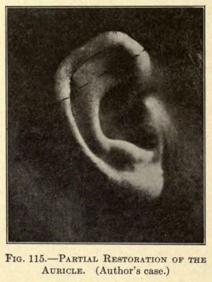
In the illustration shown the author restored the upper third of the ear shown above the line drawn obliquely across the ear. Seventeen delicate operations were necessary to obtain the result (see Fig. 115).
Where the loss of substance is not too great and along the helix of the ear, a flap can be taken from the back of the ear, leaving it attached at its cicatrized union with the primary wound, and sliding this flap upward or outward until the defect of the helix is overcorrected to allow for contraction and suturing the flap in its new position.
The secondary wound if too large to permit of direct union with sutures may at once be covered with a flap taken from the anterior
FIG. 115. PARTIAL RESTORATION OF THE AURICLE. (Author’s case.)
border of the arm, or, if preferred, from the inner aspect of the calf of the leg. The wound occasioned by the removal of the graft can easily be closed by suture, leaving simply a linear scar of little consequence. Usually such defects of the rim can be hidden by the combing of the hair, especially in women.
AURICULAR PROTHESES
When the injury has resulted in complete loss of the organ or so much of it that its remaining stump will not permit of otoplasty, protheses or artificial ears or parts of ears may be employed to render the patient less unsightly. These protheses are usually made of aluminum, papier maché, or rubber, and painted to match the good ear. They are attached with a special kind of gum, termed zincleim, which makers of such protheses furnish, or are held by metal springs, which are inserted under strips or bridges of skin surgically created for the purpose. The esthetic effect is surprisingly good in most cases.
COLOBOMA
A very common injury observed in women is laceration of the lobule of the ear or ears, generally due to the wearing of heavy earrings, which gradually cut their way through the tissues. Coloboma may be occasioned by the forcible tearing out of the earrings; it has also been found to be congenital in rare cases.
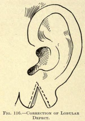
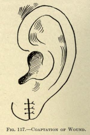
The simplest method for correcting this deformity is to cut away both cicatrized edges of the defect by the aid of the angular scissors, exposing fully the width of the lobular tissue on both sides (Fig. 116), as the cicatricial edges are likely to be thinner than the lobule proper, and if brought together would leave a depression along the line of union. The freshened cut surfaces are brought together with fine silk sutures, an inferior one being taken in the outer border, so as to establish perfect coaptation at this point (Fig. 117).
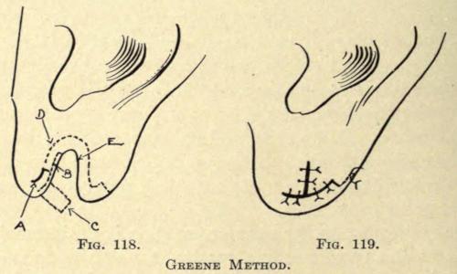
The objection to the above operation is that invariably owing to the resultant contraction a notch is formed at the union of the angles of the freshened wound. To avoid this the operation shown in Fig. 118 is to be employed (Greene).
FIG. 116. CORRECTION OF LOBULAR DEFECT.
FIG. 117.—COAPTATION OF WOUND.
The bistoury is thrust through the lobule at the point A and an incision is made to follow at a little distance the defect along the line D. This frees the cicatrix except at the pedicle A. A transverse incision is now made above the point Acorresponding to the curved exsection of the opposite side except for a thin strip of tissue B. This delicate little flap is preserved and severed a short distance beyond.
The raw edges when now brought in apposition will assume the form in Fig. 119. The wound is sutured as in the simpler operation.
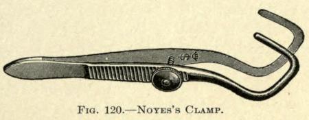
These operations are best performed under local anesthesia, the two-per-cent eucain being preferred. There is practically little bleeding, but even this may be avoided by applying a large Noyes’ compression clamp with its angular arms so placed as to include the entire lobule (Fig. 120).
MALFORMATION OF THE LOBULE
There may be an enlargement of or an absence of the lobule.
In the enlargement of the lobule the operation last described may be resorted to, making the now supposed coloboma the triangular
FIG. 118. FIG. 119.
GREENE METHOD.
FIG. 120. NOYES’S CLAMP.
ENLARGEMENT OF THE LOBULE
amount of tissue to be removed. It will be found that the upper curve of the incisions must be carried much higher in cases of this kind, furthermore, that they should define a sharper angle at this point.
The simple exsection of a triangular piece of the lobule and suturing is commonly practiced, with the objection of the notch previously referred to. This operation is very quickly done, and if care be taken in bringing the raw surfaces together neatly a splendid result is attained, especially if the incisions are made obliquely to the plane of the skin.
ATTACHMENT OF THE LOBE
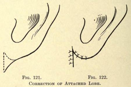
There may be a shortening of the lobule, or, as is more frequently seen, the attachment of the inner lateral border of the lobe to the skin opposite.
This attachment of the lobe has been alleged by criminologists to be a mark of the degenerate. If this be so it can scarcely apply to the Japanese, in whom it is found as a racial fact.
As the defect is often objected to by patients its correction may be considered briefly.
An incision is made in the inferior auricle and in the skin below it, as shown by the dotted lines in Fig. 121, removing the triangular piece of tissue included therein.
FIG. 121. FIG. 122.
CORRECTION OF ATTACHED LOBE.
The wound is then sutured with fine silk, as shown in Fig. 122, and allowed to heal. The result is very gratifying in most cases.
MALFORMATION
OF THE AURICLE
Malformations of the ear are due to the arrest of development, termed microtia, excessive development, or macrotia, and malposition.
MICROTIA
The total absence of the auricular appendage is quite rare. One or the other part of the ear is usually found, either partially or fully developed, giving to the ear an irregular rolled-up appearance. This defect may be unilateral or bilateral.
It may be associated with congenital fistula (Fistula auris congenita), varying in length from one fourth to one inch, and secreting a serouslike fluid. These fistulæ are usually found anteriorly and above the tragus, the lobule, or more rarely at the
crus helix, or even behind the ear. Sometimes these fistulæ communicate with the middle ear or even the esophagus. They are due to imperfect development inutero. In microtia little can be done surgically, since the malformation is usually so pronounced as to exclude all methods of restoration.
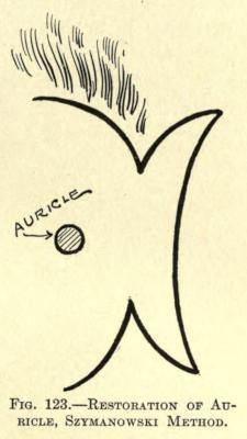
Szymanowski advises making an ear from the skin immediately back of the auditory canal if present, making the incisions of the shape shown in Fig. 123.
The flap included in these incisions is dissected up and doubled on itself posteriorly. The doubled flap thus formed is brought forward and placed as near into the linear position as the ear should have. The flap is then sutured through and through to make the raw surfaces heal together. The secondary wound and the treatment of the flap are carried out as already referred to under restoration of the auricle.
FIG. 123. RESTORATION OF AURICLE, SZYMANOWSKI METHOD.
Several later delicate operations are done to add to the shape of the newly made organ, but at best the effect is far from even good.
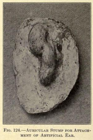
In the case of Mr. B., illustrated in Fig. 124, an attempt was made to enlarge the somewhat elastic roll of tissue corresponding to the helix by several injections of paraffin. The result proved to be anything but satisfactory; in fact, the prominence of the malformed upper ear was made more evident, and painful when subjected to pressure, so that the patient was compelled to refrain from lying on that side of the head.
IG. 124.—AURICULAR STUMP FOR ATTACHMENT OF ARTIFICIAL EAR.
F
There had been also congenital atresia of the auditory meatus, which had been operated for, leaving a hair-lined opening, leading down to a useless middle ear, a condition sometimes associated with microtia.
In presenting himself to the author for operation it was decided that the otoplastic methods for the restoration of the ear were out of the question, as is usually the fact in these cases.
The hard mass of tissue referred to and corresponding to the helix was reduced considerably, so that the stump obtained was soft and pliable, with not only the object of overcoming the sensitiveness and inconvenience of the part, but to obtain as good a base for the attachment of an artificial ear as possible (see Fig. 124).
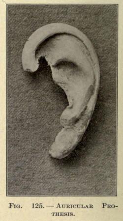
FIG. 125.—AURICULAR PROTHESIS.
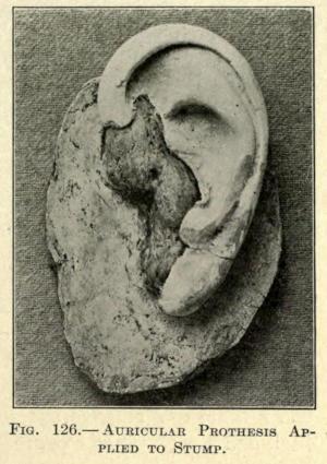
FIG. 126. AURICULAR PROTHESIS APPLIED TO STUMP.
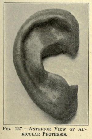
FIG. 127.—ANTERIOR VIEW OF AURICULAR PROTHESIS.
The author advises a complete amputation of such underdeveloped ears, since a better and firmer seat of attachment is offered thereby to the prothesis to be worn over it, at the same time giving the artificial organ a better position in reference to its normal relation to the face. An irregular stump makes this more or less difficult, as in the case just referred to, but even these patients are loath to part with an irregular ugly mass of tissue they consider themselves thankful to be born with.
The auricular prothesis used in this case is shown in Fig. 125, and its position and appearance when placed on the stump is shown in Fig. 126.
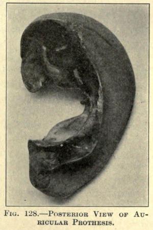
FIG. 128.—POSTERIOR VIEW OF AURICULAR PROTHESIS.
Another, showing both anterior and posterior surfaces, is given in Figs. 127 and 128.
The fistular conditions mentioned should be thoroughly dissected out and healed from the bottom when practical by antiseptic gauze packing. Those involving the middle ear require special treatment that cannot be included under plastic procedure.
MACROTIA
Abnormal enlargement of the ear is often found in the idiot, but is commonly seen as a hereditary defect in many without having the least relation to the mental development of the person. These conditions occur more frequently in men than in women.
Enlargement always depends upon overdevelopment of the cartilaginous structure of the auricle, and may also be the result of direct violence, the result of blows upon the organ, as in prize fighters, football players, and other athletes.
Following violence the auricle undergoes either an acute or chronic hypertrophy of the chondrium, resulting in the condition known as the “cauliflower ear.”
Again, there may be hematoma occasioned by direct violence, termed othematoma traumaticum, or a spontaneous development of such hematoma without any appreciable injury, as found in the insane. In the latter form the disease appears suddenly without warning or inflammatory manifestations, the hematoma reaching its full size in three or four days, after which a passive resolution in the form of absorption of the tumor takes place associated more or less with an organization of the blood mass, and leaving the auricular appendage unduly enlarged, distorted, and thickened, with here and there islands of seemingly detached or displaced cartilage firmly adherent to the overlying skin.
Early in these cases much can be done by the application of external medication, depletion, and pressure bandage, and the
removal of the effusion producing the swelling and lying between the perichondrium and the cartilage, by the introduction of a trocar cannula or by incision, as may be required.
The union between cartilage and perichondrium is always slow, requiring about three weeks in the traumatic variety and often months in the noninflammatory form.
Be the enlargement due to whatever cause, the patient not infrequently presents himself for a correction of the deformity.
The slightest of such deformities is a tiplike enlargement of the outer and upper angle of the helix, most commonly unilateral. This has been termed “foxear.”
In this condition there is more or less loss of the curl of the helix, with flattening beginning well down in the fossa, extending upward, and terminating in a triangular cartilaginous tip resembling the ear of an animal, hence the name.
The correction of this fault is quite simple. An incision somewhat larger than the base of the cartilaginous triangle is made under a local anesthetic about one fourth inch below and back of the line corresponding to the superior border of the helix. The cartilage is exposed through this incision and excised with a fine curved scissors without wounding the anterior skin of the helix, and the incision neatly sutured, leaving the now redundant skin to contract.
In this manner the fault is corrected without any appreciable scar.
The sutures can be removed in three or four days.
In the correction of macrotia various surgical methods may be employed, yet none can be emphasized, as exclusively indicated, inasmuch as the enlargements may involve one or the other part of the pinna.
The greatest fault with most of these ears lies in the overdevelopment of the triangular antihelix or that area lying posterior to the fossa of the antihelix and the fossa of the helix,
although in many cases the greatest malformation is found in the concha itself.
The following methods for operation are therefore given not so much for their individual merit, but to act as a guide in the selection of an appropriate election or modification for specific cases.
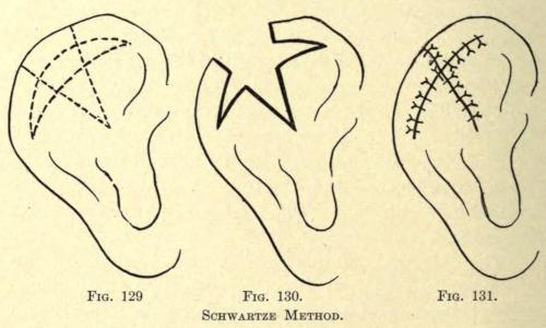
Schwartze Method.—Schwartze advises and has obtained excellent results by removing a long elliptical piece of the entire thickness of the pinna, including both skin and cartilage, from the fossa of the helix, followed by the excision of a triangular section with its base corresponding to the outer border of the helix and its apex terminating well in the concavity of the concha. The scheme of procedure is shown in Figs. 129 and 130. The raw edges are brought together by fine silk sutures, which are made to pass directly through the cartilage, and tied carefully to prevent any change of the transfixed parts, which would mar the result of the operation more or less and necessitate further interference. The arrangement of the sutures and the disposition of the parts are shown in Fig. 131.
FIG. 129. FIG. 130. FIG. 131. SCHWARTZE METHOD.
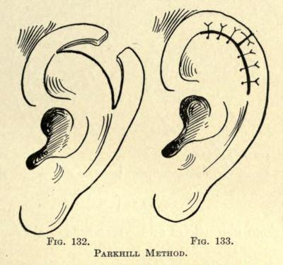
Parkhill Method.—Parkhill advises a semilunar incision from the fossa of the helix with a rhomboidal exsection of the helix, as shown in Fig. 132, and suturing the parts, as shown in Fig. 133.
FIG. 132. FIG. 133.
PARKHILL METHOD.
The tonguelike ends of the semilunar incisions must, of course, vary in length, according to the amount of tissue necessary to remove to facilitate accurate juxtaposition of the newly designed flaps.
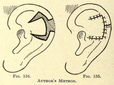
Author’s Method.—The latter operation is most successful where the upper part of the pinna is unusually flat. It does not correct this flatness, however, which is often an objection, hence the author suggests excising a section of the entire thickness of the ear from the fossa somewhat in the form shown in Fig. 134, curving the two deeper invading incisions, so that when the parts are brought together a concavity will be given the antihelix, as in the natural auricle.
The rearrangement of the parts in this event is shown in Fig. 135. The only objection to the above may be found in the two linear scars across the antihelix entirely overcome by the Parkhill operation, wherein the line of union falls just below the rim of the helix and into the groove commonly found there, yet any of these scars shows little in well-done operations and when union takes place by first intention.
There will always appear a notchlike depression where the newly cut ends of the helix are brought together, owing to the cicatrix involving the space between the cartilaginous borders.
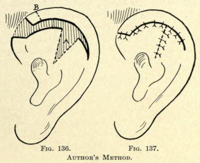
Inasmuch as this notch necessarily shows the most prominent part of the ear, the author advocates the following method in which the notch is brought anterior to the fossa of the antihelix; in other words, near to the point of the union of the helix with the skin of the face about on a line with the superior border of the zygomatic process; a point where the hair is in close proximity with the ear and where the scar can be more easily covered.
The form of incision is somewhat sickle shaped, the upper curvature of the incision following the inferior border of the helix and extending well into the fossa of the helix, as shown in Fig. 136. Where the antihelix is particularly large a triangular section may be removed, as shown at A, with a corresponding shortening of the helix flap at B. The latter gives more contour to the ear as well.
The parts are brought together and sewn into position, as shown in Fig. 137.
AURICULAR APPENDAGES
Small nipplelike projections of skin or elongated tumefactions of connective tissue are sometimes found about the tragus, the lobule, or on the neck. They are easily removed by encompassing their bases with an elliptical incision and amputating them a little below the level of the skin and suturing the wound in linear form.
FIG. 136. FIG. 137.
AUTHOR’S METHOD.
POLYOTIA
Auricular appendages may contain small pieces of cartilage or resemble crudely the auricle in miniature. This condition is termed polyotia. One or more of these supernumerary ears may be found anterior or posterior to the true ear or even below it on the skin of the neck.
In the case reported by Wilde there were four ears, the two abnormal ones being situated on the neck at either side. Langer has reported a similar case. The condition may be unilateral or bilateral.
This congenital malformation is corrected by simple amputation, as described under minor auricular appendages.
MALPOSITION OF THE AURICLE
The most common deformity met with in ears is undue prominence. The ears stand out from the head at an obtuse angle, often lopping forward and downward, giving the patient a stupid appearance. This condition is usually inherited, but may be acquired during childhood by the careless wearing of caps that crowd the pinnæ forward and away from the head. The habit of ear-pulling is also said to be a cause, also the faulty position of the head during sleep. The deformity is usually bilateral, but in the majority of cases one ear usually projects more than the other.
Where the deformity is recognized during infancy the ears should be simply bandaged to the head with a suitable bandage or ear cap, procurable for that purpose with the hope that the cartilages may thus be influenced during their period of hardening and growth.
Invariably these patients are seen too late, and operative procedures alone will restore the ears to their normal position.
The earlier in life such an operation is performed the more satisfactory is the result, inasmuch as the cartilage of the ear is more pliable, and hence more susceptible of readjustment;
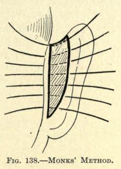
moreover, the operation when done early in life necessitates only the removal of an elliptical piece of skin from the back of the ear, according to Monks, and suturing of the wound, as shown in Fig. 138.
The elliptical form of the incision must, however, be changed according to the varied prominence of various parts of the ear. When the ear lops forward, it should be broader above and narrower below, and vice versa in the event when the concha is overprominent.
When the patient is less than fourteen or fifteen years of age a general anesthetic should be employed, but in older patients the operation can be easily undertaken under local use of two-per-cent eucain solution.
Author’s Method.—The method followed by the author is to thoroughly anesthetize the back of the ear, the patient lying in a recumbent position with the head to one side, sufficient to place the ear to be operated upon in as convenient position for operation as is possible. A rubber cap is drawn over the head to cover the hair.
An incision is now made along the whole of the back of the ear as far down as the sulcus, where the retro-aural integument joins that of the neck.
The incision should involve the skin only, and vary from three fourths to one half an inch from the outer border.
FIG. 138. MONKS’ METHOD.
At once the blood will ooze from the line of incision. The operator now presses the ear backward on the bare skin of the head, leaving an imprint of the bleeding line on the skin there.
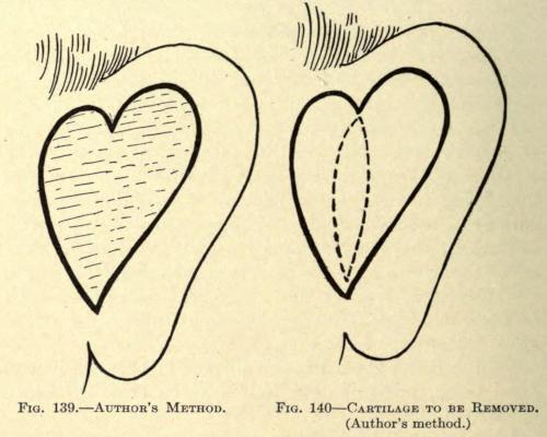
A second incision is made along this line, giving the total outlining incision a heart-shaped form, as shown in Fig. 139.
The skin within this area is now dissected up quickly. There will be more or less bleeding from the post-auricular vessels, which can easily be controlled by sponge pressure, or with one or two artery forceps of the mosquito-bill pattern. The wound should be large enough to overcorrect the fault, as the ear springs out more or less when healed.
Sutures are now introduced. When necessary one or two catgut sutures are taken through the concha, not going through the anterior skin, however, and the deeper tissue back of the ear and tied. These hold the cartilage in place.
FIG. 139. AUTHOR’S METHOD. FIG. 140.—CARTILAGE TO BE REMOVED. (Author’s method.)
For the coaptation of the skin the continuous suture is to be preferred, but when the cartilage suture is employed it will be found impracticable, owing to the close position of the ear to the head. In that event interrupted sutures must be placed, as shown in the Monks operation, and tied after the cartilage has been fixed as described.
Where it is deemed necessary to fix the cartilage in this way, the author advises to remove an elongated elliptical piece of the concha, as shown in Fig. 140.
This is neatly done by outlining the section with the scalpel, and excising it with the aid of a fine pair of scissors, half rounded; the operator holding the index finger of the left hand in the depression of the concha anteriorly as a guide to avoid injuring the skin.
After the elliptical exsection a linear incision with the scissors may be made both superiorly and inferiorly to further mobilize the springy shell of the ear, which will then be found to fall easily into place.
The bleeding in the latter method is more severe, since the posterior auricular arteries and the auricular branch of the occipital have to be severed, yet ligation is rarely necessary.
The interrupted suture may now be applied, varying the site of puncture as below or above its fellow puncture, as made necessary by the droop of the ear, with the object of shifting it into a normal position; or in other words, by raising or lowering it upon tightening the sutures.
The continuous suture is to be preferred, however, when the cartilage has been removed as described, since the ear has now become quite mobile and is easily placed in position.
When the removal of these sutures, which should be of Nos. 5 or 6 twisted silk, is considered, one can comprehend the advisability of this form of wound closure.
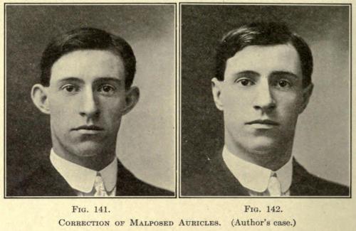
The ear will now appear to lie quite close to the head, compared with the original position, as shown in Figs. 141 and 142.
The patient is now turned so as to present the other ear, a pad of gauze and absorbent cotton being placed under the ear operated on for comfort’s sake.
The second ear is operated as was the first, the operator having taken note of the form and size of the incision of the ear just finished.
Both ears sutured, the wounds are cleansed thoroughly, though gently, with fifty-per-cent peroxid of hydrogen and dried and dusted over with aristol powder.
A pad of gauze is placed over each ear and a bandage applied around the head to protect the wounds and retain the ears, care being taken not to tighten too tightly, as this occasions great pain and possible pressure erosion of the skin.
The dressing should be changed on the second day, as there is usually some soiling of the dressings at the lower angles of the wounds. They are again powdered, using the pulverflator preferably, and rebandaged.
FIG. 141. FIG. 142.
CORRECTION OF MALPOSED AURICLES. (Author’s case.)
The ears will be found to lie very close to the head at this time, if the operation has been properly done. Anteriorly in the skin of the concha and corresponding to the line of cartilage exsection will be found a crease more or less discolored, according to the severity of injury occasioned by the operation.
This should give the surgeon no concern, as the fold will accommodate itself in a few days. There may be a persistence of the fold for some time, however, which, if desirable, can be corrected by a small secondary operation at a later date. The author has never experienced the need of such, however.
The patient at this time usually bemoans the position of his ears, and should be assured beforehand what was expected, and that the condition is only temporary.
The dressings after this can be repeated every second or third day, as may be required, although these wounds heal surprisingly well.
Moist dressings are to be avoided at all times, they soften the edges of the wound and prevent primary union.
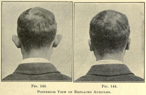
FIG. 143.
FIG. 144.
P
OSTERIOR
VIEW OF REPLACED AURICLES.
The sutures are removed on the ninth or tenth day, whereafter the patient may be allowed to go without the head bandage, but is strictly instructed to replace it at night with a band of muslin three inches wide, snugly pinned around the head to prevent the ears from being injured or torn away from their new attachment by sudden movements during sleep. This bandage should be worn at night for at least a month.
When only a part of the ear is overprominent the operation undertaken should in the main be according to the methods just described, the incisions being changed in extent accordingly.
In the illustrations above, Figs. 143 and 144, are shown the posterior view of the ears before and after operation. At no time should the ears be placed too closely to the head, as is often peculiarly requested by the patient, as it gives an unnatural appearance and predisposes toward the collection of filth in the sulcuses that is hard to remove. The distance from the head to the outer rim of the ear should be about half an inch at its widest part.
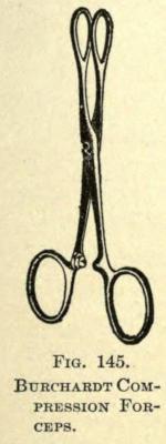
FIG. 145.
BURCHARDT COMPRESSION
FORCEPS.
CHAPTER XI
CHEILOPLASTY
(Surgery ofthe Lips)
This branch of plastic surgery has to do with the correction of deformities of the lips. These deformities usually involve one lip only, and are dependent upon direct traumatism, operative interference in the extirpation of malignant growths, particularly carcinomata, the correction of cicatricial disfigurement following tubercular or syphilitic ulceration or congenital faults, commonly met with in harelip.
Operations for the latter condition have usually been considered under a separate heading, but since the restorative procedures involve methods purely plastic they are included under this their proper classification.
Owing to the great number of blood vessels in the lips, it is advisable to resort to the bloodless method, where the defect to be corrected involves more than the superficial structure. This is accomplished:
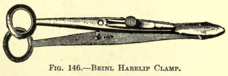
1. By compressing the coronary arteries at both angles of the mouth by digital pressure, suitable clamps or compression forceps. The fenestrated oval forceps, illustrated in Fig. 145, and designed by Burchardt, or the harelip clamp of Beinl, Fig. 146, will be found to meet the purpose well, the latter having a
sliding lock by which the pressure upon the tissue can be regulated to a nicety.
2. By clamping off the site of operation with specially made cutisector forceps. Its smooth parallel jaws should be curved outward, so that the diseased area can be fully excluded by their concavities.
3. By employing the indirect ligature of Langenbuch. This is accomplished by including the site of operation with several strong silk threads firmly tied in loops upon the skin surface, each loop including a given amount of tissue, the next encroaching upon it up to the center of this area, and so on until the entire site is rendered anemic. The advantage of this method is that with the anemia a certain amount of anesthesia is produced at the same time; a fact to be remembered when the patient is to be operated under local anesthesia, the anemia enhancing the efficacy of the latter.
HARELIP
A congenital defect of the upper lip caused by the lack of proper union of the maxillary, globular, and frontonasal processes in embryo. Treves states that from the buccal aspect of the maxillary process of either side the palatal processes arise, passing inward to combine with each other to form the soft palate and all of the hard palate, except the intermaxillary portion, and that from this same source are formed the cheeks, the outer or lateral parts of the upper