Previous editions copyrighted 2015, 2008, 2005, 1995 and 1988.
ISBN: 978-0-7020-7401-1
This translation of Sims’ Symptoms in the Mind, 6th ed., by Femi Oyebode was undertaken by Elsevier España, S.L.U. and is published by arrangement with Elsevier, Ltd.
Esta traducción de Sims’ Symptoms in the Mind, 6.ª ed., de Femi Oyebode, ha sido llevada a cabo por Elsevier España, S L U y se publica con el permiso de Elsevier, Ltd.
Cualquier forma de reproducción, distribución, comunicación pública o transformación de esta obra solo puede ser realizada con la autorización de sus titulares, salvo excepción prevista por la ley. Diríjase a CEDRO (Centro Español de Derechos Reprográficos) si necesita fotocopiar o escanear algún fragmento de esta obra (www.conlicencia.com; 91 702 19 70/93 272 04 45).
Advertencia
Esta traducción ha sido llevada a cabo por Elsevier España, S.L.U. bajo su
única responsabilidad Facultativos e investigadores deben siempre contrastar con su propia experiencia y conocimientos el uso de cualquier información, método, compuesto o experimento descrito aquí. Los rápidos avances en medicina requieren que los diagnósticos y las dosis de fármacos recomendadas sean siempre verificados personalmente por el facultativo. Con todo el alcance de la ley, ni Elsevier, ni los autores, los editores o los colaboradores asumen responsabilidad alguna por la traducción ni por los daños que pudieran ocasionarse a personas o propiedades por el uso de productos defectuosos o negligencia, o como consecuencia de la aplicación de métodos, productos, instrucciones o ideas contenidos en esta obra.
Revisión científica:
Dr. Luis Manjarrez Gutiérrez
Coordinador de Evaluación del Departamento de Psiquiatría y Salud Mental Facultad de Medicina de la Universidad Nacional Autónoma de México (UNAM)
Servicios editoriales: DRK Edición
Depósito legal: B.22.699-2019
Impreso en Italia
Dedicatoria
A mi padre, Jonathan Akinyemi Oyebode (1918-1971)
Femi Oyebode
Prefacio a la sexta edición
Este año se cumple el trigésimo aniversario de la primera publicación de Sims. Síntomas mentales. Se puede decir que, en la actualidad, se ha convertido en el libro más destacado sobre psicopatología clínica. En esta sexta edición, como ya sucedió en las cinco previas, he mantenido la estructura original del libro, pero he realizado ciertos cambios y he añadido abundante material nuevo. He introducido o desarrollado algunos temas, sobre todo acerca de la consciencia y sus trastornos, las alteraciones de la memoria, y la patología de la percepción, así como los trastornos del self y de la consciencia corporal. El material nuevo más destacado corresponde al papel de la corporalidad en la naturaleza del self y de la consciencia del cuerpo, así como de la naturaleza de la culpa y la vergüenza en los trastornos emocionales. Estos cambios se han visto motivados por mi deseo de garantizar que los lectores comprendan que la psicopatología no es una materia muerta, sino un tema vivo y con una necesidad constante de revisión para responder a los cambios conceptuales o a los nuevos hallazgos empíricos.
En mi prefacio a la quinta edición, subrayé el hecho de que la psicopatología descriptiva como método ha sobrevivido los últimos 100 años y constituye el fundamento primordial de la práctica de la psiquiatría clínica. Este método nos permite observar y describir los comportamientos y fenómenos subjetivos anómalos, así como clasificarlos a fin de poder transmitir, de un modo más preciso, cómo es el mundo en el que vive el paciente. El clínico formado en el enfoque fenomenológico es incluso más consciente de lo necesaria que resulta la comprensión empática y la adopción de una actitud ateórica y, en último término, de lo provisional que continúa siendo el estado de nuestra comprensión y nuestras explicaciones acerca de la psicopatología. La psicopatología descriptiva es incluso más relevante en la actualidad para los cometidos de los clínicos e investigadores. La nomenclatura psiquiátrica estándar es motivo de controversia. Esto significa que los fenómenos anómalos fundamentales, la infraestructura de la nosología, deben necesariamente asumir una mayor importancia en la práctica clínica. De lo contrario, la capacidad de establecer una comunicación clara entre los distintos profesionales se verá profundamente deteriorada. Estoy en deuda con más personas de las que soy capaz de nombrar. El Birmingham Philosophy Group se ha venido reuniendo mensualmente desde 1992. Sus miembros (Theo Arvantis, Lenia Constantine, Simon O’Loughlin, Kate Robertson, Sandy Robertson y Persephone Sextou) siguen influyendo en mi forma de considerar los fenómenos psiquiátricos, al igual que también lo
hacen los integrantes de la sección de psicopatología de la Asociación Psiquiátrica Europea, entre los que se incluyen John Cutting, Maria Luisa Figueira, Mircea Lazarescu, Luis Madeira, Michael Musalek, Gilberto di Petta y Pedro Varandas. Por último, este libro sería sin duda mucho peor sin los pacientes que experimentan y padecen estos fenómenos anómalos, y sin los estudiantes y residentes de psiquiatría que formulan preguntas incómodas y que, guiados por la curiosidad, investigan la naturaleza de estos fenómenos.
Femi Oyebode
SECCIÓN I
Conceptos y Métodos
Capítulo 1: Conceptos fundamentales de la psicopatología descriptiva
Capítulo 2: La evaluación de los síntomas de los padecimientos mentales
Another random document with no related content on Scribd:
There is the fact to remember in making these measures in the extreme red and the extreme violet, that the luminosities of the colours are so small that the illumination of the prism itself, by the white light falling on it, has to be taken into account, since it forms an appreciable portion of the patch of feeble colour. By placing a proper shade of blue or red glass in the front of the collimator slit this white light disappears or becomes negligible, and when the absorption of the coloured glass is known from measurement, we can get a very accurate measure of the extinction of these parts.
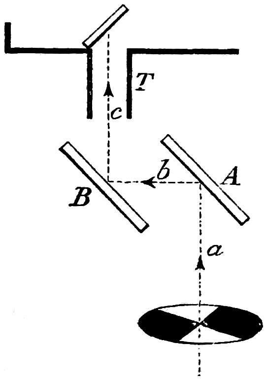
Some people may propound the idea that the rotating sectors may in such kind of measurements give a false result. Now such a criticism is quite fair, and it is absolutely necessary that it should be answered. Well, to test the accuracy or the reverse of the assumption that such measures are correct, the following small piece of simple apparatus was devised. A and B (Fig. 29) are two mirrors placed at angles of 45° to the angle of incidence of the beam. The path the beam takes can be readily ascertained from the figure. This piece of apparatus was placed in position in front of the spectrum, and the reflected beams used to form the patches of colour. For convenience only a small pencil of light was allowed to issue from the prism, a diaphragm of some ½-inch in diameter being placed in front of it. This allows a spot of any desired colour to fall on the screen, the ground glass being removed. The slit through which the spectrum colours pass is moved along the spectrum, and a position is arrived at where the last glimmer of light disappears.
FIG. 29.
The mirrors A and B may both be of plain glass blackened with smoke on one side, or one may be plain glass and one silvered, or they both may be silvered. This, with the power possessed of altering the aperture of the slit of collimator, puts us in possession of ample means of making our measures. We may also use the groundglass arrangement and use different diaphragms, which puts a
further power of variation in our hands. I may at once state that the resulting measurements fell on the curves, obtained by measurements made with the rotating sectors, a sufficient proof that the sectors may be used with confidence. There is still another method which avoids a resort to the sectors. A tapering wedge of black glass can be moved in front of the colour slit, and a different thickness of glass will be required to cause the extinction of each colour. Recently I have modified the extinction box, more particularly for the purpose of using it where the spectrum is to be formed of a feeble light, such as that of an incandescent lamp or a candle. If a really black wedge could be obtained, this would seem to be the best method, but no glass is really black. We have, therefore, to make a preliminary study of the wedge to ascertain accurately the absorption co-efficients for the different rays, a piece of work which requires a good deal of patience, but which, when done, is always at command.
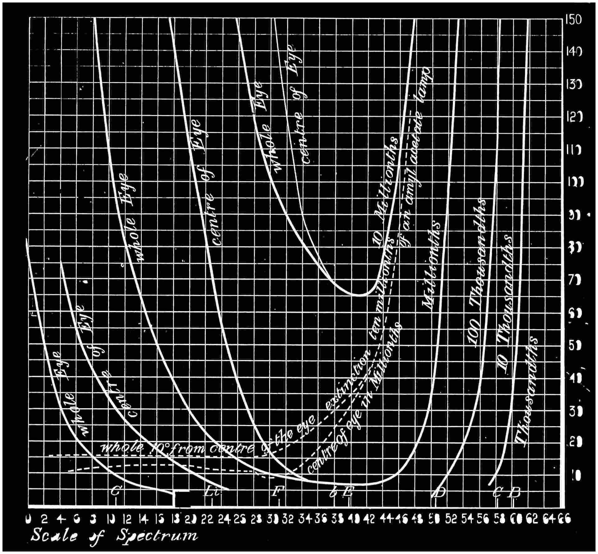
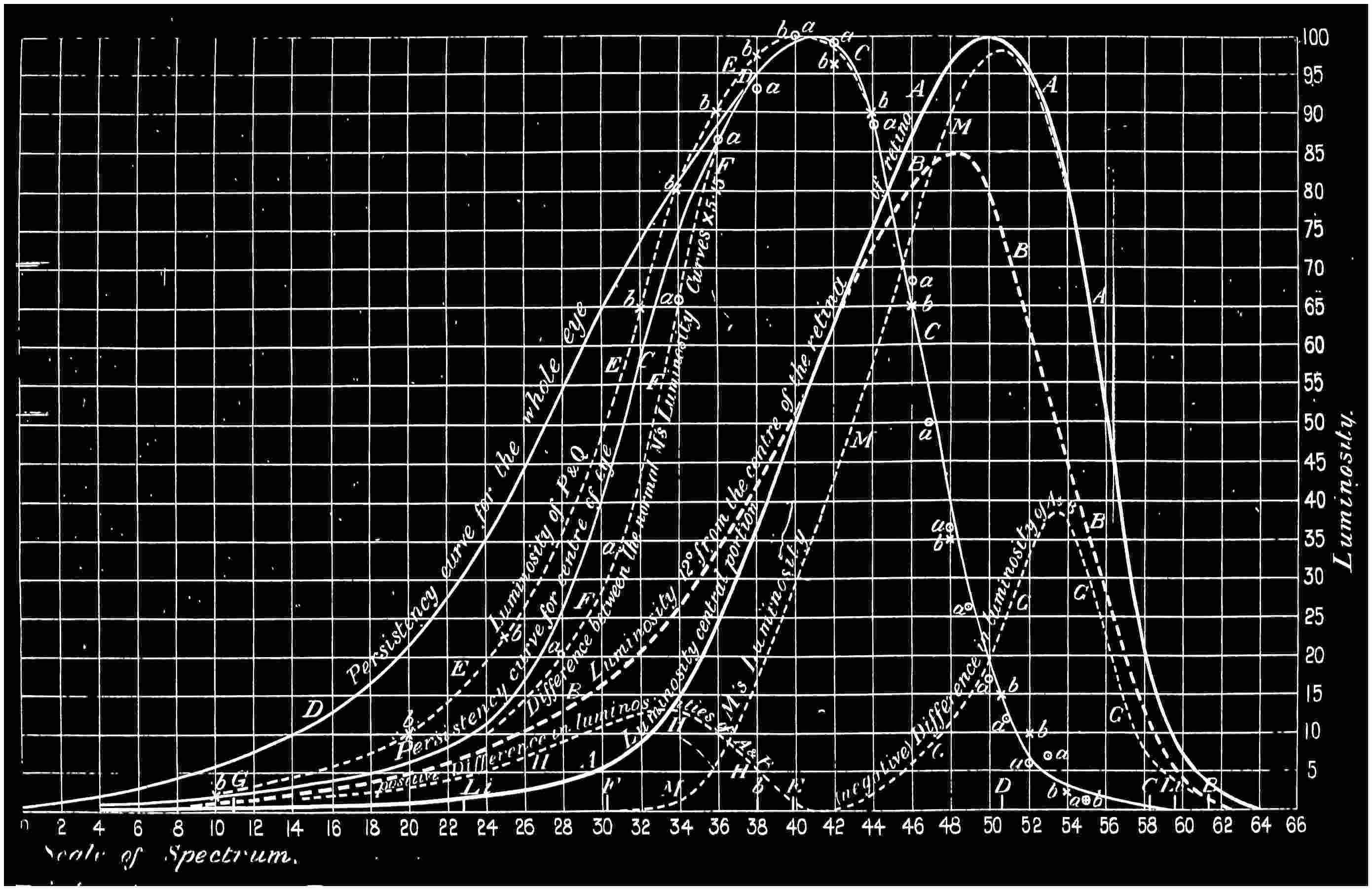
In Fig. 28 two branches of the curves are given at the blue end of the spectrum; one is shown as the extinction for the centre of the eye, and the other of the whole eye. Of course the former observations were made by looking direct at the spot. This may appear a very easy matter, but it is not really so simple as it sounds. It is curious how little control there is over the absolute direction of the eyes when the light has almost disappeared. The axes of the eyes are often directed to quite a different point. When the extinction for the whole eye is made, the readings are really much easier, as then the eye roams where it likes, and a final disappearance is noted. When the eye has once been invested with a roving commission, it is hard to control it. In making these observations it was therefore advisable to have data for the first branch of the curve, before commencing to observe for the later. The main cause of difference between the two branches of the curve is due to the absorption by the yellow spot.
It might be thought that with the curves (Fig. 28) before us, we have learnt all we can regarding the extinction of light, but is it so? Surely we ought to know something as to the reduction necessary
for extinction of the different parts of the spectrum when they are all of equal luminosities and of ordinary brightness.
We arrive at this by simple calculation. Supposing we have two luminosities, onedoubletheother, it does not require much thought to find out that you have to reduce the greater luminosity twice as much as the other in order for it to be just extinguished. In other words, if we multiply the extinction by the luminosity, we get what we want. Now, in the curves before us, we have taken the luminosity of the yellow light near D as one amyl-acetate lamp, and that has a height in the curve showing the spectrum luminosity very closely approaching 100. We may, therefore, multiply the extinctions of a ray by the value of its ordinate in the luminosity curve and divide the result by 100, and this will give us the extinction of each colour, supposing it had the luminosity of an amyl-acetate lamp. A portion of the curve so calculated is shown in the same diagram (Fig. 28) as a dotted line. It appears at the violet end as an approximately horizontal line, and then starts rapidly upwards, and would, if carried on to the same scale, reach far out of the diagram; but at the extreme red it would be found to bend and again become horizontal. I would have you notice that the same is true not only for the extinction observed with the centre of the eye through the yellow spot, but also for the whole eye. Such straight, horizontal parts of the curve must mean something.
In the diagram (Fig. 16) of colour sensations we see that in each of these two regions there is but one sensation excited, viz. the violet and the red. Now, if these sensation curves mean anything, the reduction necessary to produce the extinction of the same sensation when equally stimulated should prove to be the same, for there is no reason to the contrary, but exactly the reverse. Primâ facie, then, taking the Young theory as correct, we may suppose that these horizontal parts are due to the extinction of one sensation. Let us treat it as such, and go back to the original extinction curve shown in the continuous lines. The parts of the curve which lie over the fairly horizontal dotted line, at all events, should be the extinction curve of the same sensation, but more or less stimulated or excited. As before explained, if we have double the stimulation at one part of the spectrum to that we have at another, the reduction of the greater luminosity to give extinction will be double that of the lesser. If, then, we take the reciprocalsofthe
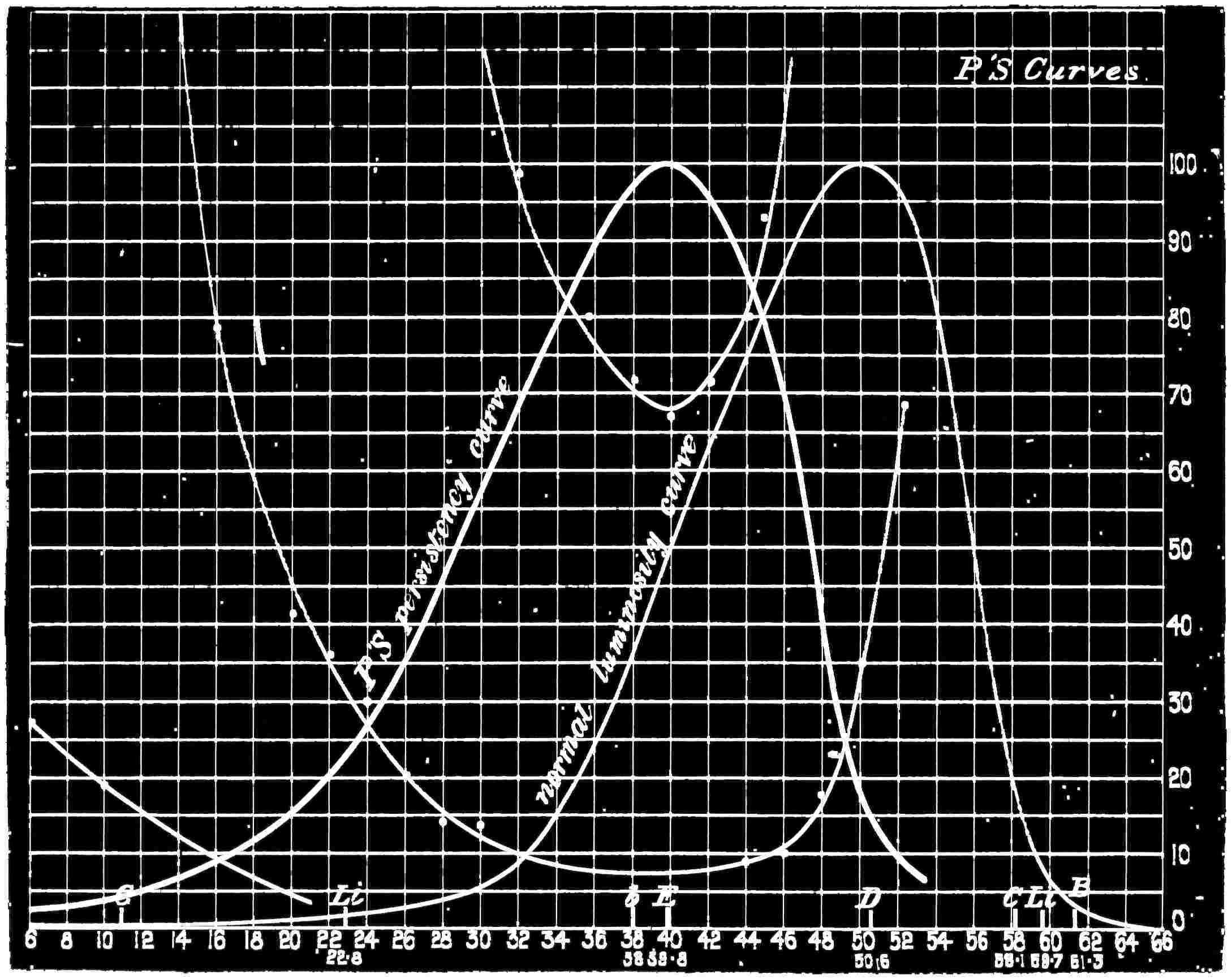
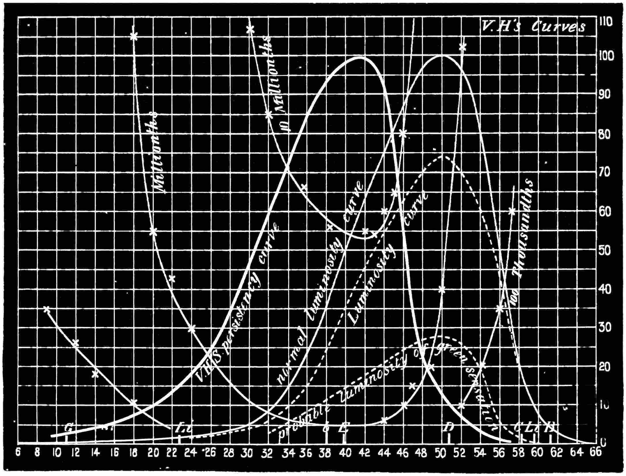
extinction, it ought to give us a curve which is of the form of some colour sensation; and when we arrive at the maximum, we may for convenience make that ordinate 100, and reduce the other ordinates proportionally. This has been done in Fig. 30 in the curves C and D. For the sake of a name my colleague and myself have named such curves “persistency curves.” Perhaps some other name might be more fitting; but still a poor name is better than none at all.
When the persistency curve was scrutinized to see what might be taken as its full signification, I must confess that the result astonished us somewhat, though we ought not to have been surprised. The persistency curve C, when applied (in a Euclidean sense) to the curve of luminosity recorded for the men who had monochromatic vision, almost exactly coincided with it. In other words, by far the largest part of the extinction was due to the extinction of the sensation which in the monochromatic vision was alone excited. If this be not the case, there is something in colour vision which no theory which I am acquainted with can account for. Then, again, the persistency curve agrees with the curve of luminosity when the intensity of the spectrum is very feeble, which is another coincidence of a remarkable character which some theory should explain. [Fig. 30 gives, besides the persistency curves, the luminosity curves of the normal eye, of monochromatic vision, and of the violet-blind; and an exaggerated curve of the difference between the normal luminosity curve and that of the violet-blind, and others which I think will be found useful for general reference.]
What sensation is it that is last extinguished, and which is possessed by a certain class of colour vision? In the Young theory it can only be the violet sensation. It is certainly not the green, and much less the red. It does not correspond, however, very well with the violet sensation shown in Fig. 16, but more with one which should be in the blue.
In making the extinctions of light, it is quite necessary that certain precautions should be taken to avoid error. All my audience know that when going from bright daylight into a cellar, in which
only a glimmer of light is admitted, but little can be seen at first, but that, as the eye “gets accustomed” to the darkness, the surroundings will begin to be seen, and after several minutes what before was blackness comes to be invested with form and detail. So it is with the extinction of light in the apparatus described. Observations carried on before the full sensibility of the eye is attained are of no value. A recorded set of observations will show this. A light of a certain character was thrown on the extinction box, to be extinguished, and the observer entered the darkened room from the full glare of daylight. The eye was placed at the eye end and kept there, and the extinctions were made one after the other till they became very fairly constant. The following is the result:—
Times of Observation. Readings. At the commencement 1·0
After 38 sec. 3·2
After 53 sec. 4·9
After 1 min. 11 sec. 6·9
After 1 min. 44 sec. 10·5
After 2 min. 43 sec. 17·0
After 3 min. 44 sec. 27·5
After 4 min. 52 sec. 43·0
After 5 min. 59 sec. 63·0
After 6 min. 41 sec. 78·0
After 7 min. 28 sec. 89·0
After 8 min. 32 sec. 96·0
After 10 min. 46 sec. 103·0
After 12 min. 103·0
(For convenience the first reading is unity; the other numbers are the inverseof the extinction value.)
The eye apparently, under the conditions in which these observations were made, was at least 100 times more sensitive to
very faint light after twelve minutes than it was at the beginning, and that then concordant readings could be made. It will now be quite understood that before any serious measures can be made this interval must elapse, and also that the light, finding its way to the end of the box to illuminate the spot, should never be strong, otherwise the eye might lose its sensitiveness.
CHAPTER X.
BEFORE considering the subject of the extinction of light by other types of colour vision, attention must be called to what has already been brought before you. The various colours of the spectrum have to be reduced to the following amounts before they suffer extinction, the orange light at D being of the value of one candle. (See appendix, page 217, for complete tables.)
Reduction in Millionths.
Remarks.
B 10,000 or 1/100 approximately pure red sensation
C 1,100 or 1/909 rather more scarlet
D 50 or 1/20000 orange light
E
6·5 or 1/154000 a green chosen by Maxwell as a standard colour
F 15·0 or 1/67000 beginning of the blue
Blue Lithium 85·0 or 1/11700 a good sample of blue
G 300·0 or 1/3300 approximately pure sensation of violet.
If we make these same colours all of the luminosity of one amylacetate lamp (·8 of a candle), we find that the numbers are as follows:
Reduction in Millionths.
These numbers are remarkable, and we may enforce what they mean in this way. The energy of radiation, and of light also when of ordinary luminosity, varies inversely as the square of the distance from an incandescent body when of small dimensions. But from the above it seems that a white screen receiving the rays from an amylacetate lamp in an otherwise perfectly dark place, and having a colour which stimulates the red sensation alone, would be invisible at 58 feet distance, for there would not be enough energy transmitted to stimulate the red perceiving apparatus sufficiently to give the sensation of light. If it were an orange light, such as sodium, of the same luminosity, we should have to move it from the screen 142 feet before the same result was attained. With the green light at E, the distance would be 550 feet, and with the violet the distance would be increased to 1000 feet. The reduction in intensity of white light, which, when of ordinary brightness, is warm, would make it colder, for the red would disappear, and finally the residue of light, just before extinction, would become a cold grey, due to the absence of all colour. The changes in hue that would occur are variable, the variation being due to the loss of colour of the different rays for different amounts of reduction, and then their final extinction. We can place two patches of white light on the screen, and gradually reduce one in intensity, keeping the other of its original value. No one would expect that the two would be dissimilar in hue, as they appear to be when the former is moderately near the extinction value. If we wish to see this perfectly, we should use an extinction box and view it away from the surroundings, which must be more or less slightly illuminated.
It has already been stated that the persistency curve for persons who have normal colour vision is closely the same as that recorded
for those who are of the monochromatic type. As this is so, we must expect to find that the persistency curve of these last is the same as their luminosity curve. We put this to the test of experiment and found that our reasoning was correct, for the persistency curve could be almost exactly fitted to it. (See table, pages 217 and 222.) The slight difference between them can be credited to the fact that the whole eye may have been brought into use during the extinction observations, the centre of the eye not being exclusively used. The Figure 31 shows both the extinction and the persistency curves, and also the curve of luminosity for the normal eye.
FIG. 31.
The former were derived from a case P. sent for examination. P. and Q. are brothers, each of whom possesses but one colour sensation, and examination showed that their vision was identical. Mr. Nettleship has kindly given me the following particulars regarding them:—“Their acutes of vision (form vision) in ordinary daylight is only one-tenth of the normal. A younger sister and brother are idiotic and almost totally blind, and in one of these the optic nerves show clear evidence of disease. Hence, the colour blindness of P. and Q. must almost without doubt be considered as the result of disease, perhaps ante-natal, involving some portion of the visual apparatus.” A lack of acuteness of vision would be expected from the small amount of light they perceive compared with normal vision. The fact that two of a family, not twins, possess exactly the same colour sense, and that their extinction curves are entirely different to those suffering from post-natal disease, but similar to those of normal vision, point to their colour blindness as falling in the same general category as that of the congenital type. To this I shall refer again.
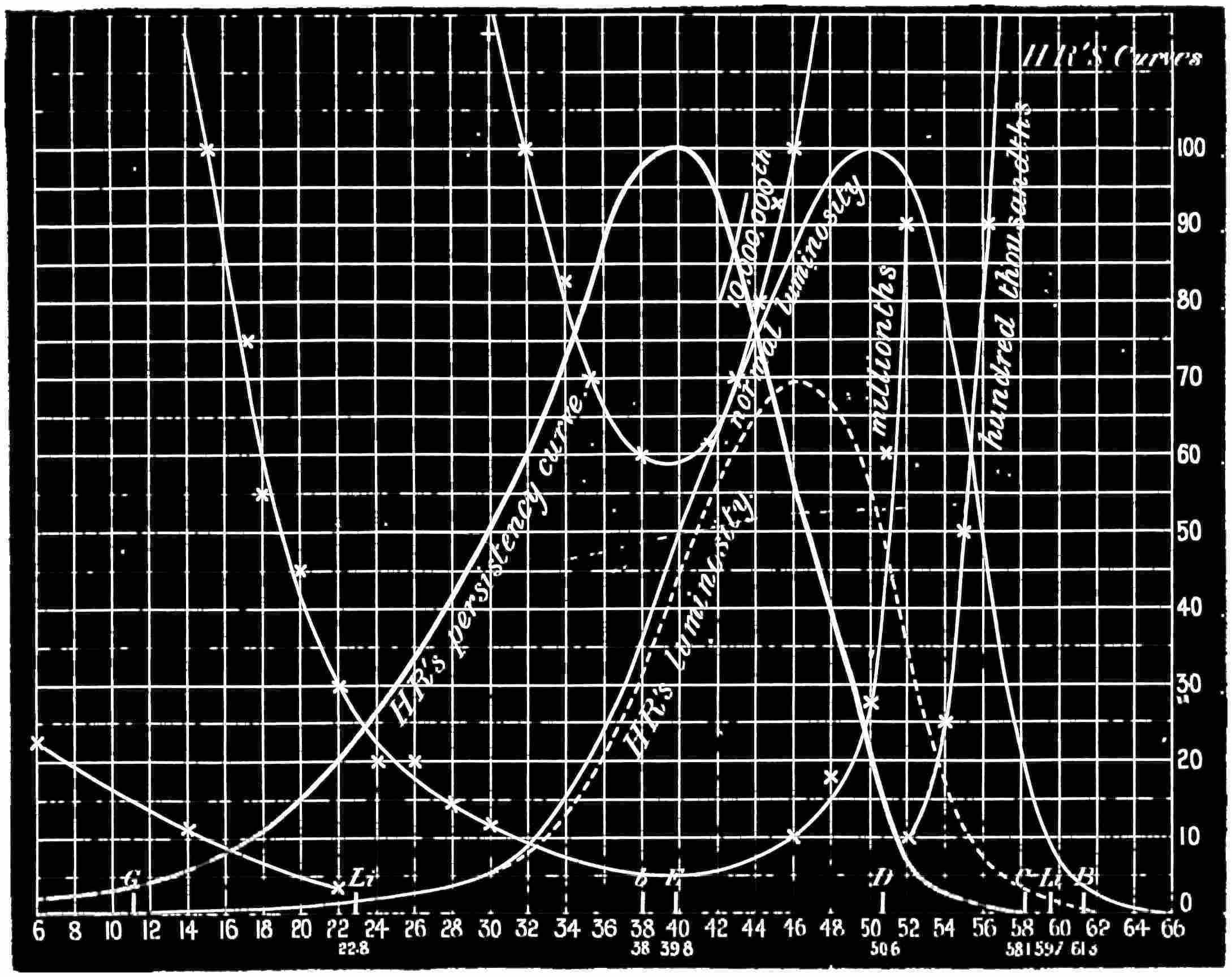
We may reason still further. With the red- and green-blind the violet sensation is still present, and we may therefore expect that their extinction curves, and consequently their persistency curves, should be alike, and should also agree with that made from your lecturer’s observations. A study of Figures 32 and 33 will tell you that such is practically the case. The former shows the luminosity, the persistency, and the extinction curves of a completely red-blind subject, and the latter the same curves for a green-blind subject (see pages 223 and 224). Both were excellent observers, and their examination was easy, owing to the acquaintance with scientific methods. The accuracy of their results may be taken as unquestionable. Each of them may be taken as a representative of
their own particular type of colour blindness. There is an agreement between them at the violet end, but a deviation at the red end of the spectrum. The general form of the curves indicates that the same sensation is extinguished last in all. Now, have we any other criterion to offer? We have. In the first instance, we have the violetblind person to compare with the others, and also another observer who had monochromatic vision, but whose sensation was different to that of the two monochromatic cases we have so far brought to your notice. We have already stated the peculiarities in colour nomenclature of the violet-blind case. His curve of luminosity for the spectrum was taken (page 227), and when compared with the curve of normal luminosity, it became evident that in the red and up to the orange his measures were those which a normal eye would make; but that the luminosity fell off in the green, and finally disappeared to an immeasurable quantity in the violet (see Fig. 30, curves M and F). If his measures of spectrum luminosity are deducted from those of the normal eye, and the ordinates be increased proportionately to make the maximum difference 100, the figure so produced, when compared with the luminosity curve obtained from the monochromatic observers, was found to be the same, and consequently with the persistency curves above referred to. Endeavours were made to gain a good extinction curve, but the results were not as successful as could be desired; but it was ascertained that, without doubt, his most persistent sensation was not more than 1/180 as lasting as that of the normal eye, or to put it in another way, his green at E was only extinguished when the energy falling on his eye was 180 times greater than that at which it vanished with the normal eye. This plainly teaches us that the missing sensation was that which, when present, is ordinarily the most persistent.
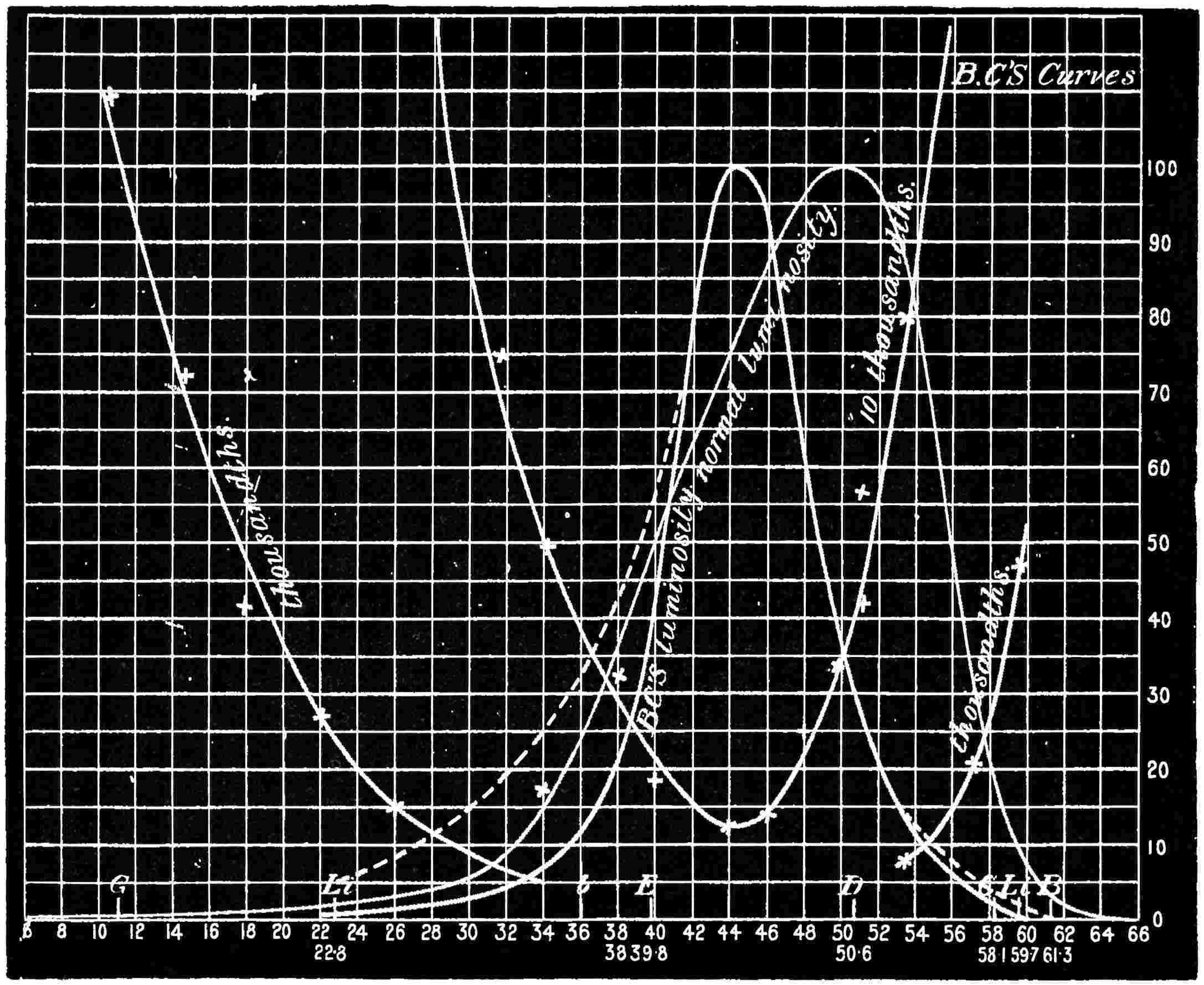
The next is a case of monochromatic vision, which differs from those previously brought before you, and I cannot do better than describe it in the words which General Festing and myself employed in our paper in the “Philosophical Transactions.”
The patient (B. C.) had been examined by Mr. Nettleship, who kindly secured his attendance at South Kensington for the purpose of being examined by the spectrum and other tests. [Mr. Nettleship states that this case is without doubt a genuine case of congenital colour blindness, without any trace whatever of disease.] B. C. is a youth of 19, who has served as an apprentice at sea. His form vision is perfect, and he is not night blind. He can see well at all times, though he states that on a cloudy day his vision seemed to be slightly more acute than in sunshine. He was first requested to make matches with the Holmgren wools in the usual manner, with the result that he was found to possess monochromatic vision. He matched reds, greens, blues, dark yellows, browns, greys, and purples together; and it was a matter of chance if he selected any
proper match for any of the test colours. Finally, when pressed, he admitted that the whole of the heap of wools were “blue” to him, any one only differing from another in brightness. The brighter colours he called “dirty” or “pale” blue, terms which eventually proved to be synonymous. We then examined him with patches of monochromatic spectrum colours by means of the colour patch apparatus. He designated every colour as “blue,” except a bright yellow, which he called white, but when the luminosity of this colour was reduced he pronounced it a good blue. So with white, as the illumination was decreased, he pronounced it to pass first into dirty blue, and then into a full blue.
Colour discs were then brought into requisition, and it was hard at first to know how to make the necessary alterations, owing to the terms he employed to express the difference which existed between the inner disc and the outer grey ring. By noting that a pale “blue” passed into a pure blue when the amount of white in the outer ring was diminished, and that the inner disc was described as “pale” or “dirty” when the outer ring was described as “a very full blue,” we were enabled to make him match accurately a red, a green, and a blue disc separately with mixtures of black and white.
The following are the equations:—
360 red = 315 black + 45 white.
360 green = 258 black + 102 white.
360 blue = 305 black + 55 white.
With these proportions he emphatically stated that all were good blues, and that the inner disc and outer ring were identical in brightness and in colour.
It may be remarked that this is a case of congenital colour blindness, and that there is reason to believe that some of his ancestors were colour blind.
Before using the discs an attempt was made to ascertain the luminosity of the spectrum as it appeared to him. His readings, however, were so erratic that nothing could be made out from these
first observations, except to fix the place of maximum luminosity, the terms “pale” and “dirty” puzzling us as to their real meanings. After the experience with the discs we had a clue as to what he wished to express by pale or dirty blue, which only meant that the colour or white was too bright, and on making a second attempt he matched the luminosities of the two shadows as easily as did P. and Q., the other cases of monochromatic vision. The method adopted was to diminish the white light illuminating one shadow to the point at which he pronounced it a good blue, when a slight alteration in the intensity was always sufficient to secure to his eye equality of luminosity between it and the coloured shadow without his perceiving any alteration in the saturation.
B. C.’s Luminosity and Extinction Curves.
The curve of luminosity, Fig. 34, is a very remarkable one, being different in character to that of P. and Q., the maximum being well on the D side of E. A great falling off in the luminosity when compared with that measured by the normal eye will be noticed both in the blue and in the red. (For measures see page 225.) The evidence was therefore presumptive that B. C.’s colour sensation was neither red nor blue, but probably a green.
The next test was made to throw light on this point. He made observations of the extinction of the different parts of the spectrum. His observations were very fair, except on the violet side of F, where
they became slightly erratic, but by requesting him to use all parts of his retina to obtain the last glimpse of light, a very concordant curve resulted, as shown in Fig. 34. Some of his observations at this part were evidently made with the centre of the retina, for they gave readings which, when the “persistency” curve was calculated, and these observations treated as part of the extinction, agreed with the luminosity curve. We may, therefore, conclude that B. C. has a region in the retina in which there is an absorbing medium corresponding to the yellow spot of the normal eyed. This is diagrammatically shown in Fig. 34 by the difference in height of ordinates in the persistency (dotted) and the luminosity curves. On the red side of the maximum the two curves are practically identical, except from Scale number 54. At this point it is probable that the white light which illuminated the prism vitiated the readings to some degree. At the violet end something similar, doubtless, occurs, but it is masked by the difference that exists in the extinction by the central part of the retina and that of the whole eye.
It must, however, be remarked that the amount of reduction of the intensity of a ray to produce extinction is very different for B. C. and for the normal eyed, or for the red- and green-blind or for P. and Q. B. C. can bear nearly 200 times less reduction for the rays near E. We have already pointed out that the same is practically the case with M., whom we presume to be violet-blind. We may therefore deduce the fact that the monochromatic vision in this case is of a totally different type to that of P. and Q., and that the last sensation to be lost is the same as that of M. If any violet sensation were present in either, the fact would be made evident by the order of the extinction. The sensation of B. C. is thus apparently the green sensation, though that this particular sensation is exactly the same as that absent in the green-blind is not certain.
The observations made by the different types of the colour blind seem to me to throw great light on the theory of colour vision. They show that when the violet sensation is present, according to the Young theory, the extinction shows its presence; and that where this sensation is absent, the reduction of light necessary to produce
extinction is greatly less, and may with great certainty be attributed to a different sensation being the final one to disappear.
CHAPTER XI.
I HAVE so far spoken only of normal, or physiological, colour blindness; a peculiarity, or defect, present at birth, and, as far as is at present known, irremediable, but not associated with any defect of the visual functions, or with any disease or any optical peculiarities. What the nature and seat of this defect may be— whether in the eye or in the sensorium—is at present unknown, although some of the characteristics of the deficiency in colour sensation, I believe, seem to indicate the existence of a special part of the brain endowed with the functions for perceiving colour.
But cases are well known to medical men in which colour vision, normal to start with, fails in greater or less degree in connection with disease. This part of the subject is large and very complex, and requires for its full elucidation an acquaintance with the diseases and disorders of the eye. Many of the phenomena accompanying acquired colour blindness, however, are of great interest to the physicist in his study of colour vision, more particularly in regard to the test of the truth of any particular theory. Through the kindness of several medical men, and Mr. Nettleship in particular, I have had the opportunity of examining by the colour apparatus several types of colour blindness due to disease. One feature, common, I understand, to all, or nearly all, cases, is the presence of some disease of the optic nerve. Defective sight—from loss of transparency of the cornea, the crystalline lens, or other transparent parts of the eye—does not interfere with the perception of colour; nor is true colour blindness, as I am informed, well marked, if present at all, in disease limited to the choroid and retina (see Fig. 1). Even in cases of the disease of the optic nerve, medical authorities tell us that great differences exist in the amount of colour defect, and that although the colour defect always goes along with
some other serious visual loss, either of form, light, or field, the relation between these several factors of the visual defect is by no means always the same, so far as can be judged by the tests commonly used by ophthalmic surgeons. They tell us that in some cases of disease of the optic nerve, colour vision when tested by the wool test, which will be described shortly, may be almost perfect, whilst the capacity for reading test letters of the alphabet may be extremely bad, and vice versâ. It seems that in some cases these discrepancies cannot be accounted for; but in others the facts can be explained by the limitation of the disease to certain fibres of the optic nerve. Thus, if those fibres which supply the yellow spot region of the retina are alone involved, direct, or central, vision will be much damaged both for form and colour, whilst a little further from the centre of the field, the visual functions in such a case are often quite normal. From what has been said in the opening chapters, this will be understood to be that the colour vision is perfect, but the definition of form more or less imperfect. We are told that cases of this type have long been known and are comparatively common, and often favourable as regards recovery; that the mischief may affect one optic nerve, or both; that when both are diseased the malady is usually due to the action of some toxic substance, and that of all substances known to have this particular effect on the optic nerves tobacco is the most important. I dwell a little on this variety— damage to form and colour sense at the centre of the visual field of each eye from limited, and usually curable, disease of the optic nerve—on account of its interest to myself in the investigations I have made, and also on account of the degree of practical importance which it assumes in connection with the proper reading of signals and coloured lights. These cases of “tobacco amblyopia,” as it is pathologically called, are, of course, always found in men; and it may occasionally happen that such a man, if an engine driver, signalman, or a look-out man on board ship, may still see form sufficiently well to see his signals, but may mistake their true colours. From evidence given before the Committee of the Royal Society on Colour Vision, it appears that the disease causing this type of colour blindness is usually produced by the over-use of
tobacco, aided by mental depression and a low state of health. As we have no sumptuary laws, cases of tobacco blindness must frequently occur, and it should be the care of all who have the management of railways or shipping to take measures for preventing persons suffering from this disease from occupying posts which require perfect colour vision in order to prevent the possibility of loss of life.
Congenital colour blindness can at once be discovered, and its possessor be excluded from any post in which normal colour perception is necessary, but with this type a single examination is no safeguard, as it may be developed at any period of a man’s career. The disease is, I believe, a progressive one, and at first is most generally unrecognised, the deficiencies of vision being usually slight at its commencement. It is very often brought to the notice of the sufferer by finding he is unable to read. The words at first seem only slightly indistinct, but later become undecipherable, and as time goes on he is unable to even see the letters. He or his friends then usually think it time to consult the specialist. In tobacco amblyopia the area of insensibility is central, and it may subtend a very small angle or one which covers a considerable portion of the field. I am not aware that it ever extends over it all, but it very generally covers the yellow spot. Now as the eye naturally receives the image on the centre of the retina, it follows that, as the ability to distinguish some colours is absent in that particular region, the patient is practically colour blind, though he can distinguish them on most parts of the retina which are not affected. As regards form vision, it was mentioned in the first chapter that in a healthy eye it is much more acute at the centre than towards the periphery, and instances were given of the angular distances apart that black dots on a white ground were required to be placed to allow their being seen as separate objects when the images were received on the centre of the retina, and at the periphery. Sharp definition may be said to be almost confined to 3° of angular distance at the centre, and most probably this is a happy state of affairs, for if we could see equally
distinctly with the whole field of vision, the mind would be distracted from the object which it wished primarily to contemplate.
Bearing in mind the want of definition beyond 3°, and the indistinctness caused by a diseased central area, it will not be surprising to find that form vision in these cases is imperfect throughout, though the colour perception outside such area may be unimpaired. But, practically, men suffering from this disease are colour blind to coloured objects, such as a signal light on a railway or a ship’s light at sea. They may see that there is light at the distant signal or on the bow of a vessel, but will be unable to interpret correctly the colour. The colours which fail to make visual impressions are the reds and greens. Some will distinguish yellow, and very nearly all will distinguish blue with the centre of the eye. If a bright spectrum be thrown on the screen, and a tobacco-blind person be requested to name the colours of the different parts pointed out to him, it is often the case that as his eyes follow the pointer he will tell you that in the extreme red he sees no light, but in the bright red he sees dull white. The bright yellow he will tell you is a pale yellow or white, according as his case is a moderate or bad one; the green he will call white, and the blue and violet he will designate correctly. At the same time that his eye is turned away to another colour, he will see the true colour of the part of the spectrum which he has just incorrectly named, but it will disappear again as he turns his eyes back again. This tells us that his sense of colour is apparently unaffected outside the diseased area.
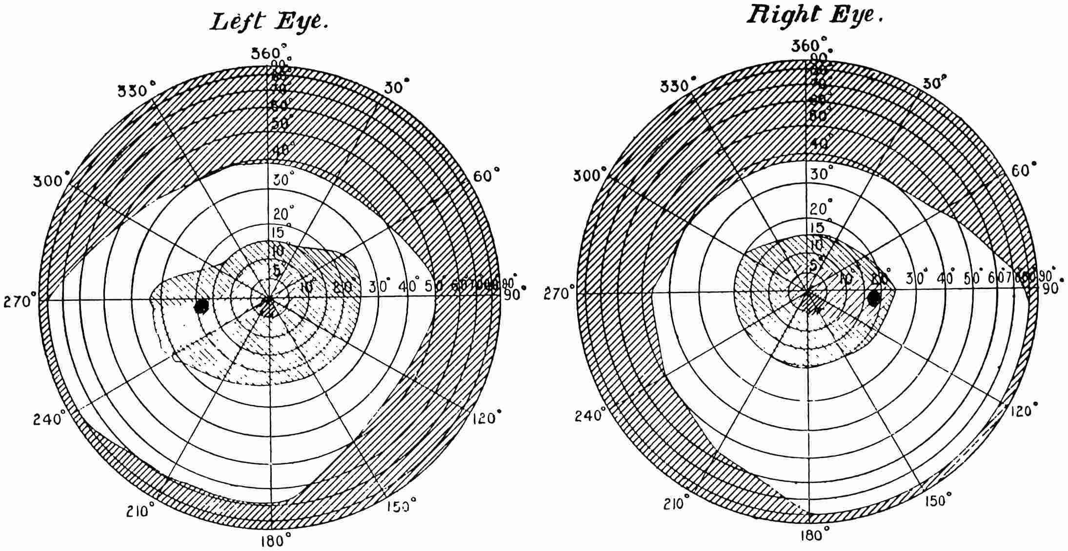
LeftEye. RightEye.
At page 10, a description has been given of the manner in which the field for colour and light has been determined, and if this same method be pursued with persons suffering from this form of colour blindness we get some remarkable results. Fig. 35 is the chart of the eye for red and for white, which was made by a case of tobacco blindness. The yellow spot is entirely affected, and, as is very common, it extends to the blind spot in the eye. At no place within that area can red be seen, though blue is immediately recognised. The extent of the field for white is that found under normal conditions, and except for the diseased area the same is true for the red. The fields for both eyes are given: that for the left eye in the left-hand chart, and that for the right eye in the right-hand chart. The small dark spots within the 5° area are places where the colour sensation is most defective. The part in the central dark area shaded with lines in this direction //// shows the portion of the field which is insensitive to red, though not to light, whilst the remainder of the shaded central area indicates the extent of the field which is sensitive to red. The field for light generally is also shown by the (approximately) rectangular unshaded area. Although the area
occupied by the insensitive part of the retina is small compared with the whole, yet it is in that part which is used for distinct vision.
For testing for colour the apparatus, Fig. 3, arranged so that the patch of colour has the white patch alongside, is the most useful, but it is as well then to use a surface of patch about ½ inch square only, and thus to confine the image as nearly as may be to the spot on the retina which is defective. These cases of central scotoma are by no means very easy to test; for it frequently happens that before they are able to distinguish that there are two patches side by side, they have to approach very close to the screen. If this be the case, however, it will usually be found that the patches of ½ inch side are still efficient, as the near approach of the eyes to the screen indicates a wide area as being affected, so that the image still lies within the diseased retinal area. In some instances the colours named will vary very considerably; sometimes, for instance, a red will be named as grey, and then immediately after as pale red. This is generally due to the diseased area being small, and a very slight change in the direction of the axis of the eye causes it to be seen in nearly its true colour, part being viewed with the diseased and part with the healthy portion of the retina. With the wool test, which we shall describe later, it is the commonest thing possible for colourblind persons who have a central scotoma to match accurately the different test-skeins, for the reason that the images of the skeins of wool are so large that they are received on the parts of the retina which are not diseased. These same colours, however, if presented to them in small patches, will inevitably show the defect in vision.
With this end in view, I have had a set of brick-clay pellets some 3/16-inch in diameter, painted with water-colours mixed with soluble glass solution of the same colours as the wools. These are placed in a shallow tray, and presented to patients affected with this central colour blindness to pick out all the pellets which match reds and greens. They will tell you that they see neither one nor the other, though they will pick out the blue pellets unerringly. A red pellet they will match with a red, green, grey, or a brown one, and a green one with the same. If, however, you instruct them to direct their eyes a
few degrees away from the tray, they will tell you they see all the colours, and as they endeavour to pick them out, they, with a natural instinct, direct their eyes again to the collection, when once more the colours vanish. It is almost piteous sometimes to see the distress which this simple test occasions. The sight of the colours for an instant and their immediate disappearance in the cases that I have tried, seem indicative of something terrible, for they usually have no idea of the cause of this (to them almost miraculous) phenomenon. I have seen these colour blind tested with a pair of ordinary bull’s-eye lanterns, placed side by side, with diaphragms of moderate size with coloured glasses, which can be changed at will, in front. At twelve feet distance they will often see both lights as one, but as they approach they will make out two lights and call them both white, or sometimes they will make a guess and call a green red, or viceversâ. It goes without saying that such eyesight is useless for reading signals, and indeed for any purpose whatever. Sometimes, but I believe this is rare, no colour whatever can be distinguished.
CHAPTER XII.
I WILL now give in full the result of the examination of a patient who was suffering from tobacco blindness. X., aged thirty-six, a commercial traveller, was suffering from rather severe tobacco amblyopia. The scotoma was a very marked one, and the loss of colour sensation most complete. Mr. Nettleship, who furnished the case, has kindly added the following remarks on the case:
His acuteness of vision was 6/36 with the right eye and 6/60 with the left. He smoked half-an-ounce of “shag” daily and drank about four pints of beer. His sight had been failing for about two months. As is common in early stages of this disease the ophthalmoscope revealed no decided changes at the optic discs.
He passed the test of the Holmgren wools satisfactorily, proving that the usual vision was normal for colour, but failed at once with the pellet test.
The objects in view were to test his perception of the spectrum colours, and then the extent of his retinal field for colour. This last is not recorded here. The spectrum colours were reduced to uniform luminosity between λ 4600 and λ 6600. Diaphragms containing holes of different sizes were placed in front of the last prism, and thus a round spot of monochromatic light of the same luminosity was produced upon the screen when a slit was passed through the spectrum. From the red end to λ 5270 he called the whole of the colours white, and from that point he began to see blue, called the colours bluish and blue. When the full illumination for all the colours was used, the same results were obtained. From this examination it would appear that he was totally deprived of the sensation of any colour except of blue. A subsequent examination of his perception of the luminosity of different rays, however, has to be taken into
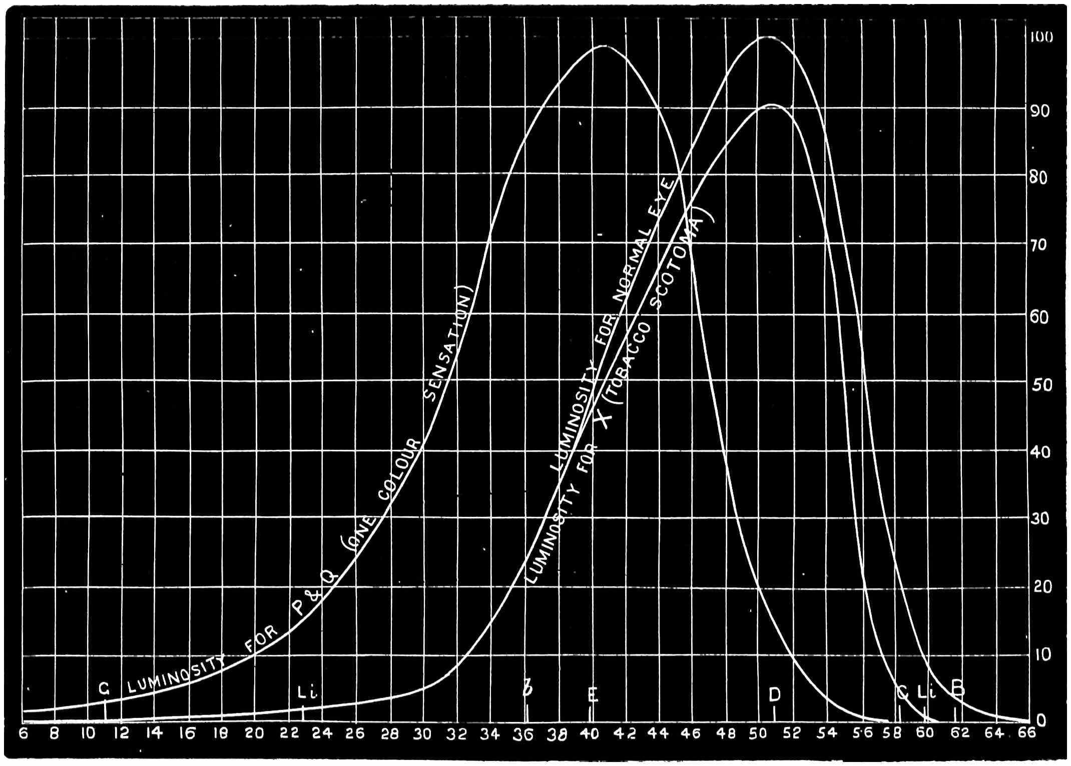
account, for in the first examination he had no light of pure white with which to compare the colours. In the next experiments, a strip of white light was placed in juxtaposition to the colour, and the results were slightly different. The table below gives his luminosity measures (Fig. 36). Col. I. is the empyric scale number, II. is the wave-length, III. the luminosity of the colour to the normal eye, IV. the luminosity to X., and V. the ratios of III. to IV.
In the diagram, his luminosity curve X. is shown, its area being 1400 against 1650 for the normal eye. His central perception of light, as arrived at by the extinction method, was only two-thirds of that of the normal eye; hence his area of luminosity should be 1100. As it is 1400, the ordinates of the above curve should be multiplied by 0·8, to compare with that of the normal eye.