Professor of Surgery, University of Edinburgh; Clinical Director Breast Cancer Now Research Laboratory; Consultant Surgeon, NHS Lothian Edinburgh Breast Unit, Western General Hospital, Edinburgh, UK
Matthew D. Barber,
BSc(Hons) MBChB(Hons) MD FRCS(Gen Surg)
Consultant Breast Surgeon, NHS Lothian Edinburgh Breast Unit, Western General Hospital, Edinburgh, and St John’s Hospital, Livingston, UK
No part of this publication may be reproduced or transmitted in any form or by any means, electronic or mechanical, including photocopying, recording, or any information storage and retrieval system, without permission in writing from the publisher. Details on how to seek permission, further information about the Publisher’s permissions policies and our arrangements with organizations such as the Copyright Clearance Center and the Copyright Licensing Agency, can be found at our website: www.elsevier.com/permissions
This book and the individual contributions contained in it are protected under copyright by the Publisher (other than as may be noted herein).
Notice
Practitioners and researchers must always rely on their own experience and knowledge in evaluating and using any information, methods, compounds or experiments described herein. Because of rapid advances in the medical sciences, in particular, independent verification of diagnoses and drug dosages should be made. To the fullest extent of the law, no responsibility is assumed by Elsevier, authors, editors or contributors for any injury and/or damage to persons or property as a matter of products liability, negligence or otherwise, or from any use or operation of any methods, products, instructions, or ideas contained in the material herein.
ISBN: 978-0-7020-7241-3
Printed in China
Last digit is the print number: 9 8 7 6 5 4 3 2 1
Content Strategist: Laurence Hunter
Content Development Specialist: Lynn Watt
Project Manager: Umarani Natarajan
Design: Miles Hitchen
Illustration Manager: Nichole Beard
Illustrator: MPS North America LLC
Evidence-based practice in surgery
Critical appraisal for developing evidence-based practice can be obtained from a number of sources, the most reliable being randomised controlled clinical trials, systematic literature reviews, metaanalyses and observational studies. For practical purposes three grades of evidence can be used, analogous to the levels of ‘proof’ required in a court of law:
1. Beyond all reasonable doubt. Such evidence is likely to have arisen from high-quality randomised controlled trials, systematic reviews or high-quality synthesised evidence such as decision analysis, cost-effectiveness analysis or large observational datasets. The studies need to be directly applicable to the population of concern and have clear results. The grade is analogous to burden of proof within a criminal court and may be thought of as corresponding to the usual standard of ‘proof’ within the medical literature (i.e. P <0.05).
2. On the balance of probabilities. In many cases a high-quality review of literature may fail to reach firm conclusions due to conflicting or inconclusive results, trials of poor methodological quality or the lack of evidence in the population to which the guidelines apply. In such cases it may still be possible to make a statement as to the best treatment on the ‘balance of probabilities’. This is analogous to the decision in a civil court where all the available evidence will be weighed up and the verdict will depend upon the balance of probabilities.
3. Not proven. Insufficient evidence upon which to base a decision, or contradictory evidence.
Depending on the information available, three grades of recommendation can be used:
a. Strong recommendation, which should be followed unless there are compelling reasons to act otherwise.
b. A recommendation based on evidence of effectiveness, but where there may be other factors to take into account in decisionmaking, for example the user of the guidelines may be expected to take into account patient
preferences, local facilities, local audit results or available resources.
c. A recommendation made where there is no adequate evidence as to the most effective practice, although there may be reasons for making a recommendation in order to minimise cost or reduce the chance of error through a locally agreed protocol.
Evidence where a conclusion can be reached ‘ beyond all reasonable doubt’ and therefore where a strong recommendation can be given. This will normally be based on evidence levels:
• Ia. Meta-analysis of randomised controlled trials
• Ib. Evidence from at least one randomised controlled trial
• IIa. Evidence from at least one controlled study without randomisation
• IIb. Evidence from at least one other type of quasiexperimental study.
Evidence where a conclusion might be reached ‘on the balance of probabilities’ and where there may be other factors involved which influence the recommendation given. This will normally be based on less conclusive evidence than that represented by the double tick icons:
• III. Evidence from non-experimental descriptive studies, such as comparative studies and case–control studies
• IV. Evidence from expert committee reports or opinions or clinical experience of respected authorities, or both.
Evidence that is associated with either a strong recommendation or expert opinion is highlighted in the text in panels such as those shown above, and is distinguished by either a double or single tick icon, respectively. The references associated with doubletick evidence are listed as Key References at the end of each chapter, along with a short summary of the paper's conclusions where applicable. The full reference list for each chapter is available in the ebook. The reader is referred to Chapter 1, ‘Evaluation of surgical evidence’ in the volume Core Topics in General and Emergency Surgery of this series, for a more detailed description of this topic.
Anatomy and physiology of the breast 1
It is essential that clinicians endeavouring to prevent, diagnose, or treat breast cancer possess a fundamental understanding of the basic anatomical and physiological precepts that are the foundation of our knowledge base. This chapter presents a concise review of basic knowledge, and a summary of clinical considerations for application to modern clinical practice.
Normal breast development: embryology and physiology
Embryology
The breast is a modified sweat gland of ectodermal and mesodermal origin. Fetal development is modulated by local factors, and has three key stages:
1. By week 5, two parallel ectodermal ridges (milk lines) appear along the ventral surface of the embryo extending from the primitive axilla to the inguinal region.
2. By week 9, the paired ectodermal ridges begin to disappear; however, they remain in the pectoral region, forming a pair of ‘primary buds’. These primary buds divide into many (15–20) smaller ‘secondary buds’ that individually extend into the underlying, vascularised connective tissue mesoderm.
3. During the third trimester, these ectodermal extensions epithelialise and branch, developing lumens and terminal pouches (acini), thereby acquiring the classical ‘ductal and lobular’
Mary Morrogh
structure that typifies the mammary gland. At birth, there are no differences (morphological or physiological) between the sexes.1,2
Clinical considerations
Failure of ectodermal regression results in the formation of accessory nipples (‘supernumerary nipples’ or polythelia) or supernumerary breasts (polymastia). Complete regression of the primary bud leads to congenital absence of breast tissue (amastia) and nipple (athelia). Failure of one or both breasts to develop fully can be congenital or acquired. Genetic causes include Poland's syndrome, which is a group of conditions associated with absence or hypoplasia of the pectoralis major muscle, and deformity of the underlying chest wall and varying degrees of syndactyly.3,4 It is rare, usually only partial in nature and is more common in men than in women. Breast development can be affected by trauma, including surgery or radiotherapy.
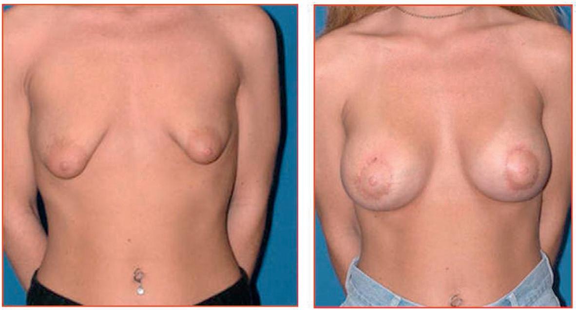
Mild asymmetry is a common problem and usually only reassurance is needed. Lipofilling has now become an important part of treating breast asymmetry. It may need to be combined with tissue expansion to increase the amount of skin for placement of subsequent breast implants.5,6 If asymmetry is marked, increasing the size of the smaller breast with or without tissue expansion and/ or reduction or augmentation of the opposite breast may be required. A pedicled or free myocutaneous flap, with or without an implant, can be used to reconstruct any muscle defect and produce symmetry in cases of severe hypoplasia or aplasia (Fig. 1.1).
• The most common breast tumours of mesenchymal origin are phyllodes tumours (see Chapter 3).
Puberty
The adult female breast is under constant influence of autocrine (systemic hormones) and paracrine (growth factors, cytokines) modulators of growth and development. Thelarche marks the onset of adult breast development. The release of GnRH (gonadotrophin releasing hormone) from the hypothalamus controls the release of the pituitary hormones FSH (follicle stimulating hormone) and LH (luteinising hormone). The early menstrual cycles are anovulatory, thus the effects of oestrogen are unopposed. Development begins with a ‘ductal growth phase’ involving ductal elongation, an increase in epithelial height and number and an increase in stromal density, and is followed by the addition of lobular units. Terminal end buds form new small alveolar buds which branch and divide into ductules. A type 1 lobule is a terminal duct with 10–12 associated alveolar buds and is the characteristic feature of a peri-menarchal breast. These lobules continue to develop for up to 15 years, at which point they begin to be replaced by more mature lobules. In the adult breast, cyclical changes occur during each menstrual cycle, with an increase in the rate of proliferation, especially during the luteal phase, followed by a wave of apoptosis. Breast size may increase by up to 15% during this phase.7,8
Clinical considerations
• Breast development prior to 8 years of age (premature thelarche) is abnormal, and most often due to dysregulation of the hypothalamic pituitary–adrenocortical axis. Conversely, a lack of oestrogen, whether congenital (such as in Turner’s syndrome, Kallman syndrome) or acquired (malnutrition, chemotherapy,
radiotherapy) may result in a failure of the ductal system to develop.
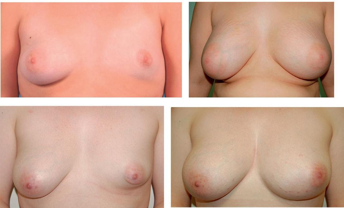
• Tuberous breasts are a common congenital breast deformity defined by a failure of breasts to develop normally. The tuberous breast is not simply a small or underdeveloped breast. Classical features include: an enlarged, swollen areola, wide spacing between the breasts, minimal breast tissue, high inframammary folds and a narrow breast base. The deformity can be classified based on location (grade I inferomedial quadrant; grade II both inferior quadrants; grade III affecting the whole breast). While breastfeeding may be affected, gonadogenesis or fertility is unlikely to be compromised (Fig. 1.2). Surgical intervention for tuberous breasts is challenging and frequently unsatisfactory and tends to involve lipomodelling and tissue expansion. Reduction of the large nipple–areolar complex may be required.
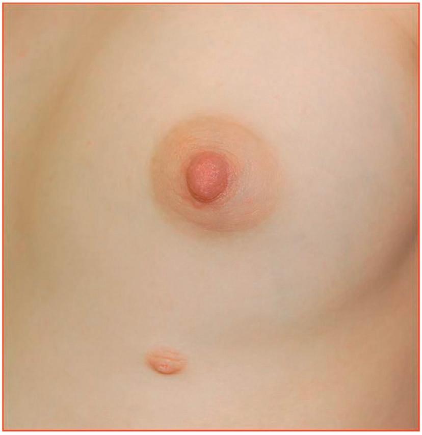
• Accessory nipples are most commonly seen below the breast and above the level of the umbilicus whereas accessory breast tissue is usually found in the axilla. They are present in up to 6% of the population.9 Although they rarely cause problems, accessory nipples in the bra line can be excised if they cause irritation (Fig. 1.3).
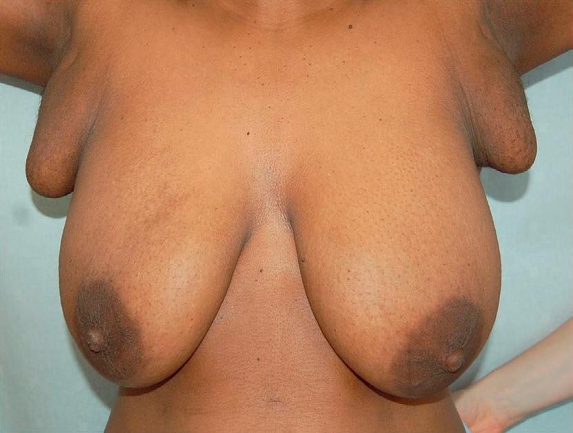
• Accessory breast tissue is a relatively common congenital condition and can present as a mass anywhere along the course of the embryologic ectodermal ridges, but most frequently (>90%) presents with accessory tissue in the axilla (Fig. 1.4). It can become more prominent during
Figure 1.1 • Hypoplasia pre- (a) and post- (b) surgery with expansion followed by implant, and pre- (c) and post(d) surgery with lipofilling.
pregnancy. Reassurance and an explanation are usually all that is required. Surgical excision should be reserved for truly symptomatic women, as accessory breast tissue is difficult to excise cosmetically and surgery is associated with significant morbidity.10 Liposuction with or without excision of skin and accessory breast tissue ensuring that the fascia of the axilla is not disturbed gives the best cosmetic outcomes. Both benign and malignant conditions can develop within accessory breast tissue.11
• Macromastia is defined as breast weight exceeding 2–3% of total body weight. It can be progressive, is more often bilateral than unilateral, and is thought to be due to rapidly developing connective tissue resulting from excess levels of growth factors or hormones. It typically presents at puberty or postpartum. Reduction mammaplasty after adolescence is the treatment of choice.12
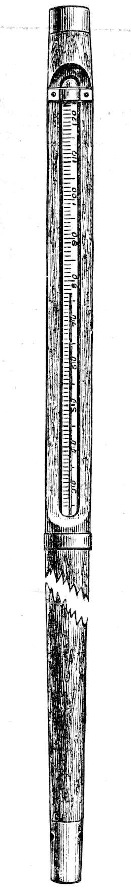
• In women <30 years of age, the stroma and lobules may respond to hormonal stimulus in an exaggerated fashion with the development of fibroadenomas (single or multiple)13 (see Chapter 3).
• Gynaecomastia (see Chapter 2) describes enlargement of the male breast. ‘Physiological’ gynaecomastia refers to ‘bilateral enlargement of breast tissue within 1 year of the onset of testicular development’ and is a normal finding in up to twothirds of pubertal males. Nonphysiological gynaecomastia is typically a result of oestrogen excess, androgen deficiency, or drug effects (see Chapter 3).
Figure 1.2 • Tubular breasts before (a) and after (b) tissue expansion and implant placement.
Figure 1.3 • Supernumerary nipple below breast.
Figure 1.4 • Bilateral accessory axillary breast tissue. Most cases are less prominent than this.
• Such is the variation in blood flow and epithelial proliferation throughout the menstrual cycle that it influences the efficacy of MRI as a breastimaging tool.14 For this reason elective MRI scans should be performed after menstruation and before ovulation.
Pregnancy and lactation
During pregnancy, autocrine and paracrine factors prepare the breast for lactation. The first phase of growth is driven primarily by progesterone, and results in proliferation of distal ducts and lobular units. During the second phase, lobular units mature by differentiating into secretory units (acini) which at the end of pregnancy become engorged with colostrum while the fat and connective tissue of the breast become almost entirely replaced by glandular epithelium. Lactogenesis is a twophase process driven by prolactin and glucocorticoids: phase 1 involves production of milk components by basal cells, engorgement of acini with colostrum, and proliferation of myoepithelial cells. Phase 2 occurs at or just after parturition with initiation of milk secretion and is marked by a rise in oxytocin, and a fall in placental hormones (progesterone), citrate and Alactalbumin. Mature milk production usually begins at 3648 hours postpartum, and the rate of lactation is constant for the first 6 months. Weaning decreases the size and number of lobules and acini; the ducts are not affected.
Clinical considerations
• Lactation disorders include failure to lactate, delayed onset of lactation, or galactorrhoea (defined as ‘inappropriate secretion of milky discharge in the absence of pregnancy/breastfeeding for more than 6 months’). Retained placenta can lead to delayed onset of lactation due to the continued secretion of progesterone which suppresses lactation and may lead to lifethreatening postpartum haemorrhage. Lactation failure may be the first presentation of postpartum hypopituitarism (Sheehan’s syndrome).
• A galactocele is a milkfilled cyst that usually occurs at the cessation of lactation and is thought to be the result of blocked drainage to the nipple, sometimes with thickened milk. These present clinically as a wellcircumscribed, fluctuant mobile mass that typically resolves with aspiration.
• Pregnancyassociated fibroadenoma is a not uncommon finding. Preexisting fibroadenomas
frequently increase in size and lactate. In this setting, FNA frequently reports atypical cells and core biopsy is therefore recommended. They will usually shrink again after breastfeeding.
• Pregnancy and breast cancer is discussed in detail in Chapter 11
Menopause
Menopause is described as ‘a cessation of ovarian function and withdrawal of steroid hormones’. ‘Involutional changes’ of the breast (i.e. involution of breast epithelium and connective tissue) become evident approximately 20 years after the menarche, and can be quite extensive by the time menopause is reached. Although there is an increase in fat deposition, the overall volume of the breast decreases during the menopause. The incidence of breast cancer increases with age, and most breast cancers are seen in postmenopausal women. 15
Clinical considerations
• In postmenopausal women, the principal source of circulating oestrogen is conversion of adrenally generated androstenedione to oestrone by aromatase in peripheral tissues with further conversion of oestrone to oestradiol.
• New onset breast pain around the time of menopause is a very common presentation to the primary physician/breast clinic. It is more likely to signify oestrogen withdrawal than underlying pathology. Most ‘breast’ pain in perimenopausal and postmenopausal women does not originate from the breast but from the chest wall (see Chapter 3).
• Exogenous hormones (hormone replacement therapy or HRT) may result in the persistence of breast epithelium. HRT increases breast tenderness, discomfort, nodularity and increases breast density. Breast cysts, which usually resolve after menopause, may persist. Epidemiological studies show an increased risk in developing breast cancer in women who take combined (oestrogen and progesterone) HRT compared with women who take oestrogen only, or placebo. As such, HRT should be avoided in women who have had a prior oestrogen receptorpositive breast cancer, or a significant family history.16
Breast anatomy
Microscopic anatomy
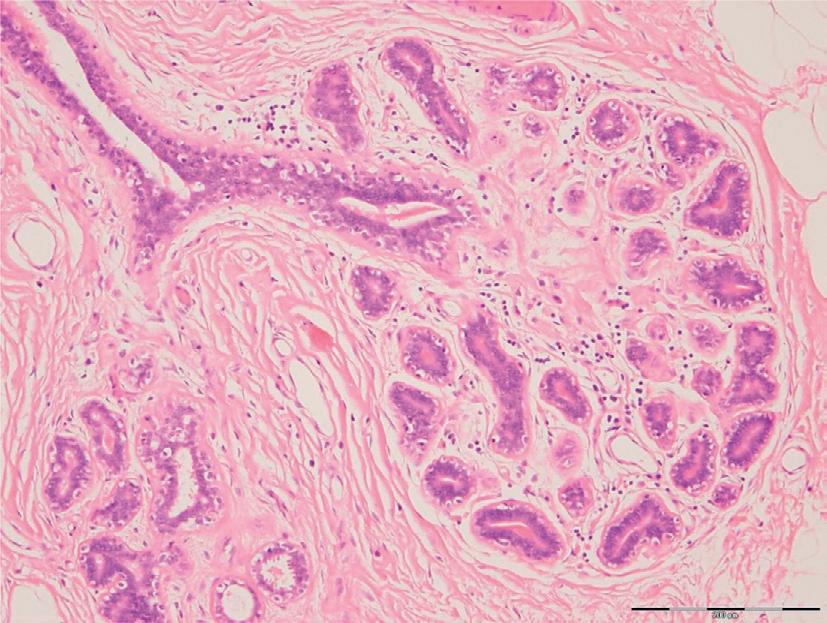
The breast gland is composed of glandular epithelium, fibrous stroma and connective tissue surrounded by fat. The relative amount of each of these tissues is under the control of circulating hormones and, as such, varies according to age, menstrual cycle, pregnancy, parity and breastfeeding (Fig. 1.5). The glandular epithelium forms a complex branching ductal system that radiates outward from the nipple (10–20 primary ducts, 30–40 segmental ducts, and 10–100 subsegmental ducts), each terminating in a lobular unit (terminal duct lobular unit or TDLU) that consists of clusters of ductules and acini.17 The breast epithelium is made up of three different cell types with distinct morphological features: (i) the superficial luminal A cells: characterised by dark nuclei, thought to be responsible for milk production; (ii) the basal (B) cells which have large, clear nuclei with distinctive intracellular filaments (these are the most common cell type); and (iii) the myoepithelial cells which contain contractile myofilaments, and are most abundant during lactation. Together, these cells assume a bilayered configuration, which is surrounded by a thick basement membrane. This basement membrane is composed of collagen and laminin, and serves to separate the breast epithelium from the blood and lymphatic vessels which lie within the stroma.18
Clinical considerations
• Breast cancers are malignant proliferations of epithelial cells. The basement membrane separates the normal breast epithelium from stroma, which
contains blood and lymph vessels. An intact basement membrane that encloses the cancer cells thereby preventing lymphovascular invasion is seen in ductal carcinoma in situ. Conversely, disruption of the basement membrane allows cancer cells to invade the stroma and come into contact with the lymphovascular system, which defines invasive breast cancer.
• Non-epithelial tumours of the breast include sarcomas, lymphoma and phyllodes tumours.
• Mammography can be limited by density of the breast tissue. Younger women tend to have more glandular or dense breasts, limiting the sensitivity of mammography in this age group.
Gross anatomy
Breast shape/skin
The breast tissue lies within the superficial fascia of the anterior chest wall, and is separated from the skin by a layer of superficial fascia and subcutaneous fat. It consists of approximately 15–20 duct/lobular units that open individually onto the nipple areolar complex (NAC). The retroareolar space contains smooth muscle, but no subcutaneous fat. The suspensory ligaments of Cooper are fascial bands that run from the deep layer of the superficial fascia to the skin between the duct lobular units and, together with the skin envelope, provide some support to the weight of the breast. There is no distinct fascial compartmentalisation of breast parenchyma. The deep layer of the superficial fascia is separated from the pectoral fascia by a distinct space known as the ‘retromammary space.’ Both the suspensory ligaments and the retromammary space contribute to the mobility of the gland. The glandular portion of the peripubertal, nulliparous breast lies almost entirely over the pectoralis muscle, extending into the lower axilla as the socalled axillary tail of Spence. At this stage of development, the breast assumes a classical, hemispheric shape. Increasing age, variations in body weight, pregnancy and lactation alter the consistency and density of the breast significantly; the mature breast becomes more lax and extends inferolaterally, assuming a somewhat flattened, pendulous shape.19
The precise determinants of breast shape and size are largely unknown. The relative volume of adiposetoglandular tissue varies greatly, as does the degree of support that is provided by the skin envelope and suspensory ligaments of Cooper. Breast ptosis is a natural consequence of ageing, and varies significantly among women both in terms of degree, rate and age of onset (Fig. 1.6). It is influenced by a multitude of factors
Figure 1.5 • Micrograph of haematoxylin and eosin stained section of breast tissue showing terminal duct lobular unit leading to duct. Note surrounding connective tissue and adjacent fat. Courtesy of Dr Jeremy Thomas.
Clinical considerations
• Appreciation of skin crease lines (Kraissl and Langer’s lines) will allow optimal selection of skin incisions for the patient.
ab c
including: BMI (Body Mass Index), weight loss/ gain, smoking, pregnancies, breastfeeding, breast size and reduction in skin elasticity that comes with ageing.20 When performing surgery on the breast, inspection, measurement and accurate marking of skin and breast gland extent are essential. Preoperative marking includes an appreciation of skin crease lines (Kraissl) and Langer’s lines as well as surface measurements such as: position of the nipple relative to the midclavicular point and inframammary fold, degree of ptosis, size of the NAC, distance from the sternal notch to the nipple and from the midline to the nipple. The degree of ptosis is assessed based on (a) the position of the NAC relative to the inframammary fold, and (b) the point direction the nipple.19
• The skin and subcutaneous fat can be separated from underlying breast tissue and breast along a relatively avascular plane. Dissection through this subdermal plane preserves the blood supply to the skin flaps and minimises blood loss. Careful dissection in this plane is especially important for patients undergoing skinsparing/nipplesparing mastectomies. Some surgeons advocate sharp dissection with scissors versus diathermy to prevent secondary thermal injury to skin flaps. Others hydrodissect this natural plane using a saline/epinephrine solution. The thickness of the skin flap varies significantly between patients, and often the flap over the inferior part of the breast is considerably thinner
than the upper. This is likely due to gravity and weight of the breast leading to stretching of the skin, and should be considered when planning the initial incision. Preoperative review of the breast imaging, either digital mammogram or MRI, can help determine the thickness of skin flaps and can help in identifying the major vessels supplying the breast and overlying skin.
• In cancer surgery, removal of the deep layer of the superficial fascia that encompasses the breast is recommended to optimise clearance at the posterior margin. This layer of fascia is separate from the pectoral fascia, which can be left intact. There is no anatomical basis for routine removal of pectoral fascia during breastconserving surgery or mastectomy; however, affected pectoral fascia and underlying muscle should be excised if involved by tumour.
• Tumours involving or adjacent to the suspensory ligaments can pull or shorten these ligaments causing skin retraction or dimpling.
• The inferolateral extension of the breast means that the upperouter quadrant of the breast has the greatest proportion of glandular tissue, and explains the increased incidence of breast cancer in this quadrant.
• Reduction of the ptotic skin envelope is a useful technique, not only for aesthetic breast surgeons but also in the oncoplastic setting when undertaking a therapeutic or symmetriation procedure. It allows the resection of larger areas of breast tissue while leaving a satisfactory breast shape.
Figure 1.6 • (a) Grade 1 ptosis – nipple at level of inframmary fold. (b) Grade 2 ptosis – nipple below level inframmary fold but remains at anterior pole of breast. (c) Grade 3 ptosis – nipple below level of inframammary fold with nipple below anterior pole of breast.
Blood supply to the breast
The breast is a highly vascular organ deriving its blood supply from three principal sources:
1. The internal mammary artery (IMA). The IMA arises from the subclavian artery near its origin. It travels inferiorly with its accompanying vein in a paramedian plane about 1–2 cm from the midline along the lateral boarder of the sternum, between the internal intercostal and transverse thoracic muscles. Ventral branches of this artery (a. anterior rami mammarii) penetrate the intercostal muscles of the second to the fifth intercostal spaces and then enter the breast. The IMA continues downward until it divides into the musculophrenic artery and the superior epigastric artery around the sixth intercostal space.
2. The lateral thoracic artery (a. thoracalis lateralis; long thoracic artery; external mammary artery) branches off the axillary artery between the subscapularis anteriorly, and the cords of the brachial plexus posteriorly. It follows the lower border of the pectoralis minor to the side of the chest, supplying the serratus anterior and pectoralis muscles, and sends branches across the axilla to the axillary nodes and subscapularis muscle. It supplies an external mammary branch, that turns around the free edge of the pectoralis major to supply the lateral aspect of the breast.
3. The costocervical trunk and the thoracic aorta also supply blood to the breast via the lateral branches of the posterior intercostal arteries. Other arteries that supply the breast include the thoracoacromial, subscapular, upper thoracic and thoracodorsal arteries. Extensive collateralisation occurs between these vessels within the breast tissue.
The venous drainage of the breast follows the primary arterial supply. The superficial veins form an extensive anastomotic network and assume a circular configuration around the nipple, known as the ‘ circulus venosus ’ . The deep veins drain almost entirely into the axilla. The principal deep veins include the perforating branches of the internal thoracic vein, tributaries of the axillary vein, and perforating branches of the posterior intercostal veins. As with all venous drainage networks, while common anatomical patterns exist, congenital variations such as absence/ duplication/alternative origins of blood vessels can be present and these may be a technical issue during surgery. 19
Clinical considerations
• The major blood supply is from a series of perforating vessels (anterior rami mammarii) situated in the upper inner quadrant. These branches of the IMA are highpressure vessels, and are the most frequent source of postoperative bleeding. These perforating branches supply the skin of the breast and need to be preserved during mastectomy. Branches passing medially into the breast need to be divided when performing a breastconserving procedure in the medial breast or mastectomy without damaging the branch to the skin. The perforating branches are in the breast skin flaps not the breast tissue. Damage to the blood supply to the skin can result in skin flap necrosis.
• The pectoralis major derives its blood supply via two branches (medial and lateral) of the lateral thoracic artery. Injury to both will result in atrophy of the muscle. This is an important consideration when placing a subpectoral implant.
• The IMA is a common recipient vessel for autologous breast reconstruction with microsurgical free tissue transfer. At the recipient site, partial rib resection for access to the internal mammary artery/vein has a risk of morbidity (chest pain, pneumothorax), and as such, reconstructive surgeons will use the internal mammary intercostal perforating vessels if they are adequate.
• The thoracodorsal branch of the subscapular artery contributes little to the blood supply of the breast, but is a key vessel in breast reconstruction. Not only does this vessel supply blood to the latissimus dorsi muscle (an option for myocutaneous pedicle flap reconstruction), but it is an important choice for recipient microvessels anastomosis for free flaps. The central and scapular lymph nodes lie adjacent to this vessel, so it is essential that during axillary lymph node dissection, the surgeon identifies and protects this artery and its associated vein. In addition, the long thoracic and thoracodorsal nerves lie nearby and may be injured during dissection.
• Knowledge of the blood supply of the breast is fundamental to understanding the pattern of metastasis in breast cancer. While the lymphatic system is the primary channel for metastasis, cancer cells can also metastasise directly through the venous system. A valveless, venous
plexus surrounding the vertebrae extends from the base of the skull to the sacrum and is in direct communication with the posterior intercostal arteries. This connection provides a potential pathway for metastasis to the vertebral column and central nervous system.
• Clear appreciation of the blood supply to the breast is an important preoperative consideration for reduction mammaplasty and volumedisplacement/replacement conservation surgery.
Lymphatic drainage of the breast
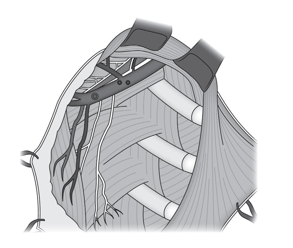
The axilla is a complex pyramidal compartment between the thoracic walls and the upper limb. A thorough understanding of axillary anatomy is essential for any surgeon treating patients with breast cancer. The borders consist of four sides (lateral –intertubercular groove of the humerus; medial –serratus anterior and thoracic wall; anterior – pectoralis and subclavius; posterior – subscapularis, teres major and latissimus dorsi) and a base with an opening at the apex, known as the axillary inlet. The contents of the axilla lie underneath a fascial layer within the axillary fat pad. The fat of the axilla is distinguishable from the subcutaneous fat as it has a smooth texture. The contents of the axilla include: axillary artery and vein, brachial plexus, lymph nodes, short head of biceps brachii and coracobrachialis tendons. Exposure of the axillary contents varies depending on the position of the arm, decreasing with full abduction, at which point the contents are at most risk during surgery. Careful positioning (80–90°) and preparation (some like to drape the arm so that it may be moved during surgery) of the upper limb provides optimal access during axillary surgery (Fig. 1.7).
All four quadrants of the breast drain as a unit into a few common nodes in the axilla. A small proportion of the breast (<10–15%) drains to the internal mammary lymphatic chain and there is also some drainage direct from the upper and deeper part of the breast to the interpectoral nodes. The lymphatics of the skin overlying the breast are in direct contact with the underlying deeper lymphatics. There are many classification systems to describe the distribution of lymph nodes in the axilla. The ‘surgical’ classification defines each group with respect to their relationship with the pectoralis minor:
• level I – lateral to the lateral border of the pectoralis minor muscle;
• level II – beneath the pectoralis minor muscle;
• level III – infraclavicular, medial to the pectoralis minor muscle up to Halsteds ligament, a dense condensation of the clavipectoral fascia extending from the medial end of the clavicle to the first rib.
The interpectoral nodes (Rotter’s nodes) lie between pectoralis major and minor, and the internal mammary nodes run alongside the internal mammary artery.19
Clinical considerations
• Sentinel lymph node biopsy (SLNB) is based on the hypothesis that the breast drains as a single unit to a few common nodes in the axilla, thus disseminating tumour cells colonise one or a few lymph nodes before involving others. This hypothesis has been validated by prospective trials that have consistently reported sensitivity
rib
Thoracodorsal artery
Thoracodorsal vein
Thoracodorsal nerve
Long thoracic nerve
Latissimus dorsi muscle
Figure 1.7
• Axillary anatomy. The medial wall of the axilla is formed by the ribs and chest wall muscles, notably serratus anterior over which runs the long thoracic nerve. Posteriorly lie the subscapularis, teres major, and latissimus dorsi muscles over which run the thoracodorsal pedicle. The pectoral muscles lie anteriorly.
Second
of SLNB in the range of 90–98%, and falsenegative rates of 2–4%. Modern treatment of the axilla is discussed in detail in Chapter 10
• The axillary nodes are the common route of lymphatic drainage for the breast and the upper limb. Lymphoedema is an unfortunate complication of axillary node clearance, affecting between 10% and 30% of patients.20 The role of mapping of the arm lymphatics (Axillary Reverse Mapping) to reduce injury continues to be explored. Surgical treatment of lymphoedema has seen recent renewed clinical interest with ongoing exploration of techniques including lymphovenous bypass, lymphgrafting, free omental lymphatic flaps and excisional procedures.21,22
• The internal mammary nodes (IMN) run along the course of the IMA, which lies between the superficial parietal pleura and the intercostal muscles, and are found in the intercostal spaces. The clinical significance of IMN disease in addition to axillary disease continues to be debated, and IMN biopsy is not performed routinely in most centres. Postmastectomy radiotherapy to the IMN chain can be considered for patients in whom the risk of IMN involvement is high considering primary tumour location/biology and axillary node status.23,24
Innervation of the breast, and nerves of the axilla
The retroareolar space lacks subcutaneous fat, but contains a layer of smooth muscle. This layer has two ‘rings’ of smooth muscle fibres that run perpendicular to one another (radial and circular). Somatic and autonomic excitation contraction of these fibres results in erection of the nipple and a decrease in the diameter of the areola. Sensory innervation of the breast skin envelope comes from the lateral and anterior cutaneous branches of intercostal nerves II–VI, and in part from the supraclavicular nerve.19
The brachial plexus is formed by the ventral rami of the C5–8 and T1. It gives motor supply to the muscles of the upper limb (with the exception of the trapezius and levator scapula), and cutaneous innervation of the upper limb except for the axilla (which is supplied by the supraclavicular nerve). It communicates with the sympathetic trunk via grey rami communicantes, which join the roots of the plexus. The cords of the brachial plexus pass over the first rib to the dome of the lung and continue under the clavicle behind the subclavian artery and then axillary artery, and are named (medial, lateral, posterior) on their relationship with the axillary artery.19
The following nerves are significant for axillary/ breast surgery (Fig. 1.7):
• Long thoracic nerve (C5, C6, C7, C8, T1): comes from roots of C5–T1; supplies serratus anterior
• Lateral pectoral nerve (C5, C6, C7): comes from the lateral cord; supplies pectoralis muscles
• Medial pectoral nerve (C8, T1): comes from the medial cord; supplies pectoralis muscles
• Thoracodorsal nerve (C6, C7, C8): comes from the posterior cord; supplies latissimus dorsi muscle.
The lateral cutaneous branch of the second intercostal nerve does not divide into an anterior and a posterior branch, and is known as the intercostobrachial nerve. It pierces the intercostalis externus and the serratus anterior muscle crosses the axilla to the medial side of the arm, and joins the medial brachial cutaneous nerve (from the medial cord of the brachial plexus) and supplies the skin of the upper half of the medial and posterior part of the arm, communicating with the posterior brachial cutaneous branch of the radial nerve. 19 One or more branches of the lower intercostal nerves also cross the axilla.
Clinical considerations
• Skin incisions will interrupt the small cutaneous nerves and can cause numbness, which may be temporary or permanent. All patients need to be counselled about altered sensation after surgery. This is especially important for patients undergoing skinsparing (± nipple preservation) mastectomy with reconstruction. It must also be borne in mind with subcutaneous dissection from distant incisions and mobilisation to allow oncoplastic resections. What may look like a breast, may not feel like a breast.
• Interruption of the retroareolar space during duct excision or nipplesparing mastectomy may damage the layer of smooth muscle. Patients undergoing such procedures must be aware that not only may they notice altered sensation, but they may also suffer loss of erectile function of the nipple.
• The initial approach to the axilla begins with identification of the axillary vein, followed by the medial pectoral nerve. Paradoxically, the medial nerve is found lateral to the lateral nerve. Both supply the pectoralis muscle, and injury would result in difficulty in adduction of the upper limb. The medial pectoral nerve runs from superior to the axillary vein to the undersurface of the pectoralis major, passing through the axillary fat pad and across level II; it has an accompanying vein which can facilitate identification of the nerve.
If a submuscular implant reconstruction is planned, preservation of the medial pectoral nerve will prevent atrophy of the muscle (Fig. 1.7).
• The next nerve to be identified is the intercostobrachial. This is a sensory nerve only, supplying sensation to the upper, inner aspect of the arm. Attempts should be made to preserve it if oncologically safe; however, it is often damaged, leading to cutaneous anaesthesia or pain syndromes over the triceps region of the arm. Sacrificing the nerve should be done by transection (knife or scissors) rather than with electrocautery. Some surgeons advocate that the ends be buried to minimise postoperative pain, and to avoid Ligaclips, which may lead to neuroma formation.
• The long thoracic nerve comes off the upper roots of the brachial plexus and enters the
Key points
axilla deep to the axillary vein. Within the axilla, the nerve always runs posterior to the intercostobrachial nerves, and runs perpendicularly along the lateral border of the serratus anterior, beneath a layer of superficial fascia. It innervates the serratus anterior muscle, and injury results in winging of the scapula.
• The thoracodorsal nerve arises from the posterior cord of the brachial plexus, runs in the lateral aspect of the axilla, and supplies the latissimus dorsi muscle. The latissimus dorsi is innervated from a number of sources, so sacrificing the thoracodorsal nerves rarely causes complications. Indeed, some reconstructive surgeons divide the nerve when undertaking a latissimus dorsi reconstruction to avoid muscle twitching.
• Knowledge of breast development, physiology and anatomy is essential for understanding much breast pathology and for undertaking surgery to the breast or axilla.
• Awareness of potential issues with breast and axillary blood supply and innervation can help prevent surgical complications.
• Efforts should be made to avoid damage to neurovascular structures (thoracodorsal pedicle, long thoracic, pectoral and intercostobrachial nerves) in the axilla during axillary surgery.
References available at http://expertconsult. inkling.com
Assessment of patient with breast symptoms 2
Patients with breast concerns that cannot be adequately managed in primary care should be referred to a specialist breast clinic. Such clinics were traditionally run by surgeons but are now often multidisciplinary. These clinics can perform a combination of clinical, radiological and pathological examinations. Assessment of a breast lump requires triple assessment.
Triple assessment
Triple assessment is the combination of clinical, radiological and pathological evaluation of a breast lump. Triple assessment should be used in all patients with a confirmed breast lump or asymmetric localised nodularity and may be relevant in women with other symptoms.1 Imaging assessment consists of mammography in those aged 40 or over and ultrasonography for all palpable and other significant radiological findings requiring further study. Histological assessment usually involves core biopsy.2
This combination of clinical and imaging assessment with core biopsy increases the reliability of determining the cause of an abnormality.3–6 It is recommended that all elements of the assessment process are reported on a scale of 1–5 with increasing concern of malignancy7 (Table 2.1). The availability of clinical and radiological assessment and biopsy at a single clinic visit (‘onestop’ clinics) is the standard of care for assessing those referred with breast problems. Immediate reporting of
Matthew D. Barber Nisha Sharma
cytology from fineneedle aspirates or touch preparation cytology from core biopsy specimens or frozen section of core biopsy specimens is possible in some centres, but given the inability of cytology to differentiate invasive from in situ cancer has limited utility.
In the USA and European countries the BIRADS scoring system is used for radiology8 (Table 2.2). The key difference between the UK and USA classifications is that all benignlooking lesions would be biopsied in the UK rather than offered shortterm followup, as they are in the USA.
It is important to classify the level of concern independently on clinical examination, imaging and histology. This allows the clinician to determine if all the components of triple assessment are concordant or not. Results of all patients undergoing biopsy should be discussed in a multidisciplinary setting to ensure concordance of findings and minimise chances of missing a breast cancer. (See Table 2.3.)
Clinical evaluation
Clinical history
A history is taken from the patient of the duration and nature of the presenting symptom. Further specific details can be of value for certain symptoms and are outlined below. The presence and type of past personal or familial breast problems should be elucidated. General factors such as past medical history, drugs and allergies should be recorded. Hormonal risk factors for cancer, such
Table 2.1 • Scoring system for triple assessment
1 Normal (or inadequate cytology)
2 Benign (or normal cytology)
3 Suspicious but probably benign
4 Suspicious and probably malignant
5 Malignant
The score is preceded by an initial based on the relevant element of assessment (e.g. E, examination; R, mammography; U, ultrasound; M, MRI; C, cytology; B, biopsy).
Table 2.2 • BIRADS classification BIRADS
0 Incomplete imaging – further information required
I Negative
II Benign findings
III Probably benign, short interval follow-up suggested
IV Suspicious abnormality and biopsy recommended. Further subdivided into:
IVa: low level of suspicion for malignancy
IVb: intermediate suspicion for malignancy
IVc: moderate suspicion for malignancy
V Highly suggestive of malignancy
VI Known biopsy-proven malignancy
as age of menarche and menopause, parity, age of first birth, breastfeeding, oral contraceptive or hormone replacement therapy use, are traditionally documented for epidemiological purposes although they are of no specific value in achieving a diagnosis in an individual case.
The history and examination findings should be recorded legibly and contemporaneously in the medical records, aided by the use of a standard form.
Clinical examination
Breast examination (see Fig. 2.1) should be conducted in a good light with the patient stripped to the waist and in the presence of a chaperone. Examination of the male breast is similar, with particular attention paid to whether an abnormality is present within the breast tissue or whether the problem being complained of is breast tissue.
Initial examination is by inspection, with the patient in the sitting position with hands by her side, paying particular attention to symmetry, nipple inversion, skin changes and any alteration of breast contour. The breast should also be inspected both with arms raised and with the chest wall muscles tensed to show changes in the dynamic setting.
Palpation of the breasts is best performed in the supine position with the head supported and the arms above the head. Putting the hands above the head spreads the breast out over the chest wall and reduces the depth of breast tissue between the examiner’s hands and the chest wall and makes abnormal areas much easier to detect and define. All of the breast tissue is examined using the fingertips. If an abnormality is identified, then it should be assessed for size, contour, texture and any deep fixation. All palpable lesions should be measured with callipers10 and location and size should be clearly recorded.
If there is a history of nipple discharge, the nipple should be gently squeezed to determine whether a pathological discharge is present. Careful note should be taken of whether discharge is emerging from single or multiple ducts, and whether blood is present (either frankly or on dipstick testing).
All women complaining of breast pain or tenderness should be examined for tenderness of the chest wall. With the patient in the sitting position, the hand may be pushed up behind the breast from below with pressure on the chest wall. The patient may also be rolled onto their side, allowing the breast to fall medially, exposing the edge of the pectoral muscle to palpation. With the patient sitting or on their side, pressure can be placed on the breast tissue alone
Table 2.3 • Accuracy of investigations in symptomatic breast clinic9
Specificity includes assessment as malignant and probably malignant. Accuracy of mammography varies with age. Accuracy of biopsy techniques is improved by image guidance.
Another random document with no related content on Scribd:
HUMIDITY
59. Instruments. As a direct factor, humidity is intimately connected with water-content in determining the structure and distribution of plants. The one is in control of water loss; the other regulates water supply. Humidity as a climatic factor undergoes greater fluctuation in the same habitat, and the efficient difference is correspondingly greater. Accordingly, simple instruments are less valuable than automatic ones, since a continuous record is essential to a proper understanding of the real influence of humidity. As is the rule, however, the use of simple instruments, when they can be referred to an ecographic basis, greatly extends the field which can be studied. In investigation, both psychrometer and psychrograph have their proper place. In the consideration of simple instruments for obtaining humidity values, an arbitrary distinction is made between psychrometers and hygrometers. The former consist of a wet and a dry bulb thermometer, while the latter make use of a hygroscopic awn, hair,orotherobject.
Psychrometers
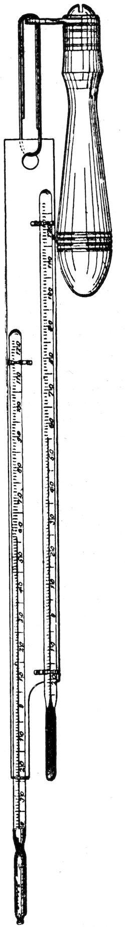
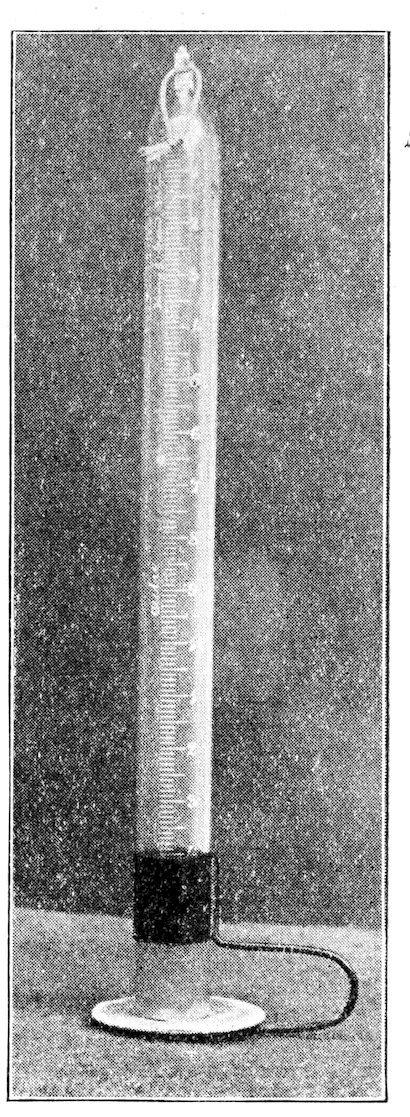
60. Kinds. There are three kinds of psychrometer, the sling, the cog, and the stationary. All consist of a wet bulb and a dry bulb thermometer set in a case; the first two are designed to be moved or whirled in the air. The same principle is applied in each, viz., that evaporation produces a decrease in temperature proportional to the amount of moisture in the air. The dry bulb thermometer is an ordinary thermometer, while the wet bulb is covered with a cloth that can be moistened. The former indicates the normal temperature of the air, the latter gives the reduced temperature due to evaporation. The relative humidity of the air is ascertained by means of the proper tables, from two terms, i. e., the air temperature and the amount of reduction shown by the wet bulb. The sling and the cog psychrometers alone are in general use. The stationary form has been found to be unreliable, because the moisture, as it evaporates from the wet bulb, is not removed, and, in consequence, hinders evaporation to the proper degree.
61. The sling psychrometer. The standard form of this is shown in the illustration, and is the one used by the Weather Bureau. This instrument can be obtained from H. J. Green, 1191 Bedford Ave., Brooklyn, or Julien P. Friez, 107 E. German St., Baltimore, at a cost of $5. It consists of a metal frame to which are firmly attached two accurately standardized thermometers, reading usually from –30° to 130°. The frame is attached at the uppermost end to a handle in such fashion that it swings freely. The wet bulb thermometer is placed lower, chiefly to aid in wetting the cloth more readily. The cloth for the wet bulb should be always of the same texture and quality; the standard used by the Weather Bureau can be obtained from the instrument makers. A slight difference in texture makes no appreciable error, but the results obtained with different instruments and by different observers will be more trustworthy and comparable if the same cloth be used in all cases. The jacket for the wet bulb may be sewed in the form of a close-fitting bag, which soon shrinks and clings tightly. It may be made in the field by wrapping the cloth so that the edges just overlap, and tying it tightly above and below the bulb. In either case, a single layer of cloth alone must be used. The cloth becomes soiled or thin after a few months’ constant use and shouldbereplaced.Itisawiseprecaution to carryasmallpieceofpsychrometerclothinthefieldoutfit.
62. Readings. All observations should be made facing the wind, and the observer should move one or two steps during the reading to prevent the possibility of error. The cloth of the wet bulb is moistened with water by means of a brush, or, much better, it is dipped directly into a bottle of water. Distilled water is preferable, as it contains no dissolved material to accumulate in the cloth. Tap-water and the water of streams may be used without appreciable error, if the cloth is changed somewhat more frequently. The temperature of the water is practically negligible under ordinary conditions. Readings can be made more quickly, however, when the temperature is not too far from that of the air. The psychrometer is held firmly and swung rapidly through the air when the space is not too confined. Where there is danger of breakage, it is swung back and forth through a short arc, pendulum-fashion. As the reading must be made when the mercury of the wet bulb reaches the lowest point, the instrument is stopped from time to time and the position of the column noted. The lowest point is often indicated by the tendency of the mercury to remain stationary; as a rule it can be noted with certainty when the next glance shows a rise in the column. In following the movement, and especially in noting the final reading,
great care must be taken to make the latter before the mercury begins to rise. For this reason it is desirable to shade the psychrometer with the body when looking at it, and to take pains not to breathe upon the bulbs nor to bring them too near the body. At the moment when the wet bulb registers the lowest point, the dry bulb should be readandtheresultsrecorded.
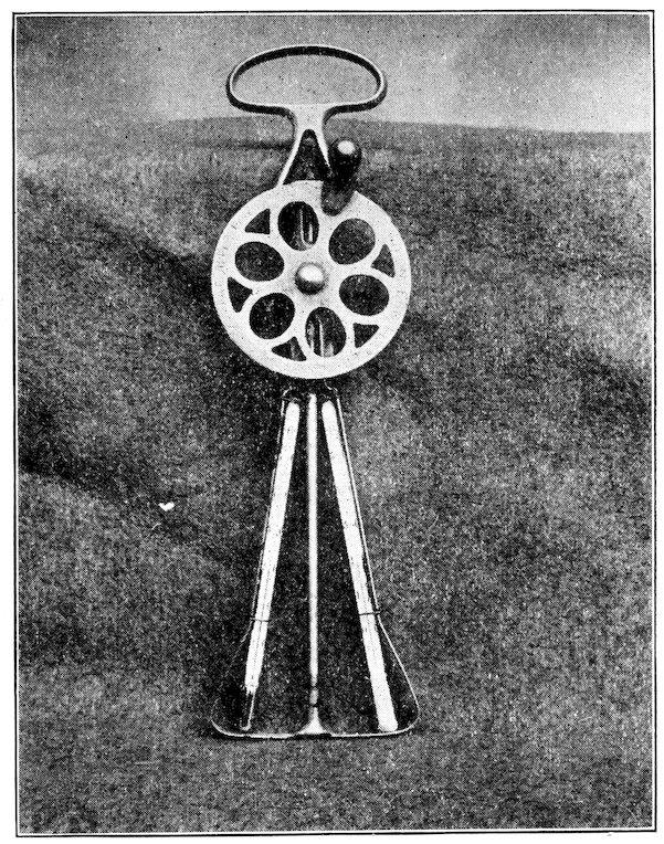
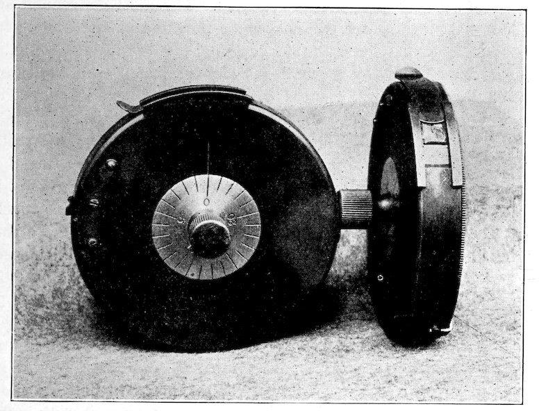
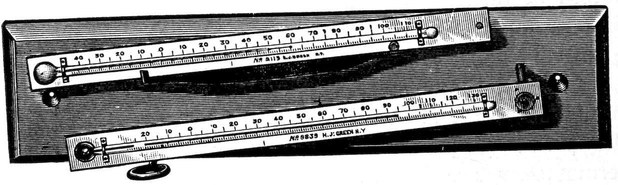
63. Cog psychrometer. This instrument, commonly called the “egg-beater” psychrometer, has been devised to obviate certain disadvantages of the sling psychrometer in field work, and has entirely supplanted the latter in the writer’s own studies. It is smaller, more compact, and the danger of breaking in carriage or in use is almost nil. It has the great advantage of making it possible to take readings in a layer of air less than two inches in thickness, and in any position. Fairly accurate results can even be obtained from transpiring leaves. The instrument can readily be made by a good mechanic, at a cost for materials of $1.75, which is less than half the price for the sling form. A single drawback exists in the use of short, Centigrade thermometers, inasmuch as tables of relative humidity are usually expressed in Fahrenheit. It is a simple matter, however, to convert Centigrade degrees into Fahrenheit, mentally, or the difficulty may be avoided by the conversion table shown on page 47, or by constructing a Centigrade series of humidity tables. The fact that the wet and dry bulbs revolve in the same path has raised a doubt concerning the accuracy of the results obtained with this instrument. Repeated comparisons with the sling psychrometer have not only removed this doubt completely, but have also proved that the standardization of the thermometers has been efficient.
64. Construction and use. A convenient form of egg-beater is the Lyon (Albany, New York), in which the revolving plates can be readily removed, leaving the axis and the frame. The thermometers used are of the short Centigrade type. They are 4½ inches long and read from –5° to 50°. Eimer and Amend, 205 Third Ave., New York city, furnish them at 75 cents each. The thermometers are carefully standardized and compared, and then grouped in pairs that read together. Each pair is used to construct a particular psychrometer. Each thermometer is strongly wired to one side of the frame, pieces of felt being used to protect the tube and increase the contact. The frame is also bent at the base angles to permit free circulation of air about the thermometer bulbs. The bulb of one thermometer is covered with the proper cloth, and the psychrometer is finished. Since the frame revolves with the thermometers, it is necessary to
pour the water on the wet bulb, or to employ a pipette or brush. The thermometer bulbs are placed in the layer to be studied, and the frame rotated at an even rate and with moderate rapidity. The observation is further made as in the case of the sling psychrometer. As the circle of rotation is less than three inches in diameter, and the layer less than an inch, in place of nearly three feet for the sling form, the instrument should not be moved at all for extremely localized readings, but it must be moved considerably, a foot or more, if it is desirable to obtain a more general reading.
65. Hygrometers. While there are instruments designed to indicate the humidity by means of a hygroscopic substance, not one of them seems to be of sufficient accuracy for use in ecological study. The difficulty is that the hygroscopic reaction is inconstant, rather than that the instruments are not sufficiently sensitive. A number of hygrometers have been tested, and in all the error has been found to be great, varying usually from 10–20 per cent. In the middle of the scale they sometimes read more accurately, but toward either extreme they are very inexact. It seems probable that an accurate hygrometer can be constructed only after the model of the Draper psychrograph. Its weight and bulk would make it an impossible instrument for field trips, and the expense of one would provide a dozen psychrometers. In consequence, it does not seem too sweeping to say that no hygrometer can furnish trustworthy results. Of simple instruments for humidity, the psychrometer alone can be trusted to give reliable readings. Crova’s hygrometer, used by Hesselmann, is not a hygrometer in the sense indicated. As it is much less convenient to handle and to operate than the cog psychrometer, it is not necessary to describe it.
Psychrographs
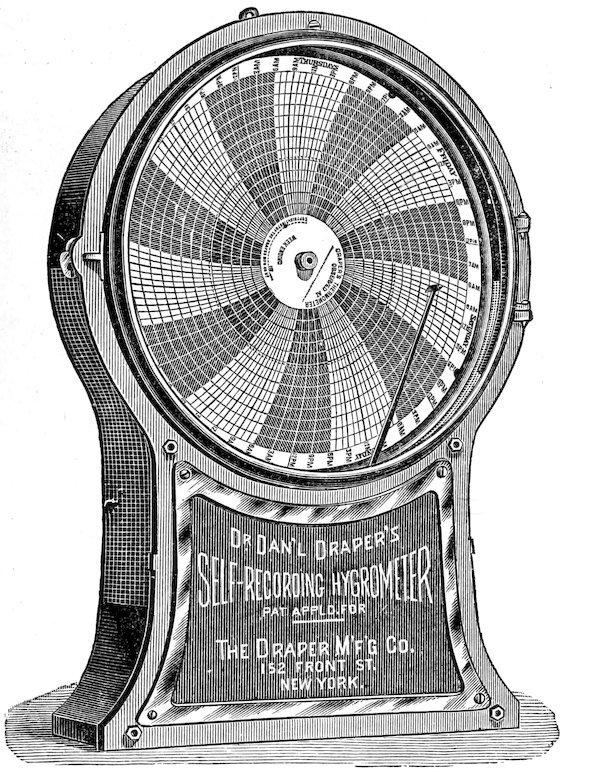
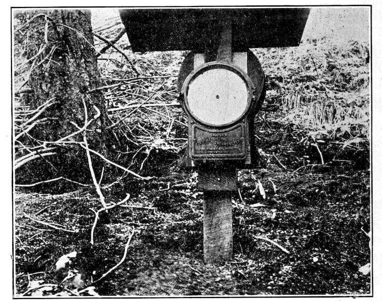
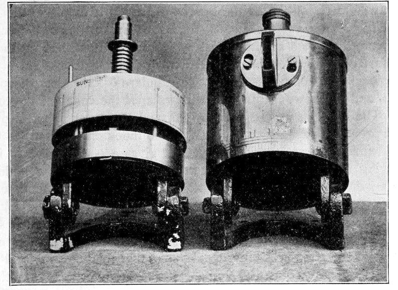
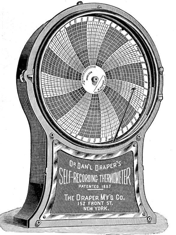
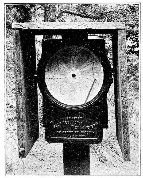
66. The Draper psychrograph. A year’s trial of the Draper psychrograph in field and planthouse has left little question of its accuracy and its great usefulness. Essentially, it consists of a band of fine catgut strings, which are sensitive to changes in the moisture-content of the air. The variations in the length of the band are communicated to a long pointer carrying an inking pen. The latter traces the record in per cent of relative humidity on a graduated paper disk, which is practically the face of an eight-day clock. The whole is enclosed in a metal case with a glass front. A glance at the illustration will show the general structure of the instrument.
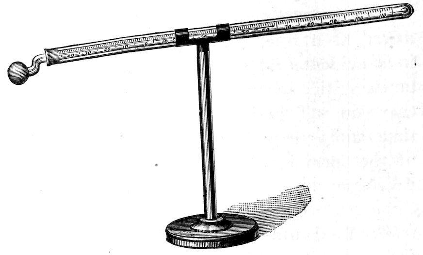
Fig.5.Slingpsychrometer.
Continued psychrometric tests demonstrate that the margin of error is well within the efficient difference for humidity, which is taken to be 5 per cent. In the field tests of the past summer, two psychrographs placed side by side in the same habitat did not vary 1 per cent from each other. The same instruments when in different habitats did not deviate more than 1 per cent from the psychrometric values, except when the air approached saturation. For humidities above 90 per cent, the deviation is considerable, but as these are temporary and incident upon rainfall, the error is not serious. For humidities varying from 10–85 per cent, the psychrograph is practically as accurate as the psychrometer. Per cents below 10 are rare, and no tests have been madeforthem.
Fig.6.Cogpsychrometer.
Fig.7.Draperpsychrograph.
67. Placing the instrument. The psychrograph should be located in a place where the circulation of the air is typical of the station observed. A satisfactory shelter will screen the instrument from sun and rain, and at the same time permit the air to pass freely through the perforations of the metal case. The form shown in figure 8 meets both of these conditions. A desirable modification is effected by fastening a strip about the cover of such depth as to prevent the sun’s rays from striking the case except when the sun is near the horizon. A cross block is fastened on the post of the shelter after being exactly leveled. The psychrograph rests upon this block, which is three feet above the ground in order to avoid the influence of radiation. The instrument is held in position by slipping the eye over a small-headed nail driven obliquely. It does not hang from the latter, but must rest firmly upon the cross block. The post is set to a depth that prevents oscillation in the wind, which is liable to obscure the record. In shallow mountain soils stability is attained by fastening a broad board at the base of the post before setting it. When two or more psychrographs are established in different habitats, great pains are taken to set them up in exactly the same way. The shelters are alike, the height above the soil the same, and the instrumentsallfacethesouth.
68. Regulating and operating the instrument. When two or more psychrographs are to be used in series, they must be compared with each other in the same spot for several days until they run exactly together with respect to per cent of humidity and to time. During this comparison they are checked by the psychrometer and so regulated that they register the proper humidity. When a single instrument is used alone as the basis to which simple readings may be referred, all regulating may well be done after the instrument is in position. This is a simple process; it is accomplished by obtaining the relative humidity beneath the shelter and at the proper height by a psychrometer. The pen hand is then moved to the proper line on the disk by means of the screws at its base. These are reached by removing the
Fig 8 Instrument shelter, showing thermograph and psychrograph in position
lettered glass face. The thumbscrew on the side opposite the direction in which the pen is to move is released, and the opposite screw simultaneously tightened, until the pen remains upon the proper line. Experience has proved that the record sheet should be correctly labeled and dated before being placed on the disk. In the press of field duties, records labeled after removal are liable to be confused. It is likewise a great saving of time to write the date of the month in the margin of each segment. Care is taken to place the sheet on the disk in the same position each time; this can easily be done by seeing that the sharp point on the disk penetrates the same spot on the paper. A single drop of ink in the pen will usually give the most satisfactory line. A thin line is read most accurately. If the pen point is too fine, however, the ink does not flow readily, and the point should be slightly blunted by means of a file. More often the line is too broad and the pen must be carefully pointed. Occasionally the pen does not touch the sheet, and it becomes necessary to bend the hand slightly. This is a frequent difficulty if the records arefoldedorwrinkled,andconsequentlythesheetsshouldalwaysbekeptflat.
69. The weekly visit. Psychrographs must be visited, checked, rewound, and inked every week. Whenever possible this should be done regularly at a specified day and hour. This is especially desirable if the same record sheet is used for more than one week. Time and energy are saved by a fixed order for the various tasks to be done at each visit. After opening the instrument the disk is removed, and the clock wound, and, if need be, regulated. The record sheet is replaced, the disk again put on the clock arbor, and the pen replenished with a drop of ink. A psychrometer reading is made, and the results in terms of relative humidity noted at the proper place on the disk sheet. If the psychrograph vary more than 1 per cent, it is adjusted to read accurately. In practice it has been found a great convenience to keep each record sheet in position for three weeks, and the time may easily be extended to four. In this event, the pen is carefully cleaned with blotting paper at each visit, and is then refilled with an ink of different color. To prevent confusion, the three different colored inks are always used in the same order, red for the first week, blue for the second, and green for the third. The advantages of this plan are obvious: fewer records are used and less time is spent in changing them. The records of several weeks are side by side instead of on separate sheets, and in working over the season’s results, it is necessary to handlebutathirdasmanysheets.
The Draper psychrograph is made by the Draper Manufacturing Company, 152 Front St., New York city. The price is $30. A few record sheets and a bottle of red ink are furnished with it. Additional recordscanbeobtainedat3centseach.Theinksare25–50centsperbottle,dependinguponthecolor.
HumidityReadingsandRecords
70. The time of readings. If simple instruments alone are used for determining humidity, readings are practically without value unless made simultaneously through several stations, or successively at one. When it is possible to combine these, and to make psychrometer readings at different habitats for each hour of the day, or at the same hour for several days, the series is of very great value. Single readings are unreliable on account of the hourly and daily variations of humidity, but when these changes are recorded by a psychrograph, such readings at once become of use, whether made in the same habitat with the recording instrument or elsewhere. In the latter case, one reading will tell little about the normal humidity of the habitat, but several make a close estimate possible. When a series of psychrographs is in use, accurate observations can be made to advantage anywhere at any time. As a rule, however, it has been found most convenient to make simple readings at 6:00 A.M., 1:00 P.M., and 6:00 P.M., as these hours afford much evidence in regard to the daily range. A good time also is that at which the temperature maximum occurs each day, but this is movable and in the press of field work can rarely be taken advantage of. A very fair idea of the daily mean humidity is obtainable by averaging the readings made at the hours already indicated. The comparison of single readings with the psychrograph record should not be made at a time when a rapid change is occurring, as the automatic instrument does not respond immediately. Such a condition is usually represented by a sudden rain, and is naturally not asatisfactorytimeforsinglereadingsinany event.
71. Place and height. As stated above, the psychrograph is placed three feet above the surface of the ground in making readings for the comparison of stations. In low, herbaceous formations, the instrument is usually placed within a few inches of the soil in order to record the humidity of the air in which the plants are growing. In forest formations, the moisture often varies considerably in the different layers. This variation is easily determined by simultaneous psychrometer readings in the several layers, or, if occasion warrants, a series of psychrographs may be used. In field work the rule has been to make observations with the psychrometer at 6 feet, 3 feet, and the surface of the soil, but the
reading at the height of 3 feet is ordinarily sufficient. Humidity varies so easily that several readings in different parts of one formation are often desirable. In comparing different formations, the readings should be made in corresponding situations, for example, in the densestportionofeach.
72. Check instruments. Humidity is so readily affected by temperature, wind, and pressure, that a knowledge of these factors is essential to an understanding of its fluctuations. Pressure, disregarding daily variation, is taken account of in the tables for ascertaining relative humidity, and is determined once for all when the altitude of a station has been carefully established. The temperature is obtained directly from the dry bulb reading. Its value is fundamental, as the amount of moisture in a given space is directly affected by it; like pressure, it also is taken account of in the formula. The movement of the air has an immediate influence upon moisture by mixing the air of different habitats and layers. So far as the plant is concerned, it has practically the effect of increasing or decreasing the humidity by the removal of the air above it. Thus, while the anemometer can furnish no direct evidence as to the amount of variation, it is of aid in explaining the reason for it. Likewise, the rate of evaporation as indicated by a series of atmometers, affords a ready method of estimating the comparative effect of humidity in different habitats. Potometers and other instruments for measuring transpiration throw much light upon humidity values. Since they are concerned with the response of the plant to humidity, they are consideredinthefollowingchapter.
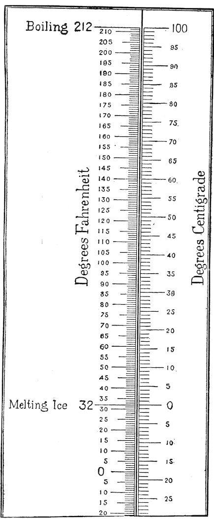
73. Humidity tables. To ascertain the relative humidity, the difference between the wet and dry bulb readings is obtained. This, with the dry bulb temperature, is referred to the tables, where the corresponding humidity is found. A variation in temperature has less effect than a variation in the difference; in consequence, the dry bulb reading is expressed in the nearest unit, and the difference reckoned to the nearest .5. The humidity varies with the air pressure. Hence, the altitude must be determined for the base station, and for all others that show much change in elevation. Within the ordinary range of growingperiod temperatures, the effect of pressure is not great. For all ordinary cases, it suffices to compute tables for pressures of 30, 29, 27, 25, and 23 inches. The following table indicates the decreaseinpressurewhichisduetoaltitude.
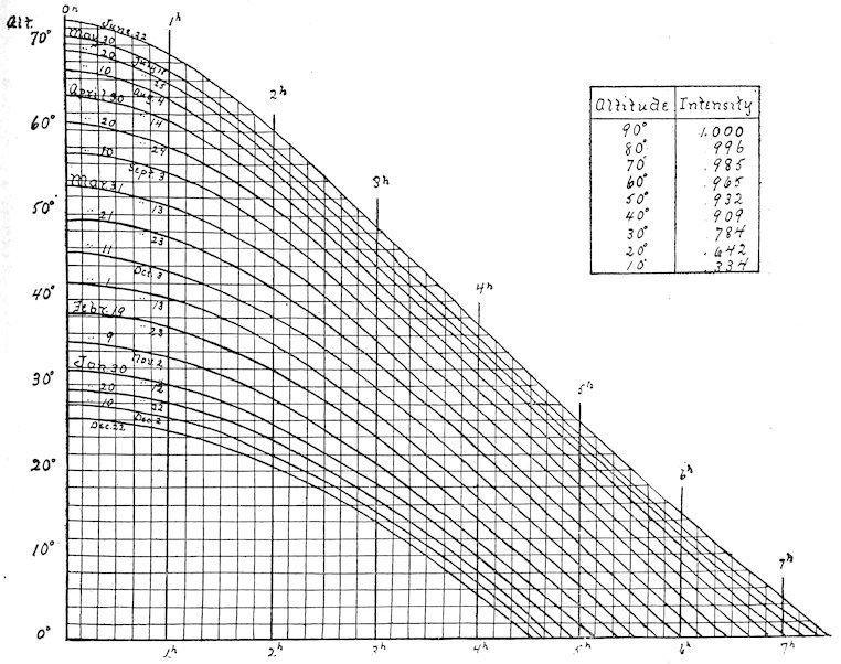
The fluctuations of pressure due to weather are usually so slight that their influence may be disregarded. An excellent series of tables of relative humidity is found in Marvin’s Psychrometric Tables, published by the U. S. Weather Bureau, and to be obtained from the Division of Publications, Washington, D. C., for 10 cents. A convenient field form is made by removing the portion containing the tablesofrelativehumidity,andbindingit instiffoilcloth.
74. Sums, means, and curves. An approximate humidity sum can be obtained by adding the absolute humidities for each of the twenty-four hours, and expressing the results in grains per cubic foot. It is possible to establish a general ratio between this sum and the transpiration sum of the plant, but its value is not great at present. Means of absolute and of relative humidity are readily determinable from the psychrograph records; the latter are the most useful. The mean of relative humidity for the twentyfour hours of a day is the average of the twenty-four hour humidities. From these means the seasonal mean is computed in the same manner. A close approximation, usually within 1 degree, may be obtained in either case by averaging the maximum and minimum for the period concerned. Various kinds of curves are of value in representing variation in humidity. Obviously, these must be derived from the psychrograph, or from the psychrometer when the series is sufficiently complete. The level curve indicates the variation in different stations at the same time. These may be combined in a series for the comparison of readings made at various heights in the stations. The day or point curve shows the fluctuations during the day of one point, and the station curve the variation at different heights in the same station. The curves of successive days or of different stations may of course be combined on the same sheet for comparison. Level and station curves based upon mean relative humidities are especially valuable.
75. Records. A field form is obviously unnecessary for the psychrograph. The record sheets constitute both a field and permanent record. The altitude and other constant features of the station and the list of species, etc., are entered on the back of the first record sheet, or, better, they are noted in the permanent formation record. For psychrometer readings, whether single or in series, the following recordformisemployed:
Fig.10.Conversionscalefortemperatures.
On page 47 is given a table for the conversion of Centigrade into Fahrenheit temperatures. This may be donementallybymeansoftheformula F = C/5×9+32°.
LIGHT
76. Methods. All methods for measuring light intensity, which have been at all satisfactory, are based upon the fact that silver salts blacken in the light. The first photographic method was proposed by Bunsen and Roscoe in 1862; this has been taken up by Wiesner and variously modified. After considerable experiment by the writer, however, it seemed desirable to abandon all methods which require the use of “normal paper” and “normal black” and to develop a simpler one. As space is lacking for a satisfactory discussion of the Bunsen-Roscoe-Wiesner methods, the reader is referred to the works cited below.[4] Simple photometers for making light readings simultaneously or in series were constructed in 1900, and have been in constant use since that time. An automatic instrument capable of making accurate continuous records proved to be a more difficult problem. A sunshine recorder was ultimately found which yields valuable results, and very recently a recording photometer which promises to be perfectly satisfactory has been devised. Since the hourly and daily variations of sunlight in the same habitat are relatively small, automatic photometers are perhaps a convenience rather than a necessity.
ThePhotometer
Fig 11 Photometer, showing front and side view
77. Construction. The simple form of photometer shown in the illustration is a light-tight metal box with a central wheel upon which a strip of photographic paper is fastened. This wheel is revolved by the thumbscrew past an opening 6 mm. square which is closed by means of a slide working closely between two flanges. At the edge of the opening, and beneath the slide is a hollow for the reception of a permanent light standard. The disk of the thumbscrew is graduated into twenty-five parts, and these are numbered. A line just beneath the opening coincides with the successive lines on the disk, and indicates the number of the exposure. The wheel contains twenty-five hollows in which the click works, thus moving each exposure just beyond the opening. The metal case is made in two parts, so that the bottom may be readily removed, and the photographic strip placed in position. The water-photometer is similar except that the opening is always covered with a transparent strip and the whole instrument is watertight. These instruments have been made especially for measuring light by the C. H. Stoelting Co., 31 W. Randolphstreet,Chicago,Ill.Thepriceis$5.
78. Filling the photometer. The photographic paper called “solio” which is made by the Eastman Kodak Company, Rochester, N. Y., has proved to be much the best for photometric readings. The most convenient size is that of the 8 × 10 inch sheet, which can be obtained at any supply house in packages of a dozen sheets for 60 cents. New “emulsions,” i. e., new lots of paper, are received by the dealers every week, but each emulsion can be preserved for three to six months without harm if kept in a cool, lighttight place. Furthermore, all emulsions are made in exactly the same way, and it has been impossible to detect any difference in them. To fill the photometer, a strip exactly 6 mm. wide is cut lengthwise from the 8 × 10 sheet. This must be done in the dark room, or at night in very weak light. The strip is placed on the wheel, extreme care being taken not to touch the coated surface, and fixed in position by forcing the free ends into the slit of the wheel by a piece of cork 8–9 mm. long. The wheel is replaced in the case, turneduntilthezeroisoppositetheindexline,andtheinstrumentisreadyforuse.
79. Making readings. An exposure is made by moving the slide quickly in such a way as to uncover the entire opening, and the standard if the exposure is to be very short. Care must be taken not to pull the slide entirely out of the groove, as it will be impossible to replace it with sufficient quickness. The time of exposure can be determined by any watch after a little practice. It is somewhat awkward for one person to manage the slide properly when his attention is fixed upon a second hand. This is obviated by having one observer handle the watch and another the photometer, but here the reaction time is a source of considerable error. The most satisfactory method is to use a stop-watch. This can be held in the left hand and started and stopped by the index finger. The photometer is held against it in the right hand in such a way that the two movements of stopping the watch and closing the slide may be made at the same instant. The length of exposure is that necessary to bring the tint of the paper to that of the standard beside it. A second method which is equally advantageous and sometimes preferable does away with the permanent standard in the field and the need for a stop-watch. In this event, the strip is exposed until a medium color is obtained, since very light or very deep prints are harder to match. This is later compared with the multiple standard. In both cases, the date, time of day, station, number of instrument and of exposure, and the length of the latter in seconds are carefully noted. The instrument is held with the edge toward the south at the level to be read, and the opening uppermost in the usual position of the leaf. When special readings are desired, as for isophotic leaves, reflected light, etc., the position is naturally changed to correspond. In practice, it is made an invariable rule to move the strip for the next exposure as soon as the slide is closed. Otherwise double exposures are liable to occur. When a strip is completely exposed it is removed in the dark, and a new one put in place. The former is carefully labeled anddatedontheback,andputawayina light-tightboxinacoolplace.
Fig. 12. Dawson-Lander sun recorder.
80. The Dawson-Lander sun recorder. “The instrument consists of a small outer cylinder of copper which revolves with the sun, and through the side of which is cut a narrow slit to allow the sunshine to impinge on a strip of sensitive paper, wound round a drum which fits closely inside the outer cylinder, but is held by a pin so that it can not rotate. By means of a screw fixed to the lid of the outer cylinder, the drum holding the sensitive paper is made to travel endwise down the outer tube, one-eighth of an inch daily, so that a fresh portion of the sensitive surface is brought into position to receive the record.” The instrument is driven by an eight-day clock placed in the base below the drum. The slit is covered by means of a flattened funnel-shaped hood, and the photographic strip is protected from rain by a perfectly transparent sheet of celluloid. The detailed structure of the instrument is shown in figure 12.Thisinstrumentmaybeobtainedfrom LanderandSmith,Canterbury,England,for$35.
In setting up the sunshine recorder, the axis should be placed in such a position that the angle which it makes with the base is the same as the altitude of the place where the observations are made. This is readily done by loosening the bolts at either side. The drum is removed, the celluloid sheet unwound by means of the key which holds it in place, the sensitive strip put in position, and the sheet again wound up. Strips of a special sensitive paper upon which the hours are indicated are furnished by the makers of the instrument, but it has been found preferable to use solio strips in order to facilitate comparison with the standards. The drum is placed on the axis, and is screwed up until it just escapes the collar at the top of the spiral. The clock is wound and started, and the outer cylinder put on so that the proper hour mark coincideswiththeindexonthefrontofthebase.
As a sunshine recorder, the instrument gives a perfect record, in which the varying intensities are readily recognizable. Since the cylinder moves one-half inch in an hour, and the slit is .01 of an inch, the time of each exposure is 72 seconds. This gives a very deep color on the solio paper, which results in a serious error in making comparisons with the standard. On account of the hood, diffuse light is not recorded when it is too weak to cast a distinct shadow. It seems probable that this difficulty will be
overcome by the use of a flat disk containing the proper slit, and in this event the instrument will become of especial value for measuring the diffuse light of layered formations. The celluloid sheet constitutes a source of error in sunlight on account of the reflection which it causes. This can be prevented by using the instrument only on sunny days, when the protection of the sheet can be dispensedwith.
81. The selagraph. This instrument is at present under construction, and can only be described in a general way. In principle it is a simple photometer operating automatically. It consists of a light-tight box preferably of metal, which contains an eight-day lever clock. Attached to the arbor of the latter is a disk 7 inches in diameter bearing on its circumference a solio strip 1 cm. wide and 59 cm. long. The opening in the box for exposure is 6 mm. square and is controlled by a photographic shutter. The latter is constructed so that it may be set for 5, 10, or 20 seconds, since a single period of exposure can not serve for both sun and shade. The shutter is tripped once every two hours, by means of a special wheel revolving once a day. Each exposure is 6 mm. square, and is separated by a small space from the next one. Twelve exposures are made every 24 hours, and 84 during the week, though, naturally, the daytime exposures alone are recorded. Comparisons with the multiple standard are made exactly as in the case of thesimplephotometer.Theselagraphismade bytheC. H.StoeltingCo.,Chicago,Illinois.
Standards
82. Use. The light value of each exposure is determined by reference to a standard. When the photometer carries a permanent standard, each exposure is brought to the tint of the latter, and its value is indicated by the time ratio between them. Thus, if the standard is the result of a 5–second exposure to full sunlight at meridian, and a reading which corresponds in color requires 100 seconds in the habitat concerned, the light of the latter is twenty times weaker or more diffuse. Usually, the standard is regarded as unity, and light values figured with reference to it, as .05. With the selagraph such a use of the standard is impossible, and often, also, with the photometer it is unnecessary or not desirable. The value of each exposure in such case is obtained by matching it with a multiple standard, after the entire strip has been exposed. The further steps are those already indicated. After the exact tint in the standard has been found, the length of the reading in seconds is divided by the time of the proper standard, and theresultexpressedasabove.
83. Making a standard. Standards are obtained by exposing the photometer at meridian on a typically clear day, and in the field where there is the least dust and smoke. Exception to the latter may be made, of course, in obtaining standards for plant houses located in cities, though it is far better to have the same one for both field and control experiment. Usable standards can be obtained on any bright day at the base station. Indeed, valuable results are often secured by immediate successive sun and shade readings in adjacent habitats, where the sun reading series is the sole standard. Preferably, standards should be made at the solstices or equinoxes, and at a representative station. The June solstice is much to be preferred, as it represents the maximum light values of the year. Lincoln has been taken as the base station for the plains and mountains. It is desirable, however, that a national or international station be ultimately selected for this purpose, in order that light values taken in different parts of the worldmaybereadilycompared.
84. Kinds of standards. The base standard is the one taken at Lincoln (latitude 41° N.) at meridian June 20–22. This is properly the unit to which all exposures are referred, but it has been found convenient to employ the Minnehaha standard as the base for the Colorado mountains, in order to avoid reducing each time. Relative standards are frequently used for temporary purposes. Thus, in comparing the light intensities of a series of formations, one to five standards are exposed on the solio strip before beginning the series of readings. Proof standards are the exposed solio strips, which fade in the light, and can, in consequence, be kept only a few weeks without possibility of error. The fading can be prevented by “toning” the strip, but in this event the exposures must be fixed in like manner before they can be compared. This process is inconvenient and time-consuming. It is also open to considerable error, as the time of treatment, strength of solution, etc., must be exactly equivalent in all instances. Permanent standards are accurate water-color copies of the originals obtained by the photometer. These have the apparent disadvantage of requiring a double comparison or matching, but after a little practice it is possible to reproduce the solio tints so that the copy is practically indistinguishable from the original. The most satisfactory method is to make a long stroke of color on a pure white paper, since a broad wash is not quite homogeneous, and then to reject such parts of the stroke as do not match exactly. Permanent standards fade after a few month’s use, and must be replaced by parts of the original
stroke. Single standards are made by one exposure, while multiple ones have a series of exposures filling a whole light strip. These are regularly obtained by making the exposures from 1–10 seconds respectively, and then increasing the length of each successive exposure by 2 seconds. Single exposures of 1–5 seconds as desired usually serve as the basis for permanent standards, but a multiple standard may also be copied in permanent form. Exposures for securing standards must be made only under the most favorable conditions, and the length in seconds must be exact. The use of the stop-watch is imperative, except where access may be had to an astronomical clock with a large second hand, which is even more satisfactory. The length of time necessary for the series desired is reckoned beforehand, and theexposuresbegunsothatthemeridian fallsinthemiddleoftheprocess.
Single standards are exceedingly convenient in photometer readings, but they are open to one objection. In the sunshine it is necessary to make instant decision upon the accuracy of the match, or the exposure becomes too deep. In the shade where the action is slower, this difficulty is not felt. For this reason it is usually desirable to check the results by a multiple standard, and in the case of selagraph records, where the various exposures show a wide range of tint, light values are obtainable only by direct comparison with the multiple standard. The exact matching of exposure and standard requires great accuracy, but with a little practice this may be done with slight chance of error by merely moving the exposure along the various tints of the standard until the proper shade is found. The requisite skill is soon acquired by running over a strip of exposures several times until the comparisons always yield the same results for each. The margin of error is practically negligible when the same person makes all the comparisons, and in the case of two or three working on the same reading the results diverge little or not at all. The efficient difference for light is much more of a variable than is the case with water-content. It has been determined so far only for a few species, all of which seem to indicate that appreciable modification in the form or structure of a leaf does not occur until the reduction in intensity reaches .1 of the meridian sunlight at the June solstice. The error of comparison is far less than this, and consequentlymaybeignored,eveninthe mostpainstakinginquiry.
Readings
85. Time. The intensity of the light incident upon a habitat varies periodically with the hour and the day, and changes in accord with the changing conditions of the sky. The light variations on cloudy days can only be determined by the photometer. While these can not be ignored, proper comparisons can be instituted only between the readings taken on normal days of sunshine. The sunlight varies with the altitude of the sun, i. e., the angle which its rays make with the surface at a given latitude. This angle reaches a daily maximum at meridian. The yearly maximum falls on June 22, and the angle decreases in both directions through the year to a minimum on December 22. At equal distances from either solstice, the angle is the same, e. g., on March 21 and September 23. At Lincoln (41° N. latitude) the extremes at meridian are 73° and 26°; at Minnehaha (39°) they are 75° and 28°. The extremes for any latitude may be found by subtracting its distance in degrees north of the two tropics from 90. Thus, the 50th parallel is 26.5° north of the tropic of Cancer, and the maximum altitude of the sun at a place upon it is 63.5°. It is73.5°northofthetropicofCapricorn,andtheminimummeridionalaltitudeis16.5°.
The changes in the amount of light due to the altitude of the sun are produced by the earth’s atmosphere. The absorption of light rays is greatest near the horizon, where their pathway through the atmosphere is longest, and it is least at the zenith. The absorption, and, consequently, the relative intensity of sunlight, can be determined at a given place for each hour of any sunshiny day by the use of chart 13. This chart has been constructed for Lincoln, and will serve for all places within a few degrees of the 40th parallel. The curves which show the altitude of the sun at the various times of the day and the year have been constructed by measurements upon the celestial globe. Each interval between the horizontal lines represents 2 degrees of the sun’s altitude. The vertical lines indicate time before or after the apparent noon, the intervals corresponding to 10 minutes. If the relative intensity at Lincoln on March 12 at 3:00 P.M. is desired, the apparent noon for this day must first be determined. A glance at the table shows that the sun crosses the meridian on this day at 9 minutes 53 seconds past noon at the 90th meridian. The apparent noon at Lincoln is found by adding 26 minutes 49 seconds, the difference in time between Lincoln and a point on the 90th meridian. When the sun is fast, the proper number of minutes is taken from 26 minutes 49 seconds. The apparent noon on March 12 is thus found to fall at 12:37 P.M., and 3:00 P.M. is 2 hours and 23 minutes later. The sun’s altitude is accordingly 36°. If the intensity of the light which reaches the earth’s surface when the sun is at zenith is taken as 1, the table of thesun’saltitudesgivestheintensityat3:00 P.M. onMarch12as.85.
For places with a latitude differing by several degrees from that of Lincoln, it is necessary to construct a new table of altitude curves from the celestial globe. It is quite possible to make a close approximation of this from the table given, since the maximum and minimum meridional altitude, and hence the corresponding light intensity, can be obtained as indicated above. For Minnehaha, which is on the 105th meridian, and for other places on standard meridians, i. e., 60°, 75°, 90°, and 120° W., the table of apparent noon indicates the number of minutes to be added to 12 noon, standard time, when the sun is slow, and to be subtracted when the sun is fast. The time at a place east or west of a standard meridian is respectively faster or slower than the latter. The exact difference in minutes is obtained from the difference in longitude by the equation, 15° = 1 hour. Thus, Lincoln, 96° 42′ W. is 6° 42′ west of the standard meridian of 90°; it is consequently 26 minutes 49 seconds slower, and this time must always be added to the apparent noon as determined from the chart. At a place east of a standard meridian, the timedifferenceis,ofcourse,subtracted.
Fig 13 Chart for the determination of the sun ’ s altitude, and the corresponding light intensity
The actual differences in the light intensity from hour to hour and day to day, which are caused by variations in the sun’s altitude, are not as great as might be expected. For example, the maximum intensity at Lincoln, June 22, is .98; the minimum meridional intensity December 22 is .73. The extremes on June 22 are .98 and .33 (the latter at 6:00 A.M. and 6:00 P.M. approximately); between 8:00 A.M. and 4:00 P.M. the range in intensity is from .90 to .98 merely. On December 22, the greatest intensity is .52, the least .20 (the latter at 8:00 A.M. and 4:00 P.M. approximately). If the growing season be taken as beginning with the 1st of March and closing the 1st of October, the greatest variation in light intensity at Lincoln within a period of 10 hours with the meridian at its center (cloudy days excepted) is from .33 to .98. In a period of 8 hours, the extremes are .65 to .98, i. e., the greatest variation, .3, is far
within the efficient difference, which has been put at .9. For the growing period, then, readings made between 8:00 A.M. and 4:00 P.M. on normal sunshiny days may be compared directly, without taking into account the compensation for the sun’s altitude. Until the efficient difference has been determined for a large number of species, however, it seems wise to err on the safe side and to compensate for great differences in time of day or year. In all doubtful cases, the intensity obtained by the astronomical method should also be checked by photometric readings. A slight error probably enters in, due to reflectionfromthesurfaceofthepaper,andtotemperature,butthisisnegligible.
86. Tablefor determining apparent noon
87. Place. The effect of latitude upon the sun’s altitude, and the consequent light intensity have been discussed in the pages which precede. Latitude has also a profound influence upon the duration of daylight, but the importance of the latter apart from intensity is not altogether clear. The variation of intensity due to altitude has been greatly overestimated; it is practically certain, for example, that the dwarf habit of alpine plants is not to be ascribed to intense illumination, since the latter increases but slightly with the altitude. It has been demonstrated astronomically that about 20 per cent of a vertical ray of sunlight is absorbed by the atmosphere by the time it reaches sea level. At the summit of Pike’s Peak, which is 14,000 feet (4,267 meters) high, the barometric pressure is 17 inches, and the absorption is approximately 11 per cent. In other words, the light at sea level is 80 per cent of that which enters the earth’s atmosphere; on the summit of Pike’s Peak it is 89 per cent. As the effect of the sun’s altitude is the same in both places, the table of curves on page 57 will apply to both. Taking into account the difference in absorption, the maximum intensity at sea level and at 14,000 feet on the fortieth parallel is .98 and 1.09 respectively. The minimum intensities between 8:00 A.M. and 4:00 P.M. of the growing period are .64 and .71 respectively. The correctness of these figures has been demonstrated by photometer readings, which have given almost exactly the same results. Such slight variations are quite insufficient to produce an appreciable adjustment, particularly in structure. They are far within the efficient difference, and Reinke[5] has found, moreover, that photosynthetic activity in Elodea is not increased beyond the normal in sunlight sixty times concentrated. In consequence, it is entirely unnecessarytotakeaccountofdifferentaltitudes inobtaininglightvalues.
The slope of a habitat exerts a considerable effect upon the intensity of the incident light. If the angle between the slope and the sun’s ray be 90°, a square meter of surface will receive the maximum intensity, 1. At an angle of 10°, the same area receives but .17 of the light. This relation between angle and intensity is shown in the table which follows. The influence of the light, however, is felt by the leaf, not by the slope. Since there is no connection between the position of the leaf and the slope of the habitat, the latter may be ignored. In consequence, it is unnecessary to make allowances for the direction of a slope, viz., whether north, east, south, or west, in so far as light values are concerned. The angle which a leaf makes with its stem determines the angle of incidence, and hence the amount of light received by the leaf surface. This is relatively unimportant for two reasons. This angle changes hourly and daily with the altitude of the sun, and the intensity constantly swings from one extreme to the other. Moreover, the extremes 1.00 and 0.17, even if constant, are hardly sufficient to produce a measurable result. When the angle of the leaf approaches 90°, there is the well-known differentiation of leaf surfaces andofchlorenchym,butthishasnorelationtotheangleofincidence.
TableofIntensityatVariousAngles
In the sunlight, it makes no difference at what height a light reading is taken. In forest and thicket as well as in some herbaceous formations, the intensity of the light, if there is any difference, is greatest just beneath the foliage of the facies. In forests especially, the light is increasingly diffuse toward the ground,
particularly where layers intervene. In woodland formations, moreover, the exact spot in which a reading is made must be carefully chosen, unless the foliage is so dense that the shade is uniform. A very satisfactory plan is to take readings in two or more spots where the shade appears to be typical, and to make a check reading in a “sunfleck,” a spot where sunlight shows through. In forests and thickets, the sunflecks are fleeting, and the light value is practically that of the shade. In passing into open woodland and thicket, the sunflecks increase in size and permanence, until finally they exceed the shade areas in amountandbecometypicaloftheformation.
Reflected andAbsorbedLight
88. The fate of incident light. The light present in a habitat and incident upon a leaf is not all available for photosynthesis. Part is reflected or screened out by the epidermis, and a certain amount passes through the chlorenchym, except in very thick leaves. The light absorbed is by far the greatest in the majority of species. Many plants with dense coatings of hairs reflect or withhold more light than they absorb, and the amount of light reflected by a thick cuticule is likewise great. As light is imponderable, the actual amount absorbed or reflected by the leaf can not be determined. It is possible, however, to express this in terms of the total amount received, by means of readings with solio paper, and the knowledge thus obtained is of great importance in interpreting the modifications of certain types of leaves. For example, a leaf with a densely hairy epidermis may receive light of the full intensity, 1; the amount reflected or screened out by the hairs may be 95 per cent of this, the amount absorbed 5 per cent, and that transmitted, nil. In the majority of cases, however, the absorbed light is considerably more thantheamountreflectedortransmitted.
Fig.14.Leafprint:exposed10 m.,11 A.M. August20. Theleavesarefromsunandshadeformsof Bursa bursa-pastoris, Rosa sayii, Thalictrum sparsiflorum,and Machaerantheraaspera.In each theshadeleafprintsmoredeeply.
89. Methods of determination. If results are to be of value, reflected and transmitted light must be determined in the habitat of the plant simultaneously with the total light which a leaf receives. An approximation of the light reflected from a leaf surface is secured by placing the photometer so that the light reflected is thrown upon the solio strip. A much more satisfactory method, however, is to determine it in connection with the amount of light transmitted through the epidermis. This is done by stripping a piece of epidermis from the upper surface of the leaf and placing it over the slit in the photometer for an exposure. An exposure in the full light of the habitat is made simultaneously with another photometer, or immediately afterward upon the same strip. When the epidermis is not too dense, both exposures are permitted to reach the same tint, and the relation between them is precisely that of their lengths of exposure. Ordinarily the two exposures are made absolutely simultaneous by placing the epidermis over half of the opening, leaving the other half to record the full light value, and the results, or epidermis prints, are referred to a multiple standard. The difference between the two values thus obtained represents the amount of reflected light together with that screened by the epidermis. The amount of light transmitted through the leaf may be measured in the same way by using the leaf itself in place of the epidermis alone. The time of exposure is necessarily long, however, and it has been found practicable to obtain leaf prints by exposing the leaf in a printing frame, upon solio paper, at the same time that the epidermis print is made. In a few species both the upper and lower epidermis can be removed and the amount of light absorbed
determined directly by exposing the strip covered with the chlorenchym. Generally, however, this must be computed by subtracting the sum of the per cents of reflected and transmitted light from 100 per cent,whichrepresentsthetotallight.
90. Leaf and epidermis prints. In diphotic leaves the screening effect of the lower epidermis may be ignored. Isophotic sun leaves, i. e., those nearly upright in position or found above lightcolored, reflecting soils, are usually strongly illuminated on both sides, and the absorbed light can be obtained only by measuring the screening effect of both epiderms. Shade leaves and submerged leaves often contain chloroplasts in the epidermis, and the above method can not be applied to them. In fact, in habitats where the light is quite diffuse, practically all incident light is absorbed. The rare exceptions are those shade leaves with a distinct bloom. In addition to their use in obtaining the amount of light absorbed, both leaf and epidermis prints are extremely interesting for the direct comparison of light relations in the leaves of species belonging to different habitats. The relative screening value of the upper and lower epidermis, or of the corresponding epiderms of two ecads or two species, is readily ascertained by exposing the two side by side in sunshine, over the slit in the photometer. For leaf prints fresh leaves are desirable, though nearly the same results can be obtained from leaves dried under pressure. The leaves are grouped as desired on the glass of a printing frame, and covered with a sheet of solio. They are then exposed to full sunlight, preferably at meridian, and the prints evaluated by means of the multiple standard. This method is especially useful in the comparison of ecads of one species. These differences due to transmitted light are very graphic, and can easily be preserved by “toning”theprintintheusualway.
ExpressionofResults
91. Light records. The actual photographic records obtained by photometer and selagraph can at most be kept but a few months, unless they are toned or fixed. “Toning” modifies the color of the exposure materially, and changes its intensity so that it can not be compared with readings not fixed. It would involve a great deal of inconvenience to make all comparisons by means of toned strips and standard, even if it were not for the fact that it is practically impossible to obtain exactly the same shade in lots toned at different times. The field record, if carefully and neatly made, may well take the place of apermanentone.Theformisthefollowing:
92. Light sums, means, and curves. Owing to the fact that the selagraph has not yet been used in the field, no endeavor has been made to determine the light value for every hour of the day in different habitats. Consequently there has been no attempt to compute light sums and means. Photometer readings have sufficed to interpret the effect of light in the structure of the formation, and of the individual, but they have not been sufficiently frequent for use in ascertaining sums and means. The latter are much less valuable than the extremes, especially when the relative duration of these is indicated. Means, however, are readily obtained from the continuous records. Light sums are probably impracticable, as the factor is not one that can be expressed in absolute terms. The various kinds and combinations of light curves are essentially the same as for humidity. The level curve through a series of habitats is the most illuminating, but the day curve of hour variations is of considerable value. The curve of daily duration, based upon full sunlight, is also of especial importance for plants, and stations which receivebothsunandshadeduringtheday.
TEMPERATURE
93. In consequence of its indirect action, temperature does not have a striking effect upon the form and structure of the plant, as is the case with water and light. Notwithstanding, it is a factor of fundamental importance. This is especially evident in the character and distribution of vegetation. It is also seen in the germination and growth of plants, in the length of season, and in the important influence of temperature upon humidity, and hence upon water-content. Because of its intimate relation with the comfort of mankind, the determination of temperature values has received more attention than that of any other factor, and excellent simple and recording instruments are numerous. For plants, it is also necessary to employ instruments for measuring soil temperatures. The latter unquestionably have much less meaning for the plant than the temperatures of the air, but they have a direct influence upon theimbibitionofwater,andupongermination.
Thermometers
94. Air thermometers. The accurate measurement of temperature requires standard thermometers. Reasonably accurate instruments may be standardized by determining their error, but they are extremely unsatisfactory in practice, since they result in a serious waste of time. Accurate thermometers which read to the degree are entirely serviceable as a rule, but instruments which read to a fraction of a degree are often very much to be desired. The writer has found the “cylindrical bulb thermometer, Centigrade scale” of H. J. Green, to be an exceedingly satisfactory instrument. The best numbers for general use are 247 and 251, which read from –15° to 50° C. and are graduated in .2°. They are respectively 9 and 12 inches long, and cost $2.75 and $3.50. These instruments are delicate and require careful handling, but even in class work this has proved to be an advantage rather than otherwise. In making readings of air temperatures with such thermometers, constant precautions must betakentoexposethebulbdirectlytothewindand to keepitawayfromthehandandperson.
95. Soil thermometers. The thermometer described above has been used extensively for soil temperatures. The determination of the latter is conveniently combined with the taking of soil samples, by using the hole for a temperature reading. When carefully covered, these holes can be used from day to day throughout the season without appreciable error, even in gravel soils. Repeated tests of this have been made by simultaneous readings in permanent and newly made holes, and the results have always been the same. It has even been found that the error is usually less than 1 degree when the hole is left uncovered, if it is more than 9 inches deep. A slight source of error lies in the fact that the thermometer must be raised to make the reading. With a little practice, however, the top of the column of mercury may be raised to the surface and read before the change of temperature can react upon it. This is especially important in very moist or wet soils where the bulb becomes coated with a film of moisture. This evaporates when the bulb is brought into the air, and after a moment or two the mercury slowly falls.
Regular soil thermometers are indispensable when readings are desired at depths greater than 12–18 inches. They possess several disadvantages which restrict their use almost wholly to permanent stations. It is scarcely possible to carry them on field trips, and the time required to place them in the soil renders them practically useless for single readings. Moreover, the instruments are expensive, ranging in price from $7 for the two-foot thermometer, to $19 for the eight-foot one. When it is recognized that deep-seated temperatures are extremely constant and that the slight fluctuations affect, as a rule, only the relatively stable shrubs and trees, it is evident that such temperatures are of restricted importance. Still, in any habitat, they must be ascertained before they can well be ignored, though it is unwise to spend much time and energy in their determination. Soil thermometers of the form illustrated may be obtained from H. J. Green, Brooklyn.
96. Maximum-minimum thermometers. These are used for determining the range of temperature within a given period, usually a day. Since they are much cheaper than thermographs, they can replace these in part, although they merely indicate the maximum and minimum temperatures for the day, and do not register the time when each occurs. The maximum is a mercurial thermometer with a constriction in the tube just above the bulb; this allows the mercury to pass out as it expands, but prevents it from running back, thus registering the maximum temperature. The minimum thermometer contains alcohol. The column carries a tiny dumbbellshaped marker which moves down with it, but will not rise as the liquid expands. This is due to the fact that the fluid expands too slowly to carry the marker upward, while the surface tension causes it to be drawn downward as the fluid contracts. The minimum temperature is indicated by the upper end of the marker. In setting up the thermometers, they are attached by special thumbscrews to a support which holds them in an oblique position. The minimum is placed in a special holder above the maximum which rests on a pin that is used also for screwing the pivot-screw into position. The support is screwed tightly to the cross-piece of a post, or in forest formations it is fastened directly to a board nailed upon a tree trunk. A shelter has not been used in ecological work, although it is the rule in meteorological observations. The minimum thermometer is set for registering by raising the free end, so that the marker runs to the end of the column. The mercury of the maximum is driven back into the bulb by whirling it rapidly on the pivot-screw after the pin has been taken out. This must be done with care in order that the bulb may not be broken. As soon as the instrument comes to rest, it is raised and the pin replaced, great care being taken to lift it no higher than is necessary. When the night maximum is sought, the thermometer should be whirled several times in order to drive the column sufficiently low. Usually, in such cases, a record is made of this point to make sure that the maximum read is the actual one. If the pivot-screw is kept well oiled, less force will be required to drive the mercury back. In practice, the thermometers have been observed at 6:00 A.M. and 6:00 P.M. each day, thus permitting the reading of the maximum-minimum for both day and night. Pairs of maximum-minimum thermometers are to be obtained from H. J. Green, 1191 Bedford Ave., Brooklyn, or Julien P. Friez, Baltimore, Maryland, at a cost of $8.25.
97. Radiation thermometers. These are used to determine the radiation in the air, and from the soil, i. e., for solar and terrestrial radiation. The latter alone has been employed in the study of habitats, chiefly for the purpose of ascertaining the difference in the cooling of different soils at night. The terrestrial radiation thermometer is merely a special form of minimum thermometer, so arranged in a support that the bulb can be placed directly above the soil or plant to be studied. It is otherwise operated exactly like the minimum thermometer, and the reading gives the minimum temperature which the air above the plant or soil reaches, not the amount of radiation. As a consequence, these instruments are valuable only where read in connection with a pair of maximum-minimum thermometers in the air, or when read in a series of instruments placed above different soils or plants.
98. Thermographs. Two types of standard instruments are in general use for obtaining continuous records of air temperatures. These are the Draper thermograph, made by the Draper Manufacturing Company, 152 Front St., New York city ($25 and $30), and the Richard thermograph sold by Julien P. Friez, Baltimore ($50). After careful trial had demonstrated that they were equally accurate, the matter of cost was considered decisive, and the Draper thermograph has been used exclusively in the writer’s own work. This instrument closely resembles the psychrograph manufactured by the same company. It is made in two sizes, of which the larger one is the more satisfactory on account of the greater distance between the lines of the recording disk. The
thermometric part consists of two bimetallic strips, the contraction and expansion of which are communicated to a hand carrying a pen. The latter traces a line on the record sheet which is attached to a metal disk made to revolve by an eight-day clock. In practice the thermograph is set up in the shelter which contains the psychrograph, and in exactly the same manner. The clock is wound, the record put in place, and the pen inked in the same way also. The proper position of the pen is determined by making a careful thermometer reading under the shelter, and then regulating the pen hand by means of the screws at the base of it A similar test reading is also made each week, when the clock is rewound. A record sheet may be left in position for three weeks, the pen being filled each week with a different ink. The fixed order of using the inks is red, blue, and green as already indicated.
Owing to the fact that they are practically stationary, soil thermographs are of slight value, except at base stations. Here, the facts that they are expensive, that the soil temperatures are of relatively little importance, and that they can be determined as easily, or nearly so, by simple thermometers, make the use of such instruments altogether unnecessary, if not, indeed, undesirable. In a perfectly equipped research station, they undoubtedly have their use, but at ordinary stations, and in the case of private investigators, their value is in no wise commensurate with their cost.
Readings
99. Time. The hourly and daily fluctuations of the temperature of the air render frequent readings desirable. It is this variation, indeed, which makes single readings, or even series of them, inconclusive, and renders the use of a recording instrument almost imperative in the base station at least. Undoubtedly, a set of simultaneous readings at different heights in one station, or at the same height in different stations, especially if made at the maximum, have much value for comparison, but their full significance is seen only when they are referred to a continuous base record. Such series, moreover, furnish good results for purposes of instruction. In research work, however, it has been found imperative to have thermographs in habitats of widely different character. With these as bases, it is possible to eke them out with considerable satisfaction by means of
maximum-minimum thermometers in less different habitats, or in different parts of the same habitat. Naturally these are less satisfactory, and are used only when expense sets a limit to the number of thermographs. In a careful analysis of a single habitat, more can be gained by one base thermograph supplemented by three pairs of maximum-minimum thermometers in dissimilar areas of the habitat than by two thermographs, and the cost is the same.
Fig. 16. Soil thermometer
Fig. 17. Maximum-minimum thermometer.
19. Draper thermograph.
Fig. 18. Terrestrial radiation thermometer.
Fig.
Fig. 20. Shelter for thermograph.
Fig. 21. Richard thermograph.
100. Place and height. For general air temperatures, thermograph and thermometer readings are made at a height of 3 feet (1 meter). Soil temperatures are regularly taken at the surface and at a depth of 1 foot. When a complete series of simultaneous readings is made in one station, the levels are 6 feet and 3 feet in the air, the surface of the soil, and 5, 10, and 15 inches in the soil. When sun and shade occur side by side in the same formation, as is true of many thickets and forests, surface readings are regularly made in both. Similarly, valuable results are obtained by making simultaneous readings on the bare soil, on dead cover, and upon a leaf, while the influence of cover is readily ascertained by readings upon it and beneath it. A full series of station readings made at the same time upon north, east, south, and west slopes is of great importance in studying the effects of exposure.
Expression of Results
101. Temperature records. Neither field nor permanent form is required for thermographic records, other than the record sheet itself, which contains all the necessary information in a fairly convenient form. Although the temperature of a particular hour and day can not be read at a mere glance, it can be obtained so easily that it is a waste of time to make a tabular copy of each record sheet. For thermometer readings, either single or in series, the following form is used: