Anoverviewoftheroleof metalsinbiology
RobertR.Crichton
CatholicUniversityofLouvain,Louvain-la-Neuve,Belgium
OUTLINE
Introduction
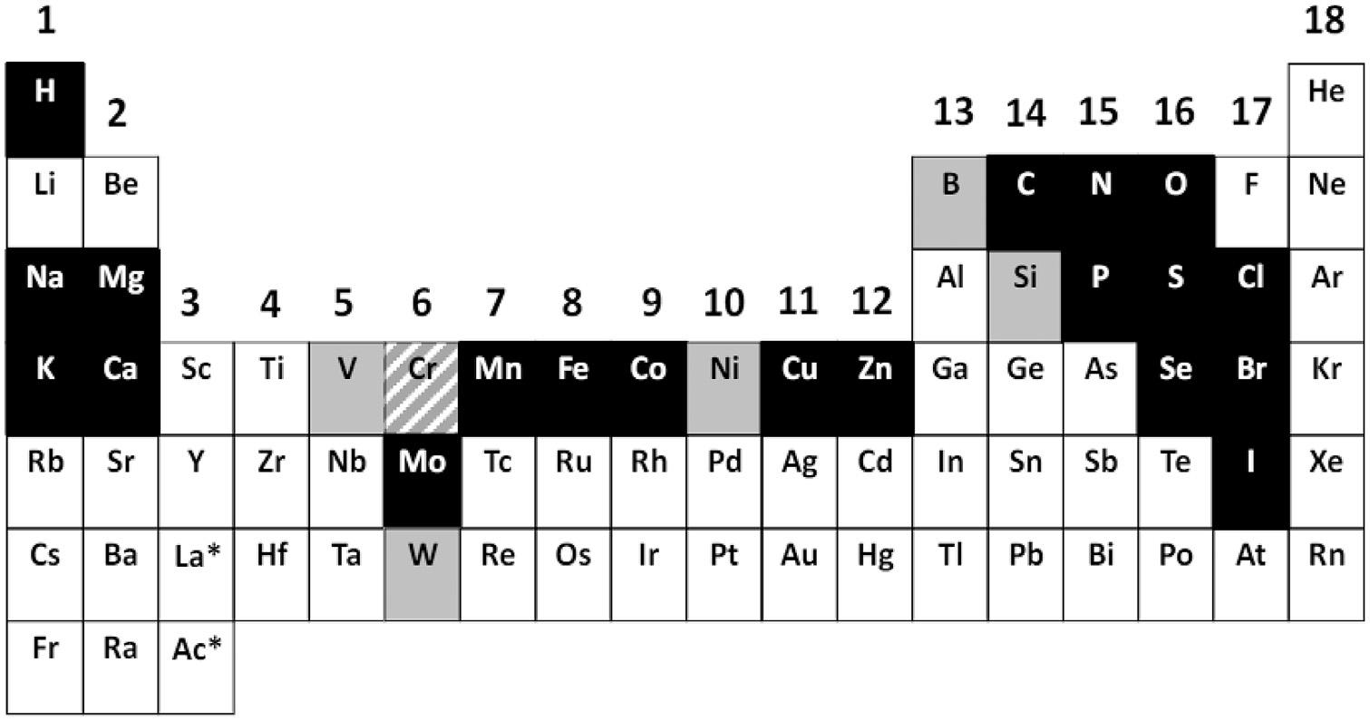
Metalsplaymanydifferentrolesinthebiologicalworld,whetherbytheirparticipation inessentialbiologicalprocesses,astoxicconstituentsofourenvironment,orasindispensablediagnosticandtherapeuticagentsinhumanmedicine.Onlyalimitednumberof metalionsareessentialformostlivingorganisms(Fig.1.1),andthisshortintroduction beginsbyillustratingthebiologicalimportanceofmetals,notonlyinvitalprocessessuch asintermediarymetabolism,electrontransfer,respiration,andphotosynthesis,butalsoin neurotransmission,cellsignaling,apoptosis,andfertilization.
Whilemanyessentialmetalscanbetoxic,particularlywhentheyareinexcess,inour modernenvironmentthereareanumberofnonessentialmetals,suchascadmium,lead, mercury,andaluminum,whicharethemselveshighlytoxic.
Finally,metalshaveassumedanextraordinarynumberofrolesinmedicine,not onlytherapeuticallyasdrugs,butalsoasnoninvasivecontrastagentsand radiopharmaceuticals.
Moredetailedaccountsoftheseaspectsofmetalionsarepresentedinthecompanion volumetothissecondedition(Crichton,2018).
FIGURE1.1 Abiologicalperiodictableoftheelementsindicatingtheessentialelements.Theessentialelementsformostformsoflifeareshowninblackwiththeexceptionofchromium(Cr),whichisshownwithan upwarddiagonalpattern,andessentialelementsthataremorerestrictedforsomeformsoflifeshowningray. Source:ReproducedfromMaret,W.,2016.Themetalsinthebiologicalperiodicsystemoftheelements:conceptsandconjectures.Int.J.Mol.Sci.17,pii:E66. doi:10.3390/ijms17010066.ThisisanopenaccessarticledistributedundertheCreative CommonsAttributionLicense(CCBY)whichpermitsunrestricteduse,distribution,andreproductioninanymedium,providedtheoriginalworkisproperlycited.
Essentialmetalionsandtheirfunctions
Mostlivingorganismsrequiresome25elements(Maret,2016;ChellanandSadler,2015) includingbetween10and14metalions(Fig.1.1).Inthecaseof Homosapiens,thereare10 essentialmetalions(sodium,potassium,calcium,magnesium,manganese,iron,cobalt, copper,zinc,andmolybdenum).Ofthese,thefirstfourareconsideredas“bulkelements” (Na1,K1,Ca21,andMg21),representing112g,160g,1.1kg,and25g,respectively,inan “average”personofbodyweight80kg(Thedataontheabundanceofelementsinthe 80kghumanbodyarethosegiveninWebElements: http://www.webelements.com/.). Together,theyconstitutesome99%ofthemetalioncontentofthehumanbody.The others,manganese,iron,cobalt,copper,zinc,andmolybdenum,designated“traceelements,”arepresentinmuchloweramountsthanthebulkelements(respectively,16mg, 4.8g,1.6mg,80mg,2.6g,and8mginan80-kgperson).
TheessentialalkalimetalionsNa1 andK1 onlyweaklybindorganicligands,rendering themextremelymobile,aswithH1 andCl .Thisenablesthemtogenerateionicgradients acrossbiologicalmembranes.ThedistributionofNa1 andK1 inmammalsisquitedifferent;Na1,togetherwithCl ,isthemajorelectrolyteintheextracellularfluid,whereasK1 isretainedwithinthecells.TheconcentrationofNa1 intheplasmaismaintainedwithin narrowlimitsatabout145mmol/L,anditsintracellularconcentrationisonlyabout 12mmol/L,whereastheintracellularconcentrationofK1 is150mmol/L,andtypically
only4 5mmol/Lintheextracellularfluids.Thisconcentrationdifferential,maintainedby the(Na1 K1)-ATPaseoftheplasmamembrane,ensuresanumberofmajorbiological processes,suchascellularosmoticbalance,signaltransduction,andneurotransmission. (Na1 K1)-ATPasetransportsthreeNa1 ionstotheoutsideofthecellandtwoK1 ionsto theinside(Figs.1.2and1.3),contributingtotheactionpotentialinvolvedintransmission ofnerveimpulsesalongneuronalaxons.Actionpotentialscanbegeneratedbypresynapticneuronsattherateofabout250persecond,accountingforbetweenone-halfand two-thirdsoftheirtotalATPconsumption.TherepetitiveG-richsequencesfoundinthe telomeresattheendsofeukaryoticchromosomesarestabilizedbyK1 andNa1 ions.The retentionofNa1 (hypernatremia)whenNa1 intakeexceedsrenalclearanceisoneof themostcommonelectrolytedisordersinclinicalmedicine.Hyperkalemiahasbecome morecommonincardiovascularpracticeduetothegrowingpopulationofpatientswith chronickidneydiseaseandthebroadapplicationofdrugsthatmodulaterenalelimination ofpotassiumbyreducingtheproductionofangiotensinII.
Thealkalineearthmetalions,Mg21 andCa21,havegreaterbindingstrengthstoorganic ligandsthanNa1 andK1,andthereforearelessmobile.Bothplayimportantstructural andcatalyticroles,with99%ofthebody’sCa21 foundinboneandteeth.AlthoughMg21 istheleastabundantofthe“bulkelements,”theintracellularconcentrationoffreeMg21 is around0.5mM,makingitthemostabundantcation,andlessthan0.5%oftotalbody Mg21 isintheplasma.HalfofcytosolicMg21 isboundtoATPandmostoftherest,along withK1,isboundtoribosomes.Unliketheotherthreebulkcations,Mg21 hasamuch slowerwaterexchangerate,allowingittoplayastructuralrole,forexample,participating inATPbindinginmanyenzymesinvolvedinphosphoryltransferreactions—6ofthe10 reactionsofglycolysisarephosphoryltransfers.
Ca21 servesasamessengerinvirtuallyalloftheimportantfunctionsofcells.Why Ca21 hasendedupinthispositionisprobablyduetoitsuniquecoordinationchemistry, whichenablesittobindtositesofirregulargeometryeveninthepresenceoflarge excessesofothercationssuchasMg21 (CarafoliandKrebs,2016).WhilethetotalCa21 concentrationinsidecellsismicromolar,inthecytosoltheconcentrationoffreeCa21 is about10,000timeslower.ThisnanometerconcentrationisachievedbyligationofCa21 by twobroadclassesofspecificproteins.(1)ThosewhichbufferCa21 inthenanometerrange, andinsomecases,alsoprocessitsinformation,byincreasing,orlessfrequentlydecreasing,theirbiologicalactivityuponCa21 bindingbyachangeinconformation,illustrated forcalmodulinin Fig.1.4—“Ca21 isnotanactivesitemetal,itistheallostericmetalpar excellence”(CarafoliandKrebs,2016).(2)Intrinsicmembraneproteinswhichtransport Ca21 inoroutofcells,orbetweenthecytosolandthelumenofcellularorganelles. Apoptosis(programmedcelldeath)playsamajorroleinthemaintenanceoftissuehomeostasis.Ca21,inadditiontoitsroleintheregulationofcellularprocesses,mayactasa proapoptoticagent,andbothintracellularCa21 depletionoroverloadmaytriggerapoptosis(Brinietal.,2013).Hypercalcemiaisacommonmetabolicperturbationandtheincrease inover-the-counterpurchaseofCa21 andvitaminDsupplements,notablytocombatosteoporosisintheagingpopulation,isacontributoryfactor.
Ofthesixessentialtracemetalions,Znhasligand-bindingconstantsintermediate betweenthoseofMg21 andCa21 andtheotherfive.Manganese,iron,cobalt,copper,and molybdenumallhavemuchstrongerbindingtoorganicligandsandarethereforeonly
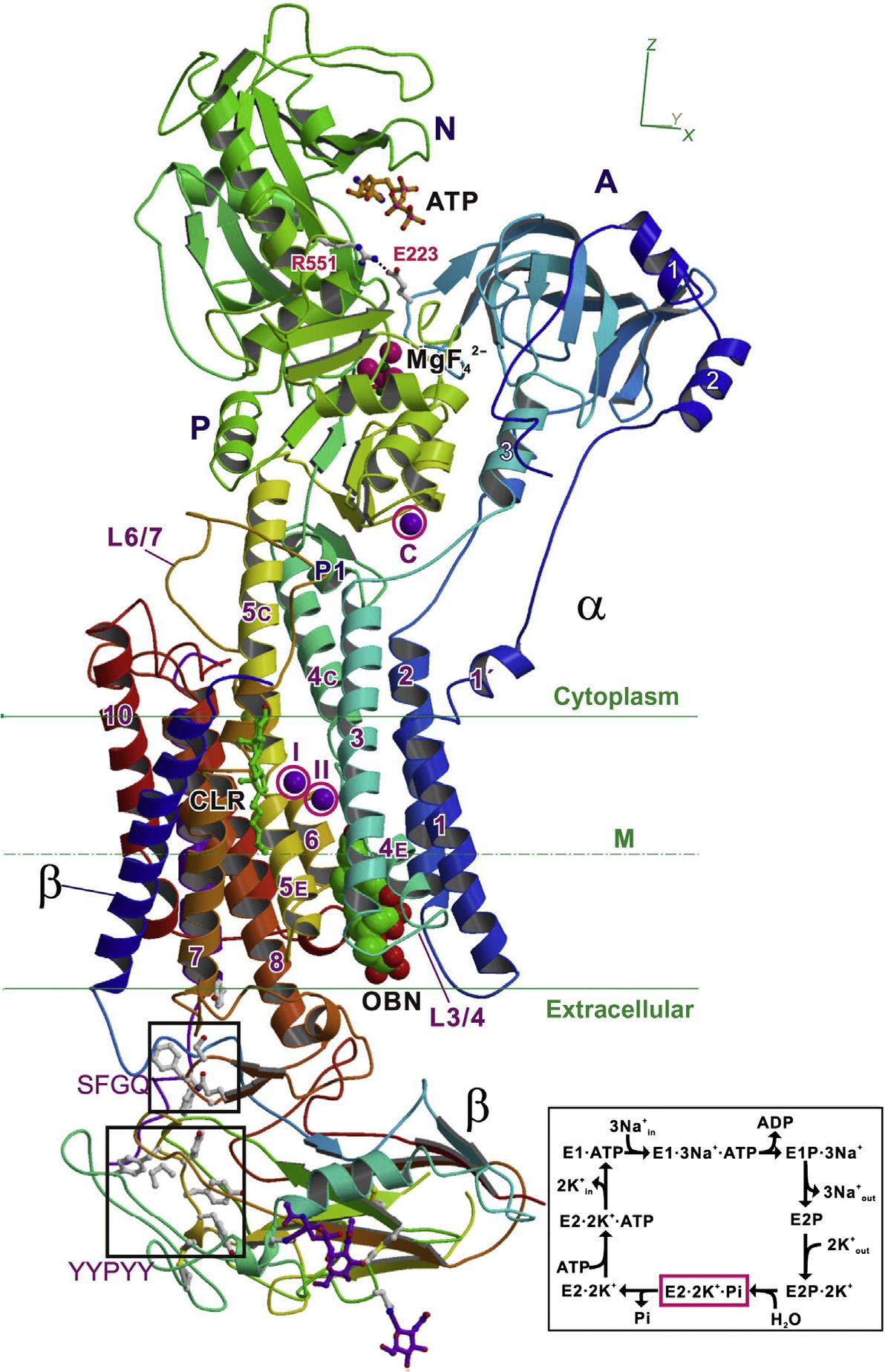
FIGURE1.2 Architecture ofNa1,K1-ATPasefromshark rectalglandwithbound MgF42 andK1,astableanalogoftheE2 Pi 2K1 state.A ribbondiagramofNKAwith ouabain(showninspacefill) boundatlowaffinity(PDB ID:3A3Y).Colorchanges graduallyfromtheNterminal(blue)totheCterminal(red).ATPistaken fromtheE2(TG) ATPcrystal structureofCa21-ATPase (SERCA1a)(PDBID:3AR4) anddockedinthecorrespondingposition.BoundK1 ionsaremarked(I,II,andC) andcircled.Insetshowsa simplifieddiagramofthe post-Albersscheme. CLR, cholesterol; OBN,ouabain. Source:From Toyoshimaetal. (2011).Copyright2011.With permissionfromElsevier.
poorlymobile.Inaddition,theyhaveaccesstoatleasttwooxidationstates,andtherefore canparticipateinelectrontransferandredoxcatalysis,whereaszinchasaccessonlytothe Zn21 state.
Manganesecanoccurinbiologicalsystemsinthreeoxidationstates,Mn(II),Mn(III), andMn(IV).Inhumans,manganeseisessentialfordevelopment,metabolism,andthe antioxidantsystemthroughitsinvolvementinanumberofenzymes,includingarginase,
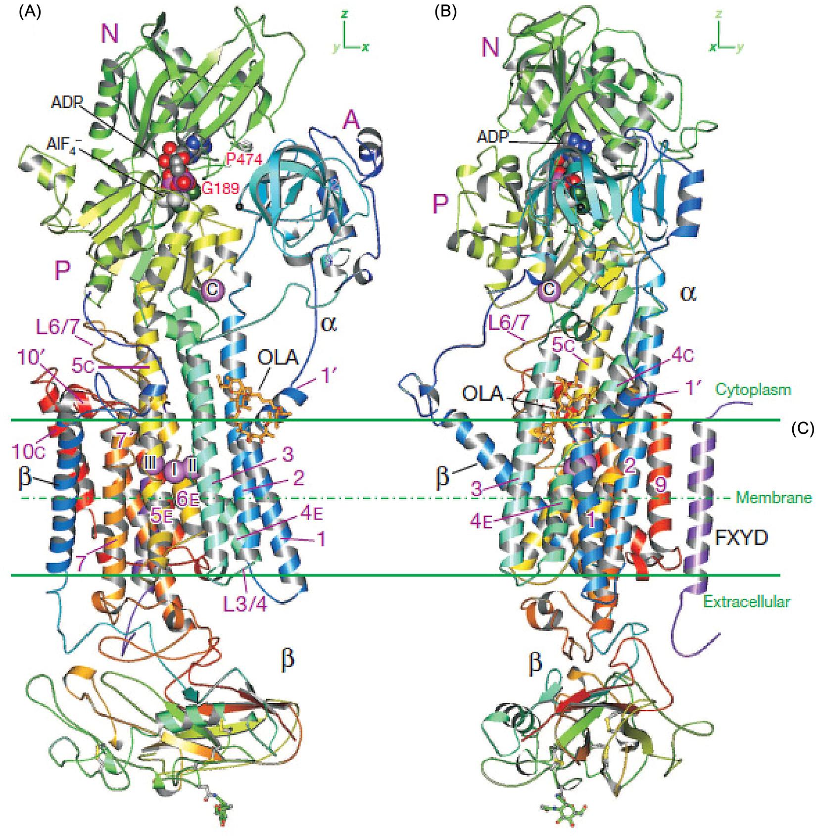
FIGURE1.3 CrystalstructureofNa1,K1-ATPaseinthetransitionstateanalogE1BP ADP 3Na1.(AandB) Ribbondiagramsviewedintwoorthogonaldirections.ColorchangesgraduallybetweentheNterminus(blue) andCterminus(red)forthe α-and β-subunits. Purple spheresshowboundNa1 ions[three(I III)inthetransmembraneregionandone(C)inthecytoplasmicregion].Sugarsattachedtothe β-subunitareshownasballand stick. OLA,oligomycinA. Source:FromKanai,R.,Ogawa,H.,Vilsen,B.,Cornelius,F.,Toyoshima,C., 2013.Crystal structureofaNa1-boundNa1,K1-ATPaseprecedingtheE1Pstate.Nature502,201 206.Copyright2013.WithpermissionfromElsevier.
theenzymeresponsibleforureaproduction,mitochondrialsuperoxidedismutase,and glutaminesynthetase,whichplaysanimportantroleinthebrain.Nevertheless,excessive exposureorintakemayleadtoaconditionknownasmanganism,aneurodegenerative

FIGURE1.4 Ribbonrepresentationshowinghowtargetbindinginduceschangesinthequaternarystructure ofcalmodulin.Theconformationofthetwodomainsof calmodulinisunaffectedbytargetbinding,buttheorientationofthedomainswithrespecttoeachother changesdrastically,bringingthetwopreviously independentdomainsintocontact.Calcium-loaded calmodulin(PDBcode1CLL)isshownatthetopand calcium-loadedcalmodulincomplexedwithapeptide derivedfromsmooth-musclemyosinlight-chainkinase (PDBcode1CDL)isshownatthebottom.TheN-terminaldomainofcalmodulinis mediumblue,theC-terminal domainis darkblue,andthelinkerloopbetweenthe domainsis lightblue.Thepeptideis red andthecalcium ionsarerepresentedas yellow balls.Thetintindicates theConnollysurfacesofthemolecules. Source:From Johnson,C.N.,Damo,S.M.,Chazin,W.J., 2014.EF-handcalcium-bindingproteins.In:EncyclopediaofLifeSciences.John Wiley&SonsLtd., https://doi.org/10.1002/9780470015902. a0003056.pub3.Copyright2014.WithpermissionfromJohn WileyandSons.
disorderthatcausesdopaminergicneuronaldeathandparkinsonian-likesymptoms(Avila etal.,2013).Clearlythemostimportantroleofmanganeseinbiologyisitsinvolvementin theoxygenevolvingcomplexofphotosystemIIincyanobacteria,algae,andgreenplants, whichoxidizeswaterintodioxygen,protons,andelectrons(Eq. 1.1).
ThedeterminationofthestructureoftheMn4CaO5 cluster(Fig.1.5)atthecenterof PSII(Sugaetal.,2015)hasprovokedanintensiveflurryofbiomimeticchemistry,with theaimofgenerating“greenenergy”usingourunlimitedaccesstosolarpower (Najafpouretal.,2015).
Ironisthemostabundantofthetransitionmetalionsinhumans,withthebulkpresent intheoxygen-bindinghemeproteins,hemoglobinandmyoglobin.Thesebothcontainiron withintheprotoporphyrinIXnucleus,requiringanumberofgenesforbiosynthesisofthe porphyrin,insertionofiron,andsubsequenthemetransport(Crichton,2016;Andreini etal.,2009).Theremainingmuchsmallerproportionofbodyironispresentinotherironcontainingproteins(hemeproteins,Fe Sproteins,andnonheme,non-Fe Sproteins)with awidevarietyoffunctions,encodedbythehumangenome(Crichton,2016).Arecentbioinformaticsapproachindicatesthatabout2%ofhumangenesencodeanironprotein(48% heme-bindingproteins,17%Fe Sproteins,and35%whichbindindividualironions).

FIGURE1.5 (A)Schematicrepresentationofthecofactorarrangementinthecoreofthereactioncenter(the viewisalongthemembraneplane).Organiccofactors(forthesakeofsimplicity,thehemegroupofcytochrome b559 isomitted)arecolored green (Chl), yellow (Pheo), magenta (plastoquinonesQA andQB),and red (carotenoids). Ca(yellow),Fe(blue),andMn(red)areshownasspheres;thefigurewasgeneratedusingPyMOL(http://www. pymol.org).ThecoordinatingproteinsubunitsD1andD2areindicatedbydottedlines.(B)StructuralarrangementoftheMn4CaOx clusterandMn O,Ca O,Mn water,andCa waterdistancesintheoxygenevolving complex(OEC)(inA ˚ )Mn1,Mn2,Mn3,andMn4denotethedifferentMnionsoftheOEC. Source:(A)Reprinted withpermissionfromReger,G., 2012.MechanismoflightinducedwatersplittinginphotosystemIIofoxygenevolvingphotosyntheticorganisms.Biochim.Biophys.Acta1817,1164 1176.Copyright2012Elsevier.(B)Reprintedwithpermission fromSuga,M.,Akita,F.,Hirata,K.,Ueno,G.,Murakami,H.,etal.,2015.NativestructureofphotosystemIIat1.95A ˚ resolutionviewedbyfemtosecondX-raypulses.Nature517,99 103.Copyright2015.NaturePublications.
Morethanhalfofthehumanironproteinshaveacatalyticfunction,andtheauthorsestimatethat6.5%ofallhumanenzymesareiron-dependent(Andreinietal.,2018).
Feisaconstituentofalargenumberofproteinsinvolvedinelectrontransferchainsin humans,notablytherespiratorychainintheinnermembraneofthemitochondria,involvingcytochromes,Fe Sproteins,andquinines,channelingelectronstotheterminalcomponent,theCu Fe-dependentcytochrome c oxidase(COX)whichmediatesthereductionof O2 (Eq.1.2).
4H1 1 4e 1 O2 -2H2 O ð1 2Þ
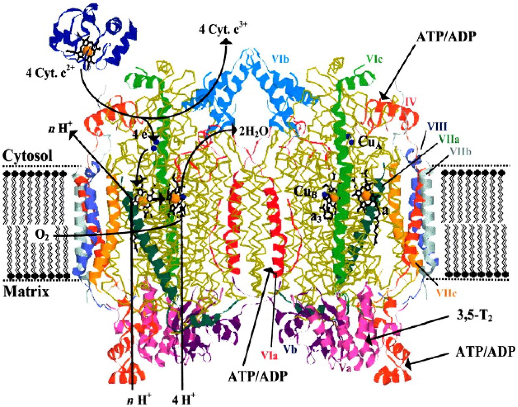
MammalianCOXiscomposedof13subunits,threecatalyticsubunitsI IIIencodedby mitochondrialDNA,and10nuclear-codedsubunitsencodedbynuclearDNA(Fig.1.6).
Electronsfromcytochrome c aretransferredtothedimetallicCuA site,whichrapidly reducestheheme a,some19A ˚ away.Heme a thentransferselectronstotheactivesite heme a3 andCuB,whereO2 binds.
Copperisthethirdmostabundantessentialtransitionmetalioninthehumanbody, involved,forexample,inrespiration,angiogenesis,andneuromodulation,yetCu proteinsrepresentlessthan1%ofthetotalproteomeinbotheukaryotesandprokaryotes
FIGURE1.6 Crystalstructureofdimericcytochrome c oxidasefrombovineheart(Tsukiharaetal.,1996).The nuclear-codedsubunitsareincolor,themitochondrial-codedsubunitsI,II,andIIIarein yellow.Indicatedschematicallyontheleftmonomeraretheelectrontransportpathwaysfromcytochrome c (Cyt.c)tooxygenaccompaniedbyuptakeofprotonsfromthematrixforwaterformationandpumpedprotons(nH1).Ontheright monomerbindingsitesfor3,5-diiodothyronine(T2)andATPorADPareindicated. Source:FromKadenbach,B., Hu¨ttemann,M., 2015.Thesubunitcompositionandfunctionofmammaliancytochrome c oxidase.Mitochondrion24, 64 76.Copyright2015.WithpermissionfromElsevier.
(Andreinietal.,2009).Coppersitesinproteinscanbeclassifiedasbelongingtooneof threeclasses.Type1(blueCuproteins)functioninsingleelectrontransfer,typeIIarecatalyticsiteswhichbinddirectlytosubstrates,whiletypeIIIsitesaredinuclearandare involvedintheactivationandtransportofoxygen.Thecopperchaperonesareaspecific classofproteinswhichensurethesafeandspecificdeliveryofpotentiallyharmfulcopper ionstoavarietyofessentialcopperproteins(Palumaa,2013).Cuisalsoinvolvedasthe catalyticcomponentindetoxification(Cu/Znsuperoxidedismutase).
Bothironandcopperarecharacterizedbygeneticdisordersassociatedwiththeaccumulationofthesemetalsinparticulartissues,withtoxicconsequences.Wilson’sdiseaseis achronicdiseaseofthebrainandliverduetoadisturbanceofcoppermetabolism,
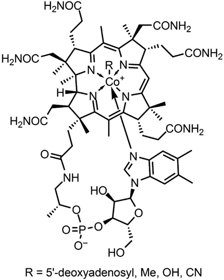
FIGURE1.7 StructureofvitaminB12 shownastheCo31 corrincomplex,whereR 5 50 deoxyadenosyl,Me,OH ,or CN (cyanocobalamin). Source:Withpermissionfrom Wikipedia(releasedintothepublicdomainbyitsauthor Ymwang42attheWikipediaproject).
accompaniedbyprogressiveneurologicaldysfunction,withprogressiveaccumulationof copperinthebrain,liver,kidneys,andthecorneaoftheeye.Ironoverloadcanresult fromgeneticdefectsinironabsorptionfromthegastrointestinaltract(hereditaryhemochromatosis),butcanalsoresultfromgeneticdysfunctionoferythropoiesis,asinthalassemia,necessitatingregularbloodtransfusions(secondaryhemochromatosis).
Althoughonly1.6mgispresentinthehumanbody,cobaltremainsanessentialtrace element,andisrequiredinthehumandietintheformofcobalamin(vitaminB12),aproductofmicrobialbiosynthesis,wheretheCoistightlyboundinacorrinring(Fig.1.7). VitaminB12 uptakefromthegutrequiresaspecificprotein,intrinsicfactor,whichis secretedbythegastricmucosaandisessentialforefficientabsorptionofthevitamin.Lack ofintrinsicfactorcausesperniciousanemia,andvitaminB12 wasidentifiedin1925asthe antiperniciousanemiafactor.ThisisduetoB12 beinganessentialcofactorforanumberof B12-dependentisomerasesandmethyltransferases(Banerjeeetal.,2009)involvedinDNA synthesis,aminoacidandfattyacidmetabolism,inthesynthesisofmyelinbyoligodendrocyteswrappedaroundtheaxonsofmotorneurons,andinthematurationofdevelopingredbloodcells.Cobaltisacutelytoxicinlargedosesandthiswasdramatically observedinthe1960samongheavybeerdrinkers(15 30pints/day),whenCo21 salts wereaddedasfoamstabilizers,resultinginsevereandoftenlethalcardiomyopathy (Kestelootetal.,1968).
Bioinformaticsanalysisofthehumangenomeindicatesthatoneproteinin10(about 3000intotal)isazincmetalloprotein(Andreinietal.,2006,2009).Zn21 isrepresentedin allsixclassesofenzymes(asdefinedbytheInternationalUnionofBiochemistry),whereit canplaybothastructuralaswellasacatalyticrole,oftenfunctioninglikeMg21 asa Lewisacid.Itcanalsofulfillaveryimportantregulatoryfunctioninthestructuralmotifs
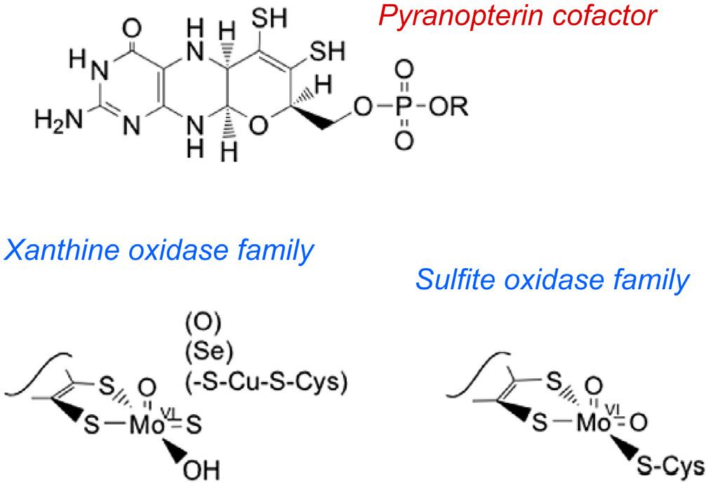
FIGURE1.8 Thestructureofthepyranopterincofactorcommontoallofthese enzymesisgivenatthetop.Activesite structuresfortwofamiliesofmononuclear molybdenumenzymes(xanthineoxidase andsulfiteoxidase). Source:Reprintedwith permissionfromHille,R.,Hall,J.,Basu,P., 2014.Themononuclearmolybdenumenzymes. Chem.Rev.114,3963 4038.Copyright2014. AmericanChemicalSociety.
knownas“zincfingers”involvedintheregulationoftranscriptionandtranslationbyits bindingtoDNAandRNA.Zn21 isthesecondmostabundantofthetracemetals(after iron),andisextensivelyinvolvedinbrainfunction,withmMconcentrationsinsynaptic vesiclesinwhichZnisstoredandfromwhichitisreleasedinacontrolledmanner (BitanihirweandCunningham,2009).Inthecourseofmeioticmaturation,oocytestakeup over2 3 109 zincatoms,andwhenaspermcellentersandfertilizestheoocyte,thistriggersthecoordinatedreleaseofzincintotheextracellularspaceinaprominent“zinc spark,”detectablebyfluorescence(Duncanetal.,2016).Thislossofzincisnecessaryto mediatetheegg-to-embryotransition.
AlthoughMoisrelativelyrareintheEarth’scrust,itisthemostabundanttransition metalinseawater,andsincetheoceansareascloseaswegettotheprimordialsoupin whichlifefirstevolved,itisnosurprisethatMohasbeenwidelyusedinbiology.While MoiswellknownasanimportantcomponentoftheFeMocofactorinnitrogenase,thekey enzymeofnitrogen-fixingorganisms,thereareanumberofMo-dependentenzymesin humans,whichallcontainMointheformofamolybdenumpyranopteridindithiolate cofactor(Fig.1.8).Theseincludexanthineoxidase,involvedinthecatabolismofpurine bases,sulfiteoxidaseinvolvedinsulfurmetabolism,andaldehydeoxidase,involvedin themetabolismofmanydrugs(Hilleetal.,2014).
Ni,V,andCrappeartobebeneficial,andhavebeenproposedtobeessentialforman. Althoughthehumanbodycontainsaround8mgofnickel,noNi-dependentenzymesare known,however,itmaybethatNiisessentialformicroorganismsthatcolonizethe humangut(Zambellietal.,2016).
Toxicmetals
Essentialmetalscanbetoxicifexcessiveconcentrationsofthemetalionsaccumulate, ofteninspecifictissuesororgans—anumberofexamplesofwhicharegivenabove. However,asaconsequenceofenvironmentalexposure,anumberofnonessentialmetal
Another random document with no related content on Scribd:
in cattle kept on distillery and brewery dregs. Lead taken in small quantities in soft water that has run through lead pipes or stood in leaden cisterns produces in cows and other animals chronic affections of the kidney. Ellenberger and Hofmeister have produced the disease experimentally with lead and copper respectively.
Microbian invasions of the kidney that advance slowly like glanders and tubercle are further causes of chronic nephritis. Other secondary microbian infections of the kidney are complications of infectious diseases in other parts, including abscess, pyæmia, septicæmia, ulcerative endocarditis of the left heart, bronchitis, pneumonia (Fröhner), and of others less directly in the line of the circulation, as omphalitis, uterine phlebitis (Lustig), abscess of the nasal sinuses, bones, and fistulæ (Trasbot).
In other cases the nephritis is evidently a result of the irritation caused by toxins in process of elimination by the kidneys, as there is no evidence of a nephritic infection.
In some instances minute emboli originating in the lungs or heart, become the starting point of the nephritis, which slowly extends by reason of infection or low condition and special susceptibility. Disease of the aorta or renal artery may lead to this condition as noticed by Cadeac and Lustig. Cadeac has also noticed its association with aneurism of the mesenteric arteries so that the strongylus (sclerostoma) armatus may be considered as a factor. Again in old horses and dogs it has been associated with atheroma of the aorta and renal vessels (Trasbot).
Overfeeding is not without its influence, especially when on animal food, which charges the kidneys with excreting an excess of the irritating urea and uric acid, and this is one reason why it is far more frequent in house dogs than in other domestic animals. When the meat is already decomposing and putrid there is the added evil of a quantity of toxins and even of microbes to be eliminated from the system by the much abused kidneys. Add to these that the dog’s urine is even in the normal condition more dense and contains more irritating ingredients than that of herbivora, and that owing to the slight activity of his perspiratory apparatus he can obtain less relief from the skin, and we find a substantial ground for the prevalence of chronic nephritis in this animal.
Disease of the valves of the right heart or dilatation with insufficiency of the auriculo-ventricular valves is a potent cause of nephritis, the reflux of blood into the veins and the increased venous tension, speedily producing passive congestion and a slow type of inflammation in the kidney. This factor is especially liable to operate in dogs, which are particularly obnoxious to rheumatism and valvular ulceration, and are very subject to nervous cardiac disorders; in horses that have contracted heaves; and in beef breeds of cattle which suffer from fatty degeneration of the heart with dilatation.
The influence of calculi must not be overlooked, whether they are lodged in the pelvis, the chalices, or the uriniferous tubules. Their tendency is to induce local irritation and exudation, with fibroid degeneration and thickening of the walls of the tubules or pelvis and of the adjacent tissue.
When to one or more of the above conditions there are added overfeeding or what is worse a low condition from starvation or unwholesome food (permeated by bacteria or cryptogams or containing vegetable acids), and when to crown all there are frequent exposures to cold or wet, we have a vicious combination especially conducive to kidney trouble.
Habitual retention of urine in mares in harness, in house dogs, or in horses in railway cars, and violent exertion, or sprains of the back are among the remaining accessory causes.
Symptoms. These are often slight or obscure, so that not only owners and attendants but even veterinarians are liable to overlook them. Loss of flesh, flabbiness of the muscles and a lack of spirit and energy are among the first symptoms. The horse appears stiff, especially in his loins and hind limbs, and fails to advance the hind feet as far under the belly as formerly, and straddles more. When put to work he is early fatigued and appears unfit for sustained exertion. His movements are slow and if urged to a trot he may even groan with every step and quickly settles back to his sluggish pace. If turned sharply round on himself he does so with difficulty and often groans. When he is mounted or when the loins are pinched he may droop to excess. If you come on him lying down, and urge him to rise he may rise on his fore limbs and sit on his haunches until urged before he makes any attempt to raise himself on his hind. The dog
may spend most of his time in the kennel, and show little disposition to run, play or hunt. On the contrary the owner may have to call him several times before he will come out and then he moves listlessly, wearily and even weakly.
In all animals the appetite is poor or capricious, and the patient gradually loses condition, at first slowly and later, after a few weeks or months, more rapidly. The advance of anæmia is also steadily progressive.
Dropsical effusion is not uncommon. It is often prominent in the horse as stocked limbs, but may be absent for a length of time. In other animals it is more likely to appear later in the disease and under the chest or abdomen or in one of the internal serous cavities. Trasbot has found it absent for months in the nephritic dog.
The exploration of the kidney through the flaccid abdominal walls in small animals, and through the rectum in small horses and cattle, may reveal renal tenderness and even swelling. If there is a tendency to frequent passage of urine in small quantities, or to straining without micturition, the indication is of value.
There may be little or no fever, and, when left at rest, little evidence of discomfort.
Any indication of urinary trouble, and especially with dropsy, weakness, flabbiness and anæmia and a subnormal temperature, should lead to examination of the urine, as a crucial test. A high density is good ground for suspicion. But this is not constant. In advanced cases (chronic interstitial nephritis, small white kidney, atrophic nephritis) it may be 1015 to 1025, in exceptional advanced cases with polyuria, it may be 1010, 1005, or even 1001. With such a condition, however, there is great anæmia, pallor of the mucosæ, and prostration. Tested with nitric acid and heat, the urine throws down an abundant precipitate of albumen. Under the microscope it shows a profusion of granular, degenerating epithelial cells, and casts of the uriniferous tubes.
Progress. The course of the disease is usually slow, extending over several months, but with a tendency to constant advance. The thirst increases and the urine increases in amount, clearness and levity. There may supervene extreme sluggishness, dropsies, anæmia, and weakness, irritability of the heart, and palpitations on slight exertion.
So long as the heart’s action is strong, elimination may be maintained and life prolonged for months (in cow, Dickinson), or years (Friedberger and Fröhner). When the heart’s action becomes weak, elimination is rendered imperfect and the animal shows catarrh of the lungs or bowels (common in dogs), local inflammation of the lungs, pleura or pericardium, or œdemas, or hæmorrhages. The toxic effect on the nerve centres is shown by stupor or lethargy, or vertigo. When an abscess forms it is associated with a temporary rise of temperature (Trasbot). The patient may die in convulsions, in a state of coma, or by gradually advancing debility and failure of the heart.
Lesions. In cases of comparatively short standing the kidney is usually of full size, or somewhat enlarged, with firmly adherent capsule and rough or even nodular surface. The surface of the cortex may be red or grayish or parti-colored, pink and gray. The cortical portion is firm and it may even be attenuated somewhat, while the medullary portion, naturally lighter, has often grayish streaks converging toward the hilus. When the gray streaks are scraped with the knife a serous fluid, mixed with fatty granules or globules, is obtained. The glomeruli may be still about the normal size with some increase of the epithelial tuft cells. The tubules contain casts (colloid, hyaline, granular), and their epithelium normally columnar, are flattened down to cubes and are swollen, granular or fatty.
In cases of older standing the connective tissue has usually undergone a marked increase. The capsule is thick, dense and adherent. The cortical substance is shrunken with a great increase of the fibrous elements, and the same holds true of the medullary portion. In consequence of this, even in the cortical substance the white or gray color predominates. The parenchymatous tissue (glomeruli, tubules) have greatly shrunken. In connection with the contraction of the forming fibrous hyperplasia, there is a general shrinkage of the kidney in size, it may be to one-half its original volume. Trasbot reports a case of nephritis, of 8 months standing, in the dog, with a kidney half the normal size. In the end the parenchyma may have practically disappeared, and the kidney may have shrunken to a small, firm, white, fibrous mass. Abscess of the kidney is exceptionally met with (Laurent, Lafosse).
Lesions of distant organs are not uncommon. Bronchitis, pneumonia, pleurisy, insufficiency of the tricuspid or mitral valves, dilated heart, hypertrophied or fatty heart, congested or fibroid liver, arteritis, and dropsies are among such morbid conditions.
Prognosis. This is almost always unfavorable. Death may be delayed for months or years, and partial transient recoveries may take place but a restoration to normal structure and function is not to be looked for.
Treatment. This cannot be expected to be much more than palliative. The avoidance of overwork, and of the exposure to cold and wet, and the securing of a free action of the skin by warm buildings and clothing, are essential. The diet should be easily digested and non-stimulating, for herbivora green food, carrots, roots, apples, silage, with a moderate allowance of oats to counteract weakness and anæmia; and for carnivora, milk, buttermilk, mush made of oat, wheat or barley meal, with, if necessary, a slight allowance of tender raw meat. Tonics fill a similar need. Iron and bitters may be combined. Or hydrochloric acid or nitromuriatic acid with bitters (nux, calumba, salicin, quassia) may be tried. These acids are especially valuable when the case has originated in or is maintained by calculi, indigestion or hepatic disorder. When the heart is defective in tone, it may be stimulated by small doses of digitalis, strophanthus, sparteine, caffein, or nitro-glycerine, or to a certain extent by strychnia or nux. These, however, must be used with judgment, if it is found that they aggravate the case by increasing the arterial tension. In those cases in which there is an excessive secretion of watery urine, the possible source of this in musty aliment should be avoided, and the flow checked by nux vomica, in moderate doses, and bromide or iodide of potassium in full doses. When, on the other hand, the urine becomes scanty and dense, the great danger of a toxic action must be met by agents that favor excretion. Pure water at will is perhaps the least objectionable of such agents, but potassium or sodium acetate or citrate, or even sodium chloride, in weak solution, may be given. In some cases benefit will come from a moderate use of the balsam of copiaba, or the leaves of buchu, which may improve the tone of the secretory elements. The most promptly effective of these agents is pilocarpin (Friedberger and Fröhner), but it has the serious drawback of
inducing profuse and dangerous depletion and debility. Yet in careful hands, and with good cardiac tone, it may often be used to advantage.
Fomentations over the loins, warm baths and mustard embrocations, may at times be beneficial. Attempts have been made to check the hyperplasia by the use of arsenic, mercury or the compounds of iodine, but their use in such cases is based on theory rather than accomplished results.
HYPERTROPHY OF THE KIDNEY.
Hypertrophy of both kidneys has not been recorded in domestic animals. On the other hand the extraordinary development of one in compensation for the loss or atrophy of the other is not uncommon. In this the organ follows the general law of adaptation, seen in the double symmetrical organs (testicle, etc.) and the more so that its functional activity is indispensable to life. Among causes are: blocking of an ureter by calculus, worms, neoplasm, nephritic abscess, gangrene, etc. The enlargement of the remaining kidney is a vicarious act and essentially a physiological one.
If compensation is perfect, it may be impossible to detect symptoms apart from those of the primary disease.
Prognosis. Life is endangered in case of any subsequent kidney disease.
ATROPHY OF THE KIDNEY.
Result of hyperplasia of connective tissues and compression and absorption of parenchyma. Unilateral or partial. Causes: chronic productive inflammation, calculus in tubes, ureter, or pelvis, tumor, retention cyst, embolism. Lesions: sclerosis of kidney, firmness, pallor, anæmia, lack of glomeruli and tubules, cysts, congenital, urinous retention, colloid. Symptoms: reduced secretion, palpation of kidney. Treatment: Prevention: arrest conditions, abundance of water, succulent food, parasiticides, operation on cysts, counteract nephritis.
Unlike hypertrophy, this is constantly the result of a pathological process. So long as a normal functional activity of the secreting elements is carried on, such parts must maintain their size and healthy characters. But with the compression of such secreting elements (glomeruli and convoluted tubes) by a hyperplasia of connective tissue, by pressure from without or from the damming back of the urine in the pelvis and tubes, the secretory elements are absorbed and removed, and the final result is a general atrophy. If such atrophy appears in both kidneys at once it can only be very partial in extent, as extreme atrophy of both, with loss of their secretory function, would entail poisoning and death from the retained urinary products. The comparative frequency of the disease may be inferred from the reports of the numbers of specimens found by Barrier and Moussu in old horses in the dissecting rooms. The latter observed a dozen cases in a single winter, other examples are recorded by Cadeac (horse), Soula (swine) and Trasbot (in various animals).
Causes. The most common source of the condition is the occurrence of chronic productive inflammation. The new product in such cases, if not pus, or a growth that rapidly passes into fatty or granular degeneration, or into gangrene, tends to form tissue of a low organization, especially fibrous. The resulting increase of the
fibrous trabeculæ, in undergoing subsequent contraction necessarily compresses the secretory tissue and the final result is a visible and, it may be, extreme wasting. Hence any slowly advancing productive inflammation is liable to result in absorption and removal of the kidney parenchyma, and distinct atrophy of the gland.
Again the obstruction of the ureter by a calculus in the pelvis which falls into the infundibuliform entrance, or a stone arrested at any part of the duct (or even of the urethra) or by worms, hydatids, cysts or tumors, throws back on the kidney the secreted urine, which distending the pelvis and uriniferous tubes leads to direct compression and absorption of the secretory parenchyma. Direct compression of the kidney by an adjacent tumor will act in a similar manner. Retention cysts by their gradual increase and augmenting pressure cause absorption of the gland tissue.
The blocking of individual uriniferous tubules by minute calculi, which is so often seen in cattle, kept on dry feeding in winter, is a cause of partial nephritis, and absorption, as noted by Röll.
A somewhat rare cause of atrophy is the diminution of the blood supply by arteritis and embolism of the renal artery, or by pressure of tumors on that vessel. Arteritis and blocking suggests at once the possible agency of the strongylus (sclerostoma) armatus in the horse. Trasbot records a striking instance of compression of the renal artery and kidney by an enormous sublumbar melanoma. This occurred in an aged horse and led to atrophy.
Lesions. In cases due to productive inflammation with sclerosis of the kidney, the firmness, pallor and bloodlessness of the organ is a marked feature. When incised it is found to be composed mainly of fibrous tissue, while the glomeruli and tubuli have to a large extent disappeared.
If there has been simple lack of circulation the kidney becomes flaccid, pale and small in size. The secretory elements (glomeruli and uriniferous tubes) are first absorbed, leaving the fibrous network, which tends to shrink and form a hard resistent mass. In extreme cases there may be absolutely no glandular tissue left, and the dense shrunken mass represents only the hyperplasia of the original fibrous network. In the different successive stages of this process the glomeruli and tubules become flattened, the epithelial cells become granular, or contain colloid casts and refrangent elements like oil
globules and finally they are represented by a small mass of fibrous material.
Of all the atrophies caused by the pressure of tumors perhaps that caused by cysts is the most characteristic. There may be a single cyst or they may be multiple; they may range in size from a pea to the size of the two fists the total size exceeding that of the normal kidney. In all such cases the cysts project visibly from the surface of the organ. They vary according to their origin and nature. Congenital cysts are said to have resulted from distension by retained urine of the capsule of the glomerulus. The arterial tuft is atrophied and flattened against the wall. Serous cysts with clear contents are found in the old. Urinous cysts again form by distension of the tubules that are obstructed by cysts or minute calculi. Colloid cysts are found in certain forms of nephritis formed by the dilatation of the capsule of the glomerulus or of the uriniferous tubules. The liquid often contains leucin, tyrosin and cholesterine. In all such cases the walls of the cyst become thick, and the glandular parenchyma is compressed leading to progressive degeneration and atrophy.
Symptoms of atrophy of the kidney are necessarily those of suppression of urine, with, in certain cases, the passage of casts of the uriniferous tubes and of crystals of salts. There are, however, no absolutely pathognomonic symptoms. When the kidney can be reached through the flaccid walls of a comparatively empty abdomen, or through the rectum, its hard, shrunken condition may assist in diagnosis.
Treatment is not successful in advanced cases. Prevention is to be sought by obviating or treating the conditions on which the atrophy depends. Nephritis must be treated on general principles. Calculi must be avoided by a liberal supply of water, by soiling, or by pasturage. Strongylus parasitism should be dealt with by destroying the parent worms in the bowels, and by securing pure drinking water free from their eggs and embryos. Cysts, and tumors are only amenable to surgical measures and not often open even to these.
FATTY DEGENERATION OF THE KIDNEY: STEATOSIS OF THE KIDNEY.
Causes: age, overfeeding, idleness, atony, retention of urine. Lesions: kidney enlarged, pale yellow, capsule loose, cut surface glistening unctuous, oil globules in scrapings, granules soluble in ether. Symptoms: in idle, overfed, obese, improved meat producing breeds, closely confined, starchy or saccharine food, fatty granules in urine, finally dropsies, anæmia, debility, sluggishness. Prognosis unfavorable in advanced stage. Treatment: butcher, restricted regimen, open air exercise, nitrogenous diet, crossing, diuretic food or drugs, oil of turpentine, balsam copiaba. Palliation only.
Fatty degeneration of the kidneys is by no means unknown in the domestic animals. It has been observed in dogs and cats (Rogers, Goubaux, Vulpain, Trasbot). In dogs it has been erroneously set down as a characteristic lesion of rabies. Like fatty degeneration of other organs, it is also met with in old and overfed individuals of meat producing breeds of animals, in which the tendency to early maturity and rapid and excessive fattening has been fostered from generation to generation. In man small, granular, fatty kidney is a common result of chronic parenchymatous nephritis, and often coincides with fatty liver. Chronic poisoning by arsenic or phosphorus is another cause, as it is of fatty degeneration in other organs.
Vulpain has attributed it to a lack of active exertion and of general tone, associated with excessive amylaceous feeding, sluggish, shallow breathing and tardy elimination. Goubaux and Trasbot attach great importance to the compulsory retention of urine in house dogs, cats and horses. The damming back of the urine in the convoluted tubes and glomeruli, temporarily arrests secretion, and the inactive and compressed cells tend at once to granular and fatty degeneration.
Lesions. The gland is sensibly increased in size, and pale, yellowish or straw yellow. The capsule is easily detached from the cortical substance, contrary to what is the case in chronic productive inflammation. The cortical substance is increased in thickness, and pale, the pallor being largely in ratio with the duration or extent of the fatty degeneration. The cut surface may be glistening and unctuous to the touch. It is softer than usual, rather friable, and if scraped, furnishes a serous or grayish pulp in which oil globules are prominent features, together with granular epithelium and free granules that dissolve readily in ether. Tubules are varicose and unequal at different parts. The medullary portion has undergone little change. It may be paler at certain points, with some shrinking of its substance and increase of firmness.
Symptoms. As a rule the disease occurs in pampered, overfed and obese animals, and in those of the improved breeds which have great power of digestion, assimilation and fattening. It is especially to be looked for after close confinement on full, stimulating, amylaceous diet. Symptoms are not usually recognized during life. There is, however, a lessening of the urinary secretion, and, as the disease advances, albuminuria. When examined microscopically this is found to contain characteristic elements, such as granular epithelial cells, the granules soluble in ether, oil globules, and at times crystals of cholesterine (Beale). A diagnosis based on the mere presence of oil globules may, however, be fallacious, as these may be present in animals that have just been heavily fed on oleaginous food, and again the oil used to smear the catheter may float in the urine and prove misleading. Under such circumstances vaseline or glycerine may be substituted on the catheter. Scriba induced fatty urine by injecting fat or oil emulsion into the blood, and Chabrie by ligating the large intestine. Trasbot says that cylindroid casts may be present. As in other grave kidney affections, dropsies supervene as the disease advances. These may show in the limbs, in the abdomen, or in other serous cavities. A steadily advancing anæmia with pallor of the mucosæ, listlessness, weakness, debility and sluggishness are to be noted.
Prognosis. Since the disease is rarely diagnosed until it has reached an advanced stage, it usually progresses steadily to a fatal issue. If, however, it can be detected at an earlier stage, it may be
palliated, or held in abeyance, for a length of time varying with the extent of the lesions. As it is very largely a disease of meat producing animals and as the subject is at first in a condition of marked obesity, it can usually be turned over to the butcher without material loss.
Treatment. If the disease has resulted from the inbred propensity to fattening, the family that shows the disposition must be subjected to a somewhat different regimen, open air exercise must take the place of confinement in warm stables, a rather bare pasturage is valuable for herbivora, and a restricted diet in which the oleaginous, saccharine, and amylaceous constituents do not predominate, is strongly indicated. Crossing with a strange male having many of the desirable qualities of the herd, but which is more vigorous may be resorted to. When the secretion of urine becomes scanty an abundance of pure water, or a diet of succulent grass or roots or ensilage or even small doses of alkaline diuretics may be resorted to. Any source of arsenic or phosphorus poisoning should be cut off, and as an antidote to phosphorus, oil of turpentine may be given in small doses. This agent may, indeed, replace the alkalies as a diuretic, bringing in an element of tone for the mucosa which is not to be despised. Or balsam of copaiba or buchu leaves may be substituted. When the small white kidney (granular, fatty) results from chronic nephritis, the prevention and treatment would be as for that disease. Little hope is to be entertained of entire restoration to health.
AMYLOID KIDNEY. LARDACEOUS OR WAXY KIDNEY.
This condition of the kidney has been found in the ox (Gerlach) and dog (Rabe, etc.). There are usually similar degenerative lesions in the liver, pancreas, intestines and other organs. It is usually a concomitant of some chronic wasting disease (chronic nephritis, tuberculosis, etc.).
Morbid Anatomy. The kidney is usually enlarged, pale and on section waxy or glistening. Soaked in dilute compound tincture of iodine it shows spots of a walnut or mahogany brown color. The glomeruli are well marked and show the earlier changes, later the tubes do so excepting the epithelium. The latter is swollen, granular, fatty.
Symptoms. There may have been those of chronic nephritis. Rabe has noticed in dogs dropsy of the limbs, ascites, emaciation, anorexia, followed by uræmia, coma, weakness, vomiting, and if the kidney alone was affected great lowering of temperature (35.9°C). With hepatic complication there was greater weakness, giddiness, and higher temperature (39.6°C). Urine is usually increased (in man albuminous) and the casts have shown the anyloid reaction. They tend to be fatty or finely granular. Casts may, however, show anyloid reaction when the kidney, post mortem, does not (Jaksch).
Diagnosis from Bright’s disease is often impossible.
Treatment is essentially the same as in chronic nephritis, and is not hopeful.
Trasbot recommends KI 3 to 7 grs., or tinct of iodine 3 drops for shepherd dog. Ol. terebinth and alkaline diuretics are also commended.
URETERITIS.
From wounds, calculus, parasites, infection, injuries in parturition. Symptoms: in wounds of ureter. Course: danger of infection of kidney or bladder. Treatment: for calculus, antispasmodics, anodynes, fomentations, for parasites arsenious acid, for catarrhal conditions, balsams, buchu, salicylates, etc. Operation. Ureterovaginal fistula.
This may arise from the passage of a rough calculus, from wounds of the ureter sustained in kicks and blows or by being run over by wheels (dogs, cats), it may be due to the blocking of the tube by a parasite such as strongylus gigas, echinococcus, etc., or it may be the result of extension of an infectious inflammation backward from the kidney or forward from the bladder. Again it may be the result of a lesion of the ureter in cases of dystokia.
The symptoms are obscure but there is likely to be frequent straining and passage of urine, tenderness of the loins, all the more significant if confined to one side, lameness or halting on the corresponding hind limb, and on examination through the rectum the swollen and tender cord representing the ureter may be recognizable. In case of calculus or other obstruction the ureter may be felt to be swollen, elastic and tender back to a slight nodular, painful, firm swelling at the seat of obstruction.
Course. In all such cases there is always danger of inflammation (infections or otherwise) of the kidney with degeneration and loss of structure and function, the organ being reduced to a simple urinous cyst (hydronephrosis). In some cases, however, the obstruction (calculus, parasite) may escape into the bladder and a recovery follow. Slight infections, too, may improve and advance to complete convalescence.
Treatment will depend much on the causative factor: Calculus must be treated by anodyne antispasmodics, and fomentations, and in case of relief by measures calculated to prevent its formation anew: parasites may be treated by arsenious acid, oil of turpentine, and other parasiticides which are secreted by the kidneys: catarrhal and infected conditions may be met by balsams, buchu, salicyclic acid and even peppers. In case of calculus which does not give promise of passing, even a surgical operation may be thought of, especially in the smaller house animals.
In rupture of the ureter in dystokia the walls of the womb or vagina have usually suffered, and a recovery with a ureterouterine or uretero-vaginal fistula is not unknown.
ACUTE CATARRHAL CYSTITIS.
Acrid diuretics, by mouth or skin, microbian infection, retention of urine, urethral calculus, parasites, spasm, enforced suspension of micturition, unclean catheter, adjacent infection, chill. Lesions: hyperæmia of mucosa, thickening, vascular distention, clouding of epithelium, muco-purulent secretion, alkaline fermentation, ammonia, liquefaction of cells, erosion. Symptoms: Slight fever, stiff, straddling gait, urine scanty, cloudy, alkaline, penis or clitoris semi-erect, smearing of tail or prepuce. Crystals of triple phosphate. Treatment: Antiseptics, boric or salicylic acid, gum arabic, astringent antiseptics, laxatives, flax seed, slippery elm, anodynes, diluents, piperazine, drainage, rest, restricted laxative diet, warmth, avoid stimulants.
Causes. Cystitis is caused in all animals by irritant diuretics like cantharides, copaiba, or oil of turpentine given by the mouth or applied to an extensive cutaneous surface. It is an error, however, to conclude with Williams that this is the sole cause. The very existence of calculi virtually implies bacterial infection, and fermentation. The presence of free ammonia in the urine usually implies fermentation, and fermentation must be looked upon as practically synonymous with microbian invasion. That bacteria may be present without serious injury is undoubted. The protective power of the healthy mucosa is very great. But when the mucosa is weakened, microbes that would otherwise be harmless, find a ready infection atrium, and triumph over the weakened tissues. Hence retention of urine and overdistension of the bladder as in urethral calculus, blocking of the urethra by a parasite, spasm of the sphincter vesicæ, compulsory retention as in the mare in harness, the dog kept indoors, or in railway car on a long journey, or in mares so travelling, may become the occasion of cystitis. Even in cases in which no microbe is present at first, this reaches the bladder by the introduction of an unclean catheter, or by extension from an uretheritis, vaginitis or metritis, or even from a peritonitis, or infected urachus. Or the infection may
descend from a suppurating kidney. Another occasion of microbian invasion is the congestion which attends on exposure to cold.
Lesions. Hyperæmia of the cystic mucosa, with dilation and tortuous deviations of the larger vessels, thickening of the membrane, and distension and clouding of the epithelial cells, with a thick covering of tenacious mucus containing epithelial, pus, or white blood cells. As the disease advances epithelium is desquamated abundantly, and degenerates with production of free nuclei and pus. Along with these are microbes, usually the bacillus coli communis, and various cocci. In the fully established disease there is liable to be alkaline fermentation, and the liberated ammonia dissolves the epithelial cells, leading to extensive desquamation and raw granulating surfaces, so that the disease tends to run in a vitiating circle, the alkali dissolving the epithelium and increasing the microbian development and fermentation, which in its turn produces an increasing quantity of ammonia.
Symptoms. There is slight hyperthermia or none, stiff or straddling gait, frequent passage of urine in small quantities and cloudy, or straining without passage, the penis or clitoris is semierect, eversion of the lips of the vulva is frequent, and the bladder is tender (through prepubian wall, vagina or rectum). If a finger is inserted into the bladder in the mare the thickening of the walls can often be recognized. The urine often contains precipitated crystals of ammonio-magnesian phosphate, and even clots of blood. It has an alkaline reaction even in herbivora.
Treatment. The danger centres around the bacteridian fermentations, and a main object must be to disinfect the bladder. This will be all the more effectual if the lotions used are of an acid reaction. Thus boric acid or salicylic acid in 3 per cent. solution, injected after evacuation of the bladder and repeated a number of times a day may soon establish a healthy action. If the bladder is especially irritable a boiled weak solution of gum arabic will form a suitable medium. Other antiseptics are often used as creosote (0.5:100), carbolic acid (3:100), chloride of zinc (3:100), chlorate of potash (3:100), mercuric chloride (1:5000), silver nitrate (0.5:100), or astringents are often better: PbA, ZnSO4 tannic acid, ferri chloridi in dilute solution so as not to cause pain.
The bowels should be kept open by an occasional saline laxative, pain moderated by codeine, and abundance of pure water and a laxative diet enjoined. Linseed tea, and infusions of slippery elm or marsh mallow have long been employed, and by soothing and relaxing the bowels they act favorably on the urinary mucosa. Stimulants of the urinary track like buchu, uva ursi or copaiba in small doses, or antiseptics like creosote, boric acid, salicylic acid, piperazine, are available in slight cases or when the acute symptoms have subsided somewhat. With prior infection of the kidneys, the latter may be used. Constant drainage may be necessary to avoid distension.
Perfect rest is absolutely essential, a restricted laxative diet, and a careful avoidance of cold, and stimulants.
When urine is retained it should be removed with a thoroughly aseptic catheter.
In case of blood clots in the bladder, wash out with a boiled normal salt solution.
ACUTE CROUPOUS CYSTITIS.
This has been found to follow the use of cantharides and other irritant diuretics, and to follow on certain specific diseases. Its nature is that of catarrhal inflammation, but with a fibrinous product or false membrane formed more or less extensively on the inflamed mucosa.
Symptoms are essentially those of catarrhal cystitis from which it is distinguished by the presence in the urine of flakes of the fibrinous membrane.
Treatment is essentially the same as in the catarrhal form, to which may be added the injection of a solution of 4 grains scale pepsin to the ounce of sterilized water. The boric acid solution may be of the strength of 20 per cent. Irrigate two or three times a day.
CHRONIC CATARRHAL CYSTITIS.
This may begin as such or it may continue after an acute attack. It has been noticed in horse, ox, and dog.
It may be associated with calculi, gravel, papilloma, and bacterial invasion especially by the colon bacillus.
Lesions. The mucosa and muscular coat are thickened, corrugated, puckered and contracted so that the bladder will not contain more than a few ounces of urine. The surface of the mucosa is discolored, mottled and variegated, slaty blue, brown, red, purple, or even black, with ulcers, encrustations of triple phosphate, and fungoid elevations. In dogs especially, the prostate is often enlarged.
Symptoms. Frequent urination accomplished with pain, groaning, or whining and it may be with sudden arrest. There may be incontinence, the urine dribbling almost continuously from the penis or vulva and in the latter case trickling down the thighs. The presence of pus and mucus tends to mat the hairs, and a strong urinous and ammoniacal odor is emitted.
Palpation of the prepubian region often, and of the vagina or rectum always causes pain and wincing. Temperature is normal.
Urine is albuminous in ratio to the amount of pus, or above that, and is then suggestive of kidney disease and likely to be complicated by casts.
Complicating lesions of the womb, vagina, prostate, and kidney are to be carefully looked for, also cystic papilloma.
Prognosis. Recovery though not uncommon is too often but partial and it is usually desirable to fatten the animal.
Treatment. Rest, moderate laxative diet, pure drinking water ad libitum, warmth, antiseptic irrigation.
CYSTITIS IN THE OX.
Special Symptoms. Beside general disorder there is a disposition to decubitus, but with frequent rising to urinate though the bladder is not filled to repletion. Then the urine is passed in a slow stream by abdominal contraction, and without pulsating contractions of the urethra at the ischium which are so marked in calculus. Cystitis is greatly aggravated by overdistension, and if the bladder is paralyzed is very liable to go on to rupture.
Galtier considers enzootic hæmaturia as essentially a hæmorrhagic cystitis, due to marshy soils, disordered liver, often distomatosis, and irritation of the urinary organs by the poisons which the liver was helpless to destroy or eliminate.
The treatment of cystitis in cattle does not differ materially from that of the horse.
The hæmorrhagic form demands prevention by drainage, cultivation and the use of phosphates to the soil.