List of Contributors
The editors would like to acknowledge and offer grateful thanks for the input of all previous editions’ contributors, without whom this new edition would not have been possible.
Hamid Abedi, DDS MS MBA
Clinical endodontist, Irvine, CA, USA
Kenneth Abramovitch, DDS, MS
Professor and Chair, Department of Oral Pathology, Radiology and Medicine, University of Missouri – Kansas City, School of Dentistry, Kansas City, MO, USA
Anita Aminoshariae, DDS, MS
Associate Professor, Endodontics, CWRU School of Dental Medicine, Cleveland, OH, USA
Ana Arias, DDS, MS, PhD
Professor of Conservative Dentistry, School of Dentistry, Complutense University, Madrid, Spain
Adham A. Azim, BDS
Division Head and Director of the Post-Graduate Program, Periodontics and Endodontics, University at Buffalo, Buffalo, New York, NY, USA
Brooke Blicher, DMD, Certificate in Endodontics
Clinical Endodontist, Upper Valley Endodontics, White River Junction, Vermont;
Clinical Instructor, Department of Restorative Dentistry and Biomaterials Science, Harvard School of Dental Medicine, Boston, MA, USA;
Assistant Clinical Professor, Department of Endodontics, Tufts University School of Dental Medicine, Boston, MA, USA; Dental Staff, Department of Oral Surgery, Dartmouth Hitchcock Medical Center, Lebanon, VT, USA
Tatiana Botero, DDS, MS
Clinical Associate Professor, Cariology Restorative Sciences and Endodontics, Ann Arbor, MI, USA
Sami Chogle, BDS, DMD, MSD
Chair and Program Director, Graduate Endodontics, Boston University, Boston, MA, USA;
Adjunct Associate Professor, Graduate Endodontics, Case Western Reserve University, Cleveland, OH, USA
William Herbert Christie, DMD, MS, FRCD(C)
Retired Professor, Senior Scholar, Department of Restorative Dentistry, University of Manitoba, Winnipeg, Manitoba, Canada
Blaine Cleghorn, DMD, MS
Professor and Assistant Dean, Faculty of Dental Clinical Sciences, Dalhousie University, Halifax, Nova Scotia, Canada
Anibal Diogenes, DDS, MS, PhD
Professor and Vice Chair, Department of Endodontics, University of Texas Health Science Center at San Antonio, San Antonio, TX, USA
Melissa Drum, DDS, MS
Professor, Advanced Endodontics Director, Division of Endodontics, College of Dentistry, The Ohio State University, Columbus, OH, USA
Brad Eli, DMD, MS
Staff, Department of Dentistry, Scripps Memorial Hospital, La Jolla, CA, USA
Mohamed I. Fayad, DDS, MS, PhD
Clinical Associate Professor, Director of Research, Department of Endodontics, University of Illinois, College of Dentistry, Chicago, IL, USA
Natasha M. Flake, DDS, PhD, MSD
Associate Professor, Department of Endodontics, University of Washington, Seattle, WA, USA
Ashraf F. Fouad, DDS, MS
Freedland Distinguished Professor, Vice-chair, Division of Comprehensive Oral Health, Adams School of Dentistry, University of North Carolina, Chapel Hill, NC, USA
Brian Goodacre, DDS, MSD
Assistant Professor, Department of Prosthodontics, School of Dentistry, Loma Linda University, Loma Linda, CA, USA
Charles J. Goodacre, DDS, MSD
Distinguished Professor, Department of Restorative Dentistry, School of Dentistry, Loma Linda University, Loma Linda, CA, USA
Robert Handysides, DDS
Dean, Department of Endodontics, School of Dentistry, Loma Linda University, Loma Linda, CA, USA
Brad Johnson, DDS, MHPE
Professor and Head, Director of Postdoctoral Endodontics, Department of Endodontics, University of Illinois at Chicago, Chicago, IL, USA
James David Johnson, DDS, MS
Clinical Professor and Chair, Department of Endodontics, University of Washington School of Dentistry, Seattle, WA, USA
Mo Kang, DDS, PhD
Professor and Chairman, Department of Endodontics, UCLA School of Dentistry, Los Angeles, CA, USA
Philip Michaelson, MS, DMD
Private Practice, Endodontics, Professional Endodontics, Inc., Chagrin Falls, OH, USA
W. Craig Noblett, DDS, MS
Volunteer Faculty, University of California, San Francisco, CA; Private practice, Fresno, CA
Ali Nosrat, DDS, MS, MDS
Clinical Assistant Professor, Department of Endodontics, School of Dentistry, University of Maryland, Baltimore, MD, USA; Private Practice, Centreville, VA, USA
John M. Nusstein, DDS, MS
Professor and Chair, Division of Endodontics, College of Dentistry, The Ohio State University, Columbus, OH, USA
Avina Paranjpe, BDS, MS, MSD, PhD
Associate Professor, Department of Endodontics, University of Washington, Seattle, WA, USA
Masoud Parirokh, DMD, MSc
Distinguished Professor, Department of Endodontics, School of Dentistry, Kerman University of Medical Sciences, Kerman, Islamic Republic of Iran;
Distinguished Professor, Endodontology Research Center, Kerman University of Medical Sciences, Kerman, Islamic Republic of Iran
Ove A. Peters, DMD, MS, PhD
Professor and Discipline Lead, Endodontics, School of Dentistry, The University of Queensland, Brisbane, Qld, Australia
Al Reader, DDS, MS
Professor, Division of Endodontics, College of Dentistry, The Ohio State University, Columbus, OH, USA
Ilan Rotstein, DDS
Professor and Chair, Endodontics, Orthodontics and General Practice Residency, Herman Ostrow School of Dentistry of USC, University of Southern California, Los Angeles, CA, USA; Associate Dean, Continuing Education, Herman Ostrow School of Dentistry of USC, University of Southern California, Los Angeles, CA, USA
Richard A. Rubinstein, DDS, MS, FACD
Adjunct Clinical Associate Professor, Cariology, Restorative Sciences and Endodontics, University of Michigan, Ann Arbor, MI, USA
Nikita B. Ruparel, MS, DDS, PhD
Associate Professor, Department of Endodontics, UTHSCSA, San Antonio, TX, USA
Mohammed Sabeti, DDS, MA
Clinical Professor, PRDS, University of California at San Francisco, San Francisco, CA, USA
Nasser Said-Al-Naief, DDS, MS
Professor and Chair, OMFP Laboratory Director, Pathology and Radiology, OHSU, Portland, OR, USA; Hospital Staff, OMFS, School of Medicine, OHSU, Portland, OR, USA;
Professor, Anatomic Pathology, School of Medicine, OHSU, Portland, OR, USA
Christine Sedgley, MDS, MDSc, FRACDS, MRACDS(ENDO), PhD
Professor and Chair, Department of Endodontology, Oregon Health and Sciences University, Portland, OR, USA
Frank Setzer, DMD, PHD, MS
Assistant Professor, Endodontic Clinic Director, and Predoctoral Endodontic Program Director, Department of Endodontics, University of Pennsylvania, Philadelphia, PA, USA
Shahrokh Shabahang, DDS, MS, PhD
Associate Professor, Department of Endodontics, School of Dentistry, Loma Linda University, Loma Linda, CA, USA
Renato Silva, DDS, MS, PhD
Associate Professor, Department of Endodontics, University of Texas Health Science Center at Houston, Houston, TX, USA
Tory Silvestrin, DDS, MSD, MSHPE
Chairman and Graduate Program Director, Department of Endodontics, School of Dentistry, Loma Linda University, Loma Linda, CA, USA
Fabricio B. Teixeira, DDS, MS, PhD
Professor and Chair, Department of Endodontics, University of Iowa, Iowa City, IA, USA
Yoshitsugu Terauchi, DDS, PhD
President, Endodontics, CT and MicroEndodontic Center, Yamato-shi, Kanagawa, Yamato City, Japan
Mahmoud Torabinejad, DMD, MSD, PhD
Affiliate Professor of Endodontics, Department of Endodontics, University of Washington Seattle, WA, USA;
Adjunct Professor, Department of Endodontics, School of Dentistry, Loma Linda University, Loma Linda, CA, USA; Adjunct Professor, Department of Endodontics, University of Pacific Arthur A. Dugoni School of Dentistry; Adjunct Professor, Department of Preventive and Restorative Dentistry, Section of Endodontics, University of California in San Francisco, School of Dentistry, San Francisco, CA, USA
Marco Aurelio Versiani, Lt. Col., DDS, MSc, PhD
Associate Researcher, Department of Restorative Dentistry, Dental School of Ribeirão Preto, University of São Paulo, Ribeirão Preto, São Paulo, Brazil
Richard Walton, DMD, MS
Professor Emeritus, Department of Endodontics, University of Iowa, Iowa City, IA, USA
Shane N White, BDentSc, MS, MA, PhD
Professor, UCLA School of Dentistry, Los Angeles, CA, USA
Anne E. Williamson, DDS, MS
Associate Professor, Endodontics, University of Iowa, Iowa City, IA, USA
1 Pathogenesis of Pulp and Periapical Diseases
CHRISTINE SEDGLEY, RENATO SILVA, AND ASHRAF F. FOUAD
CHAPTER OUTLINE
Histology and Physiology of Normal Dental Pulp, 1
Etiology of Pulpal and Periapical Diseases, 2
Microbiology of Root Canal Infections, 5
Endodontic Infections Are Biofilm Infections, 5
The Microbiome of Endodontic Infections, 6
Pulpal Diseases, 8
LEARNING OBJECTIVES
After reading this chapter, the student should be able to:
1. Describe the histology and physiology of the normal dental pulp.
2. Identify etiologic factors causing pulp inflammation.
3. Describe the routes of entry of microorganisms to the pulp and periapical tissues.
4. Classify pulpal diseases and their clinical features.
5. Describe the clinical consequences of the spread of pulpal inflammation into periapical tissues.
Histology and Physiology of Normal Dental Pulp
The dental pulp is a unique connective tissue with vascular, lymphatic, and nervous elements that originates from neural crest cells. It resides inside the tooth in a chamber with rigid walls.
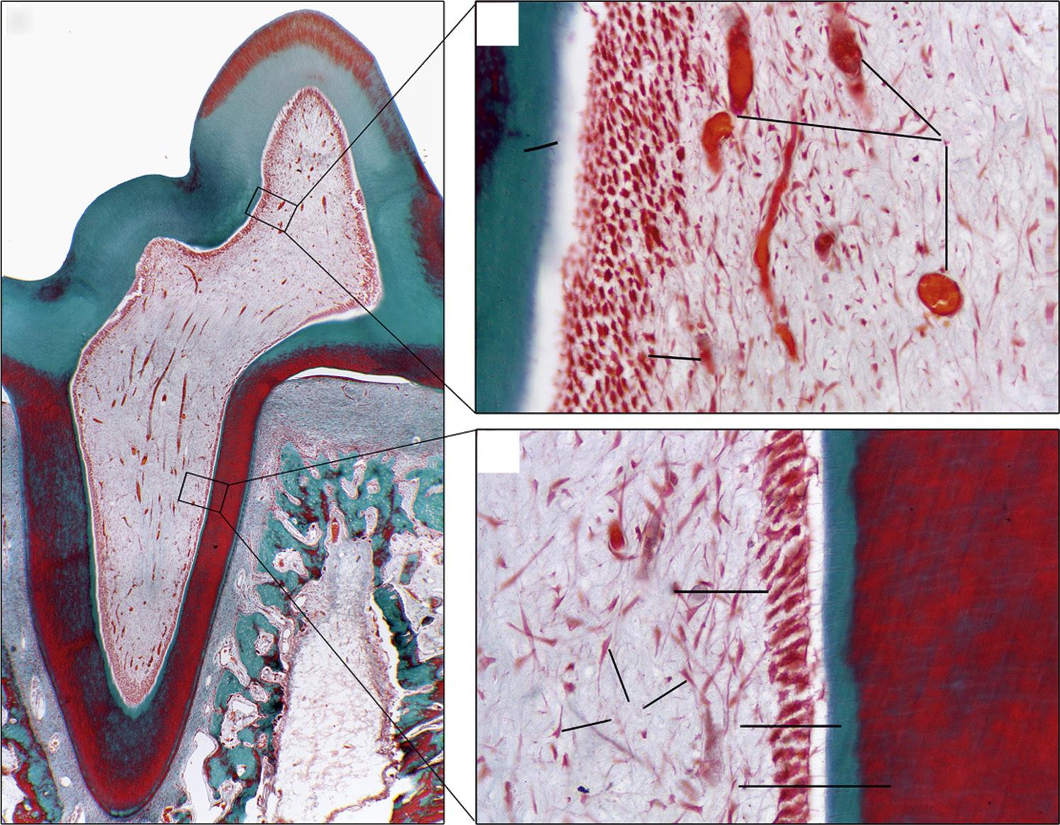
The pulp contains odontoblasts, highly specialized cells with a secretory function, which not only form dentin, but also interact with dental epithelium early in tooth development to initiate the formation of enamel. The pulp also contains fibroblasts, undifferentiated mesenchymal cells, collagen type I and II, proteoglycans, glycoproteins, and water1 (Fig. 1.1).
The histologic structure of the pulp is important, because it reflects a unique architecture suited for the formation of dentin and defense against invading pathogens. Odontoblasts form a
Normal Pulp, 11
Reversible Pulpitis, 11
Irreversible Pulpitis, 11
Pulp Necrosis, 12
Clinical Classification of Periapical (Apical) Conditions, 13
Nonendodontic Pathosis, 15
6. Describe the histopathological diagnoses of periapical lesions of pulpal origin.
7. Identify clinical signs and symptoms of acute apical periodontitis, chronic apical periodontitis, acute and chronic apical abscesses, and condensing osteitis.
8. Discuss the role of residual microorganisms and host response in the outcome of endodontic treatment.
9. Describe the steps involved in repair of periapical pathosis after successful root canal treatment.
palisading layer that lines the walls of the pulp space, and their tubules extend about two thirds of the length of the dentinal tubules. The tubules are larger at a young age and eventually become more sclerotic as the peritubular dentin becomes thicker. The odontoblasts are primarily involved in production of mineralized dentin. They are connected by gap junctions that allow them to form a semipermeable membrane. In addition, odontoblasts play an important role in defense as they express Toll-like receptors (see later), cytokines, and defensins, among other immunologic mediators.
Two main types of sensory fibers innervate the pulp: Aδ-fibers in the periphery and C-fibers in the central pulp. The Aδ-fibers are responsible for the sharp response to thermal changes. They extend between the odontoblasts, lose their myelin sheath, and extend to a distance of 100 to 200 μm into the dentinal tubules. The C-fibers are unmyelinated and are responsible for the dull
• Fig. 1.1 Histologic section of (A) rat molar tooth showing coronal (B) and (C) radicular dental pulp in higher magnification. Masson-Goldner trichrome staining. BV, Blood vessels; D, dentin; FB, fibroblasts; OD, odontoblasts in odontoblastic layer; PD, predentin. An artifact (*) separating the predentin from the odontoblastic layer. (Courtesy Dr. Claudia Biguetti.)
ache that affects patients with symptomatic irreversible pulpitis. The pulp may also have Aβ-fibers and sympathetic fibers in the walls of arterioles.
The pulp vasculature plays a critical role in its response to irritation. When the tooth first erupts into the oral cavity, the root apex is immature, and there is ample blood supply to the pulp. Eventually, the apex matures, and the ability of the pulp to withstand external irritation, such as from trauma or caries, diminishes. However, the pulp of the mature tooth has mechanisms to cope with increased blood flow during inflammation, such as arteriovenous anastomoses and loops that can circulate and increase volume of blood when the need arises. The pulp also contains an elaborate network of arterioles and capillaries around the odontoblasts, which are high-metabolic-rate cells, commonly known as the terminal capillary network.
Etiology of Pulpal and Periapical Diseases
Injury or irritation of pulpal or periapical tissues can result in inflammation. The reactions of the dental pulp to irritants are largely dictated by the type and duration of a stimulus. These irritants can be broadly classified as nonliving (mechanical, thermal, or chemical) or living (microbial) (Video 1.1).
Mechanical Irritants
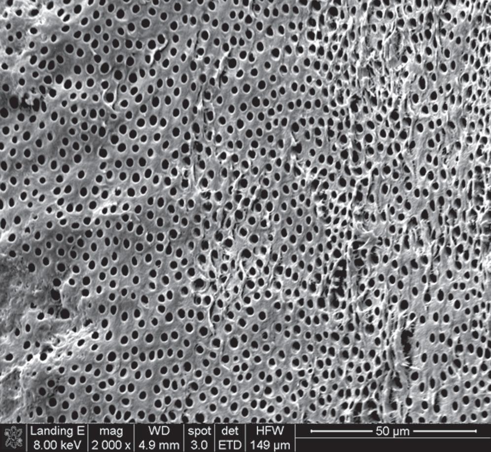
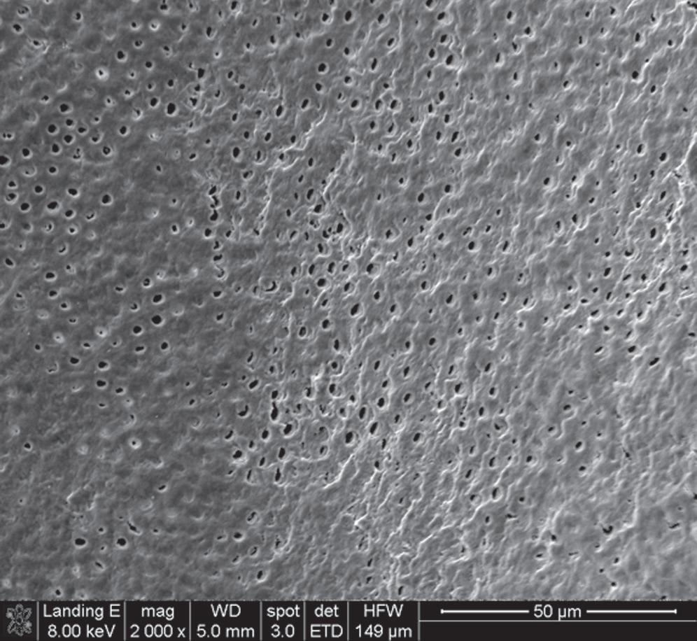
The potential for pulp irritation increases as more dentin is removed during deep cavity preparations because dentinal permeability is greater closer to the pulp2 (Fig. 1.2). The removal of tooth structure without proper cooling may also cause pulp inflammation. Deep scaling and curettage may injure apical vessels and nerves, resulting in pulpal damage.3
Pulpal damage can occur because of impact injuries. Teeth undergoing mild to moderate trauma and those with immature apices have a better chance of pulpal survival in comparison with those suffering severe injury or those with closed apices. Intrusion injuries are more likely to lead to pulp necrosis than are lateral or extrusion injuries4 (Fig.1.3).
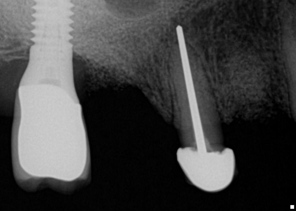
Periapical tissues can be mechanically irritated and inflamed by impact trauma, hyperocclusion, overinstrumentation of root canals, perforation of the root, and overextension of root canal filling materials (Fig. 1.4). Inaccurate determination of root canal length is usually the cause of overinstrumentation and subsequent inflammation. In addition, lack of an adequate apical resistance form created during cleaning and shaping can cause overextension of filling materials into the periapical tissues, causing physical and chemical damage (Fig. 1.5).
Application of forces beyond the physiologic tolerance of the periodontal ligament (PDL) during orthodontic tooth movement results in disturbance of the blood and nerve supply of the pulp tissue.5,6 In addition, orthodontic movement may initiate resorption of the apex, usually without a change in vitality.
Chemical Irritants
Antibacterial agents, such as silver nitrate, phenol with and without camphor, and eugenol, have been used to “sterilize” dentin after cavity preparations. The effectiveness of many of these products is questionable,7 and their cytotoxicity can cause inflammatory changes in the underlying dental pulp.8 Other irritating agents include cavity cleansers, such as alcohol, chloroform, hydrogen peroxide, and various acids; chemicals present in desensitizers, cavity liners and bases; and temporary and permanent restorative materials.
• Fig. 1.2 Scanning Electron Microscopy of Human Dentin. Dentinal permeability is greater closer to the pulp (A) than near the dentinoenamel junction (B) or the cementodentinal junction due to the higher number of tubules per unit and bigger tubule diameter. Therefore the potential for pulp irritation increases as more dentin is removed.
• Fig. 1.3 Graphic representation of pulpal circulation subsequent to various types of luxation injuries to teeth. Pulp circulation is measured in perfusion units over a 36-week observation period.
Antibacterial irrigants used during cleaning and shaping of root canals, intracanal medications, and some compounds present in obturating materials are examples of potential chemical irritants to periapical tissues.9,10 When testing the effects of antimicrobial medications on dental pulp cells, researchers showed that calcium hydroxide and lower concentrations of antibiotic pastes are conducive to cell survival and proliferation, but more concentrated forms of antibiotic pastes have detrimental effects.11
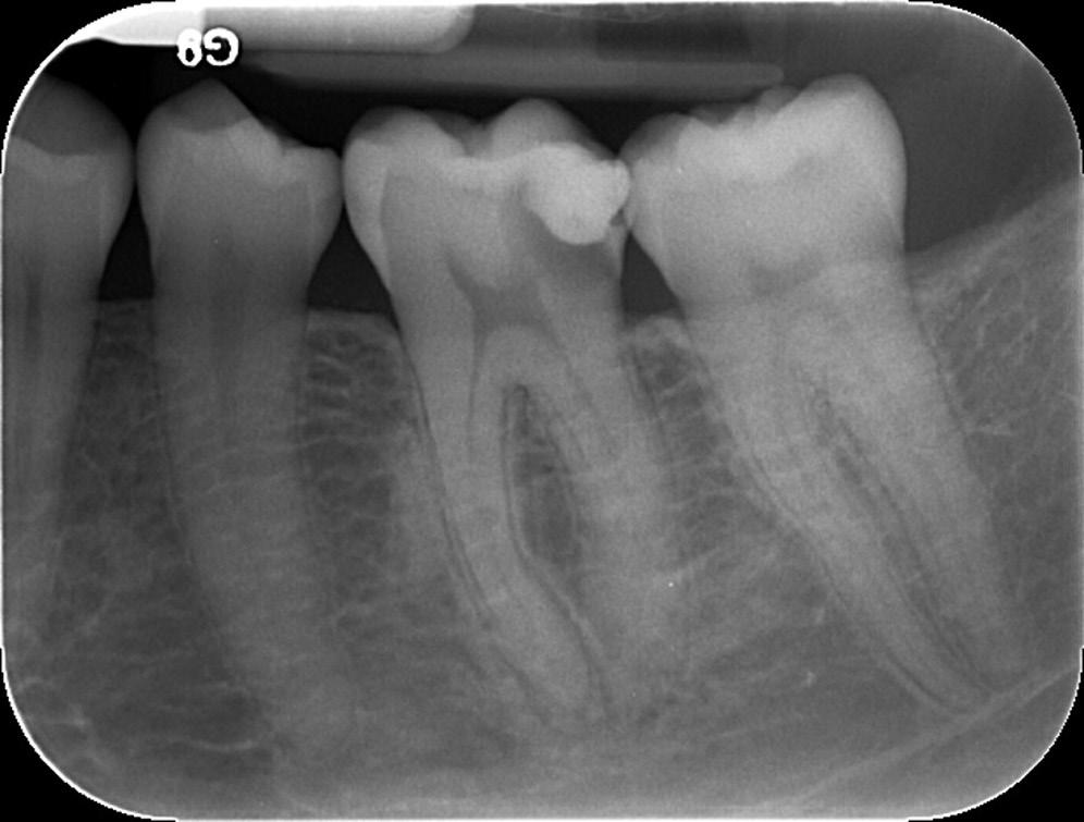
• Fig. 1.4 Periapical radiograph showing overextension of root canal filling material.
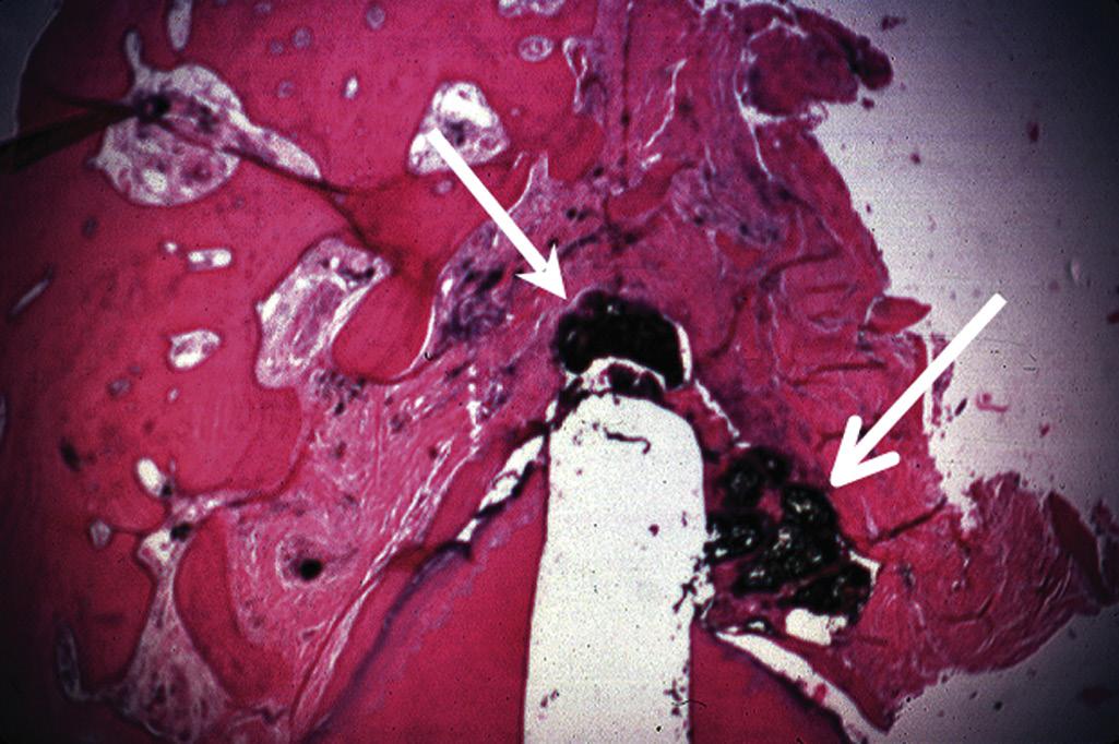
• Fig. 1.5 Improper instrumentation and extrusion of filling materials into the periapical tissues causes periradicular inflammation (arrows).
Microbial Irritants
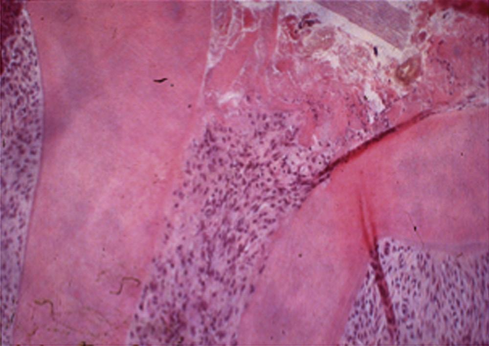
Although mechanical and chemical irritations are predominantly transient in nature, the most significant cause of inflammation is microbial. Studies have shown that even superficial carious lesions in enamel are capable of attracting inflammatory cells in the pulp.12,13 The initial reaction of the pulp to these irritants is mediated through the innate immune response. This early response to caries results in focal accumulation of chronic inflammatory cells, such as macrophages, lymphocytes, and plasma cells.14 As caries progresses toward the pulp, the intensity and character of the infiltrate change. Pulpal tissue may remain inflamed for long periods and may undergo eventual or rapid necrosis. This change depends on several factors: (1) the virulence of the microorganisms; (2) the ability to circulate inflammatory fluids to avoid a marked increase in intrapulpal pressure; (3) host resistance, including genetic variations; (4) the amount of circulation and (5) an important factor, lymphatic drainage. Subsequently, microorganisms or their byproducts and other irritants from the necrotic pulp diffuse from the canal to the periapical region, resulting in the development of an inflammatory lesion (Fig. 1.6).
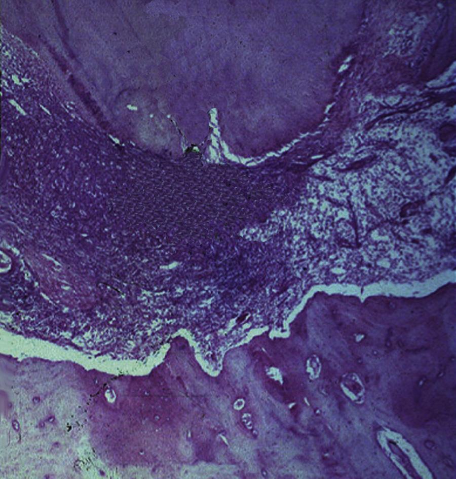
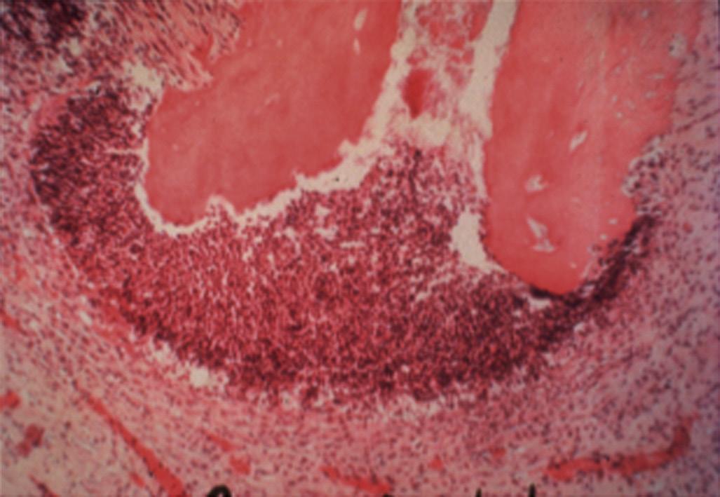
Pulpal and periapical pathoses do not develop without the presence of bacterial contamination.15,16 Kakehashi and collaborators created pulp exposures in conventional and germ-free rats.15 In the germ-free rats, minimal inflammation only occurred throughout the 72-day observation period. Further, pulpal tissue in these animals was not devitalized but rather showed calcific bridge formation by day 14, with normal tissue apical to the dentin bridge (Fig. 1.7, A). In contrast, infection, pulpal necrosis, and abscess formation occurred by the eighth day in conventional rats (Fig. 1.7, B). The bacteriological investigation by Sundqvist examining the flora of human necrotic pulps supports the findings of Kakehashi and collaborators15 and Möller and coworkers.16 Sundqvist examined previously traumatized intact teeth with necrotic pulps, with and without apical pathosis. The root canals of teeth without apical lesions were aseptic, whereas those with periapical pathosis had positive bacterial cultures.17
Several mechanisms have been proposed for identification of microorganisms as irritants by the immune system. Detection of these pathogens can occur via interaction between pathogen-associated molecular patterns (PAMPs) and specific receptors broadly
identified as pattern recognition receptors (PRRs).18 PRRs recognize PAMPs and initiate host defenses. G-protein coupled receptors and Toll-like receptors (TLRs) are part of the innate immune response and activate phagocytic functions to allow microbial ingestion. G-protein coupled receptors bind to chemokines, lipid mediators (e.g., platelet-activating factor, prostaglandin E2, and leukotriene B4) or bacterial proteins, causing extravasation of leukocytes and production of bactericidal substances. TLRs are transmembrane proteins that are expressed by cells of the innate immune system playing a central role in the initiation of cellular innate immune responses.19 These receptors recognize invading microbes and activate signaling pathways that launch immune and inflammatory responses to destroy the invaders. At least 13 TLRs have been discovered to date with different recognition abilities. Table 1.1 presents some of the currently identified TLRs and their specific interactions.
• Fig. 1.6 Egress of irritants (closed arrow) from the root canal into the periapical tissue causes inflammation (open arrow) and replacement of normal periapical structures with a granulomatous tissue.
• Fig. 1.7 A, No inflammation is seen in an exposed pulp (P) of a germ-free rat. Food particles and other debris (D) are packed into the chamber. B, Periapical lesion is apparent in a conventional rat after pulp exposure. (Courtesy Dr. H. Stanley.)
TABLE 1.1
Examples of Toll-Like Receptors (TLRS) and Associated Activators
PAMP PRR Pathogen
LPS, Lipid A TLR4 Gram-negative bacteria
Flagellin TLR5 Bacteria, flagellum
dsRNA TLR3 Virus
RNA TLR7,8 Virus
CpG DNA TLR9 Bacteria, DNA
PAMP PRR Pathogen
PAMP, Pathogen-associated molecular patterns; PRR, pattern recognition receptors.
Microbiology of Root Canal Infections
Routes of Root Canal Infection
Under normal conditions, the dental pulp and dentin are isolated from oral microorganisms by overlying enamel and cementum. When the integrity of these protective layers is breached (e.g., as a result of caries, trauma-induced fractures and cracks, restorative procedures, congenital anomalies of teeth, scaling and root planing, attrition, or abrasion) or naturally absent (e.g., because of gaps in the cementoenamel junction at the cervical root surface), the dentin-pulp complex becomes exposed to the oral environment. The pulp then becomes at risk of infection by oral microorganisms present in caries, saliva, and dental plaque. The risk increases with the depth of lesions due to the diameter of dentinal tubules increasing as they approach the pulp (see Fig. 1.2).
Caries are the most common cause of pulpal exposure (Fig. 1.8). However, microorganisms may also reach the pulp via direct pulpal exposure as a result of iatrogenic restorative procedures, as a result of trauma, and through a periodontal pocket extending to the apical foramen or lateral canal. After pulp necrosis, microorganisms can invade the entire root canal system uninhibited by host defense mechanisms. As a consequence of the interaction between microorganisms and the host defenses, inflammatory changes take place in the periapical tissues and give rise to the development of apical periodontitis.
Endodontic infections can be classified according to their anatomic location as intraradicular or extraradicular. Microorganisms that initially invade and colonize the necrotic pulp tissue cause primary intraradicular infections. Microorganisms that were not present in the primary infection but were introduced into the root canal system during or after initial treatment cause secondary infections. Secondary infections are suspected when a preoperative infection heals after treatment and then recurs at a later time. Persistent infections are caused by microorganisms from a primary infection that resisted intracanal antimicrobial procedures and remained in the prepared root canal system. Persistent and secondary infections are responsible for several clinical problems, including persistent exudation, continuation of symptoms, interappointment flare-ups, and failure of the endodontic treatment.
The goal of root canal treatment is to remove microorganisms from the root canal system. However, in the absence of a strict aseptic technique, microorganisms from caries and dental plaque can be introduced into the canal system during treatment because of lack of rubber dam usage or leakage of the rubber dam.
Contaminated endodontic files and instruments, including delivery systems for antimicrobial agents, are additional potential sources for the introduction of microorganisms into the root canal system during treatment.
Microorganisms can enter the root canal system between appointments via loss or leakage of temporary restorative materials, fracture of the tooth structure, and teeth left open for drainage. Entry of microorganisms after root canal filling occurs by loss or leakage of temporary or permanent restorative materials, during preparation of posts or other intracanal restorations without the rubber dam, fracture of the tooth structure, and by recurrent caries that expose the root canal filling material. Leakage after completion of root canal treatment is more likely to occur if the placement of the permanent restoration is delayed.
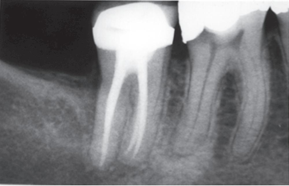
Extraradicular infection is characterized by microbial invasion and proliferation into the inflamed periapical tissues and is almost invariably a sequel to intraradicular infection. Once the intraradicular infection is properly controlled by root canal treatment or tooth extraction and drainage of pus, the extraradicular infection can be addressed by the host defenses and usually subsides (Fig. 1.9).
Endodontic Infections Are Biofilm Infections
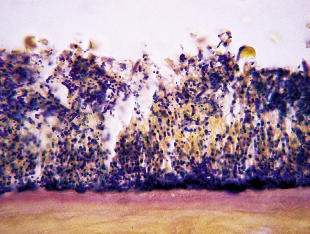
The role of microorganisms as the primary etiologic agents of root canal infections was established in seminal studies published several decades ago.15,16,20 In these and subsequent microbiological analyses of samples recovered from root canal infections, species were isolated and identified by using planktonic (liquid) culture techniques. During the past two decades, it became apparent that microorganisms exist in the root canal system not as planktonic cultures, or as single species, but rather as multispecies biofilm communities composed of microcolonies irreversibly attached to a substratum, to an interface like dentin, and to each other.21,22 All anatomic areas of the infected root canal system can harbor microbial cells organized as highly variable biofilm structures22,23 (Fig. 1.10).
Biofilm formation encompasses attachment of microbial cells to a surface, followed by cell proliferation, adherence to other microorganisms, production of matrix, and microcolony maturation. Dispersal of cells allows the formation of new biofilm microcolonies.24 Microbial cells occupy only a small proportion of the biofilm. Quorum sensing is the expression of specific microbial proteins after the bacterial cells reach a threshold number. It
• Fig. 1.8 Radiograph showing pulp exposure as a result of caries.
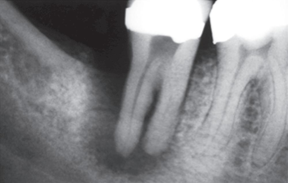
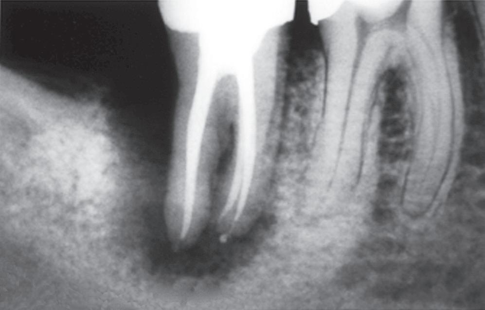
• Fig. 1.9 A, Preoperative radiograph of a second molar with pulpal necrosis and evidence of chronic apical periodontitis. B, Postoperative radiograph of the tooth. C, Postoperative radiograph 2 years after root canal therapy shows complete resolution of the periradicular pathosis.
• Fig. 1.10 Intracanal Biofilms With Predominance of Cocci. Note the high concentration of cells at the bottom of the biofilm and in direct contact with the root canal wall. (From Ricucci D, Siqueira JF Jr: Biofilms and apical periodontitis: study of prevalence and association with clinical and histopathologic findings, J Endod 36(8):1277–1288, 2010.)
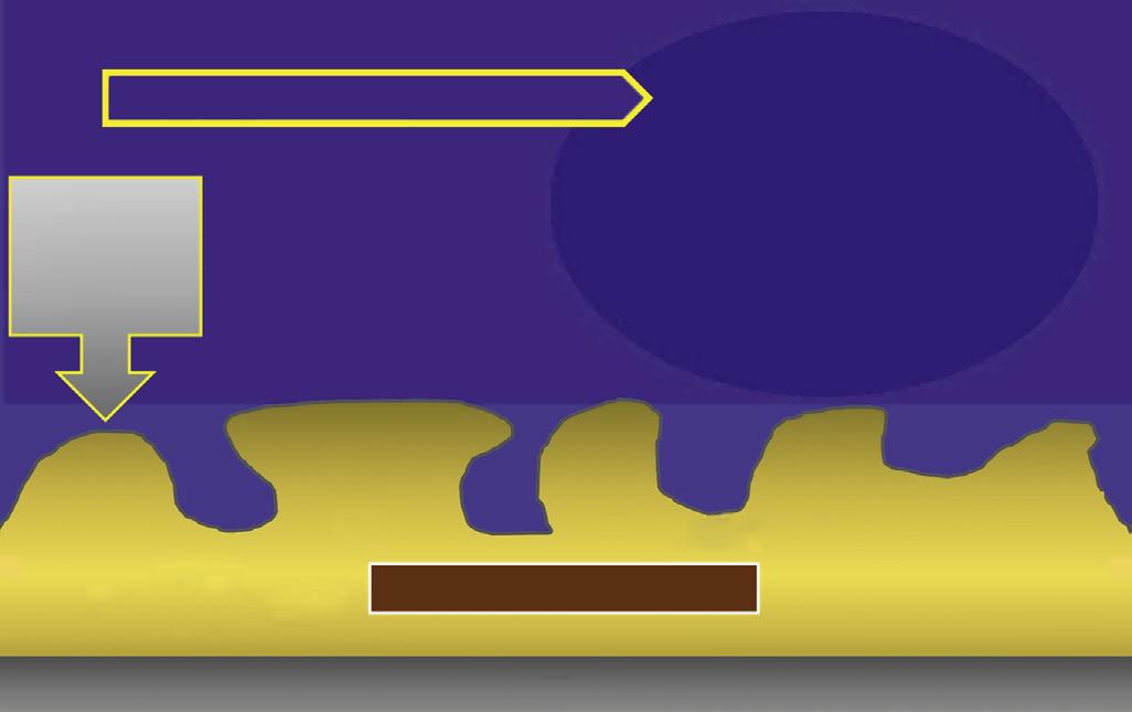
allows the coordinated regulation of expression of these proteins by microorganisms in biofilms to regulate population density and possibly virulence.25 The majority of the biofilm structure is a highly heterogeneous matrix composed of extracellular polymeric substances (EPSs) produced by cells within the biofilm. The EPS matrix provides multiple functions (Fig. 1.11). From a clinical
• Fig. 1.11 Properties of biofilms emerging from life in the extracellular polymeric substance (EPS) matrix. (From Flemming HC: EPS-then and now, Microorganisms 4[4]:pii: E41, 2016 Nov 18.)
perspective, the EPS can act as a physical barrier to antimicrobial agents such as antibiotics and disinfectants.26 Microbial organization into multispecies biofilm communities results in increased pathogenic effects on the host.27
The Microbiome of Endodontic Infections
The identity of specific microorganisms in root canal infections has been a major focus of interest for more than a century.28 Studies using culture-dependent approaches have recovered several species
Bacterial Genera Represented in Endodontic Infections
GRAM-NEGATIVE BACTERIA
Anaerobes
Dialister
Treponema TABLE 1.2
Facultatives
Anaerobes
Rods Rods
Porphyromonas
Tannerella
Prevotella
Fusobacterium
Campylobacter
Pyramidobacter
Catonella
Selenomonas
Centipeda
Capnocytophaga
Eikenella
Haemophilus
Veillonella
Megasphaera
Neisseria
Actinomyces
Pseudoramibacter
Filifactor
Eubacterium
Mogibacterium
Propionibacterium
Eggerthella
Olsenella
Bifidobacterium
Slackia
Atopobium
Solobacterium
Lactobacillus
Parvimonas
Peptostreptococcus
Finegoldia
Peptoniphilus
Anaerococcus
Streptococcus
Gemella
Spirilla
that have been identified as putative endodontic pathogens. The microbiome of carious dentin causing pulpitis and subsequent endodontic infection includes significant numbers of lactobacilli,29 gram-negative bacteria,30 and species from the Firmicutes, Actinobacteria, and Proteobacteria phyla.31 Primary root canal infections harbor a multispecies population of facultative and strict anaerobic gram-positive and gram-negative bacteria, spirochetes, yeasts, Archaea, and other unidentified species.32-36 In addition, EpsteinBarr virus may be associated with irreversible pulpitis and apical periodontitis,37 and papilloma virus and human herpes virus have been found in exudates from acute apical abscesses.38
Microorganisms have been traditionally characterized in terms of their morphology (rods, cocci, spirilla), cell wall characteristics (gram-positive and gram-negative), and oxygen tolerance (anaerobic and facultatively anaerobic). Genera cultured from symptomatic and asymptomatic root canal infections include Prevotella, Porphyromonas, Fusobacterium, Peptostreptococcus, Streptococcus, Lactobacillus, Enterococcus, Actinomyces, Propionibacterium, and Candida.20,39 (Table 1.2)
More recently, the microbiome of endodontic infections has been redefined using culture-independent molecular biology techniques. These studies have both confirmed the findings from culture studies and greatly expanded knowledge. Many species that had already been considered putative pathogens because of their frequency of detection, as reported by culture-dependent methods, have been found in a similar or even higher prevalence by using molecular approaches, strengthening associations with causation of apical periodontitis. Molecular technology has enabled the recognition of many new putative pathogens that had
GRAM-POSITIVE BACTERIA
Facultatives
Actinomyces
Corynebacterium
Lactobacillus
Streptococcus
Enterococcus
Granulicatella
not previously been found in samples from endodontic infections.40,41 A review of 12 studies that used next-generation DNA sequencing (pyrosequencing) methods to evaluate the microbiome of endodontic infections has corroborated previous multiple reports of microbial diversity. The most abundant phyla were Firmicutes, Actinobacteria, Bacteroidetes, Proteobacteria, and Fusobacteria. The most frequently detected genera were Prevotella, Fusobacterium, Porphyromonas, Parvimonas, and Streptococcus.42 (Fig. 1.12)
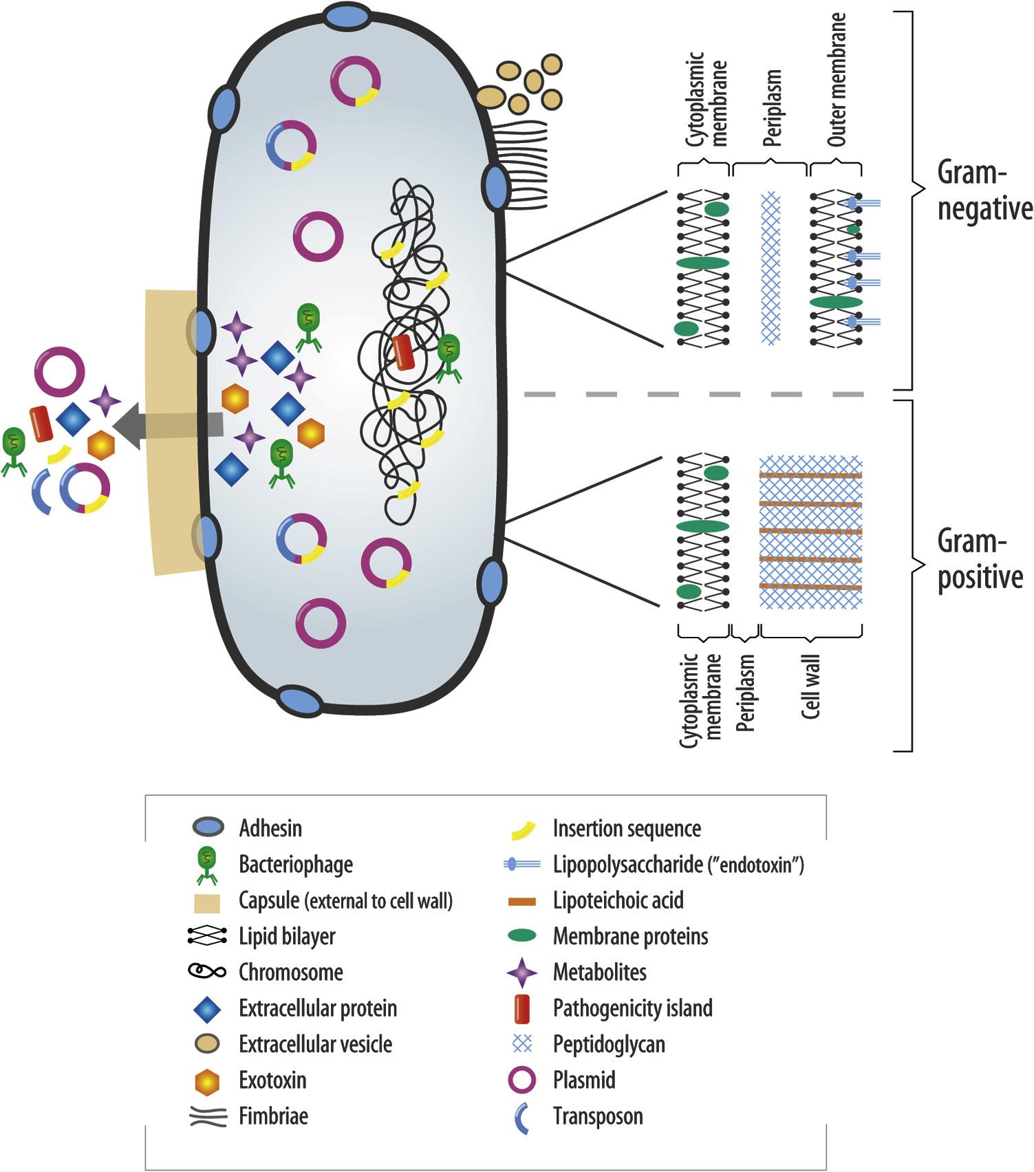
Many microorganisms isolated from endodontic infections have also been identified as commensals in the oral cavity. The transition from oral “commensal” to root canal “pathogen” likely reflects an innate ability to switch on genes that enable survival and propagation in a different environment and encode a range of virulence factors (Fig. 1.13). The first reported virulence factor associated with endodontic infections was lipopolysaccharide (“endotoxin”), a virulence factor produced by gram-negative bacteria.43
It has been suggested that symptoms increase when certain microbial species are part of the infective endodontic microbiome. Nevertheless, the same species can be found in asymptomatic cases with prevalence comparable to that of symptomatic cases; this apparent discrepancy could be explained in part by the variations in expression of virulence factors by different strains of the same species. Protein analyses (the metaproteome) of endodontic infections, along with the host response, are future steps toward better understanding of interactions between the microbiome of endodontic infections and the host throughout the infection and healing process.44,45
Cocci
Cocci
Firmicutes
Actinobacteria
Bacteroidetes
Proteobacteria
Fusobacteria
Synergistetes
General (n=7) Apical (n=5)
• Fig. 1.12 A heat map of root canal community profiles based on bacterial prevalence in 15 groups of root canal microbiomes gathered from 12 studies using next-generation DNA sequencing. Microbiome profiles are divided into general (entire root canal) and apical samples. Heat map scale ranges from 0 to 1 by taxa where “1” indicates that all samples had that specific taxa as their top 5 phyla or genera. A “0” score indicates that taxa did not appear as a top 5 phyla or genera. (From Shin JM, Luo T, Lee KH, et al: Deciphering endodontic microbial communities by next-generation sequencing, J Endod 44[7]:1080–87, 2018.)
Pulpal Diseases
Host Response in the Dental Pulp
The response of the dental pulp to microbial and other physical and chemical irritants is similar to the response in other connective tissues. An inflammatory process starts in the pulp and corresponds to the location where the irritation reaches it. For example, in an incipient carious lesion at the depth of an occlusal fissure, the pulp at the end of the affected dentinal tubules is seen to have a small inflammatory process histologically. This inflammation progresses throughout the coronal pulp as the carious lesion penetrates deeper in dentin, until the microbial irritants eventually invade the pulp in large numbers and cause severe inflammation (Fig. 1.14).
However, unlike other connective tissues, the dental pulp lacks collateral circulation and is confined within rigid dentinal walls. Therefore at a specific point in the disease process, the inflammation changes from reversible (one that would respond favorably to conservative methods of treatment and eventually heal) to irreversible pulpitis. The transition of reversible to irreversible pulpitis is important to identify clinically because it determines the optimal procedure that should be employed to treat it.
Studies have shown that the inflammatory response in the pulp is associated with several cellular and molecular changes (Video 1.2). The degree of irritation appears to trigger a corresponding level of inflammation. This titration of the
In recent years there has also been considerable interest in molecular mediators of pulpal inflammation. Some inflammatory mediators have been shown to correlate directly with increased pulpal pain and the clinical diagnosis of symptomatic irreversible pulpitis. These mediators include prostaglandins, neuropeptides, bradykinin, cytokines, chemokines, and matrix metalloproteinases.49,50 The dental pulp is heavily innervated by General
inflammatory response to the level of irritation is orchestrated by a balance of proinflammatory and antiinflammatory factors in the pulp. The condition of the pulp deteriorates or improves based on the reaction of pulpal factors to the external environment. Cellular responses include the increase in inflammatory cells, most notably neutrophils, lymphocytes, macrophages, plasma cells, mast cells, and dendritic cells (Figs. 1.15 to 1.17). The inflammatory cell response correlates in its intensity with the depth of the carious lesion.46 Noninflammatory cells, such as odontoblasts and fibroblasts, do contribute to the inflammatory response. Odontoblasts have been shown to express TLRs, cytokines, chemokines, and defensins.47 However, the degree to which noninflammatory cells contribute to the inflammation is much less than inflammatory cells like neutrophils and macrophages. A recent study showed reasonably good agreement between clinical signs and symptoms, and the histologic condition of the pulp in cases with caries.48 In this study, microbial ingress into the pulp, which is indicative of severe pathosis, seemed to be confined to cases diagnosed clinically as irreversible pulpitis.
(n=9)
(n=6)
• Fig. 1.13 Potential Virulence Factors Associated With a Bacterial Cell. (From Sedgley CM. Virulence of endodontic bacterial pathogens. In Fouad AF, editor: Endodontic microbiology, Ames, IA, 2009, WileyBlackwell Publishing, pp. 130–151.)
sensory fibers. During the inflammatory process, there is sprouting of pulp nociceptors, with an increase in secretion of neuropeptides like substance P and CGRP. These mediators lower the pain threshold and increase the permeability of blood vessels to inflammatory cells.
The compromised ability of the pulp to survive severe forms of inflammation, in comparison with other connective tissues, is illustrated by the differences in response to trauma between teeth with immature apex and those with mature apex. After luxation injuries, such as extrusion, intrusion, or even total avulsion and replantation, the tooth with immature apex is much more likely to retain (or revascularize) a vital pulp than a tooth with mature apex. This difference is primarily related to the increased collateral circulation in the immature teeth that can take advantage of the wide apical foramen.
Occasionally, radiographic analysis of the dental pulp reveals an increase in mineralization and deposition of hard dentinal tissues. Increased mineralization occurs gradually due to deposition of secondary dentin throughout life. However, as
a result of traumatic injuries, a substantial increase in mineralization may occur in the tooth or teeth that were traumatized. Increased areas of mineralization, commonly known as pulp stones , are also associated with caries or deep restorations. Excessive pulp stones have also been associated with cardiovascular diseases, 51 and with intake of statin medications. 52 Increased mineralization of the pulp, even when obliterating the entire canal space ( Fig. 1.18 ), in the absence of symptoms or apical pathosis, is not considered pathologic. However, if pulpal disease arises in these conditions, root canal treatment may become very challenging.
Less frequently, the pulp may undergo internal resorptive defects (Fig. 1.19). The cells that typically initiate the resorption, the osteoclasts or odontoclasts, are not normal inhabitants of the healthy pulp. These cells, which arise from monocytes, are normal mediators of bone turnover and are present in the PDL and alveolar bone. When resorptive lesions are detected clinically (internally or externally), they have usually attained a large size and are considered pathologic, even in the absence of symptoms.
Another random document with no related content on Scribd:
GRIMALDO, 110.
GUEVARA, 512.
GUI (Pedro de), 425.
GUILLÉN DE ÁVILA (Diego), 483.
GUILLÉN DE SEGOVIA (Pedro), 388.
GUILLERMO DE INGLATERRA (Estoria delrey), 460.
GUNDISALVO (Domingo), 120.
GUTIÉRREZ DE CEREZO (Fr. Andrés), 418.
GUTIÉRREZ DE TOLEDO (Julián), 450, 470.
HEREDIA (Pablo de), 425.
HERNÁNDEZ (Alonso), 522.
HERRERA (Fr. Diego de), 362.
HERRERA (Gabriel Alonso de), 524.
HERRERA (Martín de), 511.
HIAYA-BEN-ISAAC, 102.
HIGINO (C. Julio), 33.
HINOJOSA (D. Gonzalo de), 231.
HISPALENSE (Juan), 120.
HISPANO (Juan), 130.
HISPANO (Pedro), 180, 218.
HISPANO (Vicente), 220.
HOSPITAL (Jaime), 231.
IBARRA (Martín de), 544.
IDACIO, 72.
ILDEFONSO (S.), 80.
ILDEFONSO (Vida de S.), 226.
IMPERIAL (Micer Francisco), 346.
IRIENSE (Cronicon), 127.
ISAAQUE (Libro de), 223.
ISIDORO (S.), 78.
ISOPETE ISTORIADO (Ellibro de), 426 (Fábulas de Esopo, Burgos, 1496).
JAIME (D.), 220.
JANUARIUS (Fr. Jaime), 506.
JÁTIBA (Juan Andrés de), 541.
JERÓNIMO (Scala coelide S.), 503.
JHERONIMO (Eltransito de Sant), 445.
JIMÉNEZ CERDÁN (Juan), 312.
JIMÉNEZ DE PREJANO (D. Pedro), 541.
JIMÉNEZ DE RADA (Rodrigo), 178.
JUAN (Historia delabaddon), 506.
JUAN II (Crónica de don), 365.
JUAN MANUEL (Don), 232.
JUDA-BEN-DAVID, 100.
JUDA-LEVÍ, 112.
JULIÁN (S.), 80.
JULIANO (Presbítero), 110.
JULIANUS (Antonius), 55.
JUSTINIANUS, 75.
JUSTUS, 75.
JUVENCO, 59.
KALILA etDigna, 187.
KIMJI, 112.
LANAJA (Pedro), 231.
LANDO (Fernán Manuel de), 346.
LAPIDARIO, 187.
LARA (Gesta de los Infantes de), 147.
LARRAGA (Martín de), 231.
LATRON, 35.
LATRONIANUS, 62.
LAX (Μ. Gaspar), 506.
LEANDRO (S.), 76.
LEBRIJA (M. Elio Antonio de), 419.
LEDESMA (Fr. Francisco de), 506.
LEÓN (Francisco de), 512.
LEÓN HEBREO, 504.
LEYVA (Juan de), 512.
LI (Andrés de), 449.
LIBRO de la celestialJerarquíay infernalLaberinto, 446.
LIBRO de las marauillas delmundo, 540.
LIBRO de lospensamientos variables, 485.
LICINIANUS, 75.
LILII Medicinae, 453.
LISUARTE de Grecia (Libro...), 539.
LOAYSA (José de), 209.
LOBRAÑAN (Diego de), 312.
LOGROÑO (Bach. Juan Alfonso de), 453.
LÓPEZ (M. Fr. Juan), 430.
LÓPEZ DE AYALA (Pero), 263. (La Coronica delrey D. Pedro se imprimió en Sevilla, 1495; Toledo, 1526 (con la de Enrique II y Juan I); Sevilla, 1542, 1549; Pamplona, 1591).
LÓPEZ DE CORTEGANA (Diego), 534.
LÓPEZ DE HARO (D. Diego), 512.
LÓPEZ DE MENDOZA (D. Íñigo), Marqués de Santillana, 301.
LÓPEZ DE SEGOVIA (Juan), 425.
LÓPEZ DE TOLEDO (Fr. Diego), 470.
LÓPEZ DE VILLALOBOS (Dr. Francisco), 467.
LÓPEZ DE VIVERO PALACIOS RUBIOS (Dr. Juan), 500.
LOPIS (Juan), 503.
LUCANO, 45.
LUCAS DE TÚY (Don), 181.
LUCENA (Juan de), 379.
LUCIDARIO, 210.
LULL (Raimundo), 215.
LUNA (D. Álvaro de), 351.
LUNA (Pedro de), 273.
LUSITANO (Cronicon), 127.
LUZÓN (Juan de), 509.
LLABIA (Cancionero de Ramón de), 410.
MACÍAS, 346.
MADALENA (Historia de S. María), 539.
MADRIGAL (Alfonso de), 334.
MAGOS (Historia de los tres Reyes), 509.
MAHOMA (La Alabanza de), 131.
MAIMÓNIDES, 112.
MAINETE (Gesta de), 147.
MALLORCA (Rodrigo de), 257.
MANRIQUE (Gómez), 374.
MANRIQUE (Jorge), 401.
MANUAL de la Sancta Fe Católica, 453.
MANUEL (D. Juan), 512.
MARBRES (Juan), 400.
MARCIAL, 51.
MARINEO SÍCULO (Lucio), 415.
MARQUILLES (Jaime de), 503.
MARTÍ (Fr. Ramón), 221.
MARTÍN (S.), 74.
MARTÍN DE LEÓN (D.), 180.
MARTÍN DE LEÓN (S.), 130.
MARTÍNEZ (Μ. Ferrando), 220.
MARTÍNEZ DE AMPIÉS (Martín), 453.
MARTÍNEZ DE BIZCARGUI (Gonzalo), 509.
MARTÍNEZ DE MEDINA (Diego y Gonzalo), 346.
MARTÍNEZ SILICEO (Juan), 539.
MARTÍNEZ DE TOLEDO (Alfonso), Arcipr. de Talavera, 315.
MÁXIMO, 80.
MAYNES (Noble Cuento delemperador Carlos), 460.
MAZUELO (Fr. Vicente de), 430.
MELA (Pomponio), 39.
MELOSINA (Historia de la linda), 425.
MELLA (Juan de), 341.
MENA (Juan de), 322.
MENAHEM-BEN-SARUK, 100.
MENCIA DE CISNEROS (Cancioneiro de Dom), 204.
MENDOZA (Fr. Íñigo de), 407.
MENESES DE SILVA (Juan), 387.
ΜEROBAUDES, 72.
MEXÍA (Ferrand), 445.
MEXÍA (Hernán), 368.
MIERES (Tomás), 330.
MIGIR (Fray), 346.
MIRAVET (Juan de), 453.
MISTERIO de Elche, 440.
MODERATO DE GADES, 39.
MOHADJAR, 114.
MOHAMED-ETTEMIMY, 107.
MOISÉS-BEN-EZRA, 114.
MOISÉS-BEN-HANOCH, 100.
MOISÉS-BEN-SHEM-TOB, 231.
MOLES MARGARIT (D. Juan), 410.
MOLINA (Fr. Bartolomé de), 506.
MOLINO (Miguel del), 544.
MONCAYO (Mosén Juan de), 335.
MONJA (Fr. Alonso de la), 346.
ΜONSERRAT (Guillermo de), 330.
MONTE (Fr. Lope del), 346.
MONTESA (Fr. Bernardo), 376.
MONTESDOCA (D. Juan de), 506.
MONTESINO (Fr. Ambrosio), 478.
MONTORO (Antón de), 366.
MONTRAVA (Berenguer de), 299.
MORALES (Fernando de), 541.
MOTETES de canto d'organo (Libro de), 503.
MUJERES illustres de Boccaccio (De las), 450.
NARVÁEZ (Juan de), 511.
NEBRIDIUS, 75.
NOBLEZA óLealtat(Libro de la), 184.
NOLA (Roberto de), 339.
ΝOYA (Vidal de), 449.
NÚÑEZ (Nicolás), 512.
NÚÑEZ CORONEL (Luis), 507.
NÚÑEZ DELGADO (Pero), 499.
NÚÑEZ DE TOLEDO (Alfonso), 410.
NÚÑEZ DE TOLEDO (Hernán), 427.
NÚÑEZ DE TOLEDO (Juan), 503.
OBREGÓN (Antonio de), 523.
OCAÑA (Fr. Gonzalo de), 333.
OLID (Juan de), 372.
OLIUEROS de Castillay Artus dalgarbe (Historia de los nobles caualleros), 484.
OLIVA (Monje), 110.
OLIVER (Fr. Bernardo), 257.
OLLER (Fr. Bernardo), 257.
OROPESA (Alfonso de), 376.
OROSIO, 70.
ORTIZ (Dr. Alonso), 449.
OSIO, 57.
OSMA (M. Pedro de), 406.
OSORIO DE MOSCOSO (D. Rodrigo), 512.
OTTAS (Cuento muy fermoso delemperador), 460.
PACENSIS, 88.
PACIANO (S.), 63.
PADILLA (Fr. Juan de), 446.
PÁEZ DE RIBERA (Ruy), 346.
PALENCIA (Alfonso de), 372.
PALMERÍN de Oliva, 514.
PARDO (Jerónimo), 492.
PARIS e Viana (Historia de los amores de), 131.
PARTENOPLES (Historia delConde; en catalán), 425.
PARTINUPLES (Libro delesforçado cauallero conde), 535.
PASCUAL (S. Pedro), 217.
PATOS (Juan Pedro de), 231.
PEDRO (D.), Condestable de Portugal, 355.
PEDRO (Diego de San), 512.
PEDRO IV, 273.
PELAYO, 124.
PENTATEUCO hebraico, 426.
PEÑA (Fr. Antonio de la), 514.
PEÑAFORT (S. Raimundo de), 220.
PERALTA (Guillermo de), 484.
PÉRDIDA de España (Gesta de la), 147.
PEREGRINA, 273 y 470.
PÉREZ DE GUZMÁN (Fernán), 306.
PÉREZ DE OLIVANO (Agustín), 506.
PÉREZ DE SALANOVA (D. Ximen), 231.
PÉREZ DE VALENCIA (Jaime), 415.
PERPIÑANO DE RIU (Fr. Pedro), 231.
PERTUSA (Martín de), 231.
PINAR, 512.
PINTOR (Pedro), 484.
PLÁCIDAS (Estoria delcavallero), 460.
POLEMAR (Juan), 309.
POLÍGLOTA de Alcalá, 539.
PORIDAD de las Poridades, 184.
PORTOCARRERO (D. Luis), 512.
POTAMIO (S.), 61.
PRADILLA (Bachiller de la), 514.
PRIMALEÓN y Polendos (Libro...), 544.
PRISCILIANO, 61.
PROAZA (Alfonso de), 494.
PROVERBIOS (Libro de los buenos), 184.
PROVERBIOS en rimo de... Salomón, 207.
PRUDENCIO, 65.
PRUDENCIO GALINDO, 94.
PUENTE (Fr. Ramón de la), 217.
PUERTA (Fr. Sancho), 290.
PUERTO (Diego del), 503.
PULGAR (Hernando del), 416. (La Chronica de los Reyes Catolicos se imprimió en Granada, 1545, 1550; Valladolid, 1565; Zaragoza, 1567).
QUINTILIANO, 53.
QUIRÓS, 512.
RABI-JONAS-BEN-GANAJ, 104.
RAIMUNDO DE PEÑAFORT (S.), 220.
RAMÍREZ (D. Alfonso), 180.
RAMOS DE PAREJA (Bartolomé), 413.
RASIS (Crónica delmoro), 246.
RAZÓN de Amor, 162.
RECEMUNDO, 101.
REFRANES glosados, 510.
REGIMINE Principum (De), 450.
REINOSA (Rodrigo), 533.
RENALLO GRAMÁTICO, 123.
RESENDE (Cancionero de), 357, 542.
REVELACIÓN de un ermitaño, 272.
REYES (Crónica de Veinte), 245.
REYES dorient(Libro dels), 159.
REYES Magos (Auto de los), 149.
RIBELLES (Mosén Juan), 335.
RIBERA (Hernando), 522.
RIBERA (Páez de), 511.
RIBERA (Suero de), 335.
RIBOT (Fr. Felipe), 273.
RIMADA (Crónica), 252.
RÍO (Juan del), 231.
ROA (Fernando de), 539.
ROBERTO elDiablo (La vida de), 510.
RODERICI Campidocti(Gesta), 128.
RODRIGO (Cantar de), 252.
RODRÍGUEZ DE ALMELLA (Diego), 377.
RODRÍGUEZ DE LA CÁMARA (Juan), 327.
RODRÍGUEZ DE LENA (Pero), 310.
RODRÍGUEZ DE MONTALVO (Garci), 454.
RODRÍGUEZ DE TUDELA (Ldo. Alonso), 541.
RODULFO, 123.
ROJAS (Fernando de), 471.
ROLDÁN (Maestre), 220.
ROMANCERO, 486.
ROMANO, 98.
ROSELL (Fr. Nicolás), 257.
ROSIGNOL (Fr. Arnaldo de), 231.
RUBIO (Fr. Guillermo), 231.
RUIZ (M. Jácome), 220.
RUIZ (Juan), Arc. de Hita, 237.
RUIZ DE CORRELLA (Juan), 407.
SABAH (Rabí Abraham), 432.
SABUNDE (Raimundo), 313.
SAHAGÚN (Juan de), 360.
SAID-BEN-ABD-RABIHI, 102.
SAKTAR, 105.
SALAYA (Alonso de), 523.
SALAYA (Juan de), 499.
SALINAS (Lope), 387.
SALOMÓN (Proverbios en rimo delsabio), 207.
SAMPIRO, 103.
SAMSON, 97.
SAMUEL de Israel(Las epístolas de Rabí), 514.
SAMUEL NAGUID, 105.
SAN CRISTÓBAL (Alfonso de), 333.
SÁNCHEZ DE ARÉVALO (Rodrigo), 386.
SÁNCHEZ DE BADAJOZ (Garci), 481.
SÁNCHEZ DE BADAJOZ (Bach. Diego), 519.
SÁNCHEZ CIRUELO (M. Pedro), 493.
SÁNCHEZ TALAVERA (Fernán), 346.
SÁNCHEZ DE TOVAR Ó DE VALLADOLID (Fernando), 255. (La Crónica de Alonso XIsalió en Valladolid, 1551, 1563; Medina, 1563; Toledo, 1595).
SÁNCHEZ DE VERCIAL (Dr. Clemente), 291.
SANCHO II (Gesta de don), 147.
SANTA MARÍA (Alonso de), 295.
SANTA MARÍA (D. Gonzalo de), 298.
SANTAMARÍA (D. Pablo de), 279.
SANTA María Egipciaqua (Vida de), 159.
SANTAFÉ (Pedro de), 335.
SANTISTEBAN (Cristóbal de), 502.
SAVASORDA, 115.
SEBASTIÁN, 98.
SEGOVIA (Juan de), 309.
SEGUÍ (Pedro), 535.
SEGUNDO (Capítulo de las cosasque escribiópor rrespuestas elfilósofo), 192.
SEM TOB, 259.
SEM-TOB (Joseph-ben-), 360.
SEM-TOB-FALAGUERA, 219.
SENDEBAR, 190.
SÉNECA (Las Epístolas de), 464.
SÉNECA el filósofo, 41.
SÉNECA el retórico, 37.
SESSÉ (Juan de), 335.
SEVERO (Aquilio), 63.
SEVERUS, 75.
SEVILLA (Juan de), 93.
SEXTILIUS ENA, 31.
SILENSE (El), 126.
SILIO ITÁLICO, 49.
SILOS (Códice de S. Domingo de), 131.
SOBRARIAS (Juan), 492.
SOCARRATS (Juan), 403.
SORBÉS (Mauro Antonio), 409.
SORIA, 512.
SORIO (Fr. Baltasar), 514.
SOSA (Lope de), 512.
SOTO (Alfonso de), 430.
SPAÑON (Alonso), 485.
STANYOL (Fr. Ángel), 503.
STÚÑIGA (Cancionero de), 335.
SUERTES (Libro de), 499.
TABLANTE de Ricamonte (Crónica de...), 535.
TAFUR (Pero), 370.
TAJON, 80.
TALAVERA (Hernando de), 465.
TAPIA, 512.
TAPIA (Juan de), 335.
TARANTA (Vasco de), 290.
TARGUM de Onkelos, 432.
TEODOR (Historia de la doncella), 192.
TEODULFO, 90.
TERRENA (Fr. Guido de), 236.
TESORO (Libro del), 207.
TIBERIANO, 63.
TIRANT lo blanch(Libro delvaleros e strenu caualler), 430.
TIRANTE elBlanco, 514.
TOLEDO (Anónimo de), 131.
TOLEDO (Crónica de), 131.
TOLEDO (D. Francisco de), 406.
TOLEDO (Juan el Viejo de), 290.
TOMÁS (Álvaro), 510.
TORIBIO (S.), 72.
TORO (Arcediano de), 346.
TORQUEMADA (Juan de), 314.
TORRE (Alfonso de la), 331.
TORRE (Bachiller de la), 430.
TORRE (Fernando de la), 335.
TORRELLA (Gaspar), 466.
TORRELLA (Jerónimo), 464.
TORRELLAS (Pedro), 335.
TORRES NAHARRO (Bartolomé de), 515.
TOVAR (Fr. Luis de), 509.
TRACTADO de la viday estado de laperfeccion, 484.
TRISTAN de leonis (Libro delesforçado cauallero don), 496.
ΤROYANA (Crónica), 255.
TUNGANO (Libro delcaballero don), 509.
TURPIN (Crónica latina delseudo), 147.
TURRANIUS GRACILIS, 31.
ULIGIA (Fr. Gombaldo de), 273.
URREA (D. Pedro Manuel de), 528.
URRÍES (Mosén Ugo de), 404.
VALERA (Mosén Diego de), 411.
VALERIO (S.), 98.
VALLADOLID (Alfonso de), 217 y 236.
VALLADOLID (Juan de), 335.
VARGAS (Fr. Alonso de), 257.
VATICANA (Cancioneiro da), 204.
VÁZQUEZ DE TAPIA (Hernán), 522.
VEGA (Fr. Pedro de la), 511.
VELASCO (D. Antonio de), 512.
VERAGUE (Pedro de), 261.
VERGEL de consolación, 275 y 466.
VESPASIANO (Istoria delnoble), 430.
VIANA (Príncipe Carlos de), 340.
VICENTE (Gil), 497.
VICTORIANO Y MOLÓN (Lorenzo), 470.
VILLALPANDO (Mosén Juan de), 335.
VILLENA (D. Enrique de), 288.
VINCENCIO, 107.