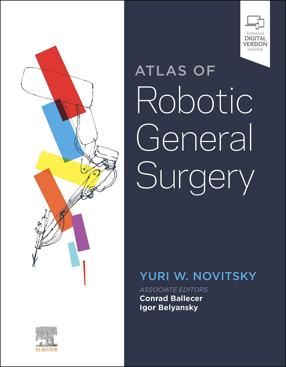
Download Atlas of robotic general surgery 1st edition yuri w. novitsky md facs (editor) ebook All C
https://ebookmass.com/product/atlas-of-roboticgeneral-surgery-1st-edition-yuri-w-novitsky-mdfacs-editor/ Download more ebook from https://ebookmass.com
More products digital (pdf, epub, mobi) instant download maybe you interests ...
Insall & Scott Surgery of the Knee, 2-Volume Set, 6e 6th Edition W. Norman Scott Md Facs
Clinical Assistant Professor Department of Surgery
Creighton University School of Medicine
Phoenix, AZ, USA
IGOR BELYANSKY MD
Chief of General Surgery, Director, Abdominal Wall Reconstruction Program, Anne Arundel Medical Center
Annapolis, MD, USA
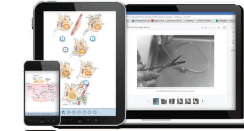
Any screen. Any time. Anywhere.
Activate the eBook version of this title at no additional charge.
Elsevier eBooks for Practicing Clinicians gives you the power to browse and search content, view enhanced images, highlight and take notes—both online and offline.
Unlock your eBook today.
1. Visit expertconsult.inkling.com/redeem
2. Scratch box below to reveal your code
3. Type code into “Enter Code” box
4. Click “Redeem”
5. Log in or Sign up
6. Go to “My Library” It’s that easy!
Place Peel Off Sticker Here
For technical assistance: email expertconsult.help@elsevier.com call 1-800-401-9962 (inside the US) call +1-314-447-8300 (outside the US)
No part of this publication may be reproduced or transmitted in any form or by any means, electronic or mechanical, including photocopying, recording, or any information storage and retrieval system, without permission in writing from the Publisher. Details on how to seek permission, further information about the Publisher’s permissions policies and our arrangements with organizations such as the Copyright Clearance Center and the Copyright Licensing Agency, can be found at our website: www.elsevier.com/permissions
This book and the individual contributions contained in it are protected under copyright by the Publisher (other than as may be noted herein).
Notices
Practitioners and researchers must always rely on their own experience and knowledge in evaluating and using any information, methods, compounds, or experiments described herein. Because of rapid advances in the medical sciences, in particular, independent verification of diagnoses and drug dosages should be made. To the fullest extent of the law, no responsibility is assumed by Elsevier, authors, editors, or contributors for any injury and/or damage to persons or property as a matter of product liability, negligence or otherwise, or from any use or operation of any methods, products, instructions, or ideas contained in the material herein.
Library of Congress Control Number: 2020950567
ISBN: 978-0-323-69780-4
E-Book: 978-0-323-69781-1
Inkling: 978-0-323-69782-8
Content Strategist: Jessica McCool
Content Development Specialist: Nani Clansey
Project Manager: Julie Taylor
Design: Brian Salisbury
Illustration Manager: Deanna Sorenson
Marketing Manager: Kate Bresnahan
Cover Art Design: Hi! Estudio. Tepatitlán, México
Section I: General Issues in Robotic Surgery
Laura Flores, Priscila Rodrigues Armijo, Salim Hosein, Dmitry Oleynikov
Vahagn C. Nikolian, Jake G. Prigoff, Dina
Sharona B. Ross, Andres Giovannetti, Iswanto Sucandy, Trenton Lippert, Alexander S. Rosemurgy
Jerald D. Wishner
Sandeep S. Vijan Section
Clark Gerhart
Kazuki N. Sugahara, John A. Chabot
Jesse Sulzer, Dioneses Vrochides, John B. Martinie
Sharona B. Ross, Alexander S. Rosemurgy, Jack Wecowski, Timothy J. Bourdeau, Iswanto Sucandy
Maureen M. Tedesco, Micaela M. Esquivel
Foreword
Over the last 100 years the role of the general surgeon has evolved as other specialties have come into their own. Not only have the procedures performed changed, but the techniques used have become less invasive while offering patients a less morbid recovery. Abdominal operations in particular have benefited from the transition from large laparotomies to a minimally invasive approach. The first step was the development of the laparoscopic approach to standard abdominal procedures. However, as surgeons became more skilled performing complex procedures using the MIS approach, it became apparent that conventional laparoscopy had some inherent limitations and its adoption was often limited.
Early adopters of a robotic MIS approach found that they could equal the results of conventional MIS procedures but extend their application beyond what had been achieved with “straight-stick” laparoscopy. The learning curve appeared shortened and utilization of the approach markedly increased. In addition, there has been a rapid progression of the complexity of procedures this new platform has allowed general surgeons to perform across general surgery and its subspecialties.
In the past there have been excellent textbooks to guide surgeons through open and more recently through laparoscopic
procedures. Until now, however, there has not been a text which accomplishes the same goals for this new robotic platform. It is essential that surgeons in training as well as accomplished surgeons learning a new technique have a text that gives them such a step-by-step guide. The Atlas of Robotic General Surgery is just such a text. From the introduction which covers the basics of robotics to its five major sections which include robotic inguinal and abdominal hernia repair, bariatric and forget surgery, intestinal and colorectal surgery, and hepatic and solid organ surgery, this book gives the reader an in-depth guide of how to safely and efficiently accomplish simple, as well as complex, procedures utilizing the robotic platform. The editors have engaged the true experts in each discipline who are not only experienced robotic surgeons but are accomplished teachers and mentors. I am confident that this text will set the standard for robotic education and is only the first of many editions to come as this field continues to grow and evolve. It is a must for any surgeon planning to begin robotic general surgery, for surgeons wanting to adopt new robotic procedures, and especially for those mentoring or teaching surgeons to incorporate this platform into their repertoire.
Edward Felix, MD
Dedication
To surgical innovators before me who paved the way, to my present colleagues who support and inspire me, to future generations of surgeons who will make it all much better, and to my girls Maya, Ella, Lily, Phoebe and Chloe who make it all worth my while.
Yuri W. Novitsky
I would like to offer my deepest gratitude to my brother Dan, my mom Amelita, and my doctor “hero” father Conrado, to my wife Michelle, and my two daughters, Madison and Emma, I love you more than you could ever imagine.
Conrad Ballecer
To all my mentors who believed in me when I needed them most.
Igor Belyansky
Preface
Surgical science is uniquely dynamic. Introduction of new techniques and technologies is a frequent occurrence. What seems one day like the ‘standard of care’ practice can rapidly become an ‘oldfashioned’ obsolete technique. Something that is viewed as revolutionary may become evolutionary in a short span. Incorporation of the robotic technology into general surgery and related fields had become that catalyst of the recent past. Robotics has fueled profound advances in surgical techniques, especially those in my field of abdominal wall and hernia surgery. The speed of surgical progress, that has occurred in the past 7–8 years largely due to development and implementation of robotic techniques, has been nothing but astounding.
My personal foray into robotics was stunted early on by several unfounded biases and preconceived hesitations. As an expert laparoscopist I felt little need for another expensive ‘toy’ to help me to deliver, what I thought, were already cutting edge minimallyinvasive operations. However, once I committed to understanding and learning the robotic platform, it became abundantly clear that I was venturing into the next era of surgery. The robotic ‘train’ was picking up steam and I had little hesitation about jumping aboard and helping drive it. I joined those surgeons, many of them contributing chapters in this textbook, who were dedicating their energy and time to integrating robotics, perfecting or pioneering techniques, developing pathways to responsible skill acquisition and implementation, and finally gathering data to demonstrate the unquestionable benefits of robotic surgery in many surgical procedures.
This textbook will hopefully become an invaluable resource to both bread-and-butter general surgeons, as well as tertiary care subspecialists. While our surgical field is advancing rapidly, the information in this textbook remains to be state of the art. We have assembled a uniquely comprehensive list of topics encompassing a wide variety of abdominal surgeries, from the most basic to the most complex robotic procedures performed today. Each chapter is authored by some of the top robotic surgeons and should provide a unique insight into their preoperative decision making, patient preparation, operative setup, and technical details. Along with vivid intraoperative photographs, the narrated video collection of this Atlas should propel it to become a ‘go-to’ resource for both trainees and practicing surgeons embarking on robotics. I am confident this textbook will help the readers to become better at their craft and that their patients will be the greatest beneficiaries.
Yuri Novitsky, MD
Even with all the attendant benefits of improved 3D visualization, precision, and dexterity, the da Vinci robotic platform struggled to penetrate the market of general surgery. In existence since the early 21st century, and initially designed for introducing minimally invasive surgery (MIS) to the field of cardiac surgery, Intuitive Surgical, “Aimed for the heart, and hit the prostate.” This innovative surgical instrument clearly advanced MIS in the fields of urology and gynecology, and attained some purchase in the field of colorectal surgery; yet its use in general surgery was generally met with contentious resistance. Many surgeons claimed that the robot had no application in the field of general surgery, and was reserved only for the few that were untrained in conventional laparoscopic surgery.
My first true exposure at the potential of robotic surgery in my general surgical practice was from watching a podium presentation by Dr. Ricardo Abdalla, showcasing his modified robotic Rives-Stoppa Repair. I was amazed at the dexterity of the instrument which allowed for dissecting layers of the anterior abdominal wall and suturing these layers back together. Immediately after the presentation I came to two conclusions: one, although I was an MIS-trained fellow, I could not readily reproduce his extraperitoneal repair with the conventional ‘straight-sticks’, and two, the robotic platform represented an advanced iteration of laparoscopy going forward.
Coincident with this time, I virtually abandoned laparoscopic ventral hernia repair, preferring open techniques where I could readily hide mesh from the visceral content, as well as reconstitute linea alba while addressing the divarication of the rectus muscle complex. As an ‘all-in’ MIS advocate, this MIS to open transformation was all too discouraging. I adopted the robotic platform to re-engage my abdominal wall practice back to MIS. With all its attendant benefits and vast improvement in ergonomics, this adoption was transformative. Essentially everything I used to do with the straight-sticks I converted to the articulating robotic platform. At the time there were no educational resources guiding my initial pursuits; pursuits that were widely rejected in private and public forums. Yet, myself and others forged on, confident we would end up on the ‘correct’ side of history.
Personally, I am very proud of how the culture of adoption of robotic surgery has evolved over the last few years and I am even more proud to serve a role, albeit a small one, in educational dissemination regarding its use. The Atlas of Robotic General Surgery serves as a culmination of all the hard work that many of the pioneering authors detail. I, and many others, will forever enjoy this Atlas as a premier educational resource in our daily practice.
Conrad Ballacer, MD
List of Contributors
Cheguevara Afaneh MD
Assistant Professor of Surgery Department of Surgery
New York-Presbyterian Hospital/Weill Cornell Medical College New York, NY
USA
Ali Ahmad MD
Clinical Assistant Professor of Surgery Division of Surgical Oncology University of Kansas School of Medicine Wichita, KS USA
Marcia Alayón-Rosario MD Department of Surgery
Division of Minimal Access and Bariatric Surgery
Prisma Health-Upstate Greenville, SC USA
Hemasat Alkhatib MD Resident Department of Surgery Cleveland Clinic Cleveland, OH USA
Vedra Augenstein MD
Associate Professor of Surgery Department of Surgery Carolinas Medical Center Charlotte, NC USA
Conrad Ballecer MD, MS
Clinical Assistant Professor Department of Surgery Creighton University School of Medicine Phoenix. AZ USA
Igor Belyansky MD
Chief of General Surgery
Director, Abdominal Wall Reconstruction Program Department of Surgery
Anne Arundel Medical Center
Annapolis, MD USA
Timothy J. Bourdeau II BSc
Surgical Research Technician
AdventHealth Tampa Tampa, FL USA
Alfredo M. Carbonell D.O.
Vice Chairman of Academic Affairs Professor of Surgery Department of Surgery Prisma Health-Upstate University of South Carolina School of Medicine Greenville, SC USA
Leandro Totti Cavazzola MD, MSc, PhD
Associate Professor of Surgery
Universidade Federal do Rio Grande do Sul
Department of Surgery
Hospital de Clínicas de Porto Alegre Porto Alegre, Rio Grande do Sul Brazil
John A. Chabot MD
David V. Habif Professor of Surgery
Columbia University Vagelos College of Physicians and Surgeons
Associate Director, Herbert Irving Comprehensive Cancer Director, The Pancreas Center Chief, GI and Endocrine Surgery Department of Surgery
Columbia University Irving Medical Center
New York, NY USA
David C. Chen MD
Professor of Clinical Surgery Department of Surgery
David Geffen School of Medicine at UCLA
Los Angeles, CA USA
Sarah Corn MD
Clinical Assistant Professor Division of Surgical Oncology Department of Surgery
University of Kansas School of Medicine-Wichita Wichita, KS USA
Francesca Dimou MD, MS Fellow
Department of Surgery
New York-Presbyterian Hospital/Weill Cornell Medical College
New York, NY USA
Colin Dunn MD Research Fellow Department of Surgery
Keck School of Medicine of University of Southern California
Los Angeles, CA USA
Aleeson Eka MD-MPH Candidate
Keck School of Medicine of University of Southern California Los Angeles, CA USA
Micaela M. Esquivel MD
Clinical Assistant Professor of Surgery Division of MIS/Bariatric Surgery Stanford University Stanford, CA USA
Laura E. Flores MD-PhD Scholar University of Nebraska Medical Center Omaha, NE USA
Carlos A. Galvani MD Professor and Chief
Division of Minimally Invasive and Bariatric Surgery
Tulane University School of Medicine
New Orleans, LA USA
Clark Gerhart MD
Director of Minimally Invasive Surgery and Robotics General and Bariatric Surgery
Wilkes-Barre General Hospital Wilkes-Barre, PA USA
Andres Giovannetti MD
General and Minimally Invasive Surgery
Robotic Hepatopancreaticobiliary Surgery Chicago Institute for Advanced Surgery Chicago, IL USA
Anthony Gonzalez MD Chief of Surgery
Baptist Hospital of Miami Associate Professor of Surgery
Florida International University General and Bariatric Surgery Miami, FL USA
Emily Helmick DO Resident Department of Surgery
Creighton University School of Medicine Phoenix, AZ USA
Salim Hosein MD
General and Bariatric Surgeon Department of Surgery
Southern Illinois Healthcare Herrin, IL USA
Caitlin Houghton MD
Assistant Professor of Surgery Division of Upper GI and General Surgery
Keck School of Medicine of University of Southern California Los Angeles, CA USA
Desmond Tuan-Khai Huynh MD Resident Department of Surgery
Cedars-Sinai Medical Center
Los Angeles, CA USA
Abraham Krikhely MD
Assistant Professor of Surgery Department of Surgery
Columbia University Irving Medical Center
New York, NY USA
Trenton Lippert Research Coordinator Surgery
AdventHealth Tampa Tampa, FL USA
Richard Lu MD, DABS
Assistant Professor of Surgery Department of Surgery
The University of Texas Medical Branch Galveston, TX USA
Ian T. Macqueen MD
Assistant Professor of Clinical Surgery
David Geffen School of Medicine at UCLA Department of Surgery
UCLA Health - Santa Monica Medical Center
Santa Monica, CA USA
Flavio Malcher MD, MSc
Assistant Professor of Surgery
Albert Einstein College of Medicine Director, Abdominal Wall Program Department of Surgery
Montefiore Medical Center
Bronx, NY USA
Luis A. Martin-del-Campo MD
Centro de Cirugía Robótica Departamento de Cirugía Hospital Ángeles del Carmen Guadalajara, Jalisco Mexico
John B. Martinie MD
Professor of Surgery
Division of Hepatobiliary and Pancreas Surgery Director, Robotic HPB Fellowship
Carolinas Medical Center, Atrium Health Charlotte, NC USA
David J. Morrell MD
Resident Department of Surgery
Penn State Hershey Medical Center Hershey, PA USA
Filip Muysoms MD, PhD
Head of Department of Surgery
Maria Middelares Gent Ghent Belgium
Vahagn C. Nikolian MD
Assistant Professor of Surgery Department of Surgery
Oregon Health and Science University Portland, OR USA
Yuri W. Novitsky MD
Professor of Surgery
Columbia University Vagelos College of Physicians and Surgeons
Director, Columbia Hernia Center Department of Surgery
Columbia University Irving Medical Center New York, NY USA
Dmitry Oleynikov MD
Chairman, Department of Surgery Monmouth Medical Center
Robert Wood Johnson Barnabas Health Monmouth, NJ USA
Sean M. O’Neill MD
Resident Department of Surgery
David Geffen School of Medicine at UCLA
Los Angeles, CA USA
Sean B. Orenstein MD
Associate Professor of Surgery Department of Surgery
Oregon Health and Science University
Portland, OR USA
Eduardo Parra-Davila MD Director, Minimally Invasive and Colorectal Surgery Director, Hernia and Abdominal Wall Reconstruction
Robotic Surgery Institute
Good Samaritan Medical Center
West Palm Beach, FL USA
Andrea Pakula MD, MPH
Medical Director of Robotic Surgery Department of Surgery
Adventist Health Simi Valley Hospital
Simi Valley, CA USA
Eric M. Pauli MD Professor of Surgery Director of Endoscopic Surgery
Chief, Division of MIS/Bariatric Surgery
Penn State Hershey Medical Center Hershey, PA USA
Dina Podolsky MD
Assistant Professor of Surgery
Columbia University Vagelos College of Physicians and Surgeons Department of Surgery
Columbia University Irving Medical Center
New York, NY USA
Ajita Prabhu MD
Associate Professor of Surgery Department of Surgery Cleveland Clinic Cleveland, OH USA
Jake G. Prigoff MD Resident Department of Surgery
Columbia University Irving Medical Center
New York, NY USA
Eric Rachlin PhD
MIS Fellow Department of Surgery
Memorial Hermann Health System Houston, TX USA
Priscila Rodrigues Armijo MD
Assistant Professor of Surgery Department of Surgery University of Nebraska Medical Center Omaha, NE USA
Alexander S. Rosemurgy MD
Director, Hepatopancreaticobiliary Surgery Department of Surgery
AdventHealth Tampa Tampa, FL USA
Sharona B. Ross MD, BS
Hepatopancreaticobiliary Surgeon Department of Surgery
AdventHealth Tampa Tampa, FL USA
Allegra Saving MD General Surgeon Department of Surgery Norton Healthcare Louisville, KY USA
William C. Sherrill III MD Resident Department of Surgery
Carolinas Medical Center, Atrium Health Charlotte, NC USA
Iswanto Sucandy MD
Clinical Instructor Department of Surgery University of Pittsburgh School of Medicine Pittsburgh, PA USA
Kazuki N. Sugahara MD, PhD Instructor in Surgery Department of Surgery
Columbia University Irving Medical Center New York, NY USA
Jesse Sulzer MD, PhD Fellow
Division of Hepatobiliary and Pancreas Surgery Department of Surgery
Carolinas Medical Center, Atrium Health Charlotte, NC USA
Hany Takla MD
General and Bariatric Surgeon Department of General Surgery
Beth Israel Lahey Health
Winchester Hospital Winchester, MA
Clinical Instructor
Tufts Medical School Boston, MA USA
Maureen Tedesco MD Chief of Surgery
Robotic and Minimally Invasive General Surgery
The Permanente Medical Group, Inc.
Kaiser Permanente Santa Clara Medical Center
Santa Clara, CA
Clinical Assistant Professor (Affiliated) Department of Surgery
Stanford University School of Medicine Stanford, CA USA
Samuel Torres-Landa MD
Resident Surgeon Department of Surgery
Oregon Health and Science University Portland, OR USA
Shirin Towfigh MD
President, Founder Beverly Hills Hernia Center Beverly Hills, CA USA
Kelly H. Tunder DO
Assistant in Clinical Surgery
Fellow, Advanced Abdominal Wall Surgery Department of Surgery
Columbia University Irving Medical Center New York, NY USA
Sandeep S. Vijan MD
Vice President of Medical Affairs & Quality Chief of Surgery
Director of Robotic Surgery Parkview Medical Center
General and Gastrointestinal Surgeon
Sangre de Cristo Surgical Associates
Pueblo, CO USA
Dionisios Vrochides MD, PhD
Professor of Surgery
Vice Chairman, Quality of Outcomes Department of Surgery
Division of Hepatobiliary and Pancreas Surgery
Carolinas Medical Center, Atrium Health Charlotte, NC USA
Another random document with no related content on Scribd:
application of ice or cold water to the head may prove useful. Apparent benefit has also followed the use of blisters on the face or back of the ear, of setons, and later of a weak electric current and strychnia. Tumors also may be advantageously removed.
But in cases marked by destruction of the retina or papilla, by congestion or atrophy of the optic nerve, by destructive disease of the optic foramen, or of the brain or its meninges, treatment is futile.
ANOPHTHALMOS. ATROPHY OF THE EYEBALL. PHTHISIS BULBI. MICROPHTHALMOS.
In some cases the eye is congenitally absent (Anophthalmos). In others it is abnormally small. One such case came under the notice of the author in which the eyeball was represented by a small black sphere about half an inch in diameter moved by the ocular muscles. The dam of the filly, born with this defect, had, during the pregnancy, a burdock entangled in the forelock and causing a violent ophthalmia which was supposed to have lasted for months. In other cases there is a fistula opening from the vitreous behind.
Cases of wasting and atrophy of the eye follow on exudates into the vitreous and their subsequent contraction, or on suppuration and granulation as noted under internal ophthalmia, recurrent ophthalmia, and panophthalmia. The condition may also result from atrophy or degeneration of the optic nerve or of its cerebral ganglia (thalamus, corpora quadrigemini, geniculata, etc.). These conditions are irremediable.
LUXATIO BULBI.
DISLOCATION OF THE GLOBE OF THE EYE.
Definition. Dog, anatomical factors. Symptoms: protrusion of bulb through palpebral orifice, orbicular spasm, vessel, muscle, nerve stretching or tearing. Sphacelus. Panophthalmia. Fracture of orbit. Treatment: early reduction, antisepsis, astringents, scarify sclera, cold, astringents, puncture aqueous with hypodermic needle, enlarge palpebral opening, suture and compress, remove foreign bodies and injurious fragments of tissues, enucleation.
Definition. Displacement of the globe of the eye out of the orbit and through the eyelids.
Causes. Among domestic animals the condition is most frequently seen in the dog, which is predisposed by reason of the normal prominence of its eye, the width of the aperture between the lids and the absence of the orbital process of the frontal bone. Blows upon the region and the insertion of pointed bodies, (teeth, horns, etc.), which can act as levers using the margin of the orbit as a fulcrum are especially liable to cause the lesion. Dog fights are the most common occasions. Other animals may also suffer but not at all frequently.
Symptoms and lesions. In the simplest form the bulb is displaced forward out of the orbit and through the palpebræ which latter contract spasmodically behind it and effectually prevent a spontaneous reduction. The optic nerve, muscles, and vessels are unduly stretched and the circulation in the bulb is seriously impaired, so that even in the least complicated cases any undue delay in reducing the dislocation will lead to serious and destructive changes in the eye. Sphacelus of the globe is not uncommon under such conditions.
In the more complicated cases, the conjunctiva, palpebræ, nictitans, muscles, nerves, etc., maybe more or less lacerated and the
globe itself may be seriously damaged either by internal lesion or by an external trauma. In all these cases there is most imminent danger of general infective inflammation of the eye, of panophthalmia, and even of secondary general infection of the system. Fracture of the bones of the orbit may also be looked for.
Treatment. When dislocation is uncomplicated and recent, say of a few hours standing only, it may be reduced and a favorable issue secured. The bulb should be first washed with water which has been sterilized by boiling or rendered antiseptic with sublimate (1 ∶ 5000), and can usually be pressed back by steady uniform pressure. The insertion under one lid of a small spatula bent at the end or the one limb of a lid speculum may assist in difficult cases. When replaced the parts may be again washed with antiseptic solution and covered by a bandage wet with an astringent collyria.
When the condition has been neglected for a day or more the bulb is congested and swollen so that its return is rendered much more difficult, and its subsequent retention may require much care and ingenuity. The reduction of the turgid globe may be assisted by opening the veins and arteries on the sclera, by astringent applications, by massage, and in obstinate cases by evacuation of a portion of the aqueous humor, by the aid of a fine aseptic needle. Finally the palpebral opening may be enlarged by incising the outer canthus with a probe pointed bistuory. When the eye has been replaced in its socket this must be closed by suture. For the retention of the eye in such cases a bandage may suffice, or this failing, the lids may be held together by strips of adhesive plaster, or by collodion. In very difficult cases Lafosse and Trasbot recommend sutures through the skin 1½ to 2 inches from the palpebral borders and the whole covered with a bandage impregnated with an antiseptic and astringent collyrium.
It is not requisite to keep the bandage in position for over four or five days as the swelling of the eyelids and other adjacent structures effectually prevents any tendency to repetition of the luxation, and the eye may be treated like an ordinary traumatic lesion.
At the outset, and later if need be, any foreign body in the orbit should be removed and any detached pieces of bone which cannot be retained firmly in position, and which are liable to prevent healing or to determine infection of the wound.
In the worst cases and in those that have been neglected until gangrene or panophthalmitis threatens, the removal of the eyeball may be the only resort. The animal may be anæsthetized by chloroform or ether, or locally by cocaine. The conjunctiva covering the sclera is then pinched up with forceps and cut through with scissors, this is continued all around the globe. Then the recti muscles, the superior and inferior oblique muscles, the retractor and finally the optic nerve are cut through with a pair of scissors curved on the flat. The divided ends of the muscles are now sutured together around the nerve which has been cut shorter, and the cavity irrigated by a cold antiseptic solution. Bleeding vessels may be twisted through with forceps if the flow is not readily checked by cold irrigation. Or a pledget of cotton dipped in tincture of muriate of iron may be loosely inserted (firm pressure would be unnecessarily painful). As a subsequent dressing, standard sulphurous acid solution, glycerine and water in equal proportions, or other antiseptic dressings may be applied.
ARTIFICIAL EYE.
These are largely in use in the human being, and have been employed in the lower animals in different cases, especially in the horse, with excellent effect. The advantages may be summed up in this, that they do away with the unsightly appearance of an empty orbit with the edges of the lids turned into the dark aperture, enhance the value by restoring the face to nearly the natural appearance, and prevent the lodgment of dust and insects in the cavity.
The artificial eye may be made to appear more natural if made of glass, yet when made of horn or still better of hard rubber, colored like the normal iris and pupil, it has the advantage of greater lightness. It must be perfectly smooth so as to cause no discomfort, and should never be introduced so long as there is any irritation in the stump or conjunctiva. It may be slipped in like a button, first beneath the deeper upper lid, and then beneath the lower, and should be worn only while at work and so long as it causes no irritation nor purulent discharge. On the return of the animal to the stable, the artificial eye is taken out, washed and placed in clean pure water. The orbit should be sponged out with a weak collyrium (boric acid 1 ∶ 100).
In man, excentration is sometimes substituted for enucleation, the cornea is removed together with the lens, vitreous, choroid and retina, leaving only the sclera which contracts into a dense scar tissue with the muscles attached. Or an artificial vitreous of glass or unoxidizable metal is introduced around which the sclera is allowed to heal. This introduces an additional element of danger over the formation of a simple sclerotic stump, but, when successful, it affords a better support to the artificial eye, turning it freely in harmony with its fellow and giving it a more natural aspect.
STRABISMUS. SQUINTING. LACK OF MUSCULAR BALANCE.
Causes: paralysis of eye muscles, bulb rolls from affected muscle, spasm of eye muscles, bulb rolls toward affected muscle, convergent squint most common. Hold head still and move object in front of eyes, imperfect movement toward paretic muscle or away from the spastic one. Ptosis. Overfatigue. Debility. Nerve or brain lesion. Dislocation of bulb. Treatment: treat any transient etiological factor, cerebral congestion, parasitisms, debility, anæmia. Tenotomy of rectus: advance of paretic rectus.
Strabismus may be due to a variety of causes, among others to the following:
Paralysis of one of the ocular muscles. When the eyes are turned in the direction away from the affected muscle the muscle is deficient in power. It may be the external rectus (abducent nerve) producing convergent squint. It may be of the superior oblique muscle (4th or pathetic nerve) causing a faulty movement of the eye downward and inward or a slightly convergent squint. Divergent squint commonly indicates paralysis of upper, lower and inner recti, and the inferior oblique (3d or oculo motor nerve): this is usually associated with ptosis or drooping of the upper lid, the levator of which is supplied by the same nerve. The existence of squint is usually so marked that no special method of examination is required. If otherwise, however, the animal’s head may be held still and some object which will attract his attention is moved before the eyes, outward and inward, when the affected eye moving in the direction of the paralytic muscle will lag visibly behind its fellow. These conditions are usually due to lesions in the respective nerves or their cerebral ganglia.
Spasmodic or Spastic Squint is the exact antithesis of the above, the eye turning toward the muscle which is the seat of spasm.
It may be seen in certain cases of rabies and is always due to disorder of the central nervous ganglia.
In some cases squinting is associated with over-fatigue, or debility, and then usually partakes of the paralytic character.
In the lower animals convergent strabismus has been most frequently observed. Brouwer records a case in the horse and Koch a congenital one in the cow. Zschokke reports a case in the cow connected with an angioma at the base of the brain. Other forms are noted by Peters, Barrier, Bayer and others. Stockfleth quotes a case in the dog following prolapsus bulbi and doubtless connected with injury to ocular muscles or nerves, sustained in the accident.
Treatment will vary with the ascertained cause. As a rule cases that depend on structural changes in the brain are hopeless. Those that depend on temporary congestion or other transient disorder of that organ may recover when that has been overcome. In cases in which debility is a prominent feature, tonics, moderate exercise in the open air and general hygienic care are demanded. The final resort in bad cases is tenotomy of the rectus on the side toward which the eye turns. In man when this is found to be insufficient the opposing weak or paretic muscle is also advanced. The tendon close to its sclerotic attachment is laid bare by incision, and a silk or catgut thread is passed through each border, upper and lower. The tendon is now cut through with scissors on the corneal side of the sutures and, by means of their needles, the latter are passed through the conjunctiva and capsule of Tenon, from within outward and close to the margin of the cornea. The sutures are now tied somewhat tighter than is absolutely necessary to properly balance the eye so as to allow some room for relaxation in healing. We are not aware that this measure of advancement has been employed in the domestic animals.
NYSTAGMUS. OSCILLATORY MOVEMENT OF THE EYE.
This consists in spasmodic involuntary oscillation of the eyeball in a horizontal, lateral, oblique or rotary direction. In animals it has been seen in connection with poisoning and brain diseases. Johné has observed it in horses in cerebro-spinal meningitis, Wenderhold in epilepsy, and Möller in chloroform anæsthesia. Möller has further seen it in puppies with congenital microphthalmos, and Siedamgrotzky in swine which had been poisoned by herring brine.
Slight cases of functional disturbance may improve under good hygiene, open air life and tonics, cases due to poisons may recover spontaneously when such poisons have been eliminated, but those which depend on structural disorder of the brain are beyond remedy.
DISEASES OF THE SKIN.
Ultimate skin lesions in man and animals similar. Masked by thick cuticle, pigment, hair, fur, feathers. White, hairless skin. Lesions and deranged functions: Maculæ, erythema, papules, nodules, blisters, blebs, pustules, boils, carbuncles, scales, crusts, sitfasts, horny growths, erosions, abrasions, chaps, fissures, ulcers, excrescences, cicatrices, neuroses, morbid secretions, changes in glands, hairs, in derma. Scleroderma. Elephantiasis. Vegetable and animal parasites.
In cutaneous diseases in man and animals the actual lesions are largely of the same nature, yet in the animal covered with hair, fur or feathers, and with the cuticle deeply pigmented, the diagnosis of the different affections becomes much more difficult. On white-skinned animals and on parts with little or no hair, the identification of the different forms is usually possible. The following list may serve to indicate the nature of the different lesions, but these must not be accepted as indicating distinct diseases, as two or more of these forms often coexist or succeed each other in the same affection:—
1st. Maculæ: Spots: Discolorations. Examples: Black, melanotic spots in skins of white horses: white spots in dourine, after pustules, etc.: ecchymosis after contusions, stings, insect bites, etc.: petechial spots in anthrax, rouget, hog cholera, rinderpest, canine distemper, swine plague, scurvy, etc.
2d. Erythema: Rash: Flush. Congestive redness usually disappearing under pressure. Physiological in blush or glow of exercise, pathological from insolation, friction, deranged innervation, etc.
3d. Papulæ: Papules: Pimples. Small, red, hard, conical elevations, not forming blister nor pustule. Due to exudation and the accumulation of leucocytes at given points, having a local or general cause, (psoriasis, intertrigo, etc.).
4th. Tuberculæ: Nodules. Larger but still circumscribed thickening of the entire skin from exudation and cell growth, from ½ inch to 2 or more inches in diameter and sometimes becoming confluent. Examples: Urticaria (surfeit) in solipeds, and cattle; petechial fever, farcy, etc. Sometimes chronic.
5th. Vesiculæ: Blisters. Rounded or conical elevations the size of a millet seed to a pea, and having a small liquid exudation under the cuticle in the centre. In inflammations of the papillary layer, of a sufficiently acute type the tendency is to the formation of vesicles. These lesions are, therefore, often present in very different forms of skin disease from those due to simple thermic irritation, to constitutional diseases like eczema, or contagious ones like sheep pox. May merge into pustules or other advanced lesions.
6th. Phlyctenæ: Bullæ: Blebs. In these the individual lesion is larger than in vesicles. They are of any size from a pea upward. The most striking example is in cantharides, blisters, scalds and burns, but in other cases it depends on a constitutional condition or a specially exudative dermatitis.
7th. Pustulæ: Pustules. These differ from vesicles in that the central exudate becomes the seat of suppuration and a limited collection of pus, at first central, though later involving, it may be the whole area of the exudate. It is often merely an advanced stage of the papule or vesicle. We find examples in the different forms of variola, in lesions caused by tartar emetic or croton oil, and in several forms of dermatitis. It is essentially microbian.
8th. Furunculus: Furuncle: Boil. Inflammatory nodosity of the derma, resulting in a necrotic central core and suppuration. Is bacteridian and common on the coronet and lower parts of the limbs in solipeds.
9th. Carbunculus: Carbuncle. An inflammatory nodosity or cluster of nodes of much greater extent, tending to necrotic change and sloughing over a much more extending area. Microbian.
10th. Squama: Scales: Dandruff. Exudation products and cells dessicate and exfoliate as branlike scales or thicker coherent laminæ. Examples are found in psoriasis, pityriasis, eczema, variola, rinderpest, etc.
11th. Crustæ: Crusts: Scabs. Hard, solidified masses of epidermis, blood, pus and serous exudate.
12th. Callositas: Callosity. Abnormal thickening of the epidermis, as a physiological protective cell growth. Examples: pads on the knees of camels, cows and even horses from kneeling on a hard, uneven surface.
13th. Sitfasts: Necrotic Callosities. Combination of dried up exudate of horny consistency, and a thickened, fibroid and partially necrotic portion of the subjacent derma with little or no disposition to spontaneous detachment.
14th. Cornu Cutaneum: Keracele: Horny Growth. Abnormal horny growth from keratogenous tissue, or from the derma in its vicinity or at some other point of the skin.
15th. Erosions: Abrasions. Lesions of the cuticle exposing the true skin, and the result of itching, scratching, friction, biting or other mechanical or thermic injury.
16th. Rimæ: Cracks: Chaps. These are linear breaches often confined to the epidermis in the bends of joints, under congestion and suppression of sebaceous secretion, in elephantiasis, dropsy, petechial fever, etc. Unless they have ulcerated they may heal without cicatrix.
17th. Crevasses: Fissures. These are chaps, which extend into the derma, giving rise to destruction of tissue and leaving a cicatrix on healing. Examples are found in the hollow of the pastern, behind the knee (Mallenders), in front of the hock (Sallanders), in the swellings of petechial fever, malignant catarrh, stocked legs, grease, etc.
18th. Ulcus: Ulcer. A sore that extends by the continual molecular breaking down of the forming granulations and of the adjacent and subjacent diseased tissue.
19th. Excrescences: Hyperplasiæ: Phymata: Dermatomata. These may include over luxuriant granulations which rise above the level of the skin and become organized into projecting fibro-cellular, raw or scabby masses: tumors of all kinds— warty, papillomatous, horny, epidermic, cancerous, sarcomatous, pigmentary, angeiomatous, tuberculosis, etc.
20th. Cicatrices: Scars. These are puckered, raised or sometimes depressed, lines or areas of condensed connective tissue with a covering of epidermis, taking the place of the normal dermis and epidermis and their appendages, which have been destroyed. They result from traumatic, ulcerous, or atrophic destruction of the skin.
21st. Neurosis. These may be exemplified by the intense itching of the skin without appreciable structural change. So in cutaneous anæsthesia and hyperæthesia.
22d. Modified Secretions. These include absence of perspiration—anidrosis, excessive perspiration—hyperidrosis, suppressed sebaceous secretion—asteatosis, excessive sebaceous secretion—steatorrhea or seborrhea, fœtid sweat—bromidrosis, colored sweat—chromidrosis, urinous sweat—uridrosis.
23d. Structural alterations in glands and ducts. Cystic ducts —hydrocystoma, blocked ducts—acne, inflamed glands— hidrosadenitis.
24th. Abnormal conditions of the hair. This embraces baldness, hypotrichosis, alopecia, excessive growth of hair, hypertrichosis, white patches, canities, nodular hairs, piedra, brittle hair, fragilitas crinium, felted hair, plica, trichoma.
25th. Scleroderma. Hard, leathery, thickened skin. Examples in old boars on shoulders, and in other animals.
26th. Elephantiasis Pachydermia. Enormous hypertrophy of the skin, with usually distention of the lymph plexuses and vessels (lymphangiectasis: see Vol. I).
26th. Vegetable parasites. Trichophyton, achorion, microsporon, actinomyces, etc. (See parasites).
27th. Animal parasites. Lice, fleas, diptera, trombidium, Acari, ixodes, cimex, filaria, coccidia, etc. (See parasites).
GENERAL CAUSES OF SKIN DISEASES.
External. Internal. Traumas, abrasions, excoriations, lacerations, contusions, compression, radiating heat, scalding, incandescent objects, solar heat, chemical caustics, cold, freezing, kicks, bites, tusk horn or claw wounds, stings, venoms, envenomed bites, road dust, sweat, excretions, sebum, mineral and vegetable poisons, essential oils, fungi. Hyperæmia, exudation, depilation, sudation, moulting, climatic changes, unwholesome or irritating foods, ptomaines, alimentary fermentations, hepatic, renal or blood disorders, altered innervation, youth, age, temperament, heredity. Experimental nervous cases.
These may be external or internal or both.
External Causes. Some affections of the skin are due to external causes exclusively, while in others the local cause of irritation is accessory but no less important in maintaining the trouble. Among the more prominent external factors may be named: traumatisms, abrasions, excoriations, lacerations, contusions, compression, radiating heat, boiling water, hot or incandescent solids or liquids, solar heat, chemical caustics and irritants, cold, freezing, injuries by harness, kicks, lacerations with teeth, tusks, horns or claws, stings, bites, (leeches, snakes, etc.), venoms, (snake, toad, etc.), road dust and sweat, liquid fæces or urine, excess of sebum in sheath or vulva, mineral poisons (mercurial, iodides, bromides, arseniates, caustic alkalies, caustic salts, etc.), vegetable poisons (croton, bryonia alba and dioica, heracleum or cow parsnip, polygala or milk wort, cyclamen or sow bread, polygonum hydropiper, mustard, œnanthe, cicuta, hypericum perforatum and androsaemum, rhus toxicodendron, radicans and venenata, capsicum, pepper, radish, Indian syringa, anemone nemorosa and patens, ranunculus acris, scelerata, flammula, mericatus and bulbosus, cytisus, euphorbium and the essential oils of turpentine, origanum, lavender, etc.), fungi of musty food, ergot, etc.
Internal Causes. Among these are all conditions that induce stasis in the capillaries or lymph vessels, active hyperæmia, exudation, depilation, profuse perspiration, shedding the coat, exposure to cold, chill, etc., sudden access of warm weather, poor and insufficient or rich, stimulating food, cotton seed meal, indian corn, buckwheat, purple clover, animal food (in dogs), spiced food, food spoilt by wet and cryptogams, indigestions, gastric and intestinal fermentations, hepatic disorders, renal disorders with imperfect elimination, blood disorders, and nervous disorders which entail vaso-motor changes. Early age predisposes to some affections (variola, warts); old age to others (eczema). A nervous temperament in horses favors the drier eruptions (pityriasis), a lymphatic temperament the exudative (grease, canker, moist eczema). A hot, moist season favors most skin affections (eczema, acariasis, etc.), dry insolation others (erythema) and cold still others (chillblains, frost bite, chaps, etc.). Some eruptions are at first summer troubles, disappearing on the advent of cold weather, yet in time the predisposition increases, or the inflamed skin becomes less resistant and the disease becomes permanent. A marked predisposition in certain animals, appears to inhere in the constitution and proves hereditary in the family (Blain, Lafosse, Cadeac). Nervous causes have not been satisfactorily traced in the lower animals, yet the dermatitis of the face and neck caused by the experimental lesion of the cervical sympathetic or its connecting ganglia shows clearly enough how any portion must be affected by disorder of its trophic or vaso-motor nerves. Charcot found that experimental lesions causing inflammation of the spinal cord, led to trophic changes and finally gangrene of a corresponding part of the skin. Babesiu and Israï injected oil of mustard in one side of the cord, in three dogs, and found in several days a vesicular eruption and atrophy of the skin on the same side, a result they supposed of the myelitis.
DIAGNOSIS OF SKIN DISEASES.
Diagnosis. Clip or shave skin. Examine in warmth: skin and mucosæ, where uninjured by rubbing, moisture, dryness, color, odor, discharge. Soapy wash. Exudation into skin: pliancy: rigidity: eruption: tenderness, itching, history, association, feeding, watering, exposure, housing, harnessing, driving. Coincident disease. Prognosis. Microbian dermatosis, parasitic dermatosis, external irritants, ingested irritants, toxic systemic products, constitution, renal disease, movement of joints, harness.
The thick hairy covering of animals, and the vicious energy with which they often rub, scratch and bite themselves, thus turning simple into extensive and severe lesions, interfere seriously with a satisfactory diagnosis. The following precautions are usually demanded:
1st. Clip the animal close to the skin to allow of careful examination. In some affections, this may be dispensed with, but as a rule it should be followed. What appears to be a circumscribed eruption may be shown to be general, or at least extensively diffused over different regions. Or what was shown only by scurf or scab may be seen in its earlier and more characteristic stage as erythema, papule or vesicle. It may even be desirable to shave the affected part, care being taken, not to slice off the characteristic papules, etc.
2d. Make the examination in a clear day in full sunshine if possible. In dark, cloudy weather, and in dimly lighted stables it is impossible to identify the different lesions. Artificial light is very unsatisfactory. Warmth, as in sunshine, or in a warm day or room, increases any itching and the cutaneous circulation and congestion, and renders more lively and active the animal parasites that may be present. These may be found in the surface scrapings taken in warmth, and not at all if taken in cold. A hand lens will assist in the
discovery of the larger parasites, while for the smaller ones the microscope must be employed.
3d. Examine carefully all parts of the skin and even the visible mucosæ, estimating whether any lesions of the latter indicate extension from the skin, by proximity, or a general constitutional affection. Scrutinize particularly such parts as have not been abraded by mechanical injury—those which show the primary character of the lesion. Is the affected portion of the skin dry or moist? Some eruptions like impetigo or grease are always moist, others like pityriasis or dry eczema are habitually dry apart from mechanical injuries. Ascertain the color, odor and consistency of any discharge. It may be a limpid or reddish serum in grease, honey like in impetigo, oily in farcy, greasy in swine plague. The odor is fœtid in grease, canker and thrush of the frog, cheesy in variola, and mousy in favus.
4th. To learn the true nature of the eruption a warm, soapy wash may be essential to remove scurf, scab, and other encrustations.
5th. Note the depth and extent of the skin lesions, the thickening of the skin, its pliancy or rigidity, its adhesions to subjacent parts or free movement upon them, whether it is contracted into folds or ridges, the degree of congestion, the nature of the eruption, uniform congestive redness, papule, vesicle, pustule, squama, sore, ulcer, nodule, slough, etc. Are the individual lesions isolated or confluent?
6th. The presence of itching and its degree are important data. Pruritus is always excessive in ordinary acariasis, marked in eczema, phthiriasis, and some neuroses, and very slight in a number of skin affections (pityriasis, ringworm, grease, thrush, contagious acne). The abrasions and sores caused by rubbing, scratching, etc., will usually give a key to the degree of pruritus, and handling the part will render the condition evident.
7th. The history of the case is always important. Is it chronic or acute? Continuous or intermittent? Associated with any special conditions of proximity to other diseased animals, to special feeding, watering, exposure, housing, harnessing, driving, which might account for it? Did there coincide with its eruption any indigestion, gastric or intestinal, or any hepatic, urinary or nervous disorder on which it might be dependent?
Prognosis. This is subordinate to the nature, causes, course, duration and complications of the disease.
Microbian dermatosis (variola, aphthous fever, rouget) usually follows a rapid course and recovery is perfect with some measure of immunity.
Parasitic dermatosis (acariasis, phthiriasis) is liable to have serious secondary results (infection to man or animals, loss of wool, tender skin), and to run a chronic course.
Maladies from external irritants (chafing, caustics, traumas, vegetable, or animal irritants), do not tend to chronicity and are often promptly curable.
Maladies due to ingested irritants (urticaria, distillery waste eruptions), also tend to recovery when the source of irritation is cut off.
Maladies due to toxic products of the system will be obstinate or incurable, in ratio with the incurability of the causative factor. Those due to the absorbed products of a simple indigestion, will tend to terminate with the removal of the cause, while those dependent on chronic and perhaps irremediable disease of the digestive organs, liver, or kidneys will be correspondingly inveterate or incurable.
Maladies due to a constitutional vice, in sanguification, nutrition, innervation, etc., are likely to be irremovable or only temporarily curable.
Burns and some other skin diseases are liable to become complicated by renal embolisms, albuminuria, indigestions, etc., which may render the skin affection inveterate or incurable.
Dermatitis on the folds of articulations or on the seats of harness, are sustained by the local irritation, and may necessitate long rest, or abstention from work requiring the use of such harness.
All dermatites are liable to show special features of inveteracy, or amenability to treatment according to surrounding conditions— hygienic or otherwise.
These must be general and local, and the first hygienic, dietetic and medicinal.
The diet is especially important in eruptions due to poisons such as green food, distillery refuse, silage, roots, ergoted or smutty food, musty fodder, irritant plants in hay or grain feed, buckwheat, etc. In many cases a change to sound fodder and a laxative to clear the alimentary canal of the irritant, may be all that is required. In cases where the feeding has been parsimonious, a judiciously gradual change to a generous diet may be required. Again when the feed has been unduly rich, or spiced as in the patent food for stock or the table leavings for dogs, a plainer, simpler and less exciting diet will be called for. Indigestions, urinary and hepatic disorders due to diet may be often corrected by a more judicious ration.
Rest is a most important element in horses and hunting dogs. When pressure of the harness keeps up the irritation, or when active movement reopens cracks in the tense rigid congested skin of the heel, carpus or tarsus of the horse, the parts must be kept quiescent. When on the other hand chaps and fissures are caused and maintained by stocking, the patient may do much better with exercise. In skin congestions which are aggravated by work and increased cutaneous circulation, rest is imperative.
Cleanliness is no less imperative. Many cases are started and maintained by filth on the skin and in the air of the stable and hence sponging, currying, brushing, rubbing, are directly therapeutic. Yet care must be taken to avoid irritation where the skin is tender. In the sensitive heels of the horse congestion, chaps, and stocking are often determined by washing in ice cold water and leaving to dry uncovered, in a draught of air, or by washing with common laundry soap having alkali in excess. Even tar soap will sometimes keep up the trouble in a specially sensitive skin. Apart from such exceptional conditions, thorough grooming is commendable, not only in cleaning the skin, but in improving its circulation and nutrition.
Diuretics are often beneficial in eliminating from the system the irritant products generated from disorders in sanguification, digestion, urinary secretion and hepatic function, as well as those that are derived from the cutaneous disorder. They tend further to reduce any existing fever, and to cool and relieve the burning integument. The alkaline diuretics are often very useful.
Purgatives act in a similar way and are especially indicated in cases due to ingested irritants, and in such as depend on morbid products of gastro-intestinal or hepatic disorder. In many acute attacks these may be said to be almost specific in their action as in urticaria, and in the eruptions due to distillery products or green food.
Tonics are often called for to correct dyspepsias, to improve the general health and vigor, the sanguification and nutrition in weak and debilitated conditions. Iron, cod liver oil, bitters, quinia, quassia, calumba, gentian, nux, are often of value in such cases.
Alteratives. Arsenic may be said to act as a tonic with a special tendency toward the skin where it affects the epidermis and epidermic products and is applicable to many subacute and chronic disorders, as psoriasis, acne, dry eczema, and pemphigus. It has been further supposed to be most useful in superficial lesions, and in those due to a neurotic origin, from the known operation of arsenic on the nerves. It is little suited to acute skin diseases, and though often valuable is not to be trusted as universally applicable.
Sulphur is often useful as a laxative, but also as a stimulant to the cutaneous secretions when these are impaired.
Antimony is similarly a cutaneous stimulant and is sometimes useful in chronic inactive conditions.
Phosphorus has been found useful in obstinate cases and probably acts on the nerve centres in improving nutrition of the integument.
Calcium sulphide is sometimes useful with free secretion from the diseased surface, but its action is somewhat uncertain.
Pilocarpin operates by securing free secretion from the skin as well as from the various mucosæ, and seems to benefit by elimination, as well as by modifying the cutaneous functions and nutrition.
Local Applications. Baths may be placed foremost among these. Cleanliness is a prime necessity in treating skin disease. Tepid or warm water is especially required in acute disease in sensitive skins. In chronic cases with accumulation of scabs a soap wash following a 24 hours inunction with oil or lard may be demanded, but as a rule castile or other non-caustic soap should be used. In certain cases the baths may be advantageously medicated, as with calcium sulphide, potassium sulphide, salt, alum, tannic acid, tar, creolin, lysol, cresol, chloro-naphtholeum, arsenic, mercury, etc. The water alone is, however, of great value in soothing and moderating inflammation, softening and dissolving scabs and epidermis, and relieving the dryness and rigidity.
Emollients are used for the same end as calmatives, and relaxing and protective agents. Fatty bodies occupy a front rank, the bland vegetable and animal oils being not only soothing but nutritive (cod, lard, olive, cotton, almond, linseed, rape, pea nut, lanolin, neats foot and goose oil). Care should be taken that these are pure and in no sense rancid. Vaseline or petrolatum are free from the risk of rancidity, yet it should be free from contamination unless a stimulating action is wanted. Glycerine often used as an emollient has the disadvantage of drawing water from the surface and of actually irritating some sensitive skins. Glycerol made with glycerine and starch is more soothing. Glycogelatine made with glycerine 5, gelatine 3, and water 9, is very emollient and protective. This can be made the basis of astringent, sedative and antiseptic preparations by adding zinc oxide, lead acetate, chrysarobin, salicyclic acid, tannin, sulphur, oil of birch or of tar, etc. An excellent emollient paste is
compounded of zinc oxide and vaseline one-half ounce of each, salicylic acid, ten grains. Oleate of lead is an excellent sedative application in irritation or pruritus.
Drying powders are found in starch, talc, magnesia, zinc oxide, lycopodium, bismuth oxide, boric acid, iodoform, aristol, salicylic acid, tannin, and, above all, magnesia carbonate. A slight addition of morphia sulphate will render them analgesic. Tar in zinc oxide or bismuth will secure antiseptic and stimulating qualities.
Protective films for irritable surfaces may be had from collodion, or from a solution of gutta percha in chloroform 1 ∶ 10 (traumaticin).
Stimulating and antiseptic applications are found in tar or oil of tar in suitable excipient and of a strength suited to the case, oil of white birch, oil of lavender, oil of cade, oil of cashew nut, oil of juniper, oil of hemlock, Canada balsam, balsam of Tolu or Peru, creolin, lysol, cresyl, creosote, carbolic acid, chloro-naphtholeum, etc. Ichthyol, of great value in chronic affections, may be used in oil or vaseline (5 ∶ 100), or in the form of Nuna’s varnish: Ichthyol 40, starch 40, concentrated albumen solution 1 to 1½, and water 20. Add the water to the starch, then rub in the ichthyol and finally the albumen. Resorcin is a useful stimulant, alterative, and antipruritic (1 ∶ 30 alcohol and oil).
As antiseptics and parasiticides, in addition to the above, are alpha- and beta-naphthol, iodized phenol, chloral camphor (rub together till they form a clear fluid), phenol camphor (add camphor gradually to the melted phenol crystals), mercuric chloride, cupric sulphate and silver nitrate. Potash (green) soap, medicated or not with tar or other agent, is of great use in many chronic affections. The phenol combinations are all more or less anæsthetic, and therefore sedatives and antipruritic. Quassia, Stavesacre, tobacco, etc., are of great use in parasitisms though not antiseptic. Sulphur fills both indications, and is a bland generally applicable agent.
Caustics (silver nitrate, antimony chloride, electric or thermocautery) are useful in luxuriant granulations, hyperplasias, and often in excessive secretion, or on infected surfaces.
Counter-irritation over the vaso-motor centres, is often of value, when the distribution of the eruption coincides with that of
particular nerves, and indicates a nervous element in the causation.
Bandages of various kinds may be demanded to afford support in threatened dropsical effusion or excessive granulation or hyperplasia, to protect the surface against outside infection, to confine volatile applications to the affected part, and to prevent injury from biting, licking, scratching, or rubbing.
For the same reason it may be desirable to employ a muzzle, beads on the neck, tying to two opposite rings by short halters, hobbles, or other means of restraint.
ERYTHEMA. ERYTHEMATOUS DERMATITIS.
Definition, congestion, heat, redness, tenderness without eruption. Sheep: swine: dogs: white horses and cattle. Causes: slight irritants. Symptoms: congestion momentarily effaced by pressure, may go on to a distinct irruption.
Definition. Congestion of the papillary and adjacent layers of the skin with heat, redness, and tenderness, or a diffuse superficial inflammation with some superadded swelling.
Genera susceptible. The affection is seen in sheep, swine, dogs, and in white horses and cattle or on white parts of the skin. It is not readily recognized on pigmented parts.
Causes. The action of any slight irritant: pressure, friction, brushing, currying, blows, vesicants, rubefacients, stings, parasitism, radiant heat, intense sunshine, cold (reaction), storm, plunging in cold streams when heated, feeding on stimulating agents, notably buckwheat.
Symptoms. On white skins there is a uniformly diffused redness, without papule or other eruption, and the color may be momentarily effaced leaving a perfectly white spot, made by the pressure of the finger. The affected part is warm, tender, and it may be, itchy. It may be but the first step of a distinct eruption of another kind, such as variola, vesicles, papules, pustules, but then the affection takes a different name. It has been named according to its seat, cause and nature as follows:
INTERTRIGO. INTERTRIGO OF CHAFING.
Causes: friction, inside thighs, side of scrotum or mammæ, inner side of elbow, between digits on clipped heels, under harness. Dried sebum, sweat, dust, clay. Pricks with stubble. Symptoms: lameness on starting, abduction, straddling, knuckling, steps on toe. Treatment: abate cause, cleanse, rest, dusting powders, zinc, lycopodium, magnesia, starch, bismuth, alum, lead, morphia, surgeon ’ s cotton. Carron oil, vaseline, antiseptics.
This occurs where the folds of skin come in contact and rub on each other as between the thighs or beside the scrotum, sheath or mammæ in fat horses and other animals, on inner side of the elbow, between the digits in ruminants, on clipped heels in horses, and under harness. Irritant perspiration and sebaceous matter dried on, and mixed with more or less gritty or septic road dust contribute to it. Drying of clay and mud in the cleft of the frog, or in the interdigital space of ruminants is a common cause, also pricking with stubble.
Symptoms. These are most marked when the animal has been standing with the raw surfaces partially dried and adherent. After moving for some time, and when the surface has been moistened by the exudate there may be little lameness. Until then he moves hesitatingly and stiffly, with the legs abducted or in case of the hind ones straddling. With intertrigo of the pastern or frog, the horse stands on the toe or with the fetlock knuckled forward, and avoids as far as possible a full extension. The same is true of cattle with interdigital intertrigo.
The affected part is hot, perhaps swollen, red, damp and exceedingly tender. Under renewed work, lameness disappears, but becomes worse on standing, and an extensive exudation may occur subcutaneously. If this becomes infected it may result in severe and even destructive lesions, but it usually remains simple and proves
readily responsive to soothing and protective treatment. It is most amenable to treatment in dogs and meat producing animals of which no work is required. On the contrary the latter when travelled long distances on foot may suffer severely.
Treatment. First abate the cause. With castile soap wash from the affected part the dried accumulations of sweat, sebum, dust and other matters, remove clay from frog, interdigital space or heels. Rest until the congestion and tenderness subside. Drying applications in the form of dusting powders are usually best: zinc oxide, lycopodium, magnesia oxide, may be dusted in freely after the affected part has been thoroughly exposed and dried. In the absence of these use gloss starch, corn starch, farina or white bismuth. To one or other of these may be added a little alum, lead acetate or morphia. The addition of a layer of surgeons’ cotton is useful, if in a place where it will be retained. Of liquid applications the veterinarians white lotion (zinc sulphate and lead acetate, of each 1 oz. water 1 qt.) is one of the best, being at once sedative and astringent. It may be applied on cotton. Astringent preparations with glycerine are useful but glycerine has the serious drawback of attracting moisture and increasing the secretion when drying is desirable. Carron oil (equal parts of lime water and linseed oil) is most effective and free from this objection. Vaseline alone or medicated with zinc oxide, lead acetate, alum or tannic acid may be resorted to when dusting powders fail. Morphia and camphor have been added when itching is violent.
In intertrigo of the frog or interdigital space the danger of infection from the floor or road is so great that the demand for antisepsis must overcome other considerations. Calomel freely applied to the surface, previously cleansed and dried, is most successful. It may be bound in place by a pledget of cotton and bandage. In other cases alum 5 parts, copper sulphate 1 part, or carbolic acid and tar may be bound to the part.
ERYTHEMA
CALORICUM:
SOLAR ERYTHEMA. WHITE FACE AND FOOT DISEASE IN HORSES.
England to Africa, on white skins, especially of face. Symptoms: local congestion, swelling, itching, desquamation, during extreme heat: in worst cases fever, dullness, inappetence, costiveness, diarrhœa, head constantly moving, rubbing, vesicles, pustules, excoriations, cracks, fissures, septic ulcers in nose and mouth, submaxillary and pharyngeal swellings, recovery with dermal thickening and tenderness. Diagnosed from petechial fever by the itching, and the absence of petechiæ. Prevention: breed solid dark colors, avoid white skinned in hot climates or work in shade, away from hot winds; sunshade; avoid friction and wetting in sunshine. Treatment: shade, astringents, cool irrigation, vaseline, zinc ointment, lamp black, antiseptics. Tie so as to prevent rubbing.
This has been observed in different latitudes from the cool climate of Great Britain, to the burning suns of Africa, though it reaches its highest intensity in the hotter regions. The parts to suffer are those that are devoid of pigment, as the white star, snip or blaze on the face, the white feet or legs, the white areas in the piebald, and the whole surface in the albino. The white face, however, suffers more than the white legs, apparently because of its more constant exposure, the absence of shadow from the trunk, and the delicacy of the skin and fineness and thinness of the hair.
Symptoms. In slight cases there may be no constitutional disorder, only redness, itching, swelling and subsequent desquamation of epidermis on the white portion of the skin, which may prove persistent so long as hot weather lasts and recover on the advent of cold.
The more violent cases seen in warmer climes, may be heralded by febrile reaction, dullness, prostration, inappetence, constipation and sometimes diarrhœa. The head may be kept in constant movement, the itchy white parts being rubbed on any object within reach, and
the limbs are stretched, the front ones forward and the hind backward, with a tendency to rub them with the nose or foot. The skin at first red, becomes later swollen, covered with vesicles which pass into pustules, burst and discharge. Meanwhile the subcutaneous connective tissue is infiltrated and gravitates toward the lowest parts, causing extensive submaxillary swellings and stocking of the limbs. In its worst forms it may go on to necrosis and sloughing, but more commonly the affected part becomes raw, excoriated, cracked and fissured. Sometimes the pituita or even the buccal membrane becomes involved, with muco-purulent discharge. In other cases the absorption of septic products causes inflammation and swelling of the submaxillary or pharyngeal lymph glands. Under favorable conditions, the secretions dry, the sores heal and the exudate is in great part absorbed, but there usually remains some thickening of the affected parts and a diminished vitality of the skin, which renders it morbidly sensitive to sources of irritation.
Severe cases might be mistaken for petechial fever, but there is much more pruritus, and there is an absence of the petechiæ, on the mucosæ, and of a tendency to the extension of the disease far beyond the patches of white.
Treatment. Prevention. This malady should be warded off by breeding or selecting for warm, sunny climes, animals of a solid color, and discarding all with white patches. Animals bred in a cooler climate should not be suddenly transferred to a hot one. When the animal with white face or feet is found in the hot sunny climate, it should be devoted as far as possible to work in the shade (indoors or in mines), or its white patches should be protected against the full unmitigated rays of the sun, and the hot winds. Sun shades are useful or in their absence leafy branches fastened to the bridle so as to protect the face. It is further important to avoid the friction of harness on the susceptible parts, or wetting of them when in the full glare of sunshine. Another obvious precaution is to keep the white patches well covered with lamp black.
When attacked the animal must be placed under cover and eruption treated with cooling astringents, constant irrigation with cool water, or lotions with acetate of lead, tannic acid, alum or sulphate of zinc. When the skin is dry and rigid it may be treated with vaseline, alone or with zinc oxide, lamp black or any one of the
astringents above named. Open sores may be treated like ordinary wounds, tense engorgements may be drained by punctures followed by antiseptic dressings, and abscesses may be opened and evacuated.
During the treatment the patient should be tied short to two sides of the stall, and other measures taken to prevent him from rubbing or otherwise injuring the affected parts.
BUCKWHEAT ERYTHEMA: FAGOPYRISM. WHITE SKIN DISEASE.
Form of white face disease, with irritating ingesta, buckwheat, etc., occurs from dried products, no insects; growing potatoes; sunshine; idiosyncrasy. Symptoms: as in white face disease: in winter itching and rubbing: in summer may go on to nervous symptoms. Treatment: stop feeding buckwheat. Give laxative and diuretics. Local treatment as in white face disease.
This may be held to be but a form of the last named affection, in which, however, certain irritating ingesta (buckwheat, maize, wheat), are essential factors in addition to the white skin and strong sunshine. It is seen only on white skins or the white portions of particolored skins, while the blacks, browns and other colors usually escape. Black breeds of hogs (Essex) escape under the same feeding and exposure, as do solid colored horses of the darker shades. Of the different food factors, buckwheat (Polygonum fagopyrum, persicare, etc.), is the most to be feared, and the poison seems to be inherent in all the products (green vegetable, dry seeds, bran and straw) and is not destroyed by cooking. Buckwheat cakes sometime produce erythema in man. This excludes the idea of the transfer of a living cryptogam to the skin, though not the theory of pathogenic products of the fungi. The invoking of bee stings and the bites of insects, which are strongly attracted to the buckwheat, is untenable because the affection occurs from the dried seeds, bran and straw, and has been known to break out weeks after the buckwheat was withdrawn from the ration.
In addition to buckwheat, maize and even wheat when liberally fed have been known to cause erythema. Hemminger records a similar outbreak in horses working among growing potatoes.