Tabledesmati'res
Instructionspourl'accèsenligne
Imagedecouverture
Titredepage
droitsdauteur
Collaborateurs
Tabledesmati'res
Abréviations
CHAPITRE1Modalités,techniques,résultatsnormaux
Échocardiographietransthoracique
Échocardiographiedestress
Échocardiographietransœsophagienne
Référence
CHAPITRE2Commentévaluerlafonctionsystoliqueduventriculegauche?
Évaluationdelafonctionglobale
Évaluationdelafonctionrégionale
Référence
CHAPITRE3Commentévaluerlafonctiondiastoliqueduventriculegauche?
Rappelsdephysiologie
Outilsdel'évaluationéchographique
Interprétationduprofilmitral(selonAppleton)enrythmesinusal(cf figure31)
Applicationpratique
CHAPITRE4Commentévaluerleventriculedroit?
Incidencesducœurdroit
Dimensionsventriculairesdroites(tableau41)
Évaluationdelafonctionsystolique(tableau42)
Évaluationdelafonctiondiastolique
CHAPITRE5Commentévaluerlespressionspulmonaires?
Dépisterunehypertensionpulmonaire
Évaluerleretentissementdunehypertensionpulmonaire
Orientationétiologiquedevantunehypertensionpulmonaire
CHAPITRE6Commentévalueruneinsuffisancemitraleprimaire?
Diagnosticpositif
Évaluationdumécanisme
Diagnosticétiologique
Quantification
Évaluationduretentissement
Évaluationdelafaisabilitédelaplastiemitraleenprésenced'unprolapsus
CHAPITRE7Commentévalueruneinsuffisancemitralesecondaire?
Physiopathologie
Diagnosticpositif
Quantification
Priseencharge
CHAPITRE8Commentévalueruneinsuffisanceaortique?
Diagnosticpositif
Évaluationdumécanisme
Diagnosticétiologique
Quantification(tableaux81et82)
Évaluationduretentissement
Chirurgiedelaorteascendante
Référence
CHAPITRE9Commentévalueruneinsuffisancetricuspide?
Diagnosticpositif
Évaluationdumécanisme
Diagnosticétiologique
Quantification(tableau91)
Évaluationduretentissement
Indicationsopératoires(ESC2017)(figure910)
Référence
CHAPITRE10Commentévaluerunrétrécissementaortique?
Techniqued'examen
Diagnosticsdifférentiels
Interprétationdesrésultats/quantification
Autrestechniquesd'imagerie
Indicationsetpriseencharge
Référence
CHAPITRE11Commentévaluerunrétrécissementmitral?
Diagnosticpositif
Diagnosticétiologique
Diagnosticdesévérité
Évaluationduretentissement
Priseenchargethérapeutique
Critèreséchographiquesdefaisabilitédelacommissurotomiemitralepercutanée
Populationsspécifiques
CHAPITRE12Commentévaluerunepolyvalvulopathie?
Physiopathologie
Diagnosticpositif
Diagnosticétiologiqueetmécanismes
Principalesassociationstableau121
Polyvalvulopathiesmixtes
Évaluationduretentissementdespolyvalvulopathies
Stratégiethérapeutique
CHAPITRE13Commentévaluerunebicuspidie?
Phénotypesvalvulairesaortiques
ClassificationdeSieversetSchmidtke
Échocardiographietransthoracique
Autrestechniquesd'imagerie
Indicationsopératoiresetpriseencharge
Référence
CHAPITRE14Commentexplorerunedissectionetunhématomeaortiques?
Dissectionaortique
Hématomeaortiqueintramural
Référence
CHAPITRE15Commentévalueruneprothèsevalvulairenormale?
Considérationsgénérales
Principesdel'examen
Évaluationdesprothèsesvalvulaires
CHAPITRE16Commentévaluerunedysfonctiondeprothèsevalvulaire?
Thrombosedeprothèse
Dégénérescencedebioprothèse
Désinsertiondeprothèse
CHAPITRE17Commentexplorerunecardiomyopathiehypertrophique?
Imageriebidimensionnelle
Étudedel'obstructionintraventriculairegauche
Rechercheetévaluationd'uneinsuffisancemitraleassociée
Évaluationdelafonctiondiastolique
Échocardiographied'effort(figure1710)
Formesparticulièresdecardiomyopathiehypertrophiqueprimitive
Échocardiographiedecontraste(agentsdecontrastespécifiques)
Évaluationdurisquedemortsubiteà5ansenpréventionprimaire:scoreeuropéen
Diagnosticsdifférentiels
CHAPITRE18Commentexplorerunecardiomyopathiedilatée?
Physiopathologie
Évaluationdeladysfonctionsystoliqueduventriculegaucheetdudébitcardiaque
Évaluationdeladilatationduventriculegauche
Évaluationdeladysfonctiondiastolique
Signesassociés
Diagnosticdifférentiel(tableau181)
Diagnosticétiologique
Suivi
CHAPITRE19Commentévaluerunepéricarditeconstrictiveetunecardiomyopathierestrictive?
Péricarditeconstrictive
Cardiomyopathiesrestrictives
Diagnosticdifférentielentreconstrictionetrestriction(tableau191)
CHAPITRE20Commentévalueruninfarctusdumyocardeetsescomplications?
Écarterlesdiagnosticsdifférentiels
Quantifierlafractiond'éjectionventriculairegaucheetdécrirelestroublesdelacinétique
Rechercherdescomplications
Évolutionàdistance
CHAPITRE21Commentrechercheruneischémiemyocardiqueenéchocardiographiedestress?
Protocolesderéalisationdel'examen
Méthodedinterprétation
CHAPITRE22Commentévaluerunasynchronisme?
Caractérisationdeladésynchronisationventriculairegauche
Autresparamètreséchocardiographiquestransthoraciquesàévaluerdanslasélectiondespatientséligiblesàuneresynchronisation
Rôledel'échocardiographietransthoraciquedanslesuivipost-implantatoirederesynchronisation
CHAPITRE23Commentévaluerlatoxicitédestraitementsanticancéreux?
Définitiondelatoxicitécardiaque
Outilsdimageriepourévaluerlatoxicitécardiaque
Commentévaluerlatoxicitédestraitementsanticancéreux?
Référence
CHAPITRE24Commentévaluerunépanchementpéricardique?
Naturedel'épanchement
Échocardiographiedel'épanchementpéricardique
CHAPITRE25Commentévalueruneendocarditeinfectieuse?
ConduiteàtenirdevantunesuspiciondEI
Lésionsàrechercher
Endocarditesurmatérielintracardiaque
Indicationsopératoires
Suivi
CHAPITRE26Commentexplorerunemasseintracardiaque?
Conditionsdexamen
Tumeurscardiaquesprimitivesbénignes
Tumeursmalignesprimitives
Tumeursmalignessecondaires(figure266)
Variantesanatomiquesdesstructuresintracardiaquesnormales
Thrombosesdescavitésgauches
Thrombosesdescavitésdroites
Endocarditeinfectieuse/végétation(cf chapitre25)
Nécrosecaséeuse
Kystehydatiquecardiaque
CHAPITRE27Commentexplorerunecardiopathiecongénitaleàlâgeadulte?
Communicationinteratriale
Communicationinterventriculaire
Coarctationdelaorte
Sténosepulmonaire
Maladied'Ebstein
TétralogiedeFallot
CHAPITRE28Commentévaluerunétatdechocenréanimation?
Spécificitésdespatientsenréanimation
Interactionscœur–poumons
Qu'est-cequ'unétatdechoc?
Causesdesétatsdechoc
Échocardiographietransthoracique
Échocardiographietransœsophagienne
Commentraisonnerfaceàunétatdechocenréanimation?
Algorithmesdanslesétatsdechoc
Conclusion
Index
Abréviations
5-HIAA5-Hydroxy-Indole-AceticAcid
ACCAmericanCollegeofCardiology
AHAAmericanHeartAssociation
AmOndeAmitrale
AoAorte
APArtèrepulmonaire
ApOndeApulmonaire
ASEAmericanSocietyofEchocardiography
ASIAAnévrismeduseptuminterauriculaire
ATCAnthracycline
AVAuriculoventriculaire
AVCAccidentvasculairecérébral
AVKAntivitamineK
BAVBlocauriculoventriculaire
BBBlocdebranche
BBDBlocdebranchedroit
BBGBlocdebranchegauche
BKBacilledeKoch
BNPBrainNatriureticPeptide
BPCOBronchopneumopathiechroniqueobstructive
CAVCanalatrioventriculaire
CCVDChambredechasseventriculairedroite
CCVGChambredechasseventriculairegauche
CDCoronairedroite
CECCirculationextracorporelle
CIACommunicationinterauriculaire
CIVCommunicationinterventriculaire
CMDCardiomyopathiedilatée
CMHCardiomyopathiehypertrophique
CMPCommissurotomiemitralepercutanée
CMRCardiomyopathierestrictive
CPCCœurpulmonairechronique
CRESTCalcinose,Raynaud,atteinteœsophagienne,sclérodactylie,télangiectasie
CRTCardiacResynchronisationTherapy
CxCirconflexe
DADissectionaortique
DEMDésynchronisationélectromécanique
DPEAoDélaidepré-éjectionaortique
DPEPDélaidepré-éjectionpulmonaire
DTDDiamètretélédiastolique
DTIDopplertissulaire
DTSDiamètretélésystolique
EACVIEuropeanAssociationofCardiovascularImaging
ECGÉlectrocardiogramme
ECMOExtracorporealMembraneOxygenation
EIEndocarditeinfectieuse
EPEmboliepulmonaire
EPRÉpaisseurpariétalerelative
ESCEuropeanSocietyofCardiology
ESVExtrasystoleventriculaire
ETOÉchocardiographietransœsophagienne
ETTÉchocardiographietransthoracique
FAFibrillationatriale
FcFréquencecardiaque
FChFauxchenal
FDRFacteurderisque
FEVDFractiond'éjectionduventriculedroit
FEVGFractiond'éjectionduventriculegauche
FMTFréquencemaximalethéorique
FOPForamenovaleperméable
FRFractionderaccourcissement
FrFractionderégurgitation
FRSFractionderaccourcissementdesurface
GdGradient
GLSGlobalLongitudinalStrain
HTAHypertensionartérielle
HTAPHypertensionartériellepulmonaire
HTPHypertensionpulmonaire
HVDHypertrophieventriculairedroite
HVGHypertrophieventriculairegauche
IAInsuffisanceaortique
ICInsuffisancecardiaque
IDMInfarctusdumyocarde
IEIndiced'excentricité
IECInhibiteurdel'enzymedeconversion
IMInsuffisancemitrale
IMTImmunothérapie
IpIndexdevitessesdoppler
IPInsuffisancepulmonaire
IRMImagerieparrésonancemagnétique
ITInsuffisancetricuspide
ITVIntégraletemps-vitesse
IVAIsovolumicAcceleration
IVDIntraveineuxdirect
KTCathétérismecardiaque
MAPSEMitralAnnularPlaneSystolicExcursion
MPRMultiplanarReconstruction
NYHANewYorkHeartAssociation
OAPŒdèmeaigupulmonaire
ODOreillettedroite
OGOreillettegauche
PAPPressionartériellepulmonaire
PAPdPressionartériellepulmonairediastolique
PAPmPressionartériellepulmonairemoyenne
PAPOPressionartériellepulmonaired'occlusion
PAPsPressionartériellepulmonairesystolique
PASPressionartériellesystolique
PCPéricarditeconstrictive
PHTPressureHalfTime
PISAProximalIsovelocitySurfaceArea
PODPressiondel'oreillettedroite
POGPressiondel'oreillettegauche
PRVGPressionsderemplissageduventriculegauche
PSGAParasternalegrandaxe
PSPAParasternalepetitaxe
PTDVDPressiontélédiastoliqueduventriculedroit
PTDVGPressiontélédiastoliqueduventriculegauche
PVPolyvalvulopathie
QcDébitcardiaque
QPDébitpulmonaire
QSDébitsystémique
RARétrécissementaortique
RAARhumatismearticulaireaigu
RACRétrécissementaortiquecalcifié
RMRétrécissementmitral
RVARemplacementvalvulaireaortique
RVPRésistancevasculairepulmonaire
RVPARetourveineuxpulmonaireanormal
SAMSystolicAnteriorMotion
SCSurfacecorporelle
SCASyndromecoronarienaigu
SIASeptuminterauriculaire
SIVSeptuminterventriculaire
SORSurfacedel'orificerégurgitant
STDSurfacetélédiastolique
SVESurfacevalvulaireeffective
SVMSurfacevalvulairemitrale
SVPSurfacevalvulaireprothétique
TATempsd'accélération
TABCTroncartérielbrachiocéphalique
TAPSETricuspidAnnularPlaneSystolicExcursion
TAVITranscatheterAorticValveImplantation
TCIVTempsdecontractionisovolumique
TDETempsdedécélérationdel'ondeE
TETempsd'éjection
TEPTomographieparémissiondepositons
TFMTempsentrelafermeturedelavalvemitraleetl'ouverturemitrale
TMTemps-mouvement
TMCThérapiemoléculaireciblée
TRIVTempsderelaxationisovolumique
UAUnitéAgatston
VaVitessed'aliasing
VARCValveAcademicResearchConsortium
VCVenacontracta
VChVraichenal
VCIVeinecaveinférieure
VCSVeinecavesupérieure
VDVentriculedroit
VEGFVascularEndothelialCellGrowthFactor
VESVolumed'éjectionsystolique
VGVentriculegauche
VIHVirusdel'immunodéficiencehumaine
VmaxPicdevitessetransvalvulaire
VOGiVolumedel'oreillettegaucheindexéàlasurfacecorporelle
VRVolumerégurgité
VSHVeinesus-hépatique
VTDVolumetélédiastolique
VTDiVolumetélédiastoliqueindexé
VTSVolumetélésystolique
ZCZonedeconvergence
Modalités,techniques,résultatsnormaux
SylvestreMaréchaux,AlexandreAltes
Commentfaireenpratique?
Examensystématique,approchestandardisée
CoupléàlECG(acquisitiondebouclesnumériques)
Stockagedesexamenspourrelectureaposteriori
▪ Patientpositionnéendécubituslatéralgaucheousurledos
▪ Échocardiographistepositionnéàdroite(habituelenFrance)ouàgauchedupatient(plusergonomique)
▪ Réglageendébutdexamen(focale,gains,compression)
Échocardiographietransthoracique
Principalesstructuresetincidencesenmodebidimensionnel
Lemodebidimensionnelclassiquepeutêtrecomplétéparuneapprochebiplanquipermetdobtenir2coupesbidimensionnellesorthogonalessimultanées(plateformes hautdegamme)
Incidencesparasternalesgauches
Coupeparasternalegauchegrandaxe(figure11A)
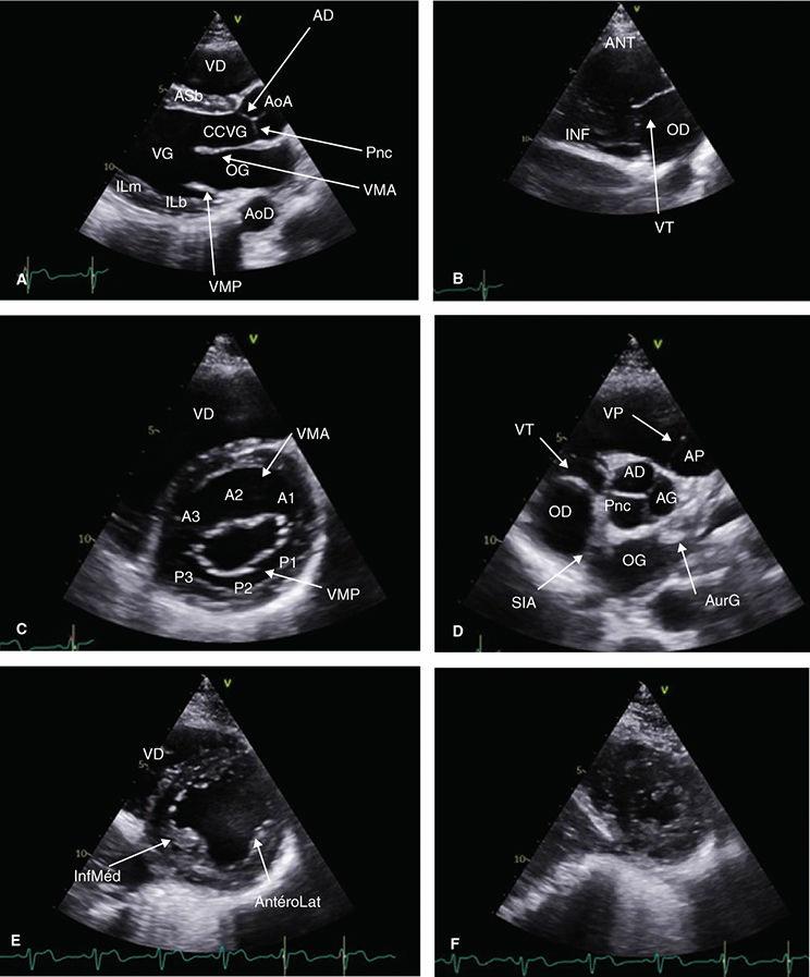
FIGURE11 Coupesparasternalesbidimensionnelles
Coupesgrandaxegauche(A),centréesurlescavtésdroites(B),petitaxecentréessuravavemtrale(C),avaveaortique(D),espiliersmitraux(E)etl'apex(F)AD:sigmoïdeantéro-droite;AG: sigmoïdeantéro-gauche;AoA:aorteascendante;AoD:aortedescendante;AP:artèrepumonaire;AS:antéroseptale;AurG:auricuegauche;b:basal;CCVG:chambredechasseventriculaire gauche;IL:inféroatéra;m:médian;OD:orellettedroite;OG:orellettegauche;Pnc:sigmoïdepostérieurenoncoronaire;SIA:septuminteratrial;VD:ventricuedroit;VG:ventriculegauche; VMA:valvemitraleantéreure(A1-A3);VMP:valvemitralepostérieure(P1-P3);VP:valvepumonaire;VT:vavetricuspde
▪ Imagelabaseducœur
▪ Sondedéchocardiographiepositionnéeleplushautpossible
▪ Curseurorientéversl'épauledroitedupatient
PSGAgauche:visualisationduVD,duSIV(paroiantéroseptale),cavitéventriculairegaucheetparoiinférolatéralebasaleetmédiane,ainsiquedelaorte ascendanteaveclavalveaortique
Enbecquantlasondeverslebas:coupe2cavitésVD:visualisationdesparoisantérieureetinférieureVDetdelavalvetricuspide(figure11B)
Coupeparasternalegauchepetitaxe
Rotationdelasondeà90°danslesenshoraireàpartird'unePSGAoptimale,sinonrisquedecoupetangentiellenonperpendiculaireaugrandaxeduVG(figure 12Aet12B),visualisationdelavalvemitraleantérieureetpostérieureenmuseaudetanche(figure11C)
▪ Enbecquantverslehaut,valveaortiqueavecses3feuillets(figure11D),signedela«Mercedes»,avecnotammentl'antérogauche,cravatéeparl'infundibulum aveclavalvepulmonaireetletroncdelartèrepulmonaire
▪ Enbecquantverslebas,leVGauniveaudespiliers(antéro-latéraletinféromédian)(figure11E)
Enbecquantencoreverslebasouendescendantdunespaceintercostal,segmentsapicauxVG(figure11F)
Another random document with no related content on Scribd:
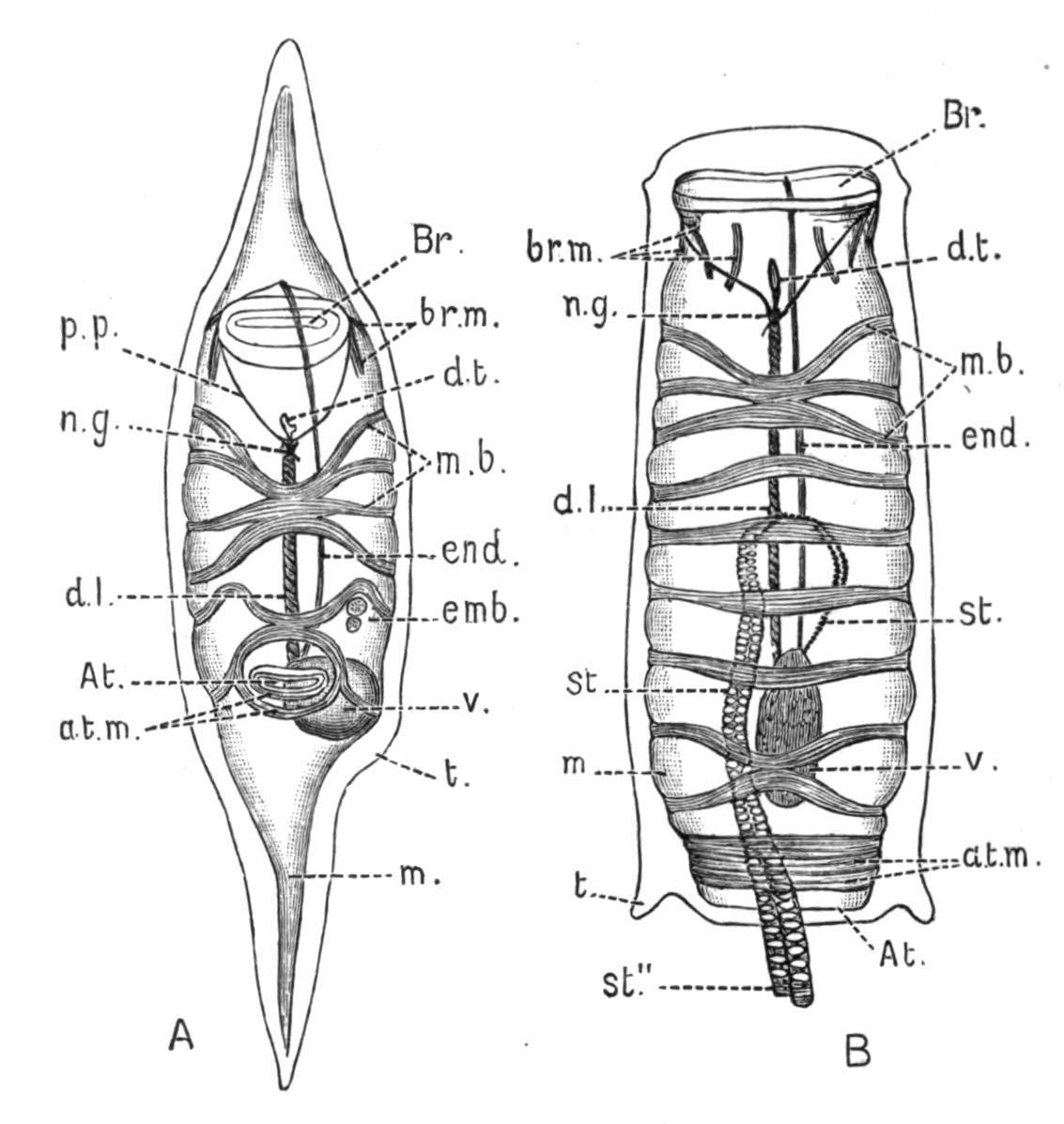
the ascidiozooids (Fig. 47, B) are provided with large incubatory pouches, opening from the peribranchial cavity, but also connected, as Bancroft[101] has recently shown, with the end of the oviduct (see Fig. 47, B). In these pouches the embryos undergo their development, and are set free by the decay of the top of the colony. The stolons pass from the ascidiozooids in the upper part of the colony down into the stalk, and there produce buds which gradually work up to the top of the stalk, where they take their places as young ascidiozooids. At the top of the colony the old ascidiozooids die and are removed (see Fig. 47, A).
Caullery has shown that in this genus there may be dimorphism in the buds, some of them placed deeply in the stalk having a large amount of reserve food-matter in their ectoderm, and remaining dormant until required to regenerate the "head" or upper part of the colony when it is lost. This genus was made known by the "Challenger" expedition. The species are mostly tropical, or from southern seas.
Fam. 2. Coelocormidae.—Colony not fixed, having a large axial cavity with a terminal aperture. Branchial apertures five-lobed. This includes one species, Coelocormus huxleyi, Herdman, which is in some respects a transition-form between the ordinary Compound Ascidians (e.g. Distomatidae) and the Ascidiae Luciae (Pyrosoma, see p. 90).
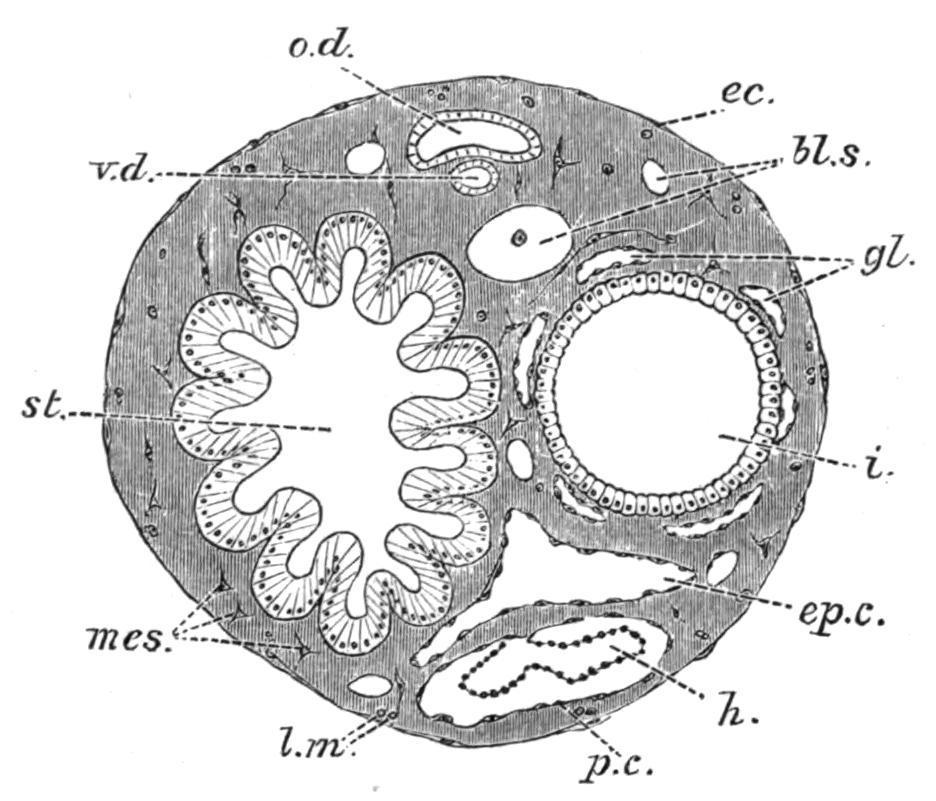
FIG. 48. Transverse section of the abdomen of a Distomid. bl.s, Bloodsinus; ec, ectoderm; ep.c, epicardium; gl, intestinal glands; h, heart; i, intestine; l.m, longitudinal muscles; mes, mesoderm; o.d, oviduct; p.c, pericardium; st, stomach; v.d, vas deferens. (After Seeliger.)
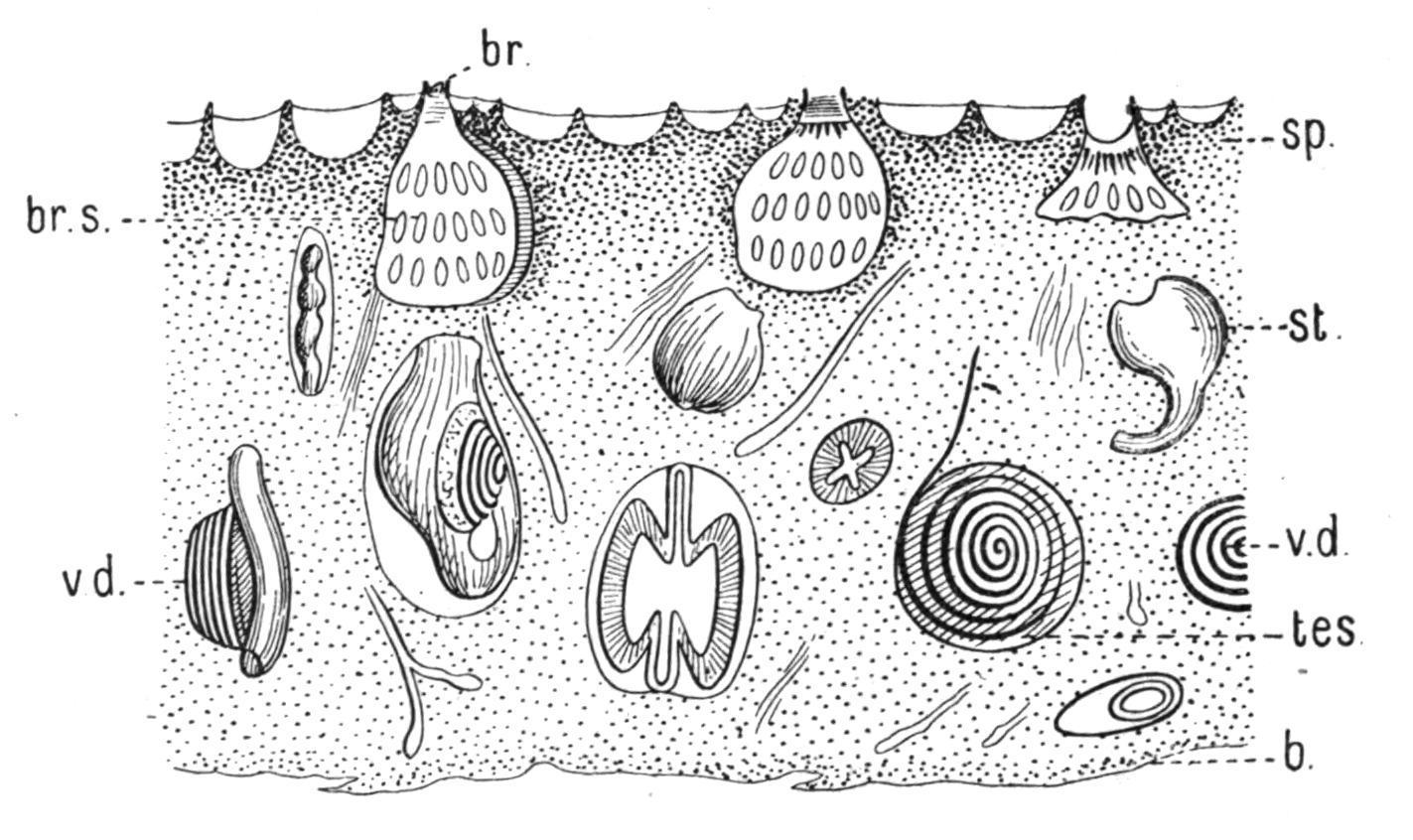
FIG. 49.—Section of Leptoclinum colony, showing the distribution of spicules and parts of the ascidiozooids. b, Base of colony; br, branchial aperture; br.s, branchial sac; sp, spicules; st, stomach; tes, testis; v.d, vas deferens.
Fam. 3. Didemnidae.—Colony usually thin and incrusting. Test containing stellate calcareous spicules (Figs. 49 and 50, B). Testis single, large; vas deferens spirally coiled (Fig. 49). The chief genera are— Didemnum, Savigny, in which the colony is thick and fleshy, and there are only three rows of stigmata on each side of the branchial sac; and Leptoclinum, Milne-Edwards, in which the colony is thin and incrusting (Fig. 49), and there are four rows of stigmata. Colonies of Leptoclinum, forming thin white, grey, or yellow crusts under stones at low water, are amongst the commonest of British Compound Ascidians.
FIG. 50.—Calcareous spicules of the Tunicata, enlarged. A, From Cystodytes; B, from Leptoclinum; C, from Culeolus; D, from Rhabdocynthia.
Fam. 4. Diplosomatidae.—Test reduced in amount (Fig. 51), rarely containing spicules. Vas deferens not spirally coiled. In Diplosoma, Macdonald, and other allied genera (Fig. 51), the larva is gemmiparous (Fig. 42, F). Some species are common British forms, especially on Zostera-beds and amongst seaweeds.
FIG. 51. Section of a colony of Diplosoma(enlarged) to show the small amount of test present. br, Branchial aperture; c.cl, common cloaca; t, test.
Fam. 5. Polyclinidae. Ascidiozooids divided into three regions—thorax, abdomen, and post-abdomen (Fig. 46, C). Testes numerous; vas deferens not spirally coiled. The chief genera are—Pharyngodictyon, Herdman, with stigmata absent or modified, containing one species, Ph. mirabile (Fig. 44, C), the only Compound Ascidian known from a depth of 1000 fathoms; Polyclinum, Savigny, with a smooth-walled stomach (Fig. 52, A); Aplidium, Savigny, with the stomach-wall longitudinally folded (Fig. 52, B); Morchellium, Giard, with an "areolated" stomach (Fig. 52, D), bearing knobs on the outside; and Amaroucium, Milne-Edwards, in which the ascidiozooid has a long post-abdomen and a large atrial languet, and where the stomach-wall shows longitudinal ridges breaking up into knobs (pseudo-areolated, Fig. 52, C). The last four genera contain many common British species.
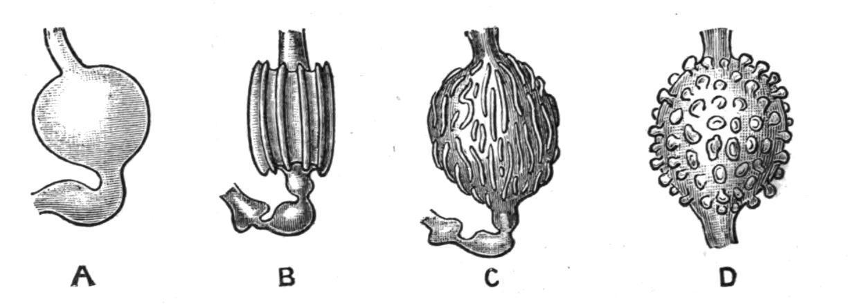
FIG. 52. Various conditions of stomach in Polyclinidae. A, Polyclinum molle, Herdman; B, Aplidium zostericola, Giard; C, Amaroucium proliferum, M.-Edw.; D, Morchelliumargus, M.-Edw.
Many of the Compound Ascidians die down in winter; but amongst Polyclinidae, as in Clavelina, a form of hibernation is found, the old ascidiozooids dying, but some of the buds in the basal part of the colony accumulating a large store of reserve-material in their ectoderm, and lying dormant until spring, when they regenerate the colony.
GROUP B. HOLOSOMATA.
Body short, compact, with viscera by the side of branchial sac; budding parietal
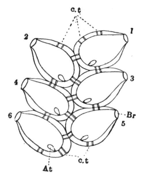
Fam. 6. Botryllidae. Ascidiozooids grouped in systems round common cloacal apertures (Fig. 53). Ascidiozooids having the intestine and reproductive organs by the side of the branchial sac (Fig. 46, A, p. 82). Dorsal lamina and internal longitudinal bars present in the branchial sac. Neural gland, as in Cynthiidae, dorsal to the ganglion in place of ventral as in the majority of Tunicata. The chief genera are—Botryllus, Gaertn. and Pall, with simple stellate systems (Fig. 53), and Botrylloides, MilneEdwards, with elongated or ramified systems. There are many species of both these genera, which form brilliantly coloured fleshy crusts under stones and on sea-weeds at low tide. They are amongst the commonest and the most beautiful of British Ascidians. Both genera contain species remarkable for the rich profusion of ectodermal "vessels" which ramify and anastomose in the colonial test. On the margins of the colony these vessels end in knob-like dilatations, the ampullae (Fig. 46, A, t.k), which are said by Bancroft to pulsate rhythmically, and so aid in keeping up the colonial circulation. They are also storage reservoirs for the blood, doubtless help in respiration, and are organs for the secretion of the testmatrix.
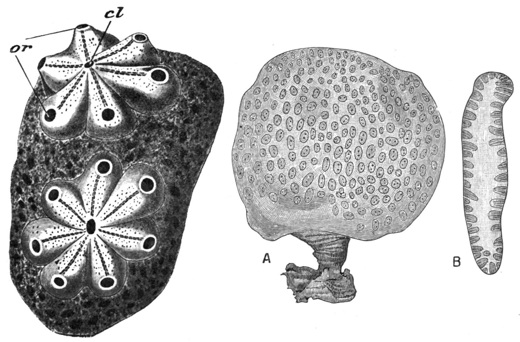
FIG. 53. Two "systems" from a colony of Botryllus violaceus, M.-Edw. cl, Common cloaca of a system; or, branchial apertures of ascidiozooids, magnified. (After H. Milne-Edwards.)
FIG. 54.—Goodsiriaplacenta, Herdman. A, Colony (half nat. size); B, section of colony showing ascidiozooids. (After Herdman, from Challenger Reports.)
Fam. 7. Polystyelidae.—Ascidiozooids not grouped in systems; branchial and atrial apertures four-lobed; branchial sac may be folded; internal longitudinal bars present. The chief genera are—Thylacium, Carus, with the ascidiozooids projecting above the general surface of the
colony; Goodsiria, Cunningham, with the ascidiozooids completely imbedded in the investing mass (Fig. 54); and Chorizocormus, Herdman, with the ascidiozooids united in little groups which are connected by stolons. The last genus contains one species, Ch. reticulatus, in some respects a transition-form between the other Polystyelidae and the Styelinae amongst Simple Ascidians.
Budding in Holosomata—In the Polystyelidae, according to Ritter,[102] the budding is of the same type as in Botryllidae, the bud arising in each case from the lateral body-wall of the parent.
In Botryllus[103] the oozooid formed from the larva gives rise at a very early period to the first blastozooid of the future colony. This then forms the two buds of the second generation on its sides (see Fig. 55), and these in their turn form the third, and these the fourth generation, in which there are thus eight blastozooids; and so the process goes on, the buds of each generation arranging themselves in a circle to form a system. As each new generation makes its appearance, the preceding one undergoes degeneration, and is eventually absorbed. Consequently, in a system there can usually be seen, in addition to the adult members, certain older ones in various stages of degeneration and removal, and certain younger ones arising as buds on the sides of their predecessors, or just separated from them, and ready to take their places as young ascidiozooids in the system. Three distinct generations are thus commonly seen in a system. Now and again one or two young ascidiozooids become squeezed by the pressure of their neighbours out of a system into the surrounding test, and so give rise to new systems which add to the extent of the colony.
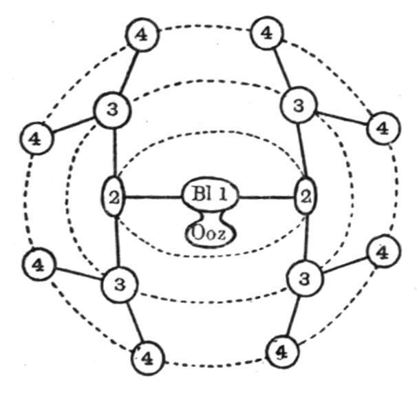
FIG. 55. Diagram to illustrate the budding and formation of a system in Botryllus. Ooz; oozooid; Bl1, first blastozooid; 2, 2, etc., successive generations of buds.
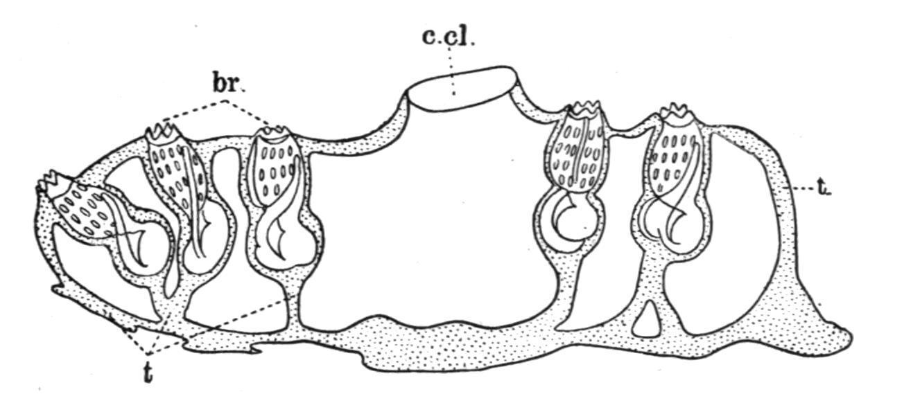
Sub-Order 3. Ascidiae Luciae.
Free-swimming pelagic colonies having the form of a hollow cylinder closed at one end (Fig. 56). The ascidiozooids forming the colony are imbedded in the common test in such a manner that the branchial apertures open on the outer surface and the atrial apertures on the inner surface next to the central cavity of the colony. They are placed with their ventral surfaces towards the closed end (Fig. 56, C). The first ascidiozooids of a colony are produced by gemmation from a stolonic prolongation of an imperfect oozooid or rudimentary larva (the "cyathozooid"), developed sexually. The subsequent ascidiozooids are formed from these as buds on a ventral stolon.
This sub-order includes a single family, the PYROSOMATIDAE, containing one well-marked genus Pyrosoma, Péron, with about six species. They are found swimming near the surface of the sea, chiefly in tropical latitudes, and are brilliantly phosphorescent. A fully developed Pyrosoma colony may be from an inch or two to upwards of twelve feet in length.
FIG. 56.—Pyrosoma. A, lateral view (nat. size); B, end view; C, diagram of longitudinal section. at, Atrial apertures; br, branchial apertures; c.cl, common cloaca; end, endostyle; t, test; v, velum or diaphragm at terminal opening.
The Colony. The shape of the colony is seen in Fig. 56, A. It tapers slightly towards the closed end, which is rounded. The opening at the opposite end may be reduced in size (see B and C), by the presence of a membranous prolongation of the common test, which can be contracted or expanded by means of the muscle-bands it receives from the atrial siphons of neighbouring zooids. The branchial apertures of the ascidiozooids are mostly placed upon short (in some cases longer)
papillae projecting from the general surface, and many of the ascidiozooids have long conical processes of the test extending outwards beyond their branchial apertures (Fig. 57, t′). There is only a single layer of adult ascidiozooids in the wall of the Pyrosoma colony, as all the fully developed ascidiozooids are placed with their antero-posterior axes at right angles to the surface and communicate by their atrial apertures with the central cavity (Fig. 56, C). Their dorsal surfaces are turned towards the open end of the colony, and the buds are given off from their ventral edges (Fig. 57).
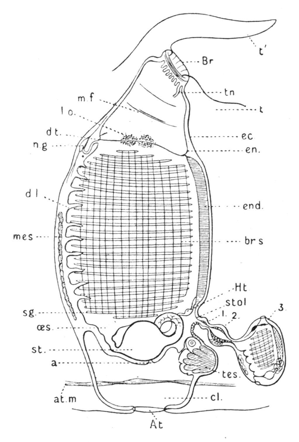
FIG. 57.—Ascidiozooid of Pyrosoma from the right side. a, Anus; At, atrial aperture; at.m, atrial muscles; Br, branchial aperture; br.s, branchial sac; cl, cloaca; d.l, dorsal lamina; d.t, dorsal tubercle; ec, ectoderm; en, endoderm; end, endostyle; Ht, heart; l.o, luminous organ; mes, mass of mesoderm cells; m.f, muscle fibre; n.g, nerve-ganglion; oes, oesophagus; sg, stigmata; st, stomach; stol, stolon; t, test; t′, projection of test near branchial aperture; tes, testis; tn, tentacle; 1, 2, 3, buds.
Anatomy. The more important points in the structure of the ascidiozooid of Pyrosoma are shown in Fig. 57. A circle of tentacles, of which one, placed ventrally (tn), is larger than the rest, is found just inside the circular branchial aperture. From this point a wide cavity, with a few circularly placed muscle-bands running round its walls, leads back to the large branchial sac (br.s.), which occupies the greater part of the body. The large stigmata are elongated transversely (dorso-ventrally), and are crossed by internal longitudinal bars running antero-posteriorly. The dorsal lamina is represented by a series of eight or ten languets. The
nerve-ganglion (on which is placed a small pigmented sense-organ, the unpaired "eye"), the neural gland, the dorsal tubercle, the peripharyngeal bands and the endostyle are placed in the usual positions. On each side of the anterior end of the branchial sac, close to the peripharyngeal bands is a mass of rounded mesodermal gland-cells (l.o), which are the source of the phosphorescence. They are apparently modified leucocytes lying in blood-sinuses. The alimentary canal is placed posteriorly to the branchial sac, and the anus opens into a large peribranchial or atrial cavity, of which only the median posterior part (cl), is shown in Fig. 57.
The heart (Ht) lies between the posterior end of the branchial sac and the intestine, close to where the endostyle is prolonged outwards to form the inner tube of the ventral stolon. The reproductive organs are developed from a cord of germinal tissue which forms a part of every budding stolon, and so establishes a continuity of origin between the ova of successive generations of Pyrosoma. On the ventral edge of the body, immediately behind the stolon, with part of which it is continuous, a portion of this germinal tissue gives rise to a lobed testis (tes), and to a single ovum surrounded by indifferent or follicle-cells.
Development and Life-History.—The development takes place within the body of the parent, in a part of the peribranchial cavity. It is a "direct" development, the tailed larval stage being omitted. The segmentation is incomplete or "meroblastic," and an elongated embryo is formed on the surface of a mass of food-yolk. Follicle-cells, or kalymmocytes, migrate into the embryo, where they aid in its nutrition. The embryo (or young oozooid),[104] after the formation of an alimentary cavity, a tubular nervous system, and a pair of laterally placed atrial tubes, divides into an anterior and a posterior part (see Fig. 58). The anterior and ventral part, or stolon, then segments into four pieces (the tetrazooids or first blastozooids),[104] which afterwards develop into the first ascidiozooids of the colony, while the posterior part remains in a rudimentary condition, and is what was called by Huxley the "cyathozooid" (Fig. 58, cy). This is really the degenerate oozooid, and eventually atrophies without having completed its development, but having precociously given rise to the budding stolon.
As the four ascidiozooids increase in size, they grow round the cyathozooid and soon encircle it (Fig. 58, B). In this condition the young colony leaves the body of the parent and becomes free. The cyathozooid absorbs the nourishing yolk upon which it lies, and distributes it to the ascidiozooids by means of a heart and system of vessels which have been meanwhile formed. When the cyathozooid atrophies and is absorbed, its original atrial aperture remains and deepens to become the central cavity[105] of the young colony, which now consists of four ascidiozooids placed in a ring, around where the cyathozooid was, and enveloped in a common test. The test is at first formed by the ectoderm cells of the cyathozooid. Later it becomes invaded by mesoblast cells from the ascidiozooids in the usual manner. The colony gradually increases by the formation of buds from these four original ascidiozooids. The young colony is, in some species, at first male, and only becomes hermaphrodite when it has attained to some size.
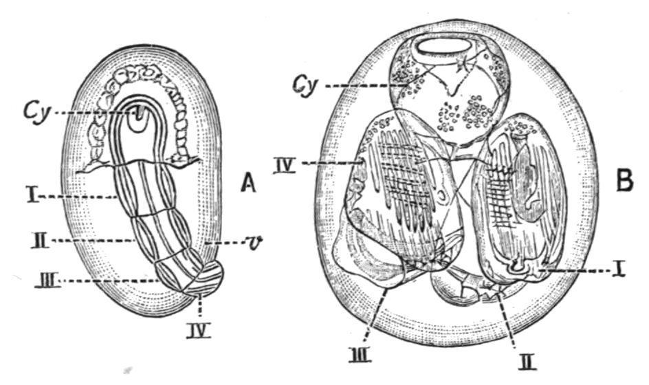
FIG. 58. Development of Pyrosomacolony. A, young stage showing oozooid or cyathozooid, cy, with stolon divided into four blastozooids (I.-IV.): v, vitellus. B, older stage showing the four blastozooids in a ring around the remains of the cyathozooid. (After Salensky.)
Occurrence. The half-dozen known species of Pyrosoma are widely distributed over the great oceans, although they are probably most abundant in tropical waters. Pyrosoma atlanticum, Péron, and P . giganteum, Lesueur, are the commonest forms. Although sometimes abundant in the Mediterranean and the North Atlantic they have apparently not been found in British seas. P . elegans, Lesueur, is a Mediterranean form allied to the last two; and P . minatum and P . aherniosum, Seeliger, were discovered during the German "Plankton" expedition in the tropical Atlantic. Finally, the enormous P . spinosum, Herdman, was found by the "Challenger" in both North and South Atlantic in 1873; and some years later (Perrier's P . excelsior) by the French "Talisman" expedition in the tropical Atlantic. The late Professor Moseley said of this ("Challenger") species, "I wrote my name with my finger on
the surface of the giant Pyrosomaas it lay on deck in a tub at night, and my name came out in a few seconds in letters of fire." Bonnier and Pérez have recently recorded that they saw an enormous profusion of a large Pyrosoma (up to four metres in length) in the Arabian part of the Indian Ocean.
Order III. Thaliacea (Salpians).
Free-swimming pelagic forms of moderate size, which may be either simple or compound, and in which the adult is never provided with a tail or notochord. Consequently the whole body here corresponds to the trunk only of the Appendicularian without the tail. The test is permanent, and may be either well developed or very slight. In all cases it is clear and transparent. The musculature of the body-wall is in the form of more or less complete circular bands, by the contraction of which water is ejected from the body, and so locomotion is effected. The branchial sac has either two large, or many small, stigmata, leading to a single peribranchial cavity, into which the anus also opens. Blastogenesis takes place from a ventral, endostylar stolon. Alternation of generations occurs in the life-history, and may be complicated by polymorphism. The Order Thaliacea comprises two groups, CYCLOMYARIA (such as Doliolum) and HEMIMYARIA (such as Salpa).
Sub-Order 1. Cyclomyaria.
Free-swimming pelagic forms which exhibit alternation of generations in their life-history, but never form permanent colonies. The body is caskshaped, with the branchial and atrial apertures at the opposite ends. The test is moderately well developed, never much thickened. The musculature is mostly in the form of complete circular bands surrounding the body. The branchial sac is fairly large, occupying the anterior half or more of the body. Stigmata are usually present in its posterior part only. The peribranchial cavity is mainly posterior to the branchial sac. The alimentary canal is placed ventrally, close to the posterior end of the
branchial sac. Hermaphrodite reproductive organs lie ventrally near the intestine.
This group is clearly distinguished from the second sub-order, the Hemimyaria, by the condition of the muscle-bands and of the branchial sac, and by the life-history. The muscle-bands are complete rings (except in Anchinia), while in the Hemimyaria they are always more or less incomplete. The branchial sac in the Cyclomyaria is a distinct cavity, and communicates with the peribranchial cavity only by small slits or stigmata. The life-history is also very characteristic, as the sexual generation in the Cyclomyaria is always polymorphic, while in the Hemimyaria it consists of one form only.
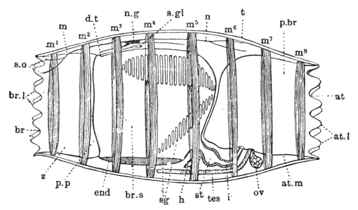
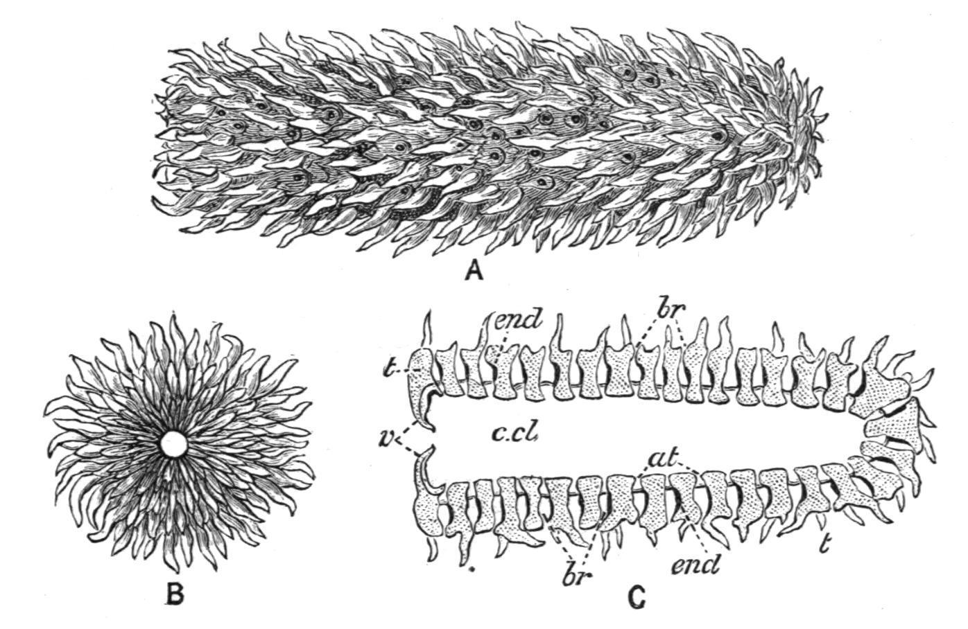
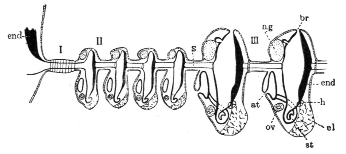
FIG. 59. Sexual generation of Doliolum tritonis, Herdman, from left side, × 10. at, Atrial aperture; at.l, atrial lobes; at.m, wall of atrium; br, branchial aperture; br.l, branchial lobes; br.s, branchial sac; d.t, dorsal tubercle; end, endostyle; h, heart; i, intestine; m, mantle; m1-m8 , circular muscle-bands; n, nerve; n.g, nerve-ganglion; ov, ovary; p.br, peribranchial cavity; p.p, peripharyngeal bands; sg, stigmata; s.gl, neural gland; s.o, sense-organ; st, stomach; t, test; tes, testis; z, prebranchial zone. (After Herdman.)
Structure of Doliolum. The single family DOLIOLIDAE includes three genera, Doliolum, Quoy and Gaimard, Dolchinia, Korotneff, and Anchinia, Eschscholtz. Doliolum, of which about a dozen species are known, from various seas, has a cask-shaped body (Fig. 59), usually from 1 to 2 cm. in length. The terminal branchial and atrial apertures are lobed, and the lobes are provided with sense-organs. The test is a thin but tough transparent layer, and contains no "test" cells. It is merely a cuticle covering the surface of the squamous ectoderm. The body-wall has eight or nine circular muscle-bands surrounding the body. The most anterior and posterior of these form the branchial and atrial sphincters. The wide branchial and atrial apertures lead respectively into branchial and
peribranchial cavities separated by the posterior and postero-lateral walls of the branchial sac which are pierced by a considerable number of small stigmata; consequently there is a free passage for the water through the body along its long axis, and the animal swims by contracting its ring-like muscle-bands so as to force out the contained water posteriorly. When stigmata are found on the lateral walls of the branchial sac (see Fig. 59) there are corresponding anteriorly directed diverticula of the peribranchial cavity. There is a distinct endostyle on the ventral edge of the branchial sac and a peripharyngeal band surrounding its anterior end, but there is no representative of the dorsal lamina along its dorsal edge; and there are neither branchial nor atrial tentacles. The oesophagus commences rather on the ventral edge of the posterior end of the branchial sac, and runs backwards to open into the stomach, which is followed by a curved intestine opening into the peribranchial cavity. The alimentary canal as a whole is to the right of the middle line. The hermaphrodite reproductive organs are to the left of the middle line alongside the alimentary canal. They open into the peribranchial cavity. The ovary is nearly spherical, while the testis is elongated, and may be continued anteriorly for a long distance. The heart is placed in the middle line ventrally, between the posterior end of the endostyle and the oesophageal aperture. The nerveganglion lies about the middle of the dorsal edge of the body, and gives off many nerves. Under it is placed the neural gland, the duct of which runs forward and opens into the anterior end of the branchial sac by a simple aperture surrounded by the spirally twisted dorsal ends of the peripharyngeal bands.
Life-History. The ova produced by the Doliolum of the sexual generation, after a complete or "holoblastic" segmentation, and normal invagination, produce tailed larvae with a relatively small caudal appendage, and a large body in which the characteristic musculature begins to appear (Fig. 60, A). These larvae after metamorphosis lose their tails and develop into oozooids, known as "nurses," which are asexual, and are characterised (Fig. 60, B) by the possession of nine musclebands, by the stigmata being few in number and confined to the posterior end of the branchial sac, by an otocyst on the left side of the body, by a ventrally-placed complex stolon or "rosette organ" near the heart, from which primary buds are produced by constriction, and by a dorsal outgrowth ("the cadophore") near the posterior end of the body. The
buds (blastozooids) give rise eventually, after further division, to the sexual generation, which is polymorphic—having three distinct forms, in two of which the reproductive organs remain undeveloped.
FIG. 60. Life-history of Doliolum. A, tailed larval stage; B, "nurse" or oozooid, showing buds (blastozooids) migrating from the ventral stolon to the dorsal process; C, posterior part of much later oozooid to show buds arranged in three rows on dorsal process; D, stolon segmenting; E, young migrating bud; F, trophozooid developed from one of the buds of a lateral row. At, Atrial aperture; b, buds; Br, branchial aperture; cl, cloaca; d.p, dorsal process; end, endostyle; ht, heart; l.b, lateral buds; m.b, median buds; n.g, nerve-ganglion; ot, otocyst; p.c, pericardium; sk, stalk; sto, stolon. (After Uljanin and Barrois.)
The primary buds are constricted off while still very young and undeveloped (Fig. 60, D, B, and E); they migrate from their place of origin on the stolon, over the surface (aided by large amoeboid test-cells which become attached to the buds) (Fig. 60, B), multiply by fission, and become attached (again by the help of amoeboid test-cells and ectoderm cells which form a slight "placenta") in three rows—a median and two lateral—to the dorsal outgrowth (Fig. 60, C) of the body of the nurse. This parent-form by this time has become greatly modified, and its structure is largely sacrificed for the good of the buds or growing zooids, for which it really forms a locomotory organ. Its muscle-bands become greatly developed in width (Fig. 60, C), and the branchial meshwork, endostyle, and alimentary canal disappear.
The three forms produced in the second generation are as follows:—(1) Nutritive forms ("trophozooids") derived from the lateral rows of buds, which remain permanently attached to the oozooid, and are sacrificed for the benefit of the rest of the colony. They serve merely to aid in respiration, and to provide the food for the nurse and the median buds.
Their development is arrested; they have the body elongated dorsoventrally with a large funnel-like branchial aperture (Fig. 60, F), and the musculature is very slightly developed.
(2) Some of the median buds become foster forms ("phorozooids"), which, like the preceding trophozooids, do not become sexually mature, but, unlike them, are eventually set free as cask-shaped bodies having the Doliolumappearance, with eight encircling muscle-bands, and having, moreover, a ventral outgrowth (not a stolon), which is formed of the stalk by which the body was formerly attached to the dorsal process of the oozooid. On this ventral outgrowth the "gonozooids" (3) are attached while still very young buds, and after the phorozooids are set free these reproductive forms gradually attain their complete development, become sexually mature, and are eventually separated off, finally losing all trace of their temporary connexion with the foster-forms. They resemble the foster-forms in having a cask-shaped body with eight muscle-bands, but differ in the absence of a ventral process, and in having the sexual reproductive organs fully developed.
Occurrence.—The best-known member of the genus is Doliolumtritonis, Herdman, which was captured in the tow-nets in thousands by Sir John Murray during the cruise of H.M.S. "Triton" in the summer of 1882 in the North Atlantic. Since then that species, or the closely allied D. nationalis, Borgert, have been found on more than one occasion in the English Channel and other parts of our south-west coast, and so Doliolummay be regarded as an occasional member of the British surface fauna.
It is probable that the occasional phenomenal swarms of Doliolumwhich have been met with in summer in the North Atlantic are a result of the curious life-history which, under favourable circumstances, allows of a small number of oozooids producing from minute buds an enormous number of phorozooids and gonozooids.
As the result of the careful quantitative work of the German "Plankton" expedition, Borgert thinks that the temperature of the water has more to do with both the horizontal and the vertical distribution of these Thaliacea in the sea than any other factor.
Other Genera. Anchinia, of which only one species is known, A.rubra, Vogt, from the Mediterranean, has the sexual forms permanently attached to portions of the dorsal outgrowth from the body of the unknown oozooid ("nurse"). The stolon is probably much longer than in Doliolum, and curves round so as to reach and lie along the dorsal outgrowth, upon which it places the buds.
The body of the adult is elongated dorso-ventrally. The test is well developed and contains branched cells. The musculature is not so well developed as in Doliolum. There are two circular bands at the anterior end, two at the posterior, and two muscles on the middle of the body, which unite to form the characteristic S-shaped lateral bands. The stigmata are confined to the obliquely-placed posterior end of the branchial sac. The alimentary canal forms a U-shaped curve. The reproductive organs are placed on the right side of the body. The lifehistory is still imperfectly known. As in the case of Doliolum the sexual generation is polymorphic, and has three forms, two of which remain in a rudimentary condition so far as the reproductive organs are concerned. They are known as the first and second sterile forms, or "trophozooids." In Anchinia, however, the three forms do not occur, so far as we know, together at the same time on the one outgrowth, but are produced successively, or in different regions, the reproductive forms of the sexual generation being independent of the "foster-forms."[106]
The third genus, Dolchinia, contains also only a single species, D. mirabilis, found by Korotneff[107] in the Gulf of Naples. It must have three different forms in its life-history—oozooid, phorozooid, and gonozooid, but the first of these is still unknown. On what must be body processes detached from the oozooid are found phorozooids somewhat like those of Doliolum, bearing sexual forms attached to ventral stalks. Dolchinia is intermediate on the whole between Anchinia, the most simple member of the family, and Doliolumthe most complex; and may eventually come to be united with the latter genus. Sub-Order 2. Hemimyaria.
Free-swimming pelagic forms which exhibit alternation of generations in their life-history, and in the sexual condition form colonies. The body is more or less fusiform, with the long axis antero-posterior, and the branchial and atrial apertures nearly terminal and opposite. The test is well developed but transparent. The musculature of the body-wall is in the form of a series of transversely-running bands which do not usually form complete independent rings as in the CYCLOMYARIA. These partiallyencircling muscles in the Salpidae (see Fig. 61, m.b) are probably to be regarded as modified branchial and atrial sphincters which have spread over the intervening body. The branchial and peribranchial (cloacal) cavities form a continuous space in the interior of the body, opening externally at the ends by the branchial and atrial apertures, and traversed obliquely from the dorsal and anterior to the ventral and posterior end by a long narrow vascular ciliated band, which represents the dorsal lamina, the dorsal blood-sinus, and the neighbouring parts of the dorsal edge of the branchial sac of an ordinary Ascidian. The alimentary canal is placed ventrally. It may either be stretched out so as to extend for some distance anteriorly, or, as is more usual, be concentrated to form along with the testis a rounded opaque mass near the posterior end of the body, known as the visceral mass or "nucleus." The embryonic development is direct, no tailed larva being formed. The embryo is united to the parent for a time by a "placenta."
This sub-order contains, in addition to its typical members, the SALPIDAE, another still somewhat problematical family the OCTACNEMIDAE, including a single very remarkable deep-water genus (Octacnemus), which in some respects does not conform with the characters given above, and exhibits a certain amount of affinity with the primitive fixed forms from which Salpidae have been derived.
FIG. 61. Salpa runcinata-fusiformis. A, aggregated or "chain" form; B, solitary form. At, Atrial aperture; at.m, atrial muscles; Br, branchial aperture; br.m, branchial muscles; d.l, dorsal lamina or "gill"; d.t, dorsal tubercle; emb, embryo; end, endostyle; m, mantle; m.b, muscle-bands; n.g, nerve-ganglion; p.p, peripharyngeal bands; st, stolon; st″, "chain" of buds; t, test; v, visceral "nucleus."
FIG. 62. Diagram to show the arrangement and connexion of the aggregated zooids in a young chain of Salps. 1, 3, 5, zooids on the right; 2, 4, 6, zooids on the left. At, Atrial aperture of a zooid; Br, branchial aperture of another; c.t at the top of the figure points to three pairs of connecting tubes; c.t at the foot, to two pairs. Each zooid is united to each of the four neighbours it touches by a pair of connecting tubes, and so has eight such tubes in all.
Occurrence and Reproduction.
The family SALPIDAE[108] includes the single genus Salpa, Forskål, which, however, may be divided into two well-marked groups of species—(1) those such as S. (Cyclosalpa) pinnata, in which the alimentary canal is stretched out ("ortho-enteric" condition) along the ventral surface of the body, and (2) those such as S. runcinatafusiformis, in which the alimentary canal forms a compact globular mass (Fig. 61, v), the "nucleus" ("caryo-enteric" condition), near the posterior end of the body. About fifteen species altogether are known; they are all pelagic in habit, and are found in nearly all seas. Each species occurs in two forms (Fig. 61, A and B), the solitary asexual (proles solitaria), and the aggregated sexual (prolesgregaria), which are in most species quite
unlike one another, the aggregated form being usually more rounded, ovoid, or fusiform (Fig. 61, A), and the solitary more quadrangular, and often provided with conical processes or projecting points.
FIG. 63.—Diagram to show the relations of the groups of young buds, when first formed on the stolon of Salpa. at, Atrial aperture; br, branchial aperture; el, elaeoblast; end, endostyle; h, heart; n.g, nerve-ganglion; ov, ovum; s, stolon; st, stomach; I, II, III, groups of buds. (After Brooks.)
FIG. 64. Transverse section through endostyle and young stolon of Salpa pinnata. ec, Ectoderm of parent reflected at ec′ to cover base of stolon; ec″, ectoderm of stolon; end, endoderm of stolon; g, ovary; mes, mesoderm cells; n, nerve-tube of stolon; p.br, peribranchial tubes of stolon. (After Brooks.)
The solitary form gives rise, by gemmation at the posterior end of the endostyle (Fig. 63), to a complex tubular stolon, containing processes from the more important organs of the parent-body, which give rise to an endodermal tube, two peribranchial tubes, a neural tube, two bloodsinuses and mesoblast cells, a genital cord, and over all the ectodermal covering (see Fig. 64). This stolon becomes segmented (Fig. 63) into a series of buds or young "chain" individuals, of which there may be several hundreds. As the stolon elongates (Fig. 61, B, st″), the buds undergo lateral shifting, and rotation round their longitudinal axis, so as to acquire the relations seen in the "chain," which then emerges from the tube in the test through which it has been growing, so as to project to the
exterior near the atrial aperture. The buds at its free end which have now become far advanced in their development are set free in groups, which remain attached together by processes of the test, each enclosing a diverticulum from the body-wall (Fig. 62), so as to form "chains." Each member of the chain is a Salpa of the sexual or aggregated form, and when mature may—either still attached to its neighbours or separated from them—produce one or several embryos (Fig. 61, A, emb), which develop into the solitary form of Salpa. Thus the two forms, different in appearance and structure and different in mode of origin, alternate regularly in the life-history of Salpa.
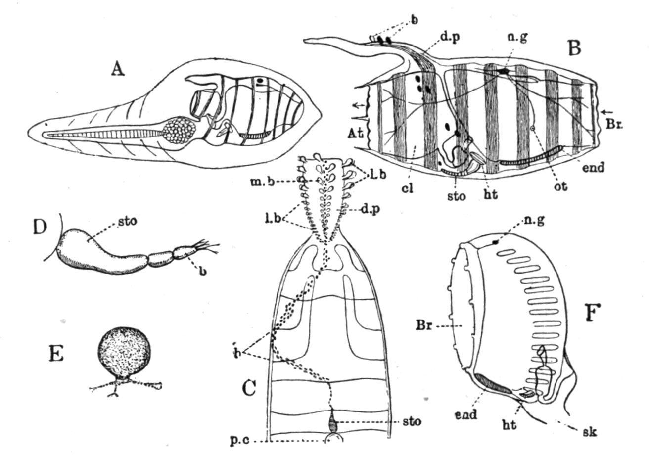
Structure.—The more important points in the structure of a typical Salpa are shown in Fig. 65. The branchial and atrial apertures are at opposite ends of the body, and lead into large cavities, the branchial and peribranchial sac respectively, which are in free communication at the sides of the obliquely-running dorsal lamina or "gill" (d.l). The transparent test is usually thick, and varies from a gelatinous to a stiff cartilaginous condition; it adheres closely to the surface of the mantle (ectoderm and body-wall). The muscle-bands (from 4 to about 20—usually 8 or 10) of the mantle do not in most cases completely encircle the body. They are present dorsally (Fig. 65, mus.bds) and laterally, but the majority do not reach the ventral surface. In many cases neighbouring bands join in the median dorsal line (Fig. 61). The muscle fibres are striated, and have rows of large equidistant nuclei. The anterior end of the dorsal lamina is in some cases prolonged to form a prominent tentacular organ, the languet or dorsal tentacle, projecting into the branchial sac, while near this opens a ciliated funnel corresponding to the dorsal tubercle, but having no connexion in the adult with either ganglion or subneural gland. The conjoined ganglion and subneural gland, the dorsal lamina, the peripharyngeal bands and the endostyle are placed in the usual positions. Eyes in the form either of a continuous horse-shoe-shaped pigmented ridge on the dorsal surface of the ganglion immediately below the ectoderm, or of one larger median and several smaller lateral ocelli are found in the various species of Salpa. These eyes have in most cases a retina formed of elongated cells, and a pigment-layer placed upon the ganglion.
The so-called otocysts of Salpahave been shown by Metcalf to be really glandular organs. They have been called lateral neural glands; they do not open at the dorsal tubercle, but separately into the pharynx. These lateral neural tubular glands have also been regarded as nephridia.
The large spaces at the sides of the dorsal lamina (often called the gill or branchia of Salpa), by means of which the cavity of the branchial sac is placed in free communication with the peribranchial cavity, are to be regarded as gigantic gill-slits formed by the suppression of the lateral walls and small stigmata of the branchial sac. The alimentary canal at the posterior end of the "gill" consists of oesophagus, stomach, and intestine, with a pair of lateral gastric glands or caeca. These viscera along with the reproductive organs, when present, make up the "nucleus" (Fig. 66, v).

FIG. 65. Diagrammatic sagittal section of a "chain" Salpa. an, Anus; at, atrial aperture; at.m, muscles of atrial aperture; atr.cav, atrial cavity; br, branchial aperture; br.m, muscles of branchial aperture; br.s, branchial sac; d.l, dorsal lamina or "gill"; d.t, dorsal tubercle; end, endostyle; ht, heart; int, intestine; l, sensory languet; mus.bds, muscle-bands; n.g, nerve-ganglion; oc, eye-spot; oe, oesophagus; ov, ovary; p.p.b, peripharyngeal band; s.gl, neural gland; stom, stomach; t, t′, test; tes, testis; z, prebranchial zone. (After Herdman.)
Alternation of Generations.—Fig. 66 represents an aggregated or sexual Salpa, which was once a member of a chain, since it shows a testis and a developing embryo. The ova (always few in number, usually only one) appear at a very early period in the developing chain Salpa, while it is still a part of the gemmiparous stolon in the body of the solitary Salpa. This gave rise to the view put forward first by Brooks that the ovary really belongs to the solitary stolon-bearing Salpa, which is therefore a female producing a series of males by asexual gemmation, and depositing in each of these an ovum, which will afterwards, when fertilised, develop in the body of the male into a solitary or female Salpa.
This idea, if adopted, would profoundly modify our conception of Salpaas an example of a life-history showing alternation of generations, but it seems to me to give a distorted view of the sequence of events. The fact that the stolon while in the solitary Salpa contains, along with representatives of other important systems of the body, a row of germinal cells, does not constitute that solitary Salpathe parent of the ova which these germinal cells will afterwards become in the body of an independent bud. We must regard as the parent the body in which the ova become mature and fulfil their function. The sexual or chain Salpa, although really hermaphrodite in its life-history, is usually[109] protogynous, i.e.the ova mature at an earlier period than the male organ or testis. This prevents self-fertilisation. The ovum is presumably fertilised by the spermatozoa of an older Salpabelonging to another chain, and the embryo is far advanced in its development before the testis is formed. The development takes place inside the body of the parent, and is "direct"—no tailed larval form being produced.
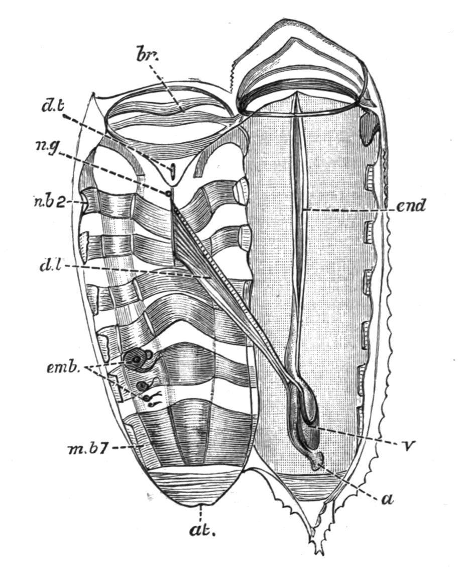
FIG. 66. Salpahexagona, Q. and G. Chain form dissected from the left side. a, Anus; at, atrial aperture; br, branchial aperture; d.l, dorsal lamina ("gill"); d.t, dorsal tubercle; emb, embryos; end, endostyle; m.b2, m.b 7, second and seventh muscle-bands; n.g, nerve-ganglion; v, visceral "nucleus." (After Traustedt.)
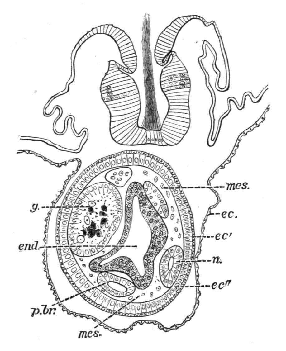
Development and Life-history.—The segmentation of the egg is holoblastic, and gives rise to a number of blastomeres, which are for a time masked by the phenomenal activity of certain cells of extraneous origin, the "kalymmocytes," derived from the follicular epithelium surrounding the ovum. These follicular kalymmocytes migrate into the ovum, surround groups of blastomeres, and arrange themselves so as to reproduce the essential structure of the future embryo for which they
form what may be termed a scaffolding or temporary support. After a time the blastomeres become active, proliferate rapidly, and finally press upon and absorb the kalymmocytes, and so eventually take their proper place in building up the organs. Some observers regard the kalymmocytes as being passive and nutritive only in function.
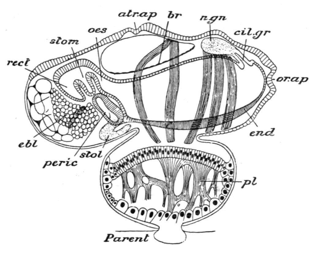
FIG. 67. Young solitary Salpa democratica-mucronata attached to the parent by the placenta. atr.ap, Atrial aperture; br, dorsal lamina; cil.gr, dorsal tubercle; ebl, elaeoblast; end, endostyle; n.gn, nerve-ganglion; oes, oesophagus; or.ap, branchial aperture; peric, pericardium; pl, placenta; rect, intestine; stol, stolon; stom, stomach. (From Parker and Haswell, after Salensky.)
At an early period in the development a part of the surface of the embryo, on its ventral edge, becomes separated off, along with a part of the wall of the cavity ("oviduct"—a diverticulum from the atrium) in which it lies, to form the "placenta" (Fig. 67, pl) in which the embryonic and maternal blood-streams circulate in close proximity, and so allow of the conveyance of nutriment to the developing embryo by means of large migrating placental cells. At a somewhat later stage a number of cells placed at the posterior end of the body alongside the future nucleus become filled up with oil-globules to form a mass of nutrient material— the "elaeoblast" (Fig. 67, ebl)—which is used up later in the development. Many suggestions have been made as to the homology and meaning of the elaeoblast; but it may now be regarded as most probable that it is reserve food-material associated with the disappearing rudiment of the tail found in the larval condition of most Ascidians. The development is direct; and it may be said, then, that this young asexual (solitary) Salpa differs from the corresponding form in the life-history of Doliolum (Fig. 60, A) in that its tail is no longer a locomotory organ, but is represented by a nutritive mass, the elaeoblast, while the body, in place of being free, is attached by its ventral surface to a special organ of nutrition—the "placenta"—in connexion with the blood-stream of the parent.
This embryo sexually produced inside the body of an aggregated form becomes a solitary Salpa (such as Fig. 61, B), which differs in appearance, structure, and habits from its parent, and has no reproductive organs. After swimming for a time, however, it develops the ventral stolon on which buds form which are eventually sexual Salpae. These are set free from the solitary form in sets, still connected together, and they may swim about together for a time as a chain of aggregated Salpae before separating to become the adult sexual individuals (such as Fig. 61, A).
Classification.—Salpamay be divided into the following subgenera:[110]
Cyclosalpa, Blainville, in which the alimentary canal is ortho-enteric, and the "chain" consists of individuals united in a circle; Iasis, Savigny, with several embryos formed at a time; and Pegea, Sav., Thalia, Blumenbach, and Salpa, Forskål, all with one embryo only, and differing from one another in the condition of the "gill" and other details: all except Cyclosalpahave the alimentary canal caryo-enteric. Cyclosalpahas three species, the best known of which is C.pinnata of the Mediterranean, a form possessing light-producing organs like those of Pyrosoma, but placed along the sides of the body. Salpahas four or five species, one of which, S. runcinata-fusiformis (Fig. 61), has occasionally been found in British seas; Thaliaincludes the species T . democratica-mucronata, which has been sometimes obtained in swarms in the Hebridean seas, or cast ashore on our southern or western coasts; Pegea has the species P . scutigera-confoederata; and Iasis contains the remaining half-dozen species, the best known of which is I.cordiformis-zonaria, the only other Salpian which has been found in British seas.
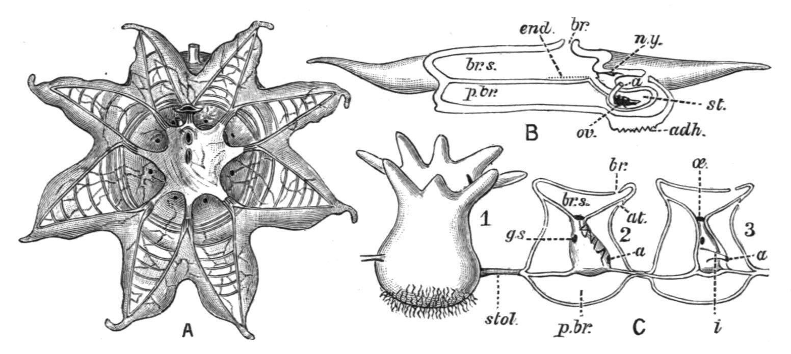
FIG. 68. A, solitary form of Octacnemus bythius (after Moseley); B, diagram of structure of Octacnemus (after Herdman); C, aggregated form of O. patagoniensis (after Metcalf). 1, from outside; 2, with test removed; and 3, with mantle removed. a, Anus; adh, area of attachment; at, atrial, and br, branchial aperture; br.s, branchial sac;
end, endostyle; g.s, gill-slits; i, intestine; n.y, nerve-ganglion; oe, oesophagus; ov, ovary; p.br, peribranchial cavity; st, stomach; stol, stolon.
The family OCTACNEMIDAE includes the single remarkable genus Octacnemus, now known in a solitary and an aggregated form. It was found during the "Challenger" expedition, and was first described by Moseley. It is apparently a deep-sea representative of the pelagic Salpidae, and may possibly be fixed at the bottom. The body in the solitary form is somewhat discoid, with its margin prolonged to form eight tapering processes, on to which the muscle-bands of the mantle are continued. The alimentary canal forms a compact nucleus, which is attached to an apparently imperforate membrane which stretches across the body, separating the branchial from the atrial cavities. The endostyle is very short, and the dorsal lamina is also much reduced. The reproduction and life-history are entirely unknown. The aggregated form consists of a small number of individuals united by a slender cord composed of test, body-wall, and endodermal tissue. Octacnemus has been found[111] in the South Pacific from depths of 1070 and 2160 fathoms, and off the Patagonian coast from 1050 fathoms. Two species have been described: O. bythius, Moseley, and O. patagoniensis, Metcalf. Metcalf, who has recently investigated the aggregated form (O. patagoniensis), considers that the genus is more nearly related to the Clavelinidae than to the Salpidae. Possibly its position might be best indicated by a line diverging from near the point (3) in the phylogenetic diagram below.
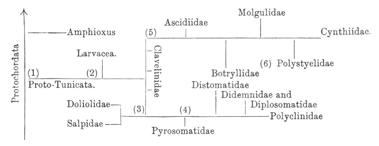
General Conclusions.
The following diagram is a graphic representation of the genetic affinities, or what is now generally supposed to have been the probable course of phylogeny of the Tunicata. It will be noticed that it shows (1) the ProtoTunicates arising from Proto-Chordata, not far from the ancestors of Amphioxus (see also, this vol. p. 112); (2) that the Larvacea are regarded as the most primitive section of the group; (3) that the Thaliacea (Doliolidae and Salpidae) are supposed to be derived not directly from primitive pelagic forms, but through the early fixed Ascidians, not far from
(4) the ancestral compound Ascidians, which gave rise to the Pyrosomatidae; (5) that the Ascidiidae and other higher Simple Ascidians are derived, like the Compound Ascidians, from ancestral Clavelinidae; and (6), that the Ascidiae Compositae are polyphyletic, the Holosomata (Botryllidae and Polystyelidae) being derived from ancestral Simple Ascidians independently of the Merosomatous families.
The Tunicata are remarkable for the variety in appearance, structure, and life-history which they present. No group illustrates in a more instructive manner so large a number of important biological principles and phenomena. They show solitary and colonial forms, fixed and free, pelagic and abyssal. The development is in some cases larval and with metamorphosis, in others abbreviated and direct. Persistent traces of ancestral characters are seen in the embryonic and larval stages, while the adults present the most varied secondary adaptations to littoral, pelagic, and deep-sea, free-swimming and sessile modes of existence. In the details of their classification they demonstrate both stable and variable species, monophyletic and polyphyletic groups. They exhibit the phenomena of gemmation and of embryonic fission, of polymorphism, hibernation, alternation of generations, and change of function. They have long been known as a stock example of degeneration; but in fact they lend themselves admirably to the exposition of more than one "Chapter of Darwinism."
NOTE TO P. 78. Oligotrema, Bourne (Quart.J. Micr . Sci. xlvii. Pt. ii. 1903, p. 233), a Molgulid from the Loyalty Islands, has a reduced branchial sac and greatly developed pinnate, muscular branchial lobes, probably used in capturing food.
CHAPTER IV
CEPHALOCHORDATA
INTRODUCTION GENERAL CHARACTERS ANATOMY OF AMPHIOXUS EMBRYOLOGY AND LIFE-HISTORY CLASSIFICATION OF CEPHALOCHORDATA SPECIES AND DISTRIBUTION
The CEPHALOCHORDATA comprise only a small group of little fish-like forms, the Lancelets, usually known as "Amphioxus," and referable to about a dozen species arranged in several closely allied genera under the single family Branchiostomatidae. The best known form is Branchiostoma lanceolatum (Pallas), the common Amphioxus or Lancelet, which has been found in British seas, and even as far north as the coast of Norway, but is much more common in warmer waters, such as the Mediterranean, and is also found in the Indian Ocean. It is abundant in the Bay of Naples, and lives and breeds in great numbers in a salt lagoon, the "Pantano," near Messina, and from these localities most of the specimens have been obtained for the numerous recent researches upon its structure and development.
Amphioxus was first discovered and described (1778) by Pallas, who regarded it as a Mollusc, and named it Limax lanceolatus. It was first correctly diagnosed as a low Vertebrate, and named Branchiostoma, by Costa, in 1834. The term Amphioxus, under which it has become so well known, was applied to it a couple of years later by Yarrell.
The anatomy was for the first time fully investigated by Johannes Müller in 1841, and this important memoir has been supplemented in regard to special systems and histological details by numerous papers by many leading zoologists, such as those by Huxley in 1874, Langerhans in 1876, Lankester in 1875 and in 1889, Retzius in 1890, and Boveri and Hatschek, both in 1892. Important papers on special points have also been written by Rolph, Rohde, Benham, Andrews, Goodrich, and others. The development was first elucidated by Kowalevsky in 1867, at about the same time when he studied the development of the Ascidians, and later again in 1877. Further papers on the development and metamorphosis we owe to Hatschek in 1881, Lankester and Willey in 1890 and 1891, Wilson in 1893, and quite recently to MacBride. Dr. Willey's book, Amphioxus and the Ancestry of the Vertebrata (1894), contains a summary of investigations on structure and development, an interesting discussion of the relations of Amphioxus to the other Chordata, and a full bibliography.
In addition to such original researches, Amphioxus is studied in more or less detail every year by countless senior and junior students in zoological laboratories and marine stations throughout the civilised world. The value of this primitive form as an object of biological education depends upon the fact that it shows the essential Vertebrate characters, and their mode of formation, in a very simple and instructive condition. Although no doubt somewhat modified, and possibly degenerate in some details of structure, in its general morphology it presents us with a persistent type probably not far removed from the ancestral line of early Chordata. There are no sufficient grounds for the view that Amphioxus is a very degenerate representative of fish-like Vertebrata.
General Characters.—The Cephalochordata (or Acrania, in contradistinction to the Craniata or Vertebrata) are marine, non-colonial Chordata, in which the notochord extends the entire length of the body, running forward into the snout beyond the nervous system. There is no skull, and the notochord is not surrounded by any vertebral column. There are no limbs nor paired fins. There is no exoskeleton, and the ectoderm is a single layer of non-ciliated columnar cells. The mouth is ventral and anterior, the anus is ventral, posterior, and asymmetrically
placed on the left side. The pharynx is a large branchial sac, having its sides perforated by many gill-slits, and is surrounded by an ectodermal enclosure, the atrium, which opens to the exterior by a median ventral atriopore. The stomach gives off a simple saccular pouch, the liver, which has connected with it a simple hepatic portal blood system. There is a respiratory circulation, the contractile ventral vessel which represents the heart sending the colourless blood forward to the respiratory pharynx to be purified. The body-wall is segmented into over fifty myotomes. There are numerous separate nephridia which develop from the mesoderm and open into the atrium. The brain remains undeveloped, being scarcely distinct from the spinal cord. There are two pairs of cerebral nerves, and many spinal, in which the dorsal and ventral roots or nerves do not unite. The sense-organs are simple; there are no paired eyes and no auditory organs. The sexes are separate; the gonads are metamerically arranged on the body-wall, and have no ducts: they burst into the atrium. In the development the segmentation is complete, a gastrula is formed by invagination, the nervous system is formed from the dorsal epiblast, the notochord from the hypoblast, and the mesoderm arises from metameric coelomic pouches. The body-cavity is an enterocoele. The gill-slits are at first perforations of the body-wall opening from the pharynx to the exterior, which later become enclosed by the development of the atrium.
ANATOMY.
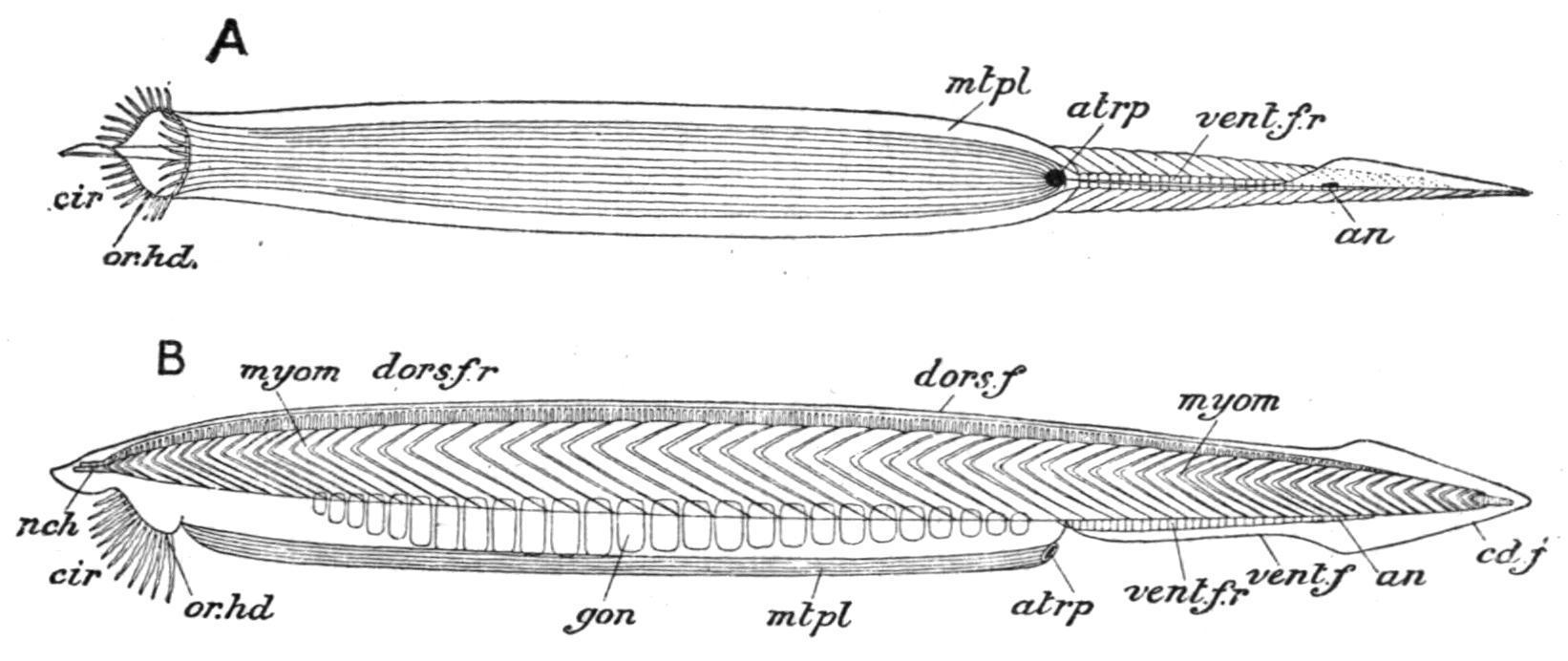
External Characters.—Amphioxus[112] is about 1½ to 2½ inches in length, slender, somewhat translucent, and pointed at both ends (Fig. 69). It lives in shallow water and burrows in the sand, head first, with great rapidity. It frequently remains with the anterior end protruding from the sand. When on the surface it lies on one side. It is said to swim freely at night. The head end is rather the thicker, and the anterior two-thirds of the ventral surface are flattened (Fig. 70, A), and may be slightly ridged longitudinally. The lateral edges of this flat area project as metapleural folds (Fig. 70, mt.pl), which begin anteriorly at the edges of the external mouth, and die away in the middle line posteriorly behind a median opening, the atriopore (Fig. 70, atrp). From this point a ventral median fin (vent.f) extends backwards around the pointed posterior end (caudal fin,
cd.f), and then forwards along the upper surface (dorsal fin, dors.f) to the anterior end of the body. These fins thus constitute a continuous median fold around a great part of the animal (Fig. 70, B, and Fig. 71).
FIG. 69. Amphioxus (Branchiostomalanceolatum) in the Pantano at Messina. (After Willey.)
FIG. 70. Branchiostoma lanceolatum. A, ventral; B, side view of the entire animal. an, Anus; atrp, atriopore; cd.f, caudal fin; cir, cirri; dors.f, dorsal fin; dors.f.r, dorsal fin-rays; gon, gonads; mtpl, metapleure; myom, myomeres; nch, notochord; or.hd, oral hood; vent.f, ventral fin; vent.f.r, ventral fin-rays. (After Kirkaldy.)
The surface is soft all over, there being no exoskeleton. The epidermis or ectoderm is formed by a single layer of epithelial cells (see Fig. 72, p. 118), some of which bear sensory processes, while others have a striated cuticular border. There is no general ciliation of the surface in the adult.
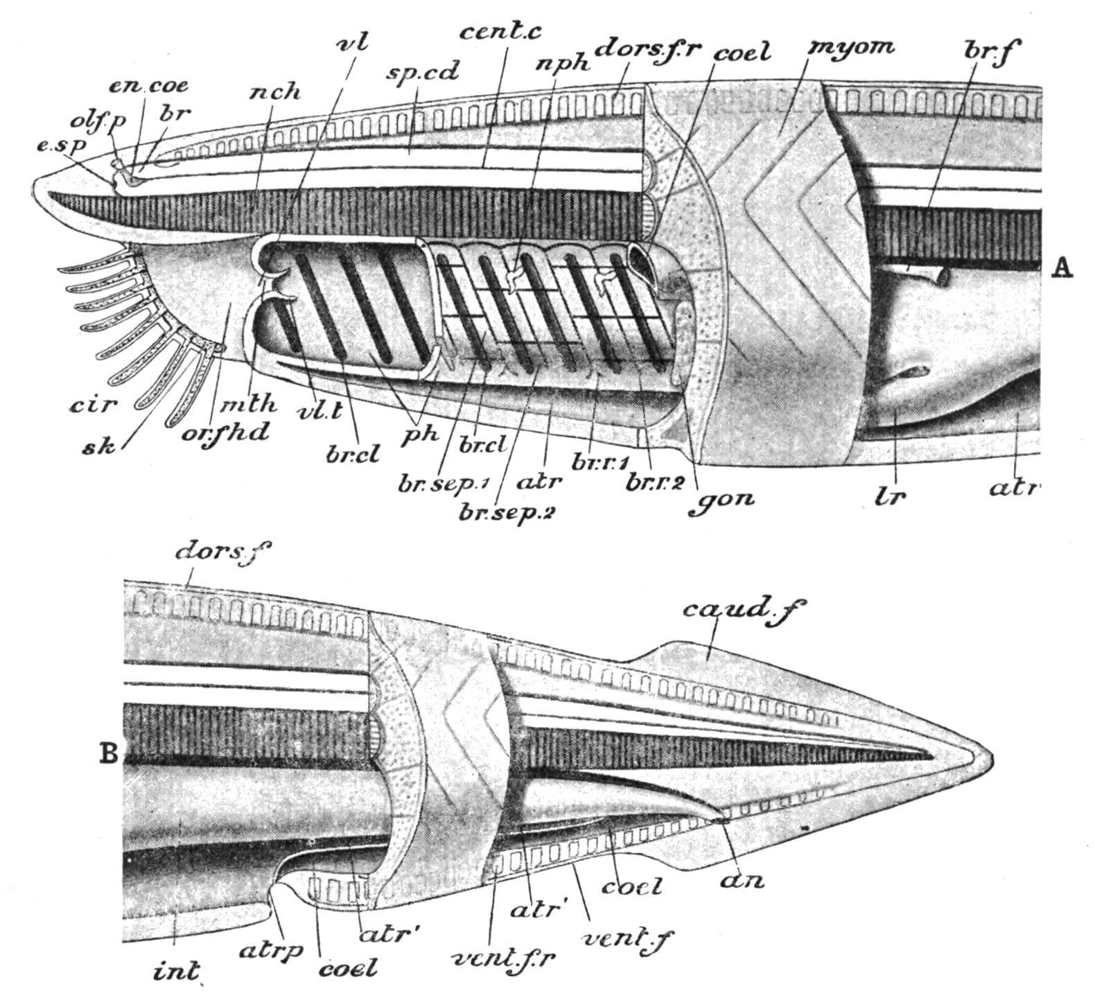
FIG. 71. Diagram of the anatomy of Amphioxus. A, anterior; B, posterior part. an, Anus; atr, atrium; atr′, its posterior prolongation; atrp, atriopore; br, brain; br.cl, branchial clefts; br.f, brown funnel; br.sep.1, primary, br.sep.2, secondary branchial lamella; br.r.1, primary, br.r.2, secondary branchial rod; caud.f, caudal fin; cent.c, central canal; cir, cirri; coel, coelom; dors.f, dorsal fin; dors.f.r, dorsal fin-ray; en.coe, cerebral vesicle; e.sp, eye-spot; gon, gonad; int, intestine; lr, liver; mth, mouth; myom, myotomes; nch, notochord; nph, nephridia; olf.p, olfactory pit; or.f.hd, oral hood; ph, pharynx; sk, skeleton of oral hood and cirri (dotted); sp.cd, spinal cord; vent.f, ventral fin; vent.f.r, ventral fin-ray; vl, velum; vl.t, velar tentacles. (From Parker and Haswell.)
The true mouth is a small pore at the bottom of a large vestibule (the stomodaeum), placed at the anterior end of the ventral surface (Figs. 70 and 71), and formed by the "oral hood," which may be a prolongation forwards of the atrial or metapleural folds at each side. The edges of the oral hood bear 12 to 20 pairs of cirri (Fig. 70, cir) or ciliated tentacles (strengthened by skeletal rods), which form a sensory fringe around the opening. The anus (Figs. 70 and 71, an), is asymmetrical, being placed on the left side of the ventral fin, some distance behind the atriopore, and not far from the posterior end of the body. The short region behind the anus and surrounded by the caudal fin may properly be called "tail." The current of water for respiratory and nutritive purposes, and which may carry the ova and spermatozoa to the exterior, usually passes in at the mouth and out at the atriopore, as in the Tunicata. On occasions, however, it is said to be reversed.