FITZGERALD ’S ClinicalNeuroanatomy and Neuroscience
1Embryology,1
2CerebralTopography,7
3Midbrain,Hindbrain,SpinalCord,27
4BloodSupplyoftheBrain,41
5Meninges,56
6NeuronsandNeuroglia,66
7ElectricalEvents,79
8TransmittersandReceptors,89
9PeripheralNerves,110
10InnervationofMusclesandJoints,119
11InnervationofSkin,128
12ElectrodiagnosticExamination,133
13AutonomicNervousSystem,148
14NerveRoots,164
15SpinalCord:AscendingPathways,176
16SpinalCord:DescendingPathways,185
17Brainstem,196
18TheLowestFourCranialNerves,214
19VestibularNerve,221
20CochlearNerve,227
21TrigeminalNerve,233
22FacialNerve,239
23OcularMotorNerves,243
24ReticularFormationandtheNeuromodulatory System,253
25Thalamus,Epithalamus,267
26BasalGanglia,274
27Cerebellum,283
28CerebralCortex,294
29Electroencephalography,310
30EvokedPotentials,319
31VisualPathways,326
32HemisphericAsymmetries,336
33OlfactoryandLimbicSystems,346
34PituitaryandHypothalamus,368
35CerebrovascularDisease,376
Glossary,387 Index,399
CHAPTERSUMMARY
SpinalCord 1
Neurulation 1
SpinalNerves 1
Brain 1
BrainParts 1
STUDYGUIDELINES
Thischapteraimstogiveyousufficientinsightintothe embryologicdevelopmentofthebrainandspinalcordto accountforthearrangementofstructuresinthemature nervoussystem.Ifnotalreadyfamiliarwithadultbrain anatomy,wesuggestyoureadthischapteragain followingyourstudyofChapters2and3.
SPINALCORD
Neurulation
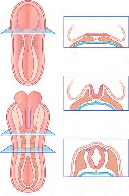
Theentirenervoussystemoriginatesfromthe neuralplate, whicharisesfromanectodermalthickeningofthefloorofthe amnioticsac(Fig.1.1).Duringthethirdweekafterfertilisation, theneuralplateformspaired neuralfolds,whichunitetocreate the neuraltube and neuralcanal.Unionofthefoldscommences inthefutureneckregionoftheembryoandproceedsrostrally andcaudally.Theopencranialandcaudalendsoftheneural tube,the neuropores,areclosedoffbeforetheendofthefourth week.Theprocessofformationoftheneuraltubefromtheectodermisknownas neurulation
Cellsattheedgeofeachneuralfoldescapefromthelineofunion andformthe neuralcrest alongsidethetube.Celltypesderived fromtheneuralcrestincludespinalandautonomicganglioncells, melanocytes,andtheSchwanncellsofperipheralnerves.
SpinalNerves
Thedorsalpartoftheneuraltubeiscalledthe alarplate andthe ventralpartiscalledthe basalplate (Fig.1.2).Neuronsdevelopingfromthealarplatearepredominantlysensoryinfunction andreceive dorsalnerveroots arisingfromthespinalganglia,and thoseinthebasalplatearepredominantlymotorneuronsand giveriseto ventralnerveroots.Atappropriatelevelsofthespinal cord,theventralrootsalsocontainaxonsfromdevelopingautonomicneurons.Thedorsalandventralrootsunitetoformthe spinalnerves,whichemergefromthevertebralcanalthroughthe intervertebralforaminalyinginbetweentheneuralarches.
Thecellsofthespinal(dorsalroot)gangliaareinitiallybipolar.Theybecomeunipolarbythecoalescenceoftheirtwoprocessesononesideoftheparentcells.
VentricularSystemand
ChoroidPlexuses 1
CranialNerves 2
CerebralHemispheres 4
Fordescriptivepurposes,theembryoisintheprone(facedown)position,wherebytheterms ventral and dorsal correspondtotheadult anterior and posterior ,and rostral and caudal correspondto superior and inferior
BRAIN
BrainParts
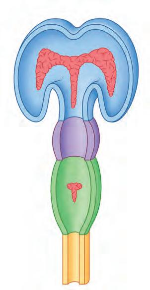
Lateinthefourthweekofembryonicdevelopment,therostralpart oftheneuraltubeundergoesflexionatthelevelofthefuturemidbrain(Fig.1.3A).Thisregionisknownasthe mesencephalon Aslightconstrictionmarksitsjunctionwiththe prosencephalon (futureforebrain)and rhombencephalon (futurehindbrain).
Thealarplateoftheprosencephalonexpandsoneachside (Fig.1.3A)toformthe telencephalon (cerebralhemispheres). Thebasalplateremainsinplaceasthe diencephalon. Finally, an opticoutgrowth fromthediencephalonistheforerunnerof theretinaandopticnerve.
Thediencephalon,mesencephalon,andrhombencephalon constitutetheembryonicbrainstem.
Thebrainstembucklesasdevelopmentproceeds.Asaresult,the mesencephaloniscarriedtothesummitofthebrain.Therhombencephalonfoldsonitself,causingthealarplatestoflareandcreating therhomboid(diamond-shaped)fourthventricleofthebrain.The rostralpartoftherhombencephalongivesrisetotheponsandcerebellum.Thecaudalpartgivesrisetothemedullaoblongata(Fig.1.4).
VentricularSystemandChoroidPlexuses
Theneuralcanaldilateswithinthecerebralhemispheres,formingthelateralventricles;thesecommunicatewiththethirdventriclecontainedwithinthediencephalon.Thetwolateral ventriclescommunicatewiththethirdventriclethroughthe foramenofMonro (interventricularforamen).Thethirdand fourthventriclescommunicatethroughthe cerebralaqueduct (or aqueductofSylvius)inthemidbrain(Fig.1.5).
Thethinroofsoftheforebrainandhindbrainareinvaginatedbytuftsofcapillaries,whichformthechoroidplexusesof
Fig.1.1 (A)Cross-sectionsfromathree-somite(20-day)embryo. (BandC)Cross-sectionsfromaneight-somite(22-day)embryo.
Alar plate
Basal plate
Neural arch
Dorsal nerve root
Spinal ganglion
Mesencephalon (midbrain)
Rhombencephalon (hindbrain)
Prosencephalon (forebrain)
Optic outgrowth
Spinal cord
Mesencephalon (midbrain)
Diencephalon Fourth ventricle
Eye
Telencephalon
Fig.1.3 (A)Earlydevelopmentofthethreeprimarybrainvesicles. (B) Asterisks indicatethesiteofinitialdevelopmentofthecerebellum.
Primary vesiclesSecondary vesiclesAdult derivatives
Telencephalon
Prosencephalon
Mesencephalon
Rhombencephalon
Diencephalon
Mesencephalon
Metencephalon
Myelencephalon
Fig.1.4 Somederivativesofthebrainvesicles.
Cerebral cortex
Corpus striatum
Thalamus
Hypothalamus
Midbrain
Cerebellum
Pons
Medulla
thelateral,third,andfourthventricles.Thechoroidplexuses secretecerebrospinalfluid(CSF),whichflowsthroughtheventricularsystem.CSFexitsthefourthventriclethroughthree aperturesinitsroofknownasforamenMagendie(inthemidline)andtherightandleftforamenLuschka(Fig.1.6).
Costal process
Centrum
Notochord
Spinal nerve
Autonomic ganglion
Ventral nerve root
Fig.1.2 Neuraltube,spinalnerve,andmesenchymalvertebraofan embryoat6weeks.
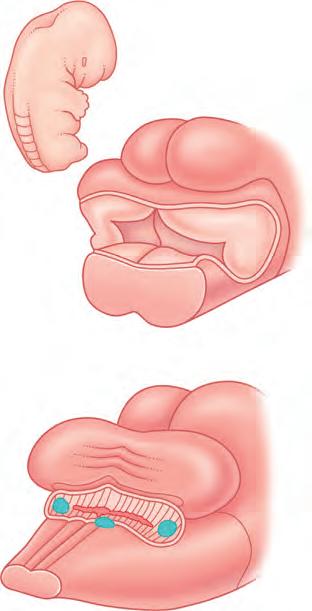
CranialNerves
Fig.1.7 illustratesthestateofdevelopmentofthecranialnerves duringthesixthweekafterfertilization.
• Theolfactorynerve(I)arisesfrombipolarneurons developingintheepitheliumliningtheolfactorypit.
• Theopticnerve(II)growscentrallyfromtheretina.
Lateral ventricle
Telencephalon (hemisphere)
Third ventricle
Aqueduct
Fourth ventricle
Central canal
Choroid plexus
Diencephalon
Mesencephalon (midbrain)
Rhombencephalon (hindbrain)
8 weeks
Spinal cord
Fig.1.5 Thedevelopingventricularsystem.Choroidplexusesare shown inred.
Foramen of Luschka Foramen of Magendie
12 weeks
Mesencephalon
Roof plate (cut)
Cerebellum
Rhombencephalon
Midbrain
Cerebellar hemisphere
Choroid plexus
Pons Medulla oblongata
Fig.1.6 Dorsalviewsofthedevelopinghindbrain (seearrowininset) (A)At8weeks,thecerebellumisemergingfromthefourthventricle. (B)At12weeks,theventricleisbecominghiddenbythecerebellum, andthreeapertureshaveappearedintheroofplate.
Trigeminal motor rootTrigeminal sensory root
Facial Abducens
Vestibulocochlear
Glossopharyngeal Vagus
Cranial accessory
Hypoglossal
Spinal accessory
Trochlear
Oculomotor
Fig.1.7 Cranialnervesofa6-week-oldembryo.(ReproducedfromBossyetal.1990,withpermissionfromSpringer-Verlag.)
• Theoculomotor(III)andtrochlear(IV)nervesarisefrom themidbrain,andtheabducens(VI)nervearisesfromthe pons;allthreewillsupplytheextrinsicmusclesoftheeye.
• Thethreedivisionsofthetrigeminal(V)nervewillprovide sensoryinnervationtotheskinofthefaceandscalp,tothe mucousmembranesoftheoronasalcavity,andtotheteeth.A motorrootwillinnervatethemusclesofmastication(chewing).
• Thefacial(VII)nervewillinnervatethemusclesoffacial expression.
• Thevestibulocochlear(VIII)nervewillinnervatetheorgans ofhearingandbalance,whichdevelopfromtheotocyst.
• Theglossopharyngeal(IX)nerveisamixednerve.Mostofits fibreswillsupplysensoryinnervationtotheoropharynxand laryngopharynxandmotorinnervationtothestylopharyngeus muscle.
• Thevagus(X)nerveisalsoamixednerve.Itcontainsa largesensorycomponentthatinnervatesthemucous membranesofthedigestivesystemandalargemotor (parasympathetic)componentthatwillinnervatetheheart, lungs,andgastrointestinaltract.
• Thecranialaccessory(XIc)nervewillbedistributedbythe vagustoinnervatethemusclesofthelarynxandpharynx.
• Thespinalaccessory(XIs)nerve willinnervatethesternocleidomastoidandtrapeziusmuscles.
• Thehypoglossal(XII)nervewillinnervatealltheintrinsic andextrinsicmusclesofthetongueexceptthepalatoglossus, whichisinnervatedbythepharyngealplexus.
CerebralHemispheres
Inthetelencephalon,mitoticactivitytakesplaceinthe ventricularzone justoutsidethelateralventricle.Daughtercellsmigrate totheoutersurfaceoftheexpandinghemisphereandformthe cerebralcortex.
Expansionofthecerebralhemispheresisnotuniform.A regiononthelateralsurface,the insula (L. ‘island’),isrelatively quiescentandformsapivotaroundwhichtheexpandinghemisphererotates.Frontal,parietal,occipital,andtemporallobes canbeidentifiedat14weeks’ gestationalage(Fig.1.8).
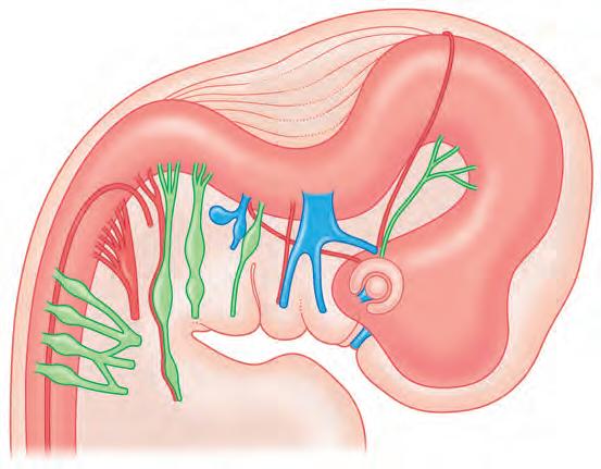
Onthemedialsurfaceofthehemisphere,apartofthecerebral cortex,the hippocampus,formsafifthlobe,the limbiclobe ofthe brain.Thehippocampusisdrawnintothetemporallobe,leaving initswakeastrandoffibresknownasthe fornix.Withintheconcavityofthisarcliesthe choroidfissure,throughwhichthechoroidplexusinvaginatesintothelateralventricle(Fig.1.9).
Corpus callosum
Anterior commissure
Corpus callosum
plexus
Fornix
Fornix
Corpus callosum
Choroid plexus
Fig.1.8 Fetalbrainat14weeks.The arrow indicatestheC-shaped growthofthehemispherearoundtheinsula. F,Frontallobe, O,occipitallobe; P,parietallobe; T,temporallobe.
nucleus
Caudate nucleus (head)
Caudate nucleus (tail)
Fig.1.9 Medialaspectofthedevelopingrighthemisphere.The hippocampus,initiallydorsaltothethalamus,migratesintothe temporallobe(arrows inA,B,andC),leavingthefornixinitswake. Theconcavityofthearchsoformedcontainsthechoroidfissure(the lineofinsertionofthechoroidplexusintotheventricle)andthetailof thecaudatenucleus.
Hippocampus
Choroid plexus
Thalamus
Hippocampus
Choroid
Caudate
Thalamus
Hippocampus
Thalamus
Central sulcus (Rolandic fissure)
Lateral sulcus (Sylvian fissure)
Choroid plexus
Lateral ventricle
Corpus striatum
Insula
Thalamus
Site of fusion
Hypothalamus
Choroid plexus
Caudate nucleus
Projection fibres
Lentiform nucleus
Third ventricle
Midbrain
Fig.1.10 Coronalsectionsofthedevelopingcerebrum.(A)At10 weeks,thecorpusstriatumistraversedbyaxonsprojectingfrom thalamustocerebralcortexandfromcerebralcortextospinalcord. (B)At17weeks,thecorpusstriatumhasbeendividedtoformthe caudateandlentiformnuclei(fusionpersistsattheanteriorend,not shownhere).
Calcarine sulcus
The anteriorcommissure developsasaconnectionlinking olfactory(smell)regionsoftheleftandrightsides.Abovethis,a muchlargercommissure,the corpuscallosum,linksmatching areasofthecerebralcortexofthetwosides.Itextendsbackward abovethefornix.
Coronalsectionsofthetelencephalonrevealamassofgrey matteratthebaseofeachhemisphere,whichistheforerunner ofthe corpusstriatum.Thediencephalondevelopsintothe thalamusandhypothalamus,aswellasformingthewallsofthe thirdventricle(Fig.1.10).
Theexpandingcerebralhemispherescomeintocontactwith thediencephalonandtheyfusewithit(see ‘siteoffusion ’ in Fig.1.10A).Oneconsequenceisthattheterm ‘brainstem’ is
Fig.1.11 Threemajorcorticalsulciina28-weekfetus.(A)Lateral surfaceand(B)medialsurfaceoftheleftcerebralhemisphere.
restrictedthereaftertotheremaining,freeparts:midbrain, pons,andmedullaoblongata.Asecondconsequenceisthatthe cerebralcortexisabletoprojectaxonsdirectlydowntothe brainstem.Togetherwithaxonsprojectingfromthalamusto thecortex,theysplitthecorpusstriatuminto caudate and lentiformnuclei (Fig.1.10B).
Bythe28thweekofdevelopment,severalsulci(fissures) appearonthesurfaceofthebrain,notablythe lateral, central, and calcarinesulci (Fig.1.11).
CoreInformation
Thenervoussystemtakestheinitialformofacellularneuraltubederived fromtheectodermandenclosinganeuralcanal.Aribbonofcellsescapes alongeachsideofthetubetoformtheneuralcrest.Themorecaudalpartof theneuraltubeformsthespinalcord.Theneuralcrestformsspinalganglion cellsthatsenddorsalnerverootsintothesensorypart(alarplate)ofthe spinalcord.Thebasalplateofthespinalcordcontainsmotorneuronsthat emitventralrootstocompletethespinalnervesbyjoiningthedorsalroots.
Themorerostralpartoftheneuraltubeformsthreebrainvesicles.Of these,theprosencephalon(forebrain)givesrisetothecerebral hemispheres(telencephalon)dorsallyandthediencephalonventrally.The mesencephalondevelopsintothemidbrain,andtherhombencephalon becomesthehindbrain(pons,medullaoblongata,andcerebellum).
Theneuraltubeexpandsrostrallytocreatetheventricularsystemofthe brain.CSFissecretedbyachoroidcapillaryplexusthatinvaginatesthe roofplatesoftheventricles.
Thecerebralhemispheresdevelopfrontal, parietal,temporal,occipital,and limbiclobes.Thehemispheresarecross-linkedbythecorpuscallosumand posteriorandanteriorcommissures.Thegreymatterinthebaseofeach hemisphereistheforerunnerofthecorpusstriatum.Thehemispheresfuse withthesidewallsofthediencephalon,whereuponthemesencephalonand rhombencephalonareallthatremainoftheembryonicbrainstem.
SUGGESTEDREADINGS
AdameykoI,FriedK.Thenervoussystemorchestratesandintegrates craniofacialdevelopment:areview. FrontPhysiol.2016;7:49.
AzaisM,AgiusE,BlancoS,etal.Timingthespinalcorddevelopmentwithneuralprogenitorcellslosingtheirproliferative capacity:atheoreticalanalysis. NeuralDev.2019; 14(7):1 19.
BossyJ,O’RahillyR,MüllerF.Ontogenèsedusystèmenerveux. In:BossyJ,ed. AnatomieClinique:Neuroanatomie.Paris: Springer-Verlag;1990:357 388.
DuboisJ,BendersM.Borradori-Tolsa,etal.Primarycorticalfolding inthehumannewborn:anearlymarkeroflaterfunctionaldevelopment. Brain.2008;131:2028 2041.
KangKH,ReichertH.Controlofneuralstemcellself-renewaland differentiationinDrosophila. CellTissueRes.2015;359(1):33 45. KieckerC,LumsdenA.Theroleoforganizersinpatterningthenervoussystem. AnnuRevNeurosci.2012;35:347 367.
LehtinenMK,WalshCA.Neurogenesisatthebrain-cerebrospinal fluidinterface. AnnuRevCellDevBiol.2011;27:653 679. LeDouarinNM,BritoJM,CreuzetS.Roleoftheneuralcrestinface andbraindevelopment. BrainResDev.2007;55:237 224.
NikolopoulouE,GaleaGL,RoloA,GreeneND,CoppAJ.Neural tubeclosure:cellular,molecularandbiomechanicalmechanisms. Development.2017;144:552 566.
O’RahillyR,MullerF.Significantfeaturesintheearlyprenataldevelopmentofthehumanbrain. AnnAnat.2008;104:105 118.
ShinotsukaN,YamaguchiY,NakazatoK,MatsumotoY,Mochizuki A,MiuraM.Caspasesandmatrixmetalloproteasesfacilitatecollectivebehaviorofnon-neuralectodermafterhindbrainneuroporeclosure. BMCDevBiol.2018;18:17.
ShiotaK.Reviewarticle prenataldevelopmentofthehumancentralnervoussystem,normalandabnormal. DonaldSchJ UltrasoundObstetricsGynecology.2015;9(1):61 66.
StilesJ,JerniganTL.Thebasicsofbraindevelopment. Neuropsychol Rev.2010;20:327 348.
SunT,HevnerRF.Growthandfoldingofthemammaliancerebral cortex:frommoleculestomalformations. NatRevNeurosci 2014;15:217 232.
YangHJ,LeeDH,LeeYJ,etal.Secondaryneurulationofhuman embryos:morphologicalchangesandtheexpressionofneuronal antigens. ChildsNervSyst.2014;30:73 82.
CHAPTERSUMMARY
SurfaceFeatures 7
Lobes 7
Diencephalon 8
MidlineSagittalViewoftheBrain 8
InternalAnatomyoftheCerebrum 8
Thalamus,Caudate,andLentiformNuclei, InternalCapsule 8
STUDYGUIDELINES
1. Themostimportantobjectiveisthatyoubecomeableto recite all thecentralnervoussystemitemsidentifiedinthe magneticresonanceimageswithoutlookingatthelabels.
2. Trytogetthenomenclatureofthecomponentpartsofthe basalgangliaintolong-termmemory.Noteasilydone!
SURFACEFEATURES
Lobes
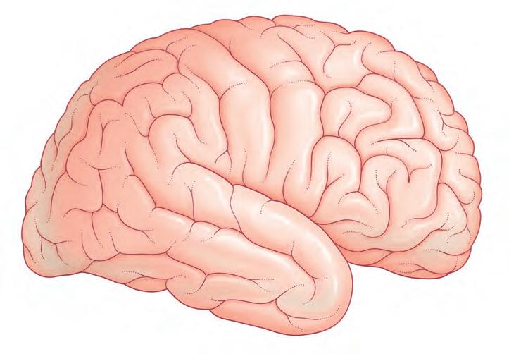
Thesurfacesofthetwocerebralhemispheresarefurrowedby sulci , andtheinterveningridgesarecalled gyri .Mostofthe cerebralcortexformsthewallsofsulciand,fromthelateral surfaceofthehemispheres,isconcealedfromview.Although thepatternsofthevarioussulcivaryfrombraintobrain,some aresufficientlyconstanttoserveasdescriptivelandmarks. Thedeepestsulciarethe lateralsulcus (Sylvianfissure) and the centralsulcus (Rolandicfissure) ( Fig.2.1A ).Thesetwo servetodividethehemisphere(lateralview)intofour lobes withtheaidoftwoimaginarylines,oneextendingbackfrom thelateralsulcus,theotherreachingfromtheupperendofthe parietooccipitalsulcus ( Fig.2.1B )toablunt preoccipital notch atthelowerborderofthehemisphere(thesulcusand notcharelabelledin Fig.2.2 ).Thelobesarecalled frontal , parietal , occipital ,and temporal .
Theblunttipsofthefrontal,occipital,andtemporallobes aretherespectivepolesofthehemispheres.
The opercula (lips)ofthelateralsulcuscanbepulledapart toexposethe insula (Fig.2.3).Theinsulawasmentionedin Chapter1asbeingrelativelyquiescentduringprenatalexpansionofthetelencephalon.
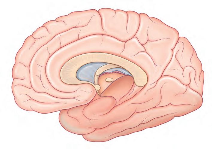
Themedialsurfaceofthehemisphereisexposedbysectioningthe corpuscallosum,amassivebandofwhitematterconnectingmatching/homotopicareasofthecortexofthetwo hemispheres.Thecorpuscallosumconsistsofarostrum,genu, bodyandspleniumfromanteriortoposterior.Theanterior commissureliesbelowtherostrum(Fig.2.2B).Thefrontallobe liesanteriortoalinedrawnfromtheupperendofthecentral sulcustothetrunkorbodyofthecorpuscallosum(Fig.2.2B).
HippocampusandFornix 12
AssociationandCommissuralFibres 12
LateralandThirdVentricles 16
Box
BrainPlanes 22
MagneticResonanceImaging 23
DiffusionTensorImaging 25
3. Becauseofitsclinicalimportance,you must beabletopop upamentalimageofthepositionandnamedpartsofthe internalcapsuleandtoappreciatethecontinuityofthe coronaradiata,internalcapsule,andcruscerebri(cerebral peduncle).
Theparietallobeliesbehindthislineandisseparatedfromthe occipitallobebytheparietooccipitalsulcus.Thetemporallobe liesinfrontofalinedrawnfromthepreoccipitalnotchtothe spleniumofthecorpuscallosum. Figs.2.2and2.4to2.6 should beconsultedalongwiththefollowingdescriptionofsurfacefeaturesofthelobesofthebrain.
FrontalLobe. Thelateralsurfaceofthe frontallobe contains the precentralgyrus boundedinfrontbythe precentral sulcus .Furtherforward, superior , middle ,and inferior frontalgyri areseparatedby superior and inferiorfrontal sulci .Onthemedialsurface,thesuperiorfrontalgyrusis separatedfromthe cingulategyrus bythe cingulatesulcus Theinferiorororbitalsurfaceismarkedbyseveral orbital gyri .Incontactwiththissurfacearethe olfactorybulb and olfactorytract
ParietalLobe. Theanteriorpartoftheparietallobecontains the postcentralgyrus boundedbehindbythe postcentralsulcus. Theposteriorparietallobeisdividedintosuperiorandinferior parietallobulesbyan intraparietalsulcus.Theinferiorparietal lobuleshowsa supramarginalgyrus cappingtheupturnedend ofthelateralsulcus,andan angulargyrus cappingthesuperior temporalsulcus.
Themedialsurfacecontainstheposteriorpartofthe paracentrallobule and,behindthis,the precuneus.Theparacentral lobule(partlycontainedinthefrontallobe)issocalledbecause ofitsrelationshiptothecentralsulcus.
OccipitalLobe. Thelateralsurfaceoftheoccipitallobeis markedbyseveral lateraloccipitalgyri.Themedialsurface containsthe cuneus (‘wedge’)betweenthe parietooccipital
Thefivelobesofthebrain.(A)Lateralsurfaceand(B)medial surfaceoftherightcerebralhemisphere.
sulcus andtheimportant calcarinesulcus.The lingualgyrus liesbetweenthecollateralsulcusandtheanteriorendofthecalcarinesulcus.Theinferiorsurfaceshowsthreegyriandthree sulci.The lateralandmedialoccipitotemporalgyri areseparatedbythe occipitotemporalsulcus
TemporalLobe. Thelateralsurfaceofthetemporallobedisplays superior , middle ,and inferiortemporalgyri separated by superior and inferiortemporalsulci .Theinferiorsurface showstheanteriorpartsofthe occipitotemporalgyri .The lingualgyrus continuesforwardasthe parahippocampal gyrus ,whichendsinabluntmedialprojection,the uncus .As willbeseenlaterinviewsofthesectionedbrain,theparahippocampalgyrusunderliesarolled-inpartofthecortex,the hippocampus .
LimbicLobe. Afifth limbiclobe ofthebrainsurroundsthe medialmarginofthehemisphere.Surfacecontributorstothe limbiclobeincludethecingulateandparahippocampalgyri. Itismoreusualtospeakofthe limbicsystem ,whichincludes thehippocampus,fornix,amygdala,andotherelements
connectedtoorrelatedinfunctiontothelimbiclobe(see Chapter33).
Diencephalon
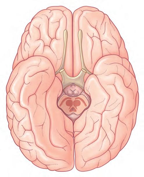
Thelargestcomponentsofthediencephalonarethe thalamus andthe hypothalamus (Figs.2.6and2.7).Thesenucleargroups formthesidewallsofthethirdventricle.Betweenthemisashallow hypothalamicsulcus,whichrepresentstherostrallimitof theembryonicsulcuslimitans.
MidlineSagittalViewoftheBrain
Fig.2.8 istakenfromamidlinesagittalsectionoftheheadofa cadaver,displayingthebraininrelationtoitssurroundings.
INTERNALANATOMYOFTHECEREBRUM
Thearrangementofthefollowingstructureswillnowbe described:thalamus,caudateandlentiformnuclei,internalcapsule;hippocampusandfornix;associationandcommissural fibres;lateralandthirdventricles.
Thalamus,Caudate,andLentiformNuclei, InternalCapsule
Thetwothalamifaceoneanotheracrosstheslot-likethird ventricle.Moreoftenthannot,theyareinterconnected acrossthethirdventricle,creatingan interthalamicadhesion/massaintermedia ( Fig.2.9 ).In Fig.2.10 ,thethalamus andrelatedstructuresareassembledinamediolateral sequence.Incontactwiththeuppersurfaceofthethalamus arethe head and body ofthe caudatenucleus .The tail ofthe caudatenucleuspassesforwardbelowthethalamusbutisnot incontactwithit.
Thethalamusisseparatedfromthelentiformnucleusbythe posteriorlimbofthe internalcapsule,whichisacommonsite fora stroke resultingfromlocalarterialembolism(blockage)or haemorrhage.Theinternalcapsulecontainsfibresrunning fromthalamustocortexandfromcortextothalamus,brainstem,andspinalcord.Intheintervalbetweencortexandinternalcapsule,theseascendinganddescendingfibresformthe coronaradiata.Belowtheinternalcapsule,the crus ofthemidbrain(cerebralpeduncle)receivesdescendingfibrescontinuing downtothebrainstem.
Thelens-shaped lentiformnucleus iscomposedoftwo parts:the putamen and globuspallidus .Whilenotpartofthe lentiformnucleus,thecaudateisofsimilarstructuretothe putamenandtheiranteriorends arefused.Inaddition,they arelinkedbystrandsofgreymatter(cellularbridges)thattraversetheanteriorlimboftheinternalcapsule,hencetheterm corpusstriatum (or,simply, striatum )usedtoincludethe putamenandcaudatenucleus.Theterm pallidum refersto the globuspallidus
Thecaudateandlentiformnucleibelongtothe basalganglia,atermoriginallyappliedtohalfadozenmassesofgrey matterlocatednearthebaseofthehemisphere.Incurrent usage,thetermdesignatesfivenucleiknowntobeinvolvedin motorcontrol:thecaudate,putamen,globuspallidus,and
Temporal lobe
Lateral sulcus
Frontal lobe
Occipital lobe
Parietal lobeCentral sulcus
Temporal lobe
Occipital lobe
Limbic lobe
Parietal lobe
Central sulcus
Frontal lobe
Fig.2.1
Postcentral gyrus
Postcentral sulcus
Intraparietal sulcus
Supramarginal gyrus
Angular gyrus
Tip of parietooccipital sulcus
Lunate sulcus
Preoccipital notch
Central sulcus
Precentral gyrus
Precentral sulcus
Temporal sulci
Cingulate sulcus
Fornix
Corpus callosum
Cingulate gyrus
Superior frontal gyrus
Septum pellucidum
Anterior commissure
Temporal gyri
Paracentral lobule
Central sulcus
Superior Middle Inferior
Precuneus
Superior frontal gyrus
Middle frontal gyrus
Inferior frontal gyrus
Lateral sulcus (Sylvian fissure)
Uncus
Parietooccipital sulcus
Cuneus
Calcarine sulcus
Lingual gyrus
Parahippocampal gyrus
Collateral sulcus
Thalamus
Fig.2.2 (A)Lateraland(B)medialviewsoftherightcerebralhemisphere,depictingthemaingyriandsulci.
subthalamicnucleus inthediencephalon,andthe substantia nigra inthemidbrain(Fig.2.11).
Inhorizontalsection,theinternalcapsulehasadog-leg shape(seephotographofafixed-brainsectionin Fig.2.12 , andinvivomagneticresonanceimage[MRI]in Fig.2.13 ).
Theinternalcapsulehasfivenamedpartsinhorizontal sections:
1. anteriorlimb,betweenthelentiformnucleusandthehead ofthecaudatenucleus; 2. genu;
3. posteriorlimb ,betweenthelentiformnucleusandthe thalamus;
4. retrolenticularpart (visualradiations),behindthelentiform nucleusandlateraltothethalamus;
5. sublenticularpart (auditoryradiations).
Cingulate sulcus (posterior end)
Superior parietal lobule
Intraparietal sulcus
Inferior parietal lobule
The corticospinaltract (CST)descendsinthelateralaspect oftheposteriorlimboftheinternalcapsule.Itisalsocalledthe pyramidaltract,becauseitpassesthroughthepyramidsofthe medulla.A tract isabundleoffibresservingacommonfunction.Over50%ofthefibresthatcontributetotheCSTmainly originatefromtheprecentralgyrusandthecorteximmediately anteriortotheprecentralgyrus.Theremainderofthefibresin theCSToriginatefromtheprimarysomatosensorycortexand theparietalassociationcortex.TheCSTdescendsthroughthe coronaradiata,thelateralpartoftheposteriorlimboftheinternalcapsule,andtheintermediate3/5th ofthecrusofthemidbrain(cerebralpeduncle)andcontinuestothespinomedullary junctionbeforecrossingtotheoppositesideofthespinalcord inthemotor(pyramidal)decussation.
Fromaclinicalstandpoint, theCSTisthemostimportant pathwayintheentirecentralnervoussystem(CNS) fortworeasons.First,itmediatesvoluntarymovementofallkinds. InterruptionoftheCSTleadstomotorweakness(called paresis) ormotorparalysis.Second,itextendstheentireverticallength oftheCNS,renderingitvulnerabletodiseaseortraumainthe cerebralhemisphereorbrainstemononesideandtospinal corddiseaseortraumaontheotherside.
Acoronalsectionthroughtheanteriorlimboftheinternal capsuleisrepresentedin Fig.2.14;acorrespondingMRIviewis shownin Fig.2.15.Acoronalsectionthroughtheposteriorlimb
Superior frontal sulcus
Precentral sulcus
Precentral gyrus
Central sulcus
Postcentral gyrus
Postcentral sulcus
Fig.2.4 ‘Thickslice’surfaceanatomybrainMagneticResonanceImagingscanfromahealthyvolunteer.(FromKatadaK.MRimagingofbrain surfacestructures:surfaceanatomyscanning(SAS). Neuroradiology. 1990;3(5):439 448.)
Parietal operculum
Temporal operculum Insula
Frontal operculum
Fig.2.3 Insula,seenonretractionoftheopercula.
Falx cerebri
Frontal pole
Orbital sulci
Temporal pole
Uncus
Inferior temporal gyrus
Occipitotemporal sulcus
Collateral sulcus
Occipital pole
Fig.2.5 Cerebrumviewedfrominferioraspect,depictingthemaingyriandsulci.
Genu CC Septum pellucidum
Tela choroidea
Interventricular foramen
Rostrum CC
Anterior commissure
Lamina terminalis
Longitudinal fissure
Olfactory bulb
Orbital gyri
Optic chiasm
Fig.2.6 Thediencephalonanditsboundaries. CC,Corpuscallosum.
FornixBody CC Thalamus
Occipitotemporal gyri
Midbrain Fusiform gyrus
Parahippocampal gyrus
Lingual gyrus
Habenular commissure
Splenium CC
Pineal gland
Posterior commissure
Aqueduct
Midbrain
Hypophysis
Hypothalamic sulcus
Hypothalamus
Mammillary body
Tuber cinereum
Infundibulum
Coronal suture
Corpus callosum
Lateral ventricle
Cingulate sulcus
Frontal bone
Subcutaneous fat
Frontal air sinus
Optic nerve
Hypophysis
Sphenoidal air sinus
Precentral sulcus
Paracentral lobule
Central sulcus
Superior sagittal sinus
Parietooccipital sulcus
Nasopharynx
Incisor teeth
Tongue
Mandible
Anterior arch of atlas
Dens of axis
SagittalMagneticResonanceImaging‘slice’ofthelivingbrain.(FromaserieskindlyprovidedbyProfessorJ.PaulFinn,Director,Magnetic ResonanceResearch,DepartmentofRadiology,DavidGeffenSchoolofMedicineatUCLA,California,USA.)
oftheinternalcapsulefromafixedbrainisshownin Fig.2.16;a correspondingMRIsectionisshownin Fig.2.17
Proceedinglaterallyfromthelentiformnucleuscanbefound the externalcapsule, claustrum,and extremecapsule,and finallytheinsularcortex.
HippocampusandFornix
Duringembryonicdevelopmentinprimates,the hippocampus (crucialformemoryformation)firstappearsabovethecorpus callosumwhereitcanbefoundpostnatallyinphylogenetically earliermammals.Beforebirth,itmigratesintothetemporal lobeasthislobedevelops,leavingatractofwhitematter,the fornix,initswake.Thematurehippocampusstretchesthefull
lengthadjacenttothefloorofthe inferior(temporal)horn ofthe lateralventricle(Figs.2.18and2.19).Postnatally,thefornix consistsofa body beneaththetrunkofthecorpuscallosum,a crus,whichentersitfromeachhippocampus,andtwo pillars (columns),whichleaveittodescendintothediencephalon. Intimatelyrelatedtothecrusandbodyisthe choroidfissure, throughwhichthechoroidplexusisinsertedintothelateral ventricle.
AssociationandCommissuralFibres
Fibresleavingthecerebralcortexfallintothreegroups: 1. associationfibres,whichpassfromonepartofasingle hemispheretoanotherpartofthesamehemisphere;
Edge of foramen magnum
Spinal cord
Medulla oblongata
IV ventricle
Cerebellum
Midbrain
Pons
Calcarine sulcus
Thalamus
C2-C3 intervertebral disk
Fig.2.7
Another random document with no related content on Scribd: