Get Ase's comprehensive echocardiography american society of echocardiography PDF ebook with Full Ch

Page 1


https://ebookmass.com/product/ases-comprehensive-

More products digital (pdf, epub, mobi) instant download maybe you interests ...

Echocardiography Review Guide: Companion to the Textbook of Clinical Echocardiography 4th Edition

Catherine M. Otto Md

https://ebookmass.com/product/echocardiography-review-guidecompanion-to-the-textbook-of-clinical-echocardiography-4thedition-catherine-m-otto-md/

Textbook of Clinical Echocardiography (Endocardiography) 5th Edition, (Ebook PDF)

https://ebookmass.com/product/textbook-of-clinicalechocardiography-endocardiography-5th-edition-ebook-pdf/

The Practice of Clinical Echocardiography 5th Edition

Catherine M. Otto

https://ebookmass.com/product/the-practice-of-clinicalechocardiography-5th-edition-catherine-m-otto/

Textbook of Clinical Echocardiography, 6e [Lingua inglese] 6th Edition Otto

https://ebookmass.com/product/textbook-of-clinicalechocardiography-6e-lingua-inglese-6th-edition-otto/

Clinical Manual and Review of Transesophageal Echocardiography 3rd Edition Joseph Mathew

https://ebookmass.com/product/clinical-manual-and-review-oftransesophageal-echocardiography-3rd-edition-joseph-mathew/

Doppler Echocardiography for the Small Animal Practitioner June A. Boon

https://ebookmass.com/product/doppler-echocardiography-for-thesmall-animal-practitioner-june-a-boon/

Practical Echocardiography for Cardiac Sonographers 1st Edition Daniel M. Shindler

https://ebookmass.com/product/practical-echocardiography-forcardiac-sonographers-1st-edition-daniel-m-shindler/

Echocardiography-A Practical Guide for Reporting and Interpretation, 4e John Chambers

https://ebookmass.com/product/echocardiography-a-practical-guidefor-reporting-and-interpretation-4e-john-chambers/

Essential Echocardiography: A Companion to Braunwald's Heart Disease 1st Edition Scott D. Solomon

https://ebookmass.com/product/essential-echocardiography-acompanion-to-braunwalds-heart-disease-1st-edition-scott-dsolomon/

ROBERTO M. LANG, MD, FASE, FACC, FESC

Professor of Medicine

Director, Noninvasive Cardiac Imaging Laboratories

Section of Cardiology

University of Chicago Heart and Vascular Center Chicago, Illinois

STEVEN A. GOLDSTEIN, MD, FASE, FACC

Professor of Medicine

Georgetown University Medical School

MedStar Heart and Vascular Institute

Washington Hospital Center Washington, DC

ITZHAK KRONZON, MD, FASE, FACC, FAHA, FESC, FACP

Professor of Medicine

Department of Cardiology

Donald and Barbara Zucker School of Medicine at Hofstra/Northwell

Northwell Health-Lenox Hill Hospital New York, New York

ASE’s Comprehensive Echocardiography

THIRD EDITION

BIJOY K. KHANDHERIA, MD, FASE, FACC, FESC, FACP

Director, Echocardiography Center for Research and Innovation – Aurora Research Institute

Aurora Cardiovascular and Thoracic Services

Aurora Sinai/Aurora St. Luke’s Medical Centers

University of Wisconsin School of Medicine and Public Health Milwaukee, Wisconsin

MUHAMED SARIC, MD, PhD, FASE, FACC

Professor of Medicine

Director, Non-invasive Cardiology

Leon H Charney Division of Cardiology

New York University Langone Health New York, New York

VICTOR MOR-AVI, PhD, FASE

Research Professor

Director of Cardiac Imaging Research

Director of Cardiovascular Research Training and Mentorship

Department of Medicine/Section of Cardiology

University of Chicago Medical Center Chicago, Illinois

Elsevier

1600 John F. Kennedy Blvd. Ste 1800 Philadelphia, PA 19103-2899

ASE’s COMPREHENSIVE ECHOCARDIOGRAPHY, THIRD EDITION

Copyright © 2022 by Elsevier, Inc. All rights reserved.

ISBN: 9780323698306

No part of this publication may be reproduced or transmitted in any form or by any means, electronic or mechanical, including photocopying, recording, or any information storage and retrieval system, without permission in writing from the publisher. Details on how to seek permission, further information about the Publisher’s permissions policies and our arrangements with organizations such as the Copyright Clearance Center and the Copyright Licensing Agency, can be found at our website: www.elsevier.com/permissions.

This book and the individual contributions contained in it are protected under copyright by the Publisher (other than as may be noted herein).

Notice

Practitioners and researchers must always rely on their own experience and knowledge in evaluating and using any information, methods, compounds or experiments described herein. Because of rapid advances in the medical sciences, in particular, independent verification of diagnoses and drug dosages should be made. To the fullest extent of the law, no responsibility is assumed by Elsevier, authors, editors or contributors for any injury and/or damage to persons or property as a matter of products liability, negligence or otherwise, or from any use or operation of any methods, products, instructions, or ideas contained in the material herein.

Previous editions copyrighted 2016, 2011.

Library of Congress Control Number: 2020945518

Content Strategist: Robin Carter

Content Development Specialist: Anne Snyder

Content Development Manager: Rebecca Gruliow

Publishing Services Manager: Shereen Jameel

Senior Project Manager: Karthikeyan Murthy

Design Direction: Brian Salisbury

Printed in Canada

Last digit is the print number: 9 8 7 6 5 4 3 2 1

Contributors

Amr E. Abbas, MD, FASE

Professor of Medicine

Director of Cardiovascular Research Department of Cardiovascular Medicine

Oakland University William Beaumont School of Medicine and Beaumont Hospital

Royal Oak, Michigan

Karima Addetia, MD, FASE

Assistant Professor of Medicine Section of Cardiology University of Chicago Heart and Vascular Center Chicago, Illinois

Jonathan Afilalo, MD, MSc

Associate Professor of Medicine

McGill University Azrieli Heart Center, Department of Medicine

Jewish General Hospital Montreal, Quebec, Canada

Hanna N. Ahmed, MD, MPH

Assistant Professor Cardiovascular Medicine University of Massachusetts Medical School

Worcester, Massachusetts

Mohamed Ahmed, MD Genesis Heart and Vascular Center Zanesville, Ohio

Ahmadreza Alizadeh, MD Chief, GI Radiology Department of Radiology Lenox Hill Hospital New York, New York

Talal S. Alnabelsi, MD Gill Heart and Vascular institute University of Kentucky Lexington, Kentucky

Carlos L. Alviar, MD

Assistant Professor of Cardiovascular Medicine Division of Cardiology University of Florida Gainesville, Florida

Bonita Anderson, DMU (Cardiac), M Appl Sc (Med Ultrasound), ACS, FASE School of Clinical Sciences

Queensland University of Technology Advanced Cardiac Scientist Cardiac Sciences Unit

The Prince Charles Hospital Brisbane, Queensland, Australia

Mohamed-Salah Annabi, MD, MSc Institut Universitaire de Cardiologie et de Pneumologie de Québec Université Laval Quebec, Canada

Reza Arsanjani, MD

Cardiovascular Medicine Mayo Clinic Scottsdale, Arizona

Federico M. Asch, MD, FACC, FASE Director, Cardiovascular Core Laboratories

MedStar Health Research Institute at Washington Hospital Center Associate Professor of Medicine Department of Cardiology Georgetown University Washington, DC

Gerard P. Aurigemma, MD, FASE Professor of Medicine and Radiology Cardiovascular Medicine

University of Massachusetts Medical School

UMassMemorial Healthcare Worcester, Massachusetts

Kelly Axsom, MD

Assistant Professor Division of Cardiology

Columbia University Irving Medical Center New York, New York

Luigi P. Badano, MD, PhD, FESC, FACC, Honorary FASE Professor School of Medicine and Surgery University of Milano-Bicocca Department of Cardiac, Neural and Metabolic Sciences

Istituto Auxologico Italiano, IRCCS Milan, Italy

Revathi Balakrishnan, MD

Director, Bellevue Cardiology Clinic

Leon H Charney Division of Cardiology

New York University School of Medicine

New York, New York

Daniel Bamira, MD

Clinical Instructor of Medicine

Leon H Charney Division of Cardiology

New York University Langone Health

New York, New York

Manish Bansal, MD, DNB Cardiology, FACC, FASE

Director, Clinical and Preventive Cardiology

Heart Institute

Medanta, The Medicity

Gurgaon, Haryana, India

Jeroen J. Bax, MD, PhD

Professor of Cardiology

Director, Cardiac Imaging Unit

Leiden University Medical Center

Leiden, The Netherlands

Roy Beigel, MD Director

Department of Cardiology

Sheba Medical Center

Sackler School of Medicine, Tel Aviv University

Tel Hashomer, Israel

Eric Berkowitz, MD, FACC

Clinical Affiliate Assistant Professor Department of Cardiovascular Disease

FAU Charles E. Schmidt College of Medicine

Boca Raton Regional Hospital Boca Raton, Florida

Samuel Bernard, MD

Cardiac Ultrasound Laboratory

Massachusetts General Hospital Boston, Massachusetts

Philippe B. Bertrand, MD, PhD

Cardiac Ultrasound Laboratory

Massachusetts General Hospital Boston, Massachusetts

Daniel G. Blanchard, MD, FASE

Professor of Medicine

Department of Cardiology

University of California

San Diego, California

Matthew Bruce, MD

Northwestern University Feinberg School of Medicine

Chicago, Illinois

Jonathan Buggey, MD

Harrington Heart & Vascular Institute University Hospital Cleveland Medical Center Cleveland, Ohio

Darryl J. Burstow, MBBS, FRACP, FASE

Associate Professor Department of Cardiology University of Queensland

The Prince Charles Hospital Brisbane, Queensland, Australia

Benjamin Byrd III, MD, FASE, FACC

Professor Department of Medicine

Vanderbilt University School of Medicine Nashville, Tennessee

Ludovica Carerj, MD

Department of Clinical and Experimental Medicine

Section of Radiology

Azienda Ospedaliera Universitaria

“Policlinico G. Martino” and Universita’ Degli Studi di Messina Messina, Italy

Scipione Carerj, MD

Professor

Department of Clinical and Experimental Medicine

Section of Cardiology

Azienda Ospedaliera Universitaria

“Policlinico G. Martino” and Universita’ Degli Studi di Messina Messina, Italy

John D. Carroll, MD

Professor of Medicine Division of Cardiology University of Colorado Director of Interventional Cardiology, University of Colorado Hospital Aurora, Colorado

Hari P. Chaliki, MD

Associate Professor of Medicine

Mayo Clinic College of Medicine

Division of Cardiovascular Medicine

Mayo Clinic Scottsdale, Arizona

Mohammed A. Chamsi-Pasha, MD, FASE

Assistant Professor

Cardiovascular Imaging Section Department of Cardiology

Houston Methodist DeBakey Heart & Vascular Center Houston, Texas

Jonathan Chan, MBBS(Hons), PhD, FRACP, FRCP, FCSANZ, FSCCT, FACC

Professor of Cardiology

Griffith University School of Medicine

Department of Cardiology

The Prince Charles Hospital Brisbane, Queensland, Australia

Kwan-Leung Chan, MD Professor of Medicine Division of Cardiology University of Ottawa Heart Institute Ottawa, Ontario, Canada

Michael Chetrit, MD

Cardiovascular Imaging Cleveland Clinic Cleveland, Ohio

Alexandra Maria Chitroceanu, MD University of Liège Hospital

GIGA Cardiovascular Sciences

Department of Cardiology

Liège, Belgium

Carol Davila University of Medicine and Pharmacy Department of Cardiology

University Emergency Hospital Bucharest, Romania

Geoff Chidsey, MD

Assistant Professor Department of Cardiology

Vanderbilt University Medical Center Nashville, Tennessee

Quirino Ciampi, MD, PhD Director, Echocardiography Laboratory Division of Cardiology

Fatebenefratelli Hospital Benevento, Italy

Marie-Annick Clavel, DVM, PhD

Associate Professor of Medicine

Institut Universitaire de Cardiologie et de Pneumologie de Québec Université Laval Quebec, Canada

Jennifer Conroy, MD

Assistant Professor

Zucker School of Medicine at Hofstra/ Northwell Department of Cardiology

Lenox Hill Hospital, Northwell Health

New York, New York

Vivian W. Cui, MD, MSc, RDCS

Research/Education Echocardiographer

Pediatric Cardiology

Advocate Children’s Hospital Heart Institute

Oak Lawn, Illinois

Maurizio Cusmà-Piccione, MD

Department of Clinical and Experimental Medicine

Section of Cardiology

Azienda Ospedaliera Universitaria “Policlinico G. Martino” and Universita’ Degli Studi di Messina

Messina, Italy

Daniel A. Daneshvar, MD

Department of Cardiology

Kaiser-Permanente

Woodland Hills, California

Jacqueline S. Danik, MD, DrPH

Clinical Director of Echocardiography

Cardiology Division

Massachusetts General Hospital

Assistant Professor of Medicine

Harvard Medical School

Boston, Massachusetts

Ravin Davidoff, MBBCh, FASE

Chief Medical Officer

Section of Cardiovascular Medicine

Boston Medical Center

Professor of Medicine

Boston University School of Medicine

Boston, Massachusetts

Brian P. Davidson, MD, FASE

Associate Professor

Knight Cardiovascular Institute

Oregon Health & Science University

VA Portland Health Care System

Portland, Oregon

Jeanne M. DeCara, MD

Professor of Medicine

Section of Cardiology

University of Chicago Medicine Chicago, Illinois

Victoria Delgado, MD, PhD

Associate Professor

Department of Cardiology

Leiden University Medical Center

Leiden, Netherlands

Anthony N. DeMaria, MD, FASE

Professor of Medicine

Department of Cardiology University of California

San Diego, California

Ankit A. Desai, MD

Assistant Professor of Medicine

Division of Cardiology

Sarver Heart Center

University of Arizona Tucson, Arizona

Neda Dianati-Maleki, MD, MSc, FACC

Division of Cardiovascular Medicine

Stony Brook University Medical Center

Stony Brook, New York

John B. Dickey, MD, FASE

Assistant Professor of Medicine Division of Cardiovascular Diseases

University of Massachusetts Medical School

Worcester, Massachusetts

Bryan Doherty, MD, FACC Non-Invasive Cardiology Dickson Medical Associates Dickson, Tennessee

Robert Donnino, MD Assistant Professor Departments of Medicine and Radiology New York University Langone Medical Center Veterans Affairs New York Harbor Healthcare System New York, New York

Pamela S. Douglas, MD, FASE

Ursula Geller Professor of Research in Cardiovascular Disease Department of Medicine (Cardiology) Duke University School of Medicine Durham, North Carolina

Adam M. Dryden, MD, FRCPC Cardiac Anesthesiologist Department of Anesthesiology and Pain Medicine

University of Ottawa Heart Institute Ottawa, Ontario, Canada

Raluca Elena Dulgheru, MD, PhD University of Liège Hospital GIGA Cardiovascular Sciences Department of Cardiology University Hospital Sart Tilman Liège, Belgium

Jean G. Dumesnil, MD, FASE (Hon) Professor of Medicine Institut Universitaire de Cardiologie et de Pneumologie de Québec Université Laval Quebec, Canada

Natalie F.A. Edwards, MCardiac Ultrasound, BExSci, ACS, AMS, FASE, FASA

Senior Cardiac Scientist Echocardiography Laboratory The Prince Charles Hospital Brisbane, Queensland, Australia

Benjamin W. Eidem, MD, FASE Professor of Pediatrics and Medicine Divisions of Pediatric Cardiology & Cardiovascular Disease Mayo Clinic Rochester, Minnesota

Nadia El Hangouche, MD Division of Cardiology

Northwestern University Feinberg School of Medicine Chicago, Illinois

Uri Elkayam, MD Professor of Medicine Division of Cardiology

University of Southern California Los Angeles, California

Francine Erenberg, MD Assistant Professor Pediatric Cardiology

Cleveland Clinic Lerner College of Medicine of Case Western Reserve University Cleveland, Ohio

Arturo Evangelista, MD, PhD Cardiac Imaging Department

Vall d´Hebron Research Institute (VHIR)

Hospital Universitari Vall d´Hebron Barcelona, Spain

Nadeen N. Faza, MD

Assistant Professor

Cardiovascular Imaging Section Department of Cardiology

Houston Methodist DeBakey Heart and Vascular Center

Houston, Texas

Afsoon Fazlinezhad, MD, RDCS, FASE Echocardiography Laboratory Department of Cardiovascular Diseases Mayo Clinic Scottsdale, Arizona

Beatriz Ferreira, MD, PhD Director

Maputo Heart Institute Maputo, Mozambique

Nowell M. Fine, MD, SM, FASE Libin Cardiovascular Institute Assistant Professor Cardiac Sciences

University of Calgary Calgary, Alberta, Canada

Laura Flink, MD Cardiologist

The Permanente Medical Group San Leandro Medical Center San Leandro, California

Nir Flint, MD Cardiology Division

Tel Aviv Sourasky Medical Center

Sackler School of Medicine, Tel Aviv University

Tel Aviv, Israel

Christopher B. Fordyce, MD, MHS, MSc

Clinical Assistant Professor

Division of Cardiology

University of British Columbia Vancouver, British Columbia, Canada

Benjamin H. Freed, MD, FASE, FACC

Associate Professor of Medicine

Division of Cardiology

Northwestern University Feinberg School of Medicine Chicago, Illinois

Christos Galatas, MD, CM Division of Cardiology

Hôpital Cité-de-la-Santé Laval, Quebec, Canada

Julius M. Gardin, MD, MBA, FASE Professor of Medicine

Division of Cardiology

Rutgers New Jersey Medical School

Newark, New Jersey

Edward A. Gill, MD, FASE Professor of Medicine

University of Colorado School of Medicine, Division of Cardiology Aurora, Colorado

Kudrat Gill, MD

Department of Radiology

Lenox Hill Hospital

New York, New York

Linda D. Gillam, MD, MPH, FASE

Dorothy and Lloyd Huck Chair of Cardiovascular Medicine

Morristown Medical Center/Atlantic Health System

Morristown, New Jersey Professor of Medicine

Thomas Jefferson University Philadelphia, Pennsylvania

Steven Giovannone, MD Cardiology Associates Schenectady, New York

Mina Girgis, MD, FRCPC Division of Cardiology

Toronto General Hospital, University Health Network

University of Toronto Toronto, Ontario, Canada

Mark Goldberger, MD

Assistant Clinical Professor of Medicine

Division of Cardiology

Columbia University Medical Center

New York, New York

Steven A. Goldstein, MD, FASE, FACC Professor of Medicine

Georgetown University Medical School MedStar Heart and Vascular Institute Washington Hospital Center Washington, DC

Fei Fei Gong, MBBS, BMedSc Division of Cardiology

Northwestern University Feinberg School of Medicine Chicago, Illinois

John Gorcsan III, MD, FASE Professor of Medicine Director of Clinical Research Division of Cardiology

Washington University in St. Louis St. Louis, Missouri

Julia Grapsa, MD, PhD, FASE Cardiology Department

Guys and St Thomas Barts Health Trust London, United Kingdom

Erin S. Grawe, MD

Assistant Professor of Anesthesia University of Cincinnati Cincinnati, Ohio

Pooja Gupta, MD, FASE

Associate Professor Pediatric Cardiology Central Michigan University Children’s Hospital of Michigan Detroit, Michigan

Vedant A. Gupta, MD Assistant Professor Internal Medicine–Cardiology Gill Heart and Vascular Institute University of Kentucky Lexington, Kentucky

Swaminatha V. Gurudevan, MD, MS Invasive Cardiology Arch Health Medical Group Escondido, California

Ezequiel Guzzetti, MD

Institut Universitaire de Cardiologie et de Pneumologie de Québec Cardiology Université Laval Quebec, Canada

Rebecca T. Hahn, MD, FASE Professor of Medicine Division of Cardiology

Columbia University Irving Medical Center

The New York Presbyterian Hospital New York, New York

Jennifer Hellawell, MD

Medical Director

Early Development, Cardiometabolic Division

Amgen Thousand Oaks, California

Brian D. Hoit, MD, FASE

Professor of Medicine, Physiology and Biophysics

Case Western Reserve University Director of Echocardiography

University Hospital Cleveland Medical Center Cleveland, Ohio

Sara Hoss, MD

Division of Cardiology

Toronto General Hospital University of Toronto Toronto, Ontario, Canada

Grace Hsieh, MD

Section of Cardiovascular Medicine Boston Medical Center Boston, Massachusetts

Richard Humes, MD

Professor Pediatric Cardiology

Central Michigan University Children’s Hospital of Michigan Detroit, Michigan

Judy Hung, MD, FASE Director of Echocardiography Cardiology Division

Massachusetts General Hospital Professor of Medicine

Harvard Medical School Boston, Massachusetts

Sabrina Islam, MD, MPH, FASE

Assistant Professor of Medicine

Temple Heart and Vascular Institute

Lewis Katz School of Medicine

Temple University Philadelphia, Pennsylvania

Eric M. Isselbacher, MD Co-director

Thoracic Aortic Center

Massachusetts General Hospital

Associate Professor of Medicine

Harvard Medical School Boston, Massachusetts

Kamari C. Jackson, MD

Northwestern University Feinberg School of Medicine

Chicago, Illinois

Renuka Jain, MD, FACC, FASE

Clinical Adjunct Associate Professor of Medicine

University of Wisconsin

Aurora St. Luke’s Medical Center

Milwaukee, Wisconsin

Bernard Kadosh, MD

Leon H. Charney Division of Cardiology

New York University School of Medicine

New York, New York

Peter A. Kahn, MD, MPH, ThM Department of Internal Medicine

Yale University School of Medicine

New Haven, Connecticut

Minako Katayama, MD Assistant Professor of Medicine

Mayo Clinic College of Medicine

Mayo Clinic Scottsdale, Arizona

Martin G. Keane, MD, FASE Professor of Medicine

Temple Heart and Vascular Institute

Lewis Katz School of Medicine

Temple University Philadelphia, Pennsylvania

Benjamin B. Kenigsberg, MD Departments of Cardiology and Critical Care

MedStar Washington Hospital Center Washington, DC

Bijoy K. Khandheria, MD, FASE, FACC, FESC, FACP Director, Echocardiography Center for Research and Innovation

Aurora Sinai/Aurora St. Luke’s Medical Centers

University of Wisconsin School of Medicine and Public Health Milwaukee, Wisconsin

Benjamin Khazan, MD

Temple Heart and Vascular Institute

Lewis Katz School of Medicine

Temple University Philadelphia, Pennsylvania

Bruce J. Kimura, MD

Medical Director

Scripps Mercy Cardiovascular Ultrasound

Department of Cardiology

University of California

San Diego, California

James N. Kirkpatrick, MD, FASE Professor of Medicine Section of Cardiology University of Washington Medical Center Seattle, Washington

Allan L. Klein, MD, FRCP(C), FACC, FAHA, FASE, FESC Professor of Medicine Cleveland Clinic Lerner College of Medicine of Case Western Reserve University Department of Cardiovascular Medicine Heart, Vascular and Thoracic Institute Cleveland Clinic Cleveland, Ohio

Arber Kodra, MD Department of Cardiology Northwell Health–Lenox Hill Hospital New York, New York

Payal Kohli, MD

Cardiologist Cherry Creek Heart Denver, Colorado

Smadar Kort, MD, FACC, FASE, FAHA

Director, Noninvasive Cardiovascular Imaging Professor of Medicine Division of Cardiovascular Disease Stony Brook University Medical Center Stony Brook, New York

Wojciech Kosmala, MD, PhD Professor Department of Cardiology Wroclaw Medical University Wroclaw, Poland

Frederick W. Kremkau, PhD Professor of Radiologic Sciences Center for Experiential and Applied Learning

Wake Forest University School of Medicine Winston Salem, North Carolina

Eric V. Krieger, MD Associate Professor Departments of Medicine and Cardiology University of Washington Seattle, Washington

Itzhak Kronzon, MD, FASE, FACC, FAHA, FESC, FACP Professor of Medicine Department of Cardiology

Donald and Barbara Zucker School of Medicine at Hofstra/Northwell Northwell Health–Lenox Hill Hospital New York, New York

Preetham Kumar, MD Department of Cardiology MedStar Washington Hospital Center Washington, DC

Agatha Kwon, BSc (Hons), GradDipCardiacUltrasound Senior Clinical Measurement Scientist Cardiac Investigations Unit

Royal Brisbane Women’s Hospital Brisbane, Queensland, Australia

Wyman W. Lai, MD, MPH, MBA Clinical Professor Department of Pediatrics

UCI School of Medicine

Irvine, California

Director of Echocardiography CHOC Children’s Orange, California

A. Stephane Lambert, MD, MBA, FRCPC

Professor of Anesthesiology Department of Anesthesiology and Pain Medicine

University of Ottawa Heart Institute Ottawa, Ontario, Canada

Patrizio Lancellotti, MD, PhD, FESC, FACC

Professor University of Liège Hospital GIGA Cardiovascular Sciences Department of Cardiology

University Hospital Sart Tilman Liège, Belgium

Roberto M. Lang, MD, FASE, FACC, FESC

Professor of Medicine Director, Noninvasive Cardiac Imaging Laboratories

University of Chicago Heart and Vascular Center

Chicago, Illinois

Katherine Lau, MBBS, FRACP

Lecturer, School of Clinical Medicine

Staff Specialist, Department of Echocardiography

The Prince Charles Hospital

The University of Queensland Brisbane, Queensland, Australia

Florent Le Ven, MD, PhD

Hopital de La Cavale Blanche Cardiology

University Hospital Brest, France

Hanna Lee, MD, FRCPC

Division of Cardiology

Peter Munk Cardiac Centre

Toronto General Hospital, University Health Network

University of Toronto Toronto, Ontario, Canada

Kyle R. Lehenbauer, MD

Saint Luke’s Mid America Heart Institute

Kansas City, Missouri

Steven J. Lester, MD, FASE

Cardiovascular Medicine

Mayo Clinic

Scottsdale, Arizona

Steve W. Leung, MD, FASE

Associate Professor Departments of Cardiovascular Medicine and Radiology

Gill Heart and Vascular Institute

University of Kentucky Lexington, Kentucky

Aaron C.W. Lin, MBChB, FRACP Department of Cardiology

The Prince Charles Hospital

Brisbane, Queensland, Australia

Jonathan R. Lindner, MD, FASE

M. Lowell Edwards Professor of Cardiology

Knight Cardiovascular Institute

Oregon National Primate Research Center

Oregon Health and Science University Portland, Oregon

Stephen H. Little, MD, FASE

Associate Professor

Cardiovascular Imaging Section Department of Cardiology

Houston Methodist DeBakey Heart & Vascular Center Houston, Texas

Shiying Liu, MD

Cardiac Ultrasound Laboratory

Massachusetts General Hospital Boston, Massachusetts

Luca Longobardo, MD Department of Clinical and Experimental Medicine

Section of Cardiology

Azienda Ospedaliera Universitaria

“Policlinico G. Martino” and Universita’ Degli Studi di Messina Messina, Italy

Leo Lopez, MD, FASE

Clinical Professor of Pediatrics

Stanford University Medical Director of Echocardiography

Lucile Packard Children’s Hospital Palo Alto, California

Ángela López Sainz, MD, PhD Cardiac Imaging Department Hospital Universitario Vall Hebrón Barcelona, Spain Vall Hebron Research Institut Universitat Autónoma de Barcelona CiBERCV Spain

Sushil Allen Luis, MBBS, FRACP, FACC, FASE

Associate Professor of Medicine Department of Cardiovascular Medicine Mayo Clinic Rochester, Minnesota

Michael L. Main, MD, FASE Co-Executive Medical Director

Saint Luke’s Mid America Heart Institute

Kansas City, Missouri

Judy R. Mangion, MD, FASE Associate Director of Echocardiography Division of Cardiovascular Medicine Brigham and Women’s Hospital Boston, Massachusetts

Sunil V. Mankad, MD, FACC, FASE Professor of Medicine Department of Cardiovascular Medicine Mayo Clinic Rochester, Minnesota

Dimitrios Maragiannis, MD, FESC, FASE, FACC, FAHA Department of Cardiology General Military Hospital of Athens Athens, Greece

Rachel Marcus, MD, FASE Medstar Union Memorial Hospital Baltimore, Maryland

Thomas H. Marwick, MD, PhD, MPH Professor Director, Baker Heart and Diabetes Institute

Melbourne, Victoria, Australia

S. Carolina Masri, MD Assistant Professor Section of Cardiology University of Wisconsin Madison, Wisconsin

Priti Mehla, MD

Assistant Professor

Zucker School of Medicine at Hofstra/ Northwell

Department of Cardiology

Lenox Hill Hospital, Northwell Health

New York, New York

Sudhir Ken Mehta, MD, MBA

Clinical Associate Professor of Pediatrics Cleveland Clinic Lerner College of Medicine of Case Western Reserve University Cleveland, Ohio

Todd Mendelson, MD, MBE

Assistant Professor of Clinical Medicine University of Pennsylvania Philadelphia, Pennsylvania

Hassan Mir, MD, FRCPC Division of Cardiology

Peter Munk Cardiac Centre

Toronto General Hospital, University Health Network University of Toronto Toronto, Ontario, Canada

Carol Mitchell, PhD, ACS, RDMS, RDCS, RVT, RT(R), FASE Associate Professor

University of Wisconsin School of Medicine and Public Health Madison, Wisconsin

Farouk Mookadam, MD Department of Cardiovascular Medicine

Mayo Clinic Scottsdale, Arizona

Tyler B. Moran, MD, PhD

Assistant Professor

Section of Cardiology, Department of Medicine

Baylor College of Medicine Houston, Texas

Michael Morcos, MD Department of Cardiology

University of Washington Medical Center Seattle, Washington

Denisa Muraru, MD, PhD, FESC, FACC, FASE

Department of Medicine and Surgery University of Milano-Bicocca Department of Cardiovascular, Neural and Metabolic Sciences

Istituto Auxologico Italiano, IRCCS Milan, Italy

Sherif F. Nagueh, MD, FACC, FAHA, FASE

Professor of Medicine

Division of Cardiology

Weill Cornell Medical College

Medical Director of Echocardiography

Laboratory

Methodist DeBakey Heart and Vascular Center

Houston, Texas

Mayooran Namasivayam, MBBS, PhD

Division of Cardiology

Massachusetts General Hospital, Harvard Medical School

Boston, Massachusetts

Tasneem Z. Naqvi, MD, FRCP(UK), MMM, FASE

Professor of Medicine, Consultant

Department of Cardiovascular Diseases

Mayo Clinic Scottsdale, Arizona

Akhil Narang, MD, FASE

Assistant Professor of Medicine

Division of Cardiology

Northwestern University Chicago, Illinois

Kazuaki Negishi, MD, PhD, FASE

Professor of Medicine

Nepean Clinical School

University of Sydney

Kingswood, New South Wales, Australia

Talha Niaz, MBBS

Assistant Professor

Division of Pediatric Cardiology

Mayo Clinic Rochester, Minnesota

Arvind Nishtala, MD

Division of Cardiology

Northwestern University Chicago, Illinois

Vuyisile T. Nkomo, MD, MPH, FASE

Associate Professor of Medicine

Mayo Clinic College of Medicine

Division of Cardiovascular Medicine

Mayo Clinic Rochester, Minnesota

Thomas F. O’Connell, MD

Department of Cardiovascular Medicine

Beaumont Hospital

Royal Oak, Michigan

Erwin Oechslin, MD

The Bitove Family Professor of Adult Congenital Heart Disease

Professor of Medicine

University of Toronto

Peter Munk Cardiac Centre, University Health Network Toronto, Ontario, Canada

Joan Olson, RDCS, RVT, FASE Echocardiography Laboratory University of Nebraska Omaha, Nebraska

Julio A. Panza, MD, FACC, FAHA Chief of Cardiology Westchester Medical Center Professor of Medicine New York Medical College Valhalla, New York

Alexander I. Papolos, MD

Assistant Professor of Medicine Department of Cardiology MedStar Washington Hospital Center Washington, DC

Roosha K. Parikh, MD

Houston Methodist DeBakey Heart & Vascular Center Houston Methodist Hospital Houston, Texas

Matthew W. Parker, MD, FASE Director of Echocardiography

UMassMemorial Healthcare

Associate Professor of Medicine

Division of Cardiovascular Medicine University of Massachusetts Medical School Worcester, Massachusetts

Amit R. Patel, MD

Associate Professor Medicine and Radiology University of Chicago Chicago, Illinois

Aneet Patel, MD Cardiology Department Kaiser Permanente Seattle, Washington

Hena N. Patel, MD Section of Cardiology University of Chicago Chicago, Illinois

Yash Patel, MD, MPH Department of Cardiovascular Medicine Morristown Medical Center/Atlantic Health System Morristown, New Jersey

Gila Perk, MD, FASE

Associate Professor of Medicine Director, Interventional Echocardiography Icahn School of Medicine at Mount Sinai New York, New York

Andrew C. Peters, MD Division of Cardiology Northwestern University Feinberg School of Medicine Chicago, Illinois

Ferande Peters, MBBCH, FCP, FESC, FACC, FRCP(London)

Associate Professor

Cardiovascular Pathophysiology and Genomic Unit

University of the Witwatersrand Medical School

Johannesburg, South Africa

Duc Thinh Pham, MD

Associate Professor of Surgery Division of Cardiac Surgery Northwestern University Feinberg School of Medicine Chicago, Illinois

Philippe Pibarot, DVM, PhD, FACC, FAHA, FASE Professor of Medicine

Institut Universitaire de Cardiologie et de Pneumologie de Québec Université Laval Quebec, Canada

Eugenio Picano, MD, PhD Professor

Biomedicine Department Institute Clinical Physiology

National Council Research Pisa, Italy

Michael H. Picard, MD, FASE, FACC, FAHA Professor of Medicine

Harvard Medical School Massachusetts General Hospital Boston, Massachusetts

Juan Carlos Plana, MD, FASE

Don W. Chapman, M.D. Endowed Chair of Cardiology Section of Cardiology Department of Medicine Baylor College of Medicine Houston, Texas

Zoran B. Popovi ć, MD, PhD Department of Cardiovascular Medicine Heart and Vascular Institute Cleveland Clinic Cleveland, Ohio

Thomas R. Porter, MD, FASE Professor of Medicine Division of Cardiovascular Medicine University of Nebraska Omaha, Nebraska

Adriana Postolache, MD

University of Liège Hospital GIGA Cardiovascular Sciences Department of Cardiology

University Hospital Sart Tilman Liège, Belgium

Shawn C. Pun, MD, FRCPC Division of Cardiology

Royal Inland Hospital

Kamloops, British Columbia, Canada

Robert A. Quaife, MD

Professor of Medicine

Division of Cardiology

University of Colorado, Anshutz Medical Campus

Director of Advanced Cardiac Imaging, University of Colorado Hospital Aurora, Colorado

Peter S. Rahko, MD, FACC, FASE Professor of Medicine

University of Wisconsin School of Medicine and Public Health Director, Adult Echocardiography Laboratory

University of Wisconsin Hospital Madison, Wisconsin

Harry Rakowski, MD, FRCPC, FACC, FASE

Professor of Medicine

University of Toronto

Douglas Wigle Chair in HCM Research

Division of Cardiology

Peter Munk Cardiac Centre

Toronto General Hospital Toronto, Ontario, Canada

Jay Ramchand, MBBS BMedSci FRACP

Cardiovascular Imaging, Heart and Vascular Institute

Cleveland Clinic Cleveland, Ohio

Kate Rankin, MBBS (Hons.), FRACP University Hospital Geelong Geelong, Victoria, Australia

Peter Munk Cardiac Centre

Toronto General Hospital, University Health Network Toronto, Ontario, Canada

Rajeev V. Rao, MD, FRCPC, FACC

Medical Director of Echocardiography Laboratory

Division of Cardiology

Royal Victoria Regional Health Centre Barrie, Ontario, Canada

Nina Rashedi, MD, FASE

Cardiovascular Imaging

University of Chicago Chicago, Illinois

Corey Rearick, MD Department of Medicine

University of Chicago Chicago, Illinois

Vera H. Rigolin, MD, FASE Professor of Medicine

Northwestern University Feinberg School of Medicine

Medical Director

Echocardiography Laboratory

Northwestern Memorial Hospital Chicago, Illinois

David A. Roberson, MD, FASE Director of Echocardiography

Advocate Children’s Heart Institute Hope Children’s Hospital Chicago, Illinois

José F. Rodríguez Palomares, MD, PhD

Director

Cardiac Imaging Department Hospital Universitari Vall d´Hebron CIBER-CV Barcelona, Spain

Sarah M. Roemer, RDCS, FASE Echocardiography Laboratory

Advocate Aurora Health Milwaukee, Wisconsin

Eleanor Ross, MD Pediatric Cardiology

Advocate Children’s Hospital Heart Institute for Children Oak Lawn, Illinois

Frederick L. Ruberg, MD

Associate Chief, Cardiovascular Medicine

Associate Professor of Medicine

Boston Medical Center

Boston University School of Medicine Boston, Massachusetts

Lawrence G. Rudski, MD, FRCPC, FASE, FACC

Professor of Medicine

McGill University Director, Azrieli Heart Center Department of Medicine

Jewish General Hospital Montreal, Quebec, Canada

Carlos E. Ruiz, MD, PhD Professor of Cardiology in Pediatrics and Medicine

Hackensack Meridian Health–Seton Hall University

Hackensack, New Jersey

Erwan Salaun, MD, PhD

Institut de Cardiologie et Pneumologie de Québec

Quebec Heart and Lung Institute Quebec, Canada

Ernesto E. Salcedo, MD, FASE

Professor of Medicine

Division of Cardiology

University of Colorado

Director of Echocardiography, University of Colorado Hospital

Aurora, Colorado

Danita M. Yoerger Sanborn, MD, MMSc, FASE

Echocardiography Laboratory, Cardiology Division

Massachusetts General Hospital Assistant Professor of Medicine

Harvard Medical School

Boston, Massachusetts

Yamuna Sanil, MD, FASE

Associate Professor Pediatric Cardiology

Central Michigan University Children’s Hospital of Michigan

Detroit, Michigan

Muhamed Saric, MD, PhD, FASE, FACC

Professor of Medicine

Director, Non-invasive Cardiology Division of Cardiology

New York University Langone Health New York, New York

Gregory M. Scalia, MBBS, MMedSc, FRACP, FCSANZ, FACC, FASE

Professor of Cardiology

University of Queensland Brisbane, Australia

Director of Echocardiography

The Prince Charles Hospital Brisbane, Queensland, Australia

Nelson B. Schiller, MD Professor Division of Cardiology San Francisco Veterans Affairs Medical Center

Cardiovascular Research Institute University of San Francisco San Francisco, California

Partho P. Sengupta, MD, DM, FACC, FASE

Professor of Cardiology

Chief of Cardiology

West Virginia University Heart and Vascular Institute

Morgantown, West Virginia

Atman P. Shah, MD

Associate Professor of Medicine

Clinical Director, Section of Cardiology

The University of Chicago

Chicago, Illinois

Jack S. Shanewise, MD, FASE

Professor of Anesthesiology

Columbia University Vagelos College of Physicians & Surgeons

New York, New York

Miriam Shanks, MD, PhD

Associate Professor Mazankowski Alberta Heart Institute University of Alberta Edmonton, Alberta, Canada

Stanton K. Shernan, MD, FAHA, FASE

Professor of Anaesthesia Department of Anesthesiology, Perioperative and Pain Medicine

Brigham & Women’s Hospital

Harvard Medical School Boston, Massachusetts

Rosa Sicari, MD, PhD

Research Director

Institute of Clinical Physiology, National Council of Research Pisa, Italy

Omar K. Siddiqi, MD

Assistant Professor

Boston Medical Center

Boston University School of Medicine

Boston, Massachusetts

Robert J. Siegel, MD, FASE

Director Cardiac Non-Invasive Laboratory

Smidt Heart Institute

Cedars-Sinai Medical Center

Los Angeles, California

Amita Singh, MD

Assistant Professor of Medicine

Section of Cardiology University of Chicago Hospitals Chicago, Illinois

Gregory J. Sinner, MD, MPT Division of Cardiovascular Medicine

Gill Heart and Vascular Institute University of Kentucky Lexington, Kentucky

Samuel Siu, MD, SM, MBA, FASE Professor of Medicine

Western University London, Ontario, Canada

Vincent L. Sorrell, MD, FASE

The Anthony N. DeMaria Professor of Medicine

Gill Heart and Vascular Institute

University of Kentucky Lexington, Kentucky

Simona Sperlongano, MD

University of Liège Hospital Department of Cardiology

University Hospital Sart Tilman Liège, Belgium

University of Campania “Luigi Vanvitelli” Department of Translational Medical Sciences Monaldi Hospital Naples, Italy

Raymond F. Stainback, MD, FASE Chief, Noninvasive Cardiology Department of Cardiology

Baylor St Luke’s Medical Center Hospital Texas Heart Institute

Associate Professor of Medicine Baylor College of Medicine Houston, Texas

Masaaki Takeuchi, MD, PhD Professor Department of Laboratory and Transfusion Medicine

University of Occupational and Environmental Health Hospital Kitakyushu, Japan

Balaji K. Tamarappoo, MD, PhD Smidt Heart Institute Cedars Sinai Medical Center Los Angeles, California

Astha Tejpal, MD Department of Cardiology Lenox Hill Hospital New York, New York

Paaladinesh Thavendiranathan, MD, MSc, FRCPC, FASE

Associate Professor of Medicine Peter Munk Cardiac Centre Toronto General Hospital University of Toronto Toronto, Ontario, Canada

James D. Thomas, MD, FASE Division of Cardiology Feinberg School of Medicine Northwestern University Chicago Illinois

Biana Trost, MD Assistant Professor of Cardiology Director of Echocardiography Zucker School of Medicine at Hofstra/ Northwell Lenox Hill Hospital, Northwell Health New York, New York

Michael Y.C. Tsang, MD

Clinical Assistant Professor Division of Cardiology

University of British Columbia Vancouver, Canada

Wendy Tsang, MD, SM Assistant Professor of Medicine, University of Toronto Division of Cardiology

Toronto General Hospital, University Health Network Toronto, Ontario, Canada

Matt M. Umland, ACS, RDCS, FASE Echocardiography Quality Director Advocate Aurora Health Milwaukee, Wisconsin

Alan F. Vainrib, MD

Assistant Professor

Leon H Charney Division of Cardiology

New York University Langone Health New York, New York

Joseph M. Venturini, MD Attending Cardiologist

Advocate Heart Institute Downers Grove, Illinois

Philippe Vignon, MD, PhD

Medical-Surgical Intensive Care Unit Limoges Teaching Hospital Faculty of Medicine University of Limoges Limoges, France

Rachel Wald, MD, FRCPC

Associate Professor University of Toronto

Peter Munk Cardiac Centre, University Health Network Toronto, Ontario, Canada

Nozomi Watanabe, MD, PhD, FJCC, FACC Director, Department of Clinical Laboratory Chief, Noninvasive Cardiovascular Imaging

Miyazaki Medical Association Hospital Cardiovascular Center Miyazaki, Japan

Kevin Wei, MD, FASE Professor of Medicine

Knight Cardiovascular Institute Oregon Health and Science University Portland, Oregon

Neil J. Weissman, MD, FASE

Chief Scientific Officer

MedStar Health Research Institute

Georgetown University Washington, DC

Mariko Welch, MD

Virginia Mason Medical Center Seattle, Washington

Brent White, MD Division of Cardiology

Northwestern University Feinberg School of Medicine Chicago, Illinois

Lynne Williams, MBBCh, MRCP, PhD Department of Cardiology

Royal Papworth Hospital NHS Foundation Trust

Cambridge, United Kingdom

Anna Woo, MD, SM, FRCPC, FACC Director, Echocardiography

Peter Munk Cardiac Centre

Toronto General Hospital, University Health Network

University of Toronto Toronto, Ontario, Canada

Feng Xie, MD Professor of Medicine

Division of Cardiovascular Medicine University of Nebraska Omaha, Nebraska

Concetta Zito, MD, PhD Department of Clinical and Experimental Medicine

Section of Cardiology

Azienda Ospedaliera Universitaria “Policlinico G. Martino” and Universita’ Degli Studi di Messina Messina, Italy

William A. Zoghbi, MD, FASE Professor and Chair Department of Cardiology

Houston Methodist DeBakey Heart & Vascular Center Houston, Texas
Preface

For more than a half a century, Doppler echocardiography has made unparalleled contributions to clinical cardiology as a tool for real-time imaging of cardiac anatomy and physiology. Echocardiography is currently used daily in hospitals and clinics around the world for assessing cardiac structure and function while simultaneously providing invaluable noninvasive information required for the diagnosis and prognosis of multiple disease states. The American Society of Echocardiography (ASE) is an organization of more than 17,000 professionals committed to advancing cardiovascular ultrasonography and improving lives through excellence in education, research, innovation, advocacy, and service to the profession and the public. ASE’s goal is to be the primary resource for education, knowledge exchange, and professional development in echocardiography. The previous editions of this book, ASE’s Comprehensive Echocardiography, constituted a major step toward the achievement of this goal while at the same time becoming a highly popular educational and clinical resource for echocardiographers worldwide.

This new edition of the book is the result of a large-scale collaborative effort of more than 100 ASE members, who have contributed chapters on their topics of expertise. In contrast to previously published textbooks on echocardiography, this book covers a full range of topics in a succinct format that is well illustrated by multiple figures, tables, and an extensive collection of online videos.

Since the preparation of the second edition of this textbook and its publication in 2016, ASE has developed and updated professional guidelines pertaining to multiple aspects of echocardiographic practice. These guidelines were mostly driven by the need to incorporate novel quantitative techniques in our practice, as well as the incorporation of myocardial strain and three-dimensional imaging technology that not only has “pushed the envelope of echocardiography” but, in fact, significantly expanded this modality into new, previously uncharted territories.

The publication of many of these guidelines, as well as the expansion of clinical knowledge in many areas, were the principal

motivations for the need to revise ASE’s Comprehensive Echocardiography and thus provide up-to-date information that includes the most recent developments. Accordingly, the material in this book, including text, figures, and references, has been extensively revised to achieve this goal.

As with the previous editions, we encourage the readers to also review the accompanying online video clips of cardiac diseases, which provide additional insights to the hundreds of images included in the print version. We believe that this combined approach is the most effective way of learning clinical echocardiography. Our hope is that physicians and cardiac sonographers will continue to use this new text and the companion material as a reference and educational aide for echocardiography laboratories around the world.

The ASE and the editors thank the authors for selflessly contributing their time, effort, and expertise for the completion and success of this project. We also wish to thank the sonographers, who with their expert hands have generated and provided us with the spectacular images that illustrate this text, without which this educational endeavor would not have been possible. Importantly, we wish to thank those who contributed to the previous editions of the book but could not participate in this most recent update. The editors also want to thank our ASE colleagues, who have tirelessly worked with us on this project from conception to fruition, including Robin Wiegerink, Alyssa Lawrentz, and Christina LaFuria, as well as the expert help from the Elsevier staff.

Roberto M. Lang, MD, FASE

Steven A. Goldstein, MD, FASE

Itzhak Kronzon, MD, FASE

Bijoy K. Khandheria, MD, FASE Muhamed Saric, MD, FASE Victor Mor-Avi, PhD, FASE

Acknowledgments

We wish to thank our families for their continuous support while we worked on this project: our spouses Lili, Simoy, Ziva, Priti, Amy, and Andy; our children Daniella, Gabriel, Lindsey, Lauren, Derek, Iris, Rafi, Shira, Vishal, Trishala, Malik, Elias, Eden, and

Yarden; and our grandchildren Ella, Adam, Lucy, Eli, Jacob, and Levi. Dr. Kronzon also thanks Dr. Arbor Kodra for his valuable help with editing and proofreading.

Section I Physics and Instrumentation

General Principles of Echocardiography

Echocardiography is diagnostic imaging with ultrasound (sonography) of the heart. Sonography comes from the Latin sonus (sound) and the Greek graphein (to write). Diagnostic sonography is medical, real-time, two-dimensional (2D) and threedimensional (3D) anatomic, motion, and flow imaging using ultrasound.

ULTRASOUND

Ultrasound is sound, a traveling pressure wave, of frequency higher than what humans can hear. Frequencies used in echocardiography range from about 2 MHz for adult transthoracic studies to about 10 MHz for higher-frequency applications such as harmonic imaging and pediatric and transesophageal studies. Higher frequencies produce images with improved detail resolution but with less penetration because the weakening of the ultrasound as it travels (attenuation) increases with increasing frequency. Ultrasound provides a live, noninvasive view of the heart. Echocardiography is accomplished with a pulse-echo technique.1 Pulses of ultrasound, two to three cycles long, are generated by a transducer (Fig. 1.1) and directed into the patient, where they produce echoes at organ boundaries and within tissues. These echoes then return to the transducer, where they are detected and presented on the display of the sonographic instrument (Fig. 1.2). The ultrasound instrument processes the echoes and presents them as visible dots, which form the anatomic image on the display. The brightness of each dot represents the echo strength (amplitude), producing what is known as a grayscale image. The location of each dot corresponds to the anatomic location of the echo-generating object. Positional information is determined by knowing the path of the pulse as it travels and measuring the time it takes for each echo to return to the transducer. From a starting point at the top of the display, the proper location for presenting each echo is determined. Because the speed of the sound wave is known, the echo arrival time can be used to determine the depth of the object that produced the echo.

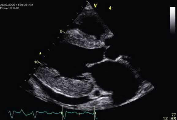
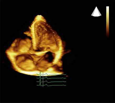
When a pulse of ultrasound is sent into tissue, a series of dots (one scan line, data line, or echo line) is displayed. Not all of the ultrasound pulse is reflected from any single interface. Rather, most of the original pulse continues into the tissue and is reflected from deeper interfaces. The echoes from one pulse appear as one scan line. Subsequent pulses go out in slightly different directions from the same origin. The result is a sector scan (sector image), which is shaped like a slice of pie (Fig. 1.3A). The resulting cross-sectional image is composed of many (typically 96 to 256) of these scan lines. For decades, sonography was limited to 2D cross-sectional scans (or slices) through anatomy such as that in Fig. 1.3A. 2D imaging has been extended into 3D scanning and imaging, also called volume imaging, as described in Chapter 2. This requires scanning the ultrasound through many adjacent 2D tissue cross sections to build up a 3D volume of echo information (see Fig. 1.3B), like a loaf of sliced bread in which each slice represents a 2D image and the loaf represents the 3D volume.

TRANSDUCER

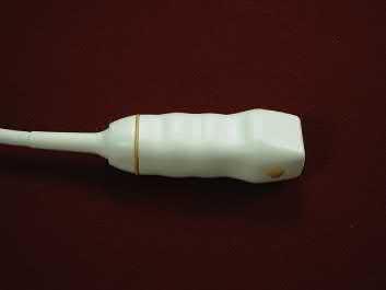
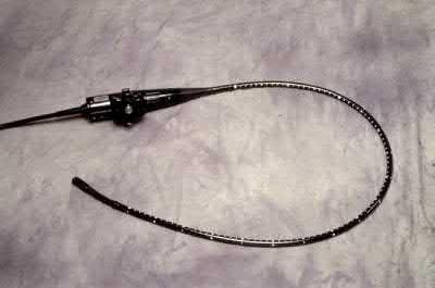
The transducer used in echocardiography is a phased array that electronically steers the ultrasound beam in the sector format. It is energized by an electrical voltage from the instrument that produces each outgoing ultrasound pulse. The returning echo stream is received by the transducer and converted to an echo voltage stream that is sent into the instrument, ultimately appearing on the display as a scan line. This process occurs a few thousand times per second (called the pulse repetition frequency [PRF]). A coupling gel is used between the transducer and the skin to eliminate the air that would block the passage of ultrasound across that boundary. Transducers (see Fig. 1.1) are designed for transthoracic (see Section II) and for transesophageal (see Section III) imaging. The latter provides a shorter acoustic path (with less attenuation, allowing higher frequencies and improved resolution) to the heart that avoids intervening lung and ribs.

Figure 1.1. A, Transthoracic transducer. B, Transesophageal transducer.

INSTRUMENT

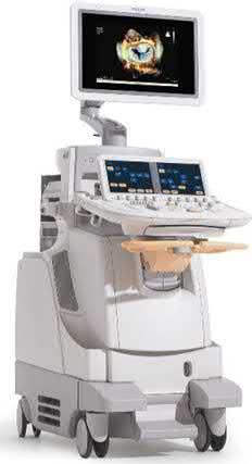
The echocardiographic instrument has a functional block diagram as shown in Fig. 1.2B. The beam former drives the transducer and receives the returning echo streams, amplifying (this is called gain) and digitizing them. Attenuation compensation occurs in the reception side of the beam former. This is an amplification process that equalizes echo strengths (amplitudes) by increasing gain with depth. This compensates for the weakening of echo amplitude with depth caused by attenuation. The signal processor, among other functions, detects the amplitude of each echo voltage. Echo amplitudes are stored digitally in the image memory, which is part of the image processor. Upon completion of a single scan (one frame of a real-time presentation), the stored image is sent to the flat-panel display. The echo information enters the image memory in ultrasound scan-line sector format but is read out to the display in horizontal-line display format, with each horizontal line on the display corresponding to a row of echo data in the image memory.

ARTIFACTS

In imaging, an artifact is any presentation that does not correctly display the structure or function (tissue motion and blood flow) imaged. Artifacts are caused by problematic aspects of the imaging process. They can hinder correct interpretation and diagnosis. These artifacts must be avoided or managed properly when encountered to avoid the errors they can cause. Some artifacts are produced by improper equipment operation or settings (e.g., incorrect gain and compensation settings). Other artifacts are inherent in the sonographic methods and can occur even with proper equipment and technique. The assumptions inherent in the sonographic process include the following:

• Sound travels in straight lines.

• Echoes originate only from objects located on the beam axis.

• The amplitude of returning echoes is related directly to the reflecting or scattering properties of the objects that produces them.

• The distance to reflecting or scattering objects is proportional to the round-trip travel time at a speed of 1.54 mm/μs.

If any of these assumptions are violated, artifacts occur. Fig. 1.4 and Video 1.4 provide examples of artifacts in echocardiography.

VIRTUAL BEAMFORMING

Two alternative fundamental principles of operation are now present in the array of commercially available echocardiographic equipment. The first, termed conventional echocardiography, has been the operating principle for more than 50 years and is described earlier in this chapter. Recently, a second principle has appeared, termed virtual-beam echocardiography 1,2 These two operating principles are fundamentally different, and there are implications for the resulting anatomic images and motion information presented. Virtual-beam sonography improves nearly every aspect of echocardiographic, anatomic imaging, motion, and flow presentation. Rather than a oneto-one relationship of pulse to scan line, characteristic of conventional echocardiography, virtual-beam echocardiography acquires each image with a few broad, unfocused beams and then through massive, high-speed, retrospective computation accomplished with graphics processing units determines the amplitude (and Doppler shift if needed) of the echo at each pixel location (Fig. 1.5). This computational, retrospective beamforming capitalizes on the fact that an echo arrives at the elements of the transducer in a sequence uniquely characteristic of the location from which it originated. The challenge is to sort out each echo from the mixed-up combination in

Figure 1.2. A, Echocardiographic instrument. B, Block diagram of an echocardiographic instrument. T, transducer.
Figure 1.3. A, Two-dimensional cardiac sector image. B, Three-dimensional

Another random document with no related content on Scribd:

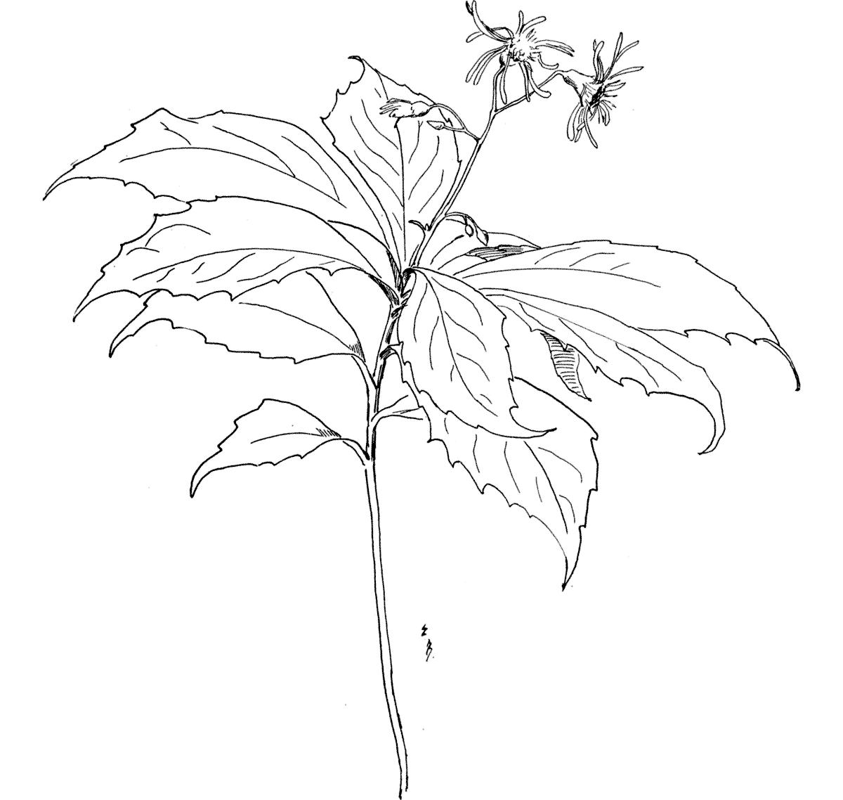
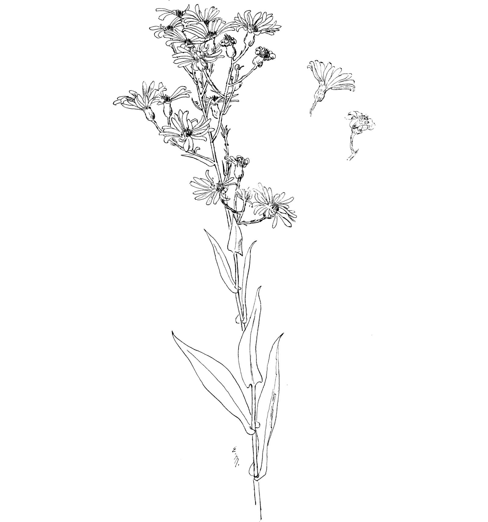
Pointed-leaved Aster. Asteracuminatus.

Found in August in rich woods (Mountains of Vermont).

It grows about 1 foot high, and has a slender, sometimes zigzag stalk, which is slightly rough, and bears the leaves and flowers clustered at the top.

The leaf, from 2 to 4 inches long, tapers at tip and base, and is coarsely toothed on the edge; the surface is a little hairy, and the texture is thin; the leaves, set directly on the stalk, are alternate at short distances, giving the effect of a whorl of leaves. Color green.

The flower-head has from 12 to 18 long, narrow, rays, white or faintly tinted with violet, while the disc flowers are dull yellow. The flower-heads are apt to be few, on slender stems in a branching cluster, on the top of the stalk.

This Aster lacks richness of color, but has a certain charm of slender growth peculiar to wood Asters; its few pale flowers set above the full cluster of the broad pale leaves have an air of selection, unknown to its more luxuriant relatives.

POINTED-LEAVED ASTER: A. acuminatus.

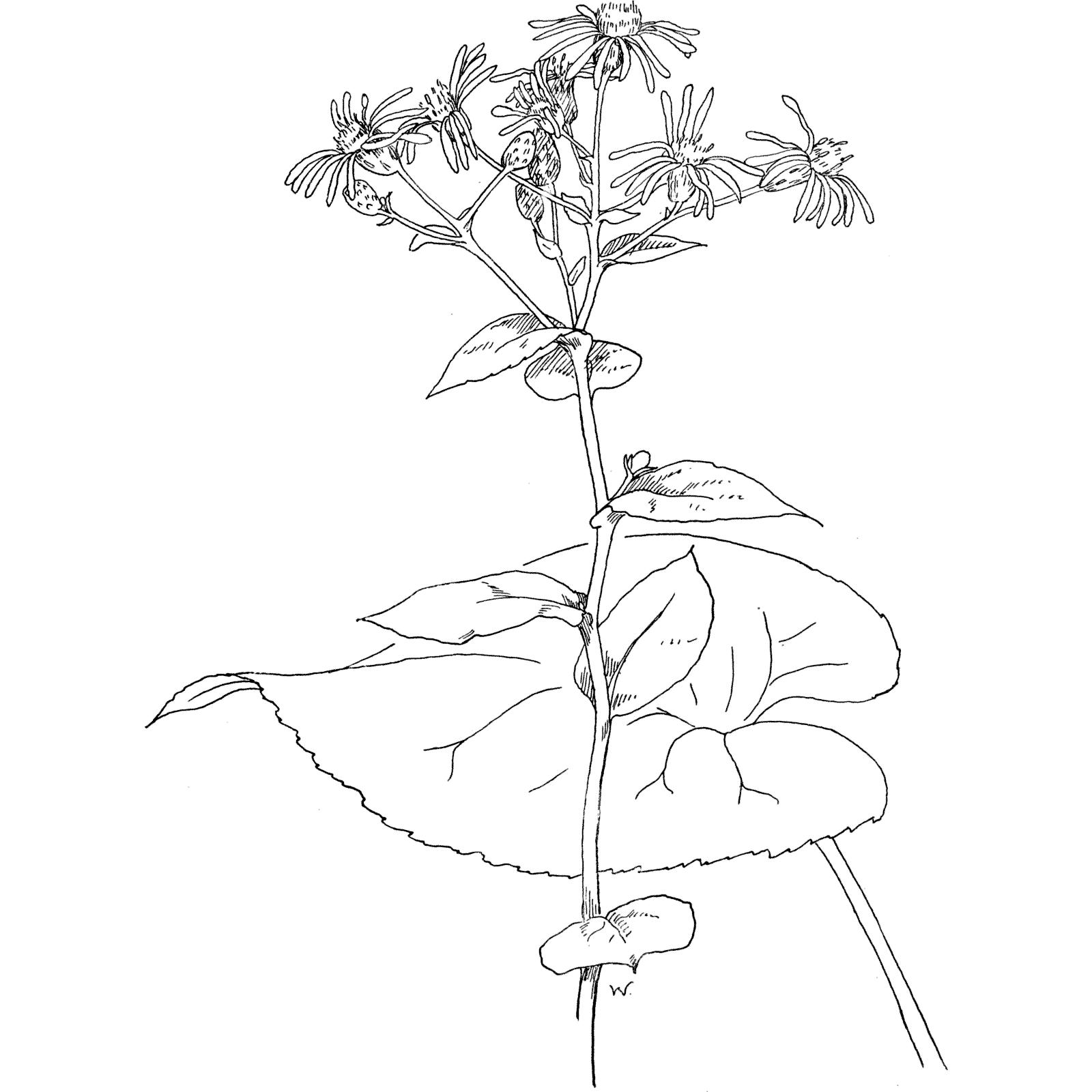
Broad-leaved Aster. Astermacrophyllus.

Found in the woods in September, generally in upland regions.

The single, very stout growing, stalk is between 1 and 2 feet in height; it is slightly rough to the touch. In color light green.

The lowest leaves (generally 3 in number) are very conspicuous, being especially large, and nearly round, with a heart-shaped base, and a suddenly sharp tip; with many ribs, and the margins prettily cut in rounding scallops, with tiny sharp teeth; these leaves are set on long trough-like stems that clasp the stalk. The upper leaves become, very abruptly, quite small; with fine-pointed tips, entire, slightly and irregularly notched margins, and showing only the midrib; they are set on very short stems, or, near the flowers, are placed immediately upon the stalk. All the foliage is exceptionally thin in texture, and rough on both surfaces; the arrangement is alternate. The color is a fine gray-toned green.

The disc flower is small; yellow turning to brown; the rays, 12 to 16 in number, are long and slender, of a very delicate texture, and pale lilac (sometimes nearly white it is asserted) in color. The cup is cylindrical, and slightly sticky, as are the foot-stems. The heads are arranged in a loose, terminal cluster.

An odor of camphor mingles with the sweet scent of these flowers, and is distinctly perceptible at a distance. The decorative quality of the leafage is of unique excellence.

BROAD-LEAVED ASTER: A. macrophyllus.

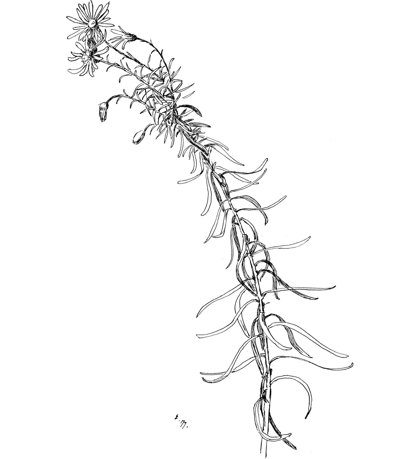
Pine Aster. Asterlinariifolius.

Found chiefly on the edges of pine woods in September.

The stalk reaches from 1 to 2 feet in height; it is single, leafy, slender, and rough to the touch. In color, pale green, tinged with red.

The leaf is long, and narrow, with an entire margin; the surface covered with a minute down that is harsh to the touch; the fibre is stiff. The color is green. The leaves are set upon the stalk, irregularly, alternating, and numerous.

The disc flowers are yellow; the ray flowers, 12 to 20 in number, are long and slender, and pale bluish-lavender in color. The heads are few, and grow, on slender stems, either singly, or in a terminal flat-topped cluster.

The stiff edges of the leaves and their rigid bearing are not unlike the needles which carpet the favorite haunts this Aster loves. Though not pleasant to pluck, on account of its harsh texture, this is a very attractive plant to look upon.

PINE

ASTER: A. linariifolius.

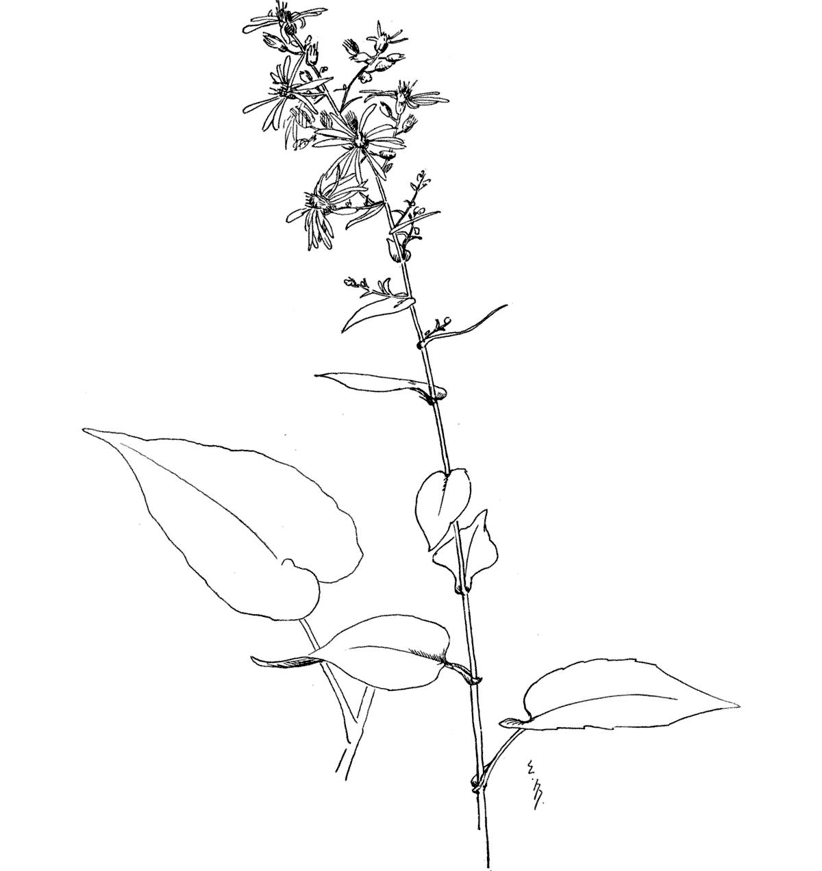
Variable-leaved Aster. Asterundulatus.

Found in dry woods, and thickets, during September.

The slender, branching stalk (2 to 3 feet high) is soft, with down, to the touch. Light green in color. Leaning and swaying in gesture.

The leaves are very variable in shape, the lowest being decidedly heart-shaped, and set on long margined stems (these margins somewhat notched) that clasp the stalk with shallow wings; above, the leaves are somewhat heart-shaped, and are set on short stems, that clasp the stalk with broad wings; while the upper leaves are set immediately upon the stalk, and clasp it by their prolonged bases. All the leaves have tapering tips, entire, or very slightly, notched margins, with a rough upper surface, while underneath it they are soft-hairy to the touch; they are placed alternately. The color is cool green.

The disc flowers are pale yellow at first, but become reddishbrown in maturity. The ray flowers, 10 to 20 in number, are pale lavender or lilac. The heads are arranged in loose clusters.

The color of this leafage, and its shapes, are very pleasing. Gray’s Manualstates the color of its rays as “bright blue”!

VARIABLE-LEAVED ASTER: A. undulatus.

Later Purple Aster. Asterpatens.

Found during September and October along the borders of dry woods and thickets.

The stalk (from 1 to 2 and ½ feet high) is branching, and leafy, with a fine texture, and smooth surface; slender and swaying in growth. Its color is sometimes green, often a dull reddish-purple.

The leaf is long, and narrow, tapering at the tip, and partly clasping the stalk at the base; the margin is slightly rough to the touch, the texture is firm and thick, and the surface is smooth to the touch. Color, a cool dark grayish-green. The arrangement is alternate.

The disc flowers are sometimes yellow, or often reddish-violet; the rays, from 20 to 30, are a deep rich violet. The heads are set in vase-shaped green cups, whose parts are overlapping; they are numerous in large terminal clusters.

As the flowers mature, the rays curl inward around the disc, and dry thus on the plant; its leafage is very fine in quality, and in beautiful harmony with the rich brilliance of the blossoms.

LATER PURPLE ASTER: A. patens.

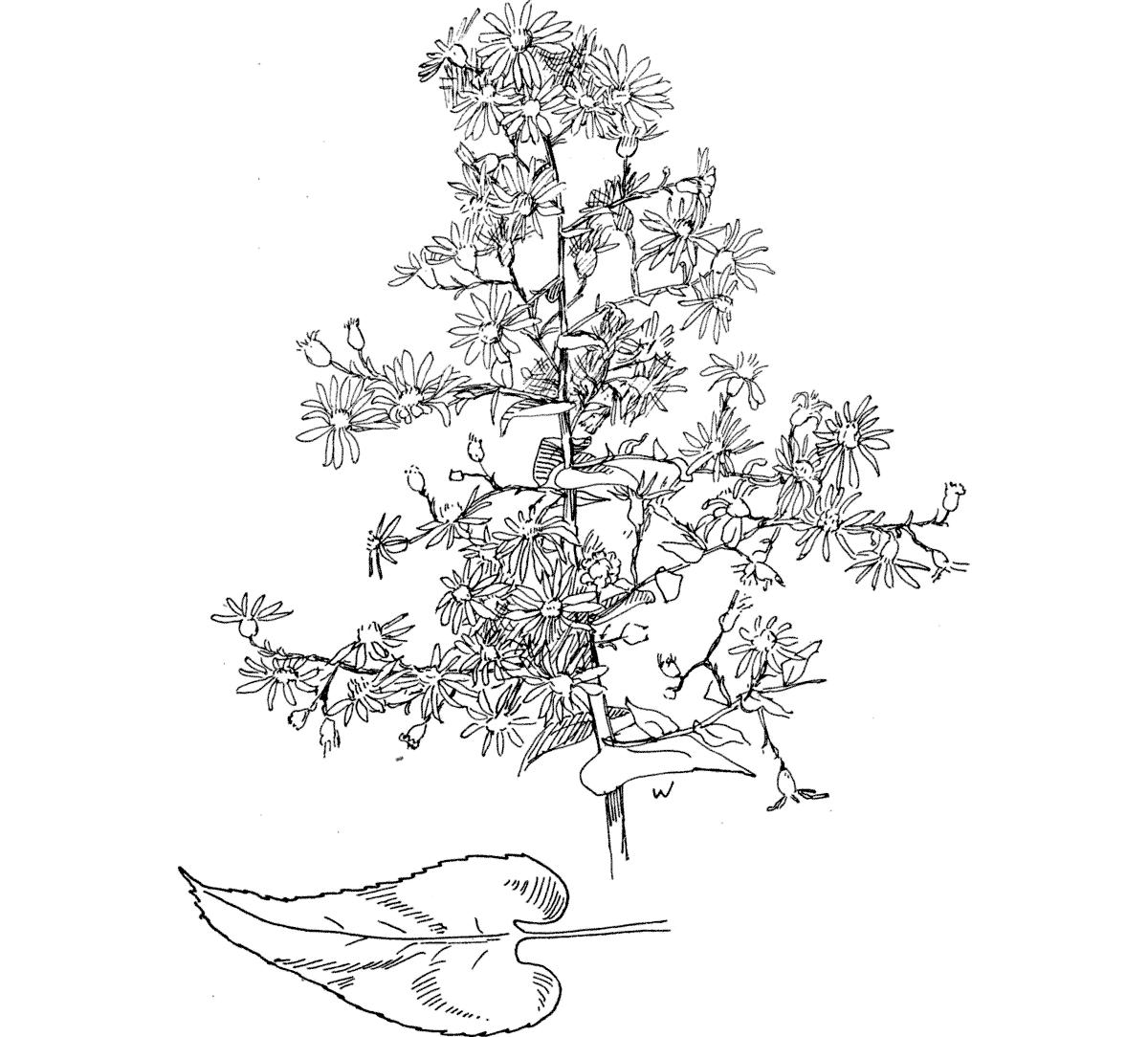
Branching Aster. Astercordifolius.

Found in the latter part of September in shaded thickets and on dry banks.

The stalk is often about 4 feet high, very widely branching, leafy, and bearing a profusion of flowers; it has a coarse, very rough surface usually. In color reddish.

The leaf has a heart-shaped base, and pointed tip, a slightly notched or entire margin, and a coarse texture, the surface generally being rough above and also on the underside of the midrib; the upper leaves clasp the stalk, while the lower, and larger, leaves are set on short hairy stems. In color, a rather dull green. They are numerous all the way from foot to tip.

The many flower-heads, are small, with 14 to 20 delicate, short, pale-lavender rays; the cups are smooth and shining, and are set on leafy foot-stems, in large loose clusters.

Coming just as its purple and white sisters are departing, and as though desiring to atone for their cessation, this delightful late Aster fills the country roads and lanes with the generous abundance of its misty gray-lavender bloom.

BRANCHING ASTER: A. cordifolius.

LOBELIA FAMILY.

LOBELIACEÆ.

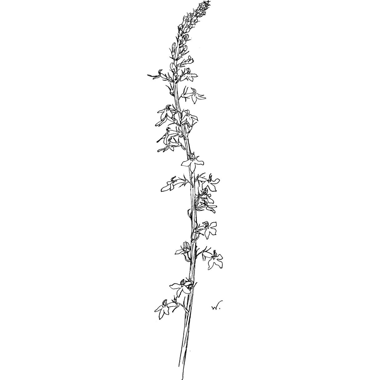
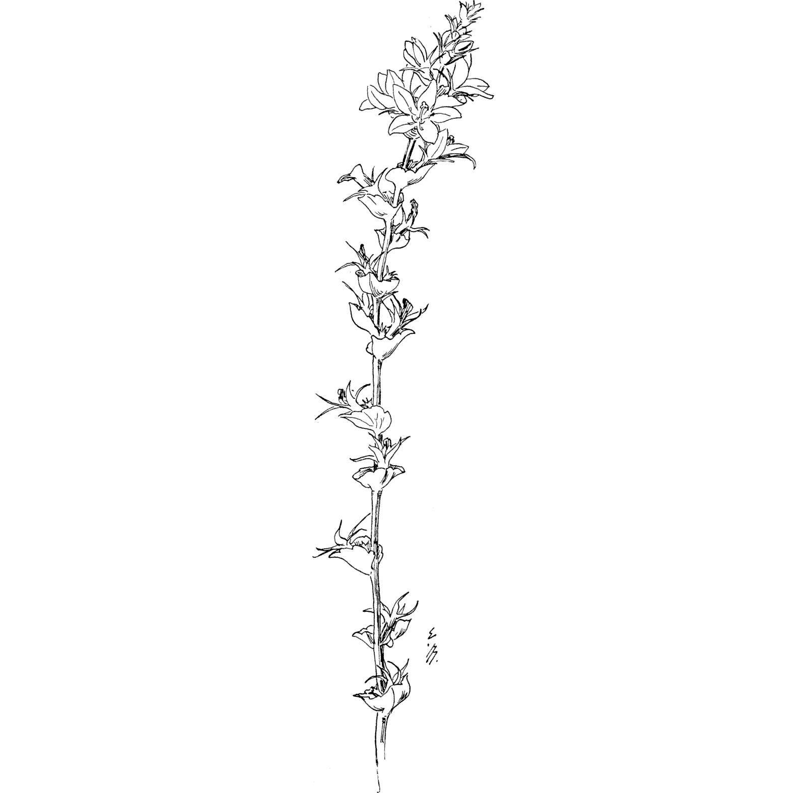
Spiked Lobelia. Lobeliaspicata.

Found in pastures and waysides during July and August.

The single, very erect stalk varies from 1 to 2 feet in height, and is very slender, grooved, and noticeably twisted; its fibre is fine, and near the foot it is hairy to the touch. In color green, with a trace of red at the leaf.

The foot-leaves are large, and a wide oval in shape, and set on margined stems; the upper leaves are a long narrow, oval shape, diminishing in size to a mere line, and are set immediately upon the stalk; the margins are irregularly notched, they are downy to the touch, and light green, tending to yellow. Their arrangement is alternate.

The small flower is 2-lipped, the lower lip being 3-parted with a pair of swellings at the throat of the tube, the upper lip divided by a cleft down the length of the tube; all parts of the corolla are sharppointed, and flaring from the 5-parted calyx; the texture is fine and smooth, and the color a pure pale lavender, faintly lined with dark, white near the throat, and pinkish in the tube, the green calyx being also tinted with pink. The flowers, on short foot-stems, are set alternately, and close, in a long, very pretty spike.

There is something individual about the alert carriage of this plant, its whole gesture is erect and animated. It grows numerously in pastures, though in less close fellowship than its little brook sister.

SPIKED LOBELIA: Lobelia spicata.

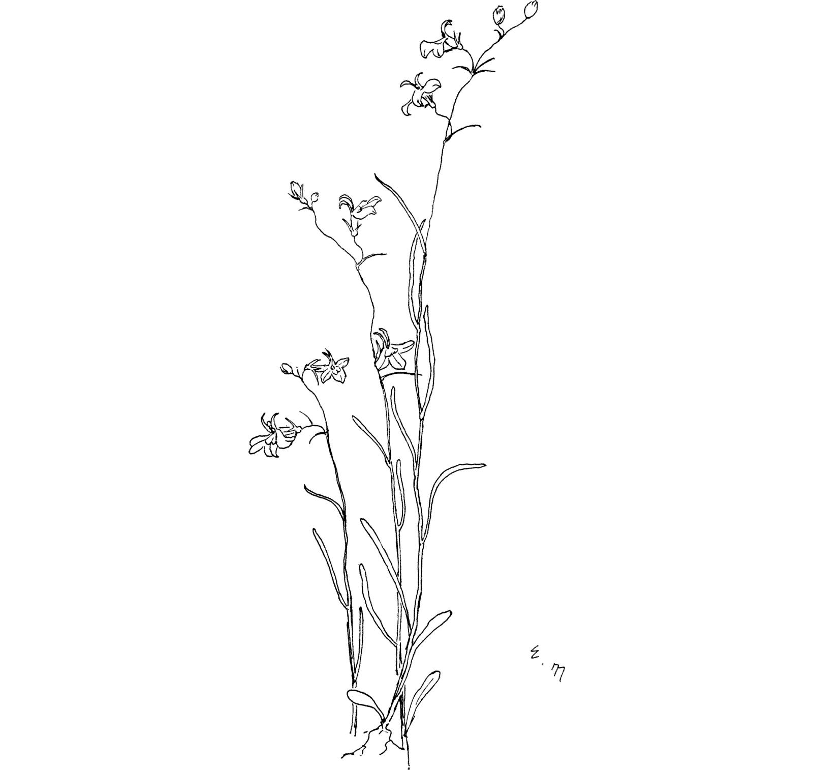
Brook Lobelia. LobeliaKalmii.

Found on moist ground in limestone soil, blossoming in July and August.

The erect, minutely angular stem, is slender, smooth and polished, fine and at the same time strong in fibre; in color a light grassgreen.

The small, narrow and grass-like leaf is about an inch long; towards the root it broadens a little at the tip; color grass-green. The growth is alternate.

The corolla of the small, irregular flower is in 2 divisions, the upper one with 2 narrow, upright parts, between which the stamens show; the lower one with 3 broad equal lobes; the color varies from deep lavender to light, and from a bluish to a pinkish tone, the lower division is marked with a broad white wedge-shaped spot near the throat. The small calyx has 5 thread-like divisions, green, tinged with lavender. From 3 to 4 flowers blossom at once on the top of the stem.

This little Lobelia flourishes especially in the limestone regions of the Green Mountains. It grows in communities among the short grass, beside some wayside spring or runlet; the flowers, all facing in the same direction, appear as though turned to greet the wayfarer. It is a plant of delicate finish and color, with a lithe and graceful gesture.

BROOK LOBELIA: L. Kalmii.

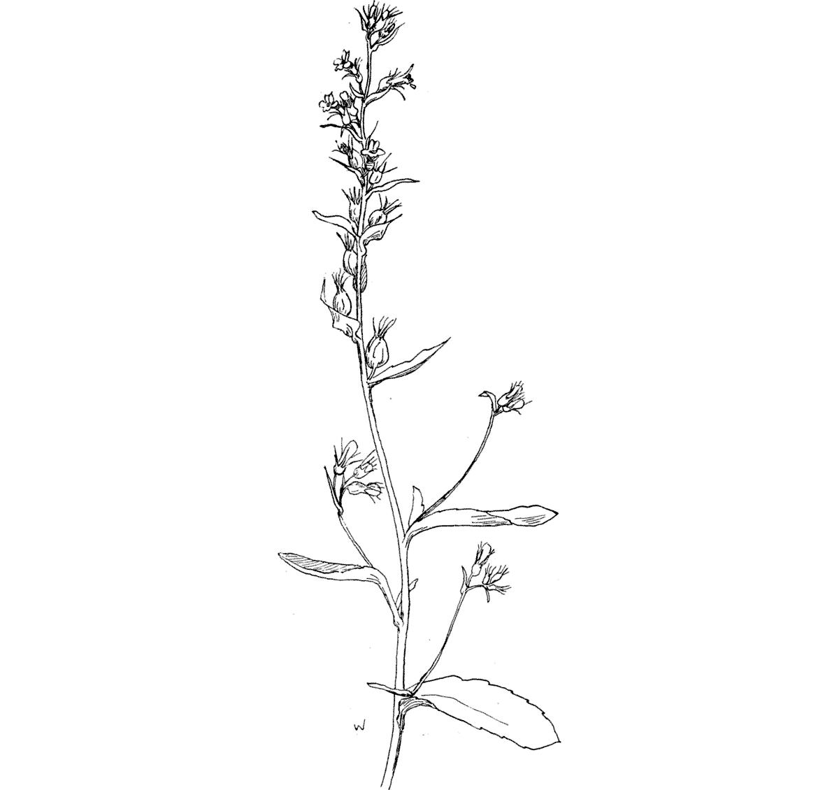
Indian Tobacco. Lobeliainflata.

Found in damp places, and along roadside ditches, in August.

The leafy, sometimes branching stalk grows from 12 to 18 inches, and is not large, though less slender than the Spiked Lobelia, its texture being also coarser; it is very slightly rough to the touch, and light green in color.

The leaves near the foot are about an inch long, and a blunt oval shape; near the top they become smaller and narrower; with coarsely toothed margins, a thin texture, and dull green color. They clasp the stalk alternately.

The flowers are smaller than the other Lobelias mentioned; the corolla is lilac, with a white spot on the lower lip; the calyx, whose 5 divisions are thread-like, is purplish-green. The flowers, on their little foot-stems, are placed in a scattering fashion upon the stalk to form a long terminal spike; each flower is supported by a little leaf or bract.

As the blossoms fall the seed-pods swell into a round and inflated shape, which, together with the scantiness of its bloom, gives the spike an untidy look, unusual in this class of delicate forms and elegant proportions.

INDIAN TOBACCO: L. inflata.

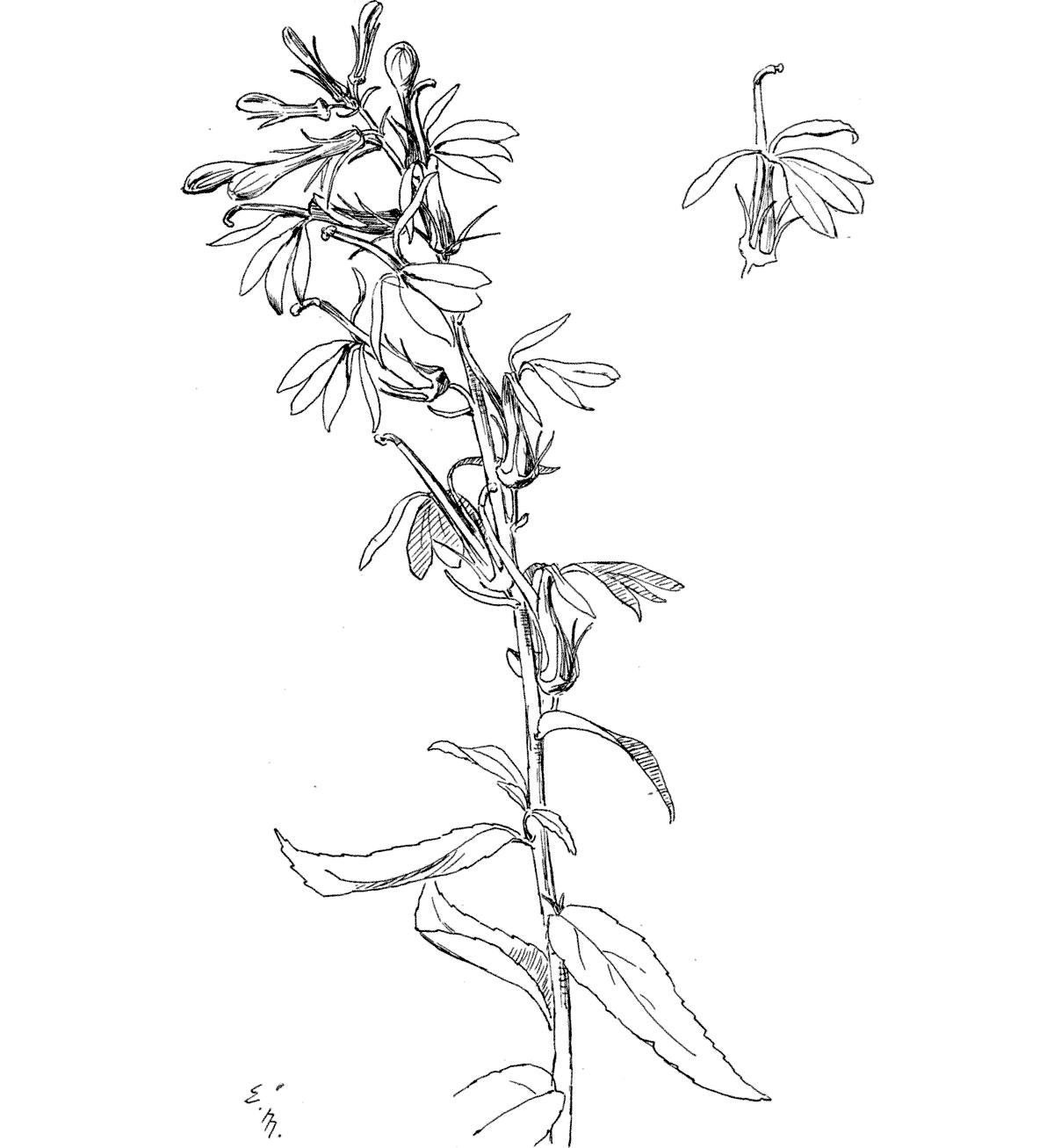
Cardinal Flower. Lobeliacardinalis.

Found on the margins of small streams, in light shade, and in low moist ground, from July to October.

The single, rather large stalk is round, and somewhat irregularly grooved, smooth to the touch, and a full green color. It grows from 2 to 4 feet high.

The leaves are generally from 2 to 3 inches in length, tapering at both ends with the veins distinctly showing, and irregularly toothed margins; their texture is fine, and the color a full dark green. They are set immediately upon the stalk, in an alternate arrangement.

The irregular corolla is somewhat 2-lipped, with a long slender tube which is split down its upper side; the lower lip is broad and 3cleft, the upper being narrow and in 2 parts; its color is a deep and glowing red, with a darker spot near the throat upon the lower lip. The pistil and 5 stamens, which curve outwards noticeably, are of a lighter red color with fine gray tips; the calyx is 5-parted and green. The flowers are arranged in a long, usually one-sided, spike.

Coming into bloom when the midsummer vegetation is growing dusty, this plant fills the eye with the richness of its dark green leafage toned into perfect accord with the glowing hue of its flowers, —their color a truly royal one, of unusual depth as well as brilliancy. The Cardinal Flower should not be plucked ruthlessly, lest it be exterminated, an ever-present danger to the beautiful annuals man appreciates. It can be cultivated from the seed with considerable success.

CARDINAL FLOWER: L. cardinalis.

CAMPANULA FAMILY.

CAMPANULACEÆ.

Venus’ Looking-glass. Speculariaperfoliata.

Found in blossom from June through July, among the grass, in open fields, and on hills.

The simple stalk grows from 12 to 20 inches high; it is somewhat angled, and roughened on the angles; rather weak in fibre it leans against the surrounding vegetation. It is of a light green color.

The small, shell-shaped leaf is broad, heart-shaped at the base, with a scalloped margin. The leaves are strung on the stalk, alternately, at short distances; they are stiffish in texture, and light green in color.

The deeply divided 5-pointed corolla is a deep reddish-violet; there are 5 stamens, and the pistil is 3-parted; the long, cylindrical, green calyx has 5 slender points. The flowers grow from the angles of the leaves, there being usually a number open at once toward the top of the stalk.

The lower flower buds do not open, ripening their seeds without the show of blossoming. As the flowering season advances the stalk lengthens out into a long wand, strung with shell-like little green leaves, each one holding in its hollow a seed-case.

VENUS’ LOOKING-GLASS: Specularia perfoliata.

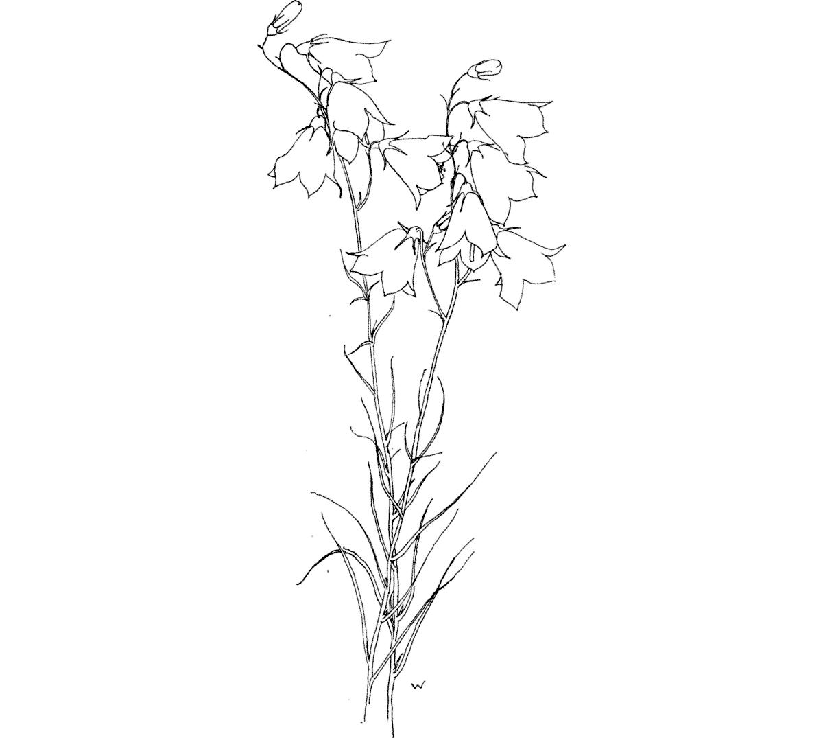
Harebell. Campanularotundifolia.

Found during July and August in rocky soil, along roadside thickets; in damp shade, or on exposed and barren uplands.

The stems of this lovely plant are very slender, like leafy wands, from 10 to 16 inches in height; they are firm and fine in fibre, smooth and shining, and bronzy-green.

The grass-like leaf is about an inch long, fine and thin and smooth; its color betrays the violet hues that temper all the green parts of the plant.

The blossom varies in size rather noticeably. Its corolla is shaped like a bell with 5 small pointed tips which curve outward to show the long lavender pistil with its malachite-green tip; the texture of the bell is delicately thin and yet firm, and the color an exquisite violet, which ranges from pale lavender, or even almost white, to a reddishpurple hue. The green calyx is 5-parted, its divisions very slender, half as long as the bell, and clinging closely to its shape. The many blossoms nod on thread-like stems.

In early spring the Harebell plant consists of a tuffet of small, round, slightly notched leaves, on spreading stems; these mostly disappear when the flowering stems begin to rise. In part, the swaying, flexile grace of gesture belonging to these flowers may be due to the light way in which the bells are caught in their tiny calices. When advanced in maturity the pistil becomes 3-parted and loses its vivid green tip. The Harebell is commonly credited with a love of shade, but it is frequently found in the crevices of exposed rock-ledges; and a favorite haunt, in one instance, is a bare mountain ridge, covered only by thick dry gray mosses, where these lovely bells nod amidst the Ebony Fern leaves (A. ebeneum), in thrifty vigor under the broad sunlight.

HAREBELL: Campanula rotundifolia.

Found in July on dry roadsides, and along walls and fences.

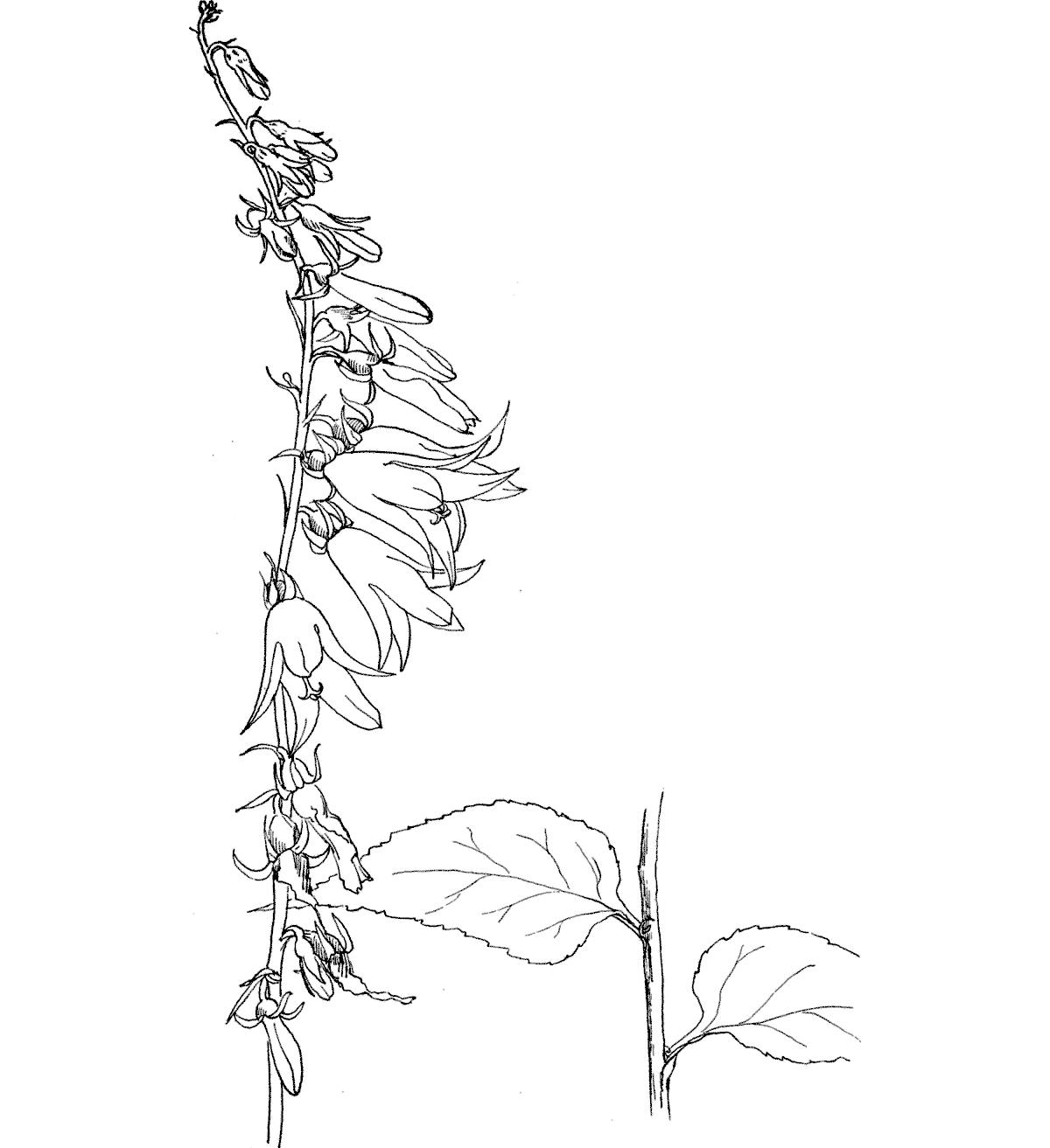
The stalk is single and leafy, slender, and slightly rough with hairs, from 2 to 4 feet in height; light green.

The root-leaves are small, and heart-shaped, with scalloped margins, set on stems that are sometimes 6 inches long. The lowest leaves along the stalk are shaped like an Indian arrow-point; the upper leaves are long and narrow: the margins are irregularly toothed, they are thin in texture, hairy, and light green; and are set on short broad stems which clasp the stalk alternately.

The corolla is bell-shaped with 5 outward-curving, tapering tips, and a beautiful violet-blue color; the pistil is 3-parted, protruding, and purple tinted; the green calyx is 5-parted, its divisions abruptly turned back. The flowers are set along one side of the stalk in the angles of the small upper leaves, forming a long wand-like spire.

Although the flowers bloom first at the base of the spire, frequently, when the tip has begun to hang out its noticeable bells, fresh buds will push forth far below, and open beside the old dried blossoms at the foot,—for the withered flower clings long to the calyx.

BELLFLOWER: C. rapunculoides.

HEATH FAMILY. ERICACEÆ.

Trailing Arbutus. Epigæarepens.

May flower.

Ground Laurel.

Found in the woods, or clearings near woods, and upland pastures, during April. It particularly favors pine woods.

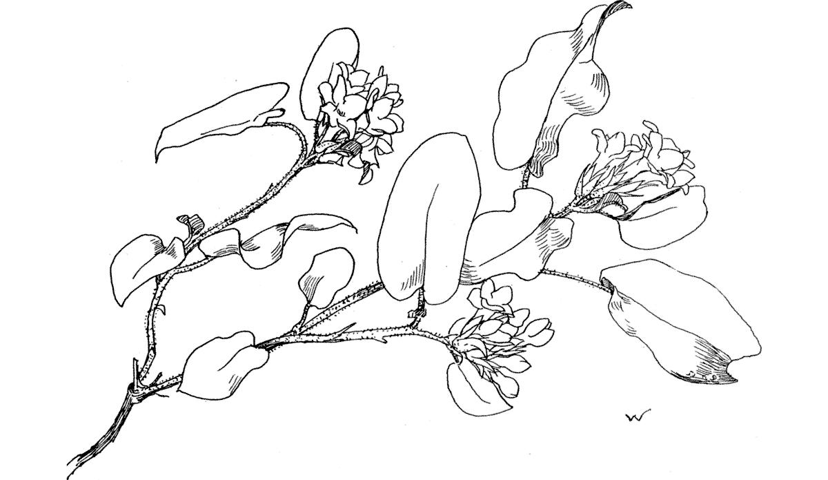
The branching leafy stalk is low, and trailing close to the ground under dry leaves and dead grass; it is tough and woody-fibred, and rough-hairy to the touch; rather slender. Its color is rusty brown, and very rich in tone.

The oval leaf is heart-shaped at the base, with a strong midrib which is somewhat taut and gives the entire margin usually an undulating character; the fibre is tough, and rough to the touch. The color is a strong positive green, more or less worm-eaten, and rustspotted; it is evergreen. The leaves are set, on slender rough stems, alternately, at irregular intervals, and more closely near the flowers. The new leaves come after the blossoming time.

The flower has a tubular corolla, spreading into 5 rounded points; its color varies from an exquisite white to a deeply tinted rose; the inconspicuous stamens show only as a feathery, pale yellow ring in the centre. The 5-parted, pale green calyx is set around with 5 similar leafy bracts. The blossoms, on their little foot-stems, are gathered in close clusters along the ends of the stalk and branches. They exhale a sweet and spicy fragrance.

The plant, with its green and rusty leaves, hugs the earth so closely that often only the tips of the flowery clusters show here and there among the dried leaves, which almost conceal them from their friendly enemies, whose love of their beauty knows no better manner of exhibiting itself than by tearing the vine up by its roots.

This selfish ignorance is fast exterminating the lovely flower in the populous districts of New England,—already it has become in certain place as mere tradition. As though in revenge, nature keeps fast the secret of its growth, and the Arbutus refuses to live in man’s care for more than one or two seasons.

ARBUTUS: Epigæa repens.

Cassandra. Cassandracalyculata.

Leather-leaf.

Found in April and May, in marshes and swamps, sometimes standing in water to the depth of two feet.

This shrub, about 3 or 4 feet high, branches often, and is toughfibred; the bark when new is smooth, but is inclined to become frayed and to split when it is old. The color is brown, of a golden hue.

The leaf is oval, widest near the tip, the midrib is prominent beneath, the margin is entire and slightly thickened on the edge, the texture is coarse grained and tough, but not thick; while the upper surface is smooth and shining, the underside is dull. It is evergreen; in color green, rusty underneath. The arrangement is alternate, and close together, and the leaves are so set as to point upwards; their short stems are often reddish.

The corolla is a small 5-pointed bell, curved inwards at the throat and spreading its points to show the thread-like, green pistil; of a fine smooth texture and pure white; the 5-parted calyx is curved closely to the bell,—white, or yellow, or tawny-tinted; at its base are found 2 little rusty, or white bracts. The flowers are set on short rusty stems which grow from the angles of the leaves, and turn downward.

It is worth the effort to discover the shy Cassandra’s haunts, in order to see the bush, its slender branches set close with pretty hanging bells beneath the alert leaves, that stand upright above them, in watchful military rows.

Turn static files into dynamic content formats.

Create a flipbook
Issuu converts static files into: digital portfolios, online yearbooks, online catalogs, digital photo albums and more. Sign up and create your flipbook.
Get Ase's comprehensive echocardiography american society of echocardiography PDF ebook with Full Ch by Education Libraries - Issuu